Embed Size (px)
Citation preview
REVIEW
Vol. 25, No. 11 November 2013 313
Abstract: Health care-associated infections (HCAIs) are infections acquired through contact with any aspect of health care. They can cause minor complications or serious disability or death, and can involve a wide variety of resistant or emergent organisms. Surgical site infections make up ap-proximately one-fifth of HCAIs, and at least 5% of patients undergoing open surgery develop an SSI. Surgical site infections are probably the most preventable HCAI but have received the least attention; although that is changing with increased surveillance and public awareness of published data of individual specialty and hospital incidence rates. Surgical site in-fection continues to be a complication of surgical care. These infections span a continuum of severity with some being quite innocent and easy to manage, while others are life-threatening. Considerable evidence provides direction in the prevention of SSI (eg, systemic antibiotic prophylaxis) but many preventive strategies need better definition, with additional clinical studies. When SSI occurs, the clinician needs to quickly recognize it and tailor management to the specific needs of the patient. In general, drain-age, debridement, and specific antibiotics for the putative pathogen are the hallmarks of management.
Key words: surgical site infection, treatment, prophylaxis
WOUNDS 2013;25(11):313-323
From the 1University of Huddersfield, Queensgate, UK; 2University of New Mexico School of Medicine, Albuquerque, NM; 3Northwestern University, Chicago, IL; 4Medical University of Vienna, Vienna, Austria
Address correspondence to:David Leaper, MD, ChM, FRCS, FACS, FLSProfessor in Clinical StudiesSchool of Human and Health SciencesUniversity of HuddersfieldQueensgate, Huddersfield HD1 3DHUnited [email protected]
Disclosure: The authors disclose no financial or other conflicts of interest.
Health care-associated infections (HCAIs) are infections that are ac-quired through contact with any aspect of health care. They can cause minor complications or serious disability or death, and can in-
volve a wide variety of resistant or emergent organisms.1 Examples of HCAIs include respiratory tract infections such as hospital- and ventilator-associated pneumonias complicated by Gram-negative, nonfermenting bacilli that are re-sistant to almost all classes of antibiotics; and urinary tract infections caused by microorganisms resistant to the quinolones and increased by the presence of a catheter, mostly involving coliforms that can produce extended spec-trum beta-lactamases (and, more worryingly, metallo-beta-lactamases, such as the newly described New Delhi-metallo-beta-lactamase 1 [NDM-1],2,3 mak-ing bacteria resistant to a wide range of beta-lactam antibiotics, including all
Perspectives in Prevention and Treatment of Surgical Site Infection – A Narrative Review of the Literature
David Leaper, MD, ChM, FRCS, FACS, FLS1; Donald Fry, BSc, MD, FACS2,3; Ojan Assadian, MD, DTMH4
DO NOT D
UPLICATE

Leaper et al
314 WOUNDS® www.woundsresearch.com
carbapenems). Infections involving prosthetic materials, as diverse as hip replacement or vascular grafts, are caused mainly by multiple-resistant coagulase negative staphylo-cocci; bacteraemias and complicated skin and soft tissue infections are associated with highly virulent meticillin sensitive Staphylococcus aureus (MSSA) or meticillin re-sistant Staphylococcus aureus (MRSA) strains; and emer-gence of Clostridium difficile is the underlying cause of antibiotic related colitis.4
Among HCAIs, surgical site infection (SSI) is of greatest recent concern.5 Surgical site infections can be caused by many organisms which may be antibiotic resistant. Patients who develop HCAIs usually have related treat-ments or underlying contributory illnesses, but the mis-use of antibiotics is a key factor in all cases. All HCAIs can be prevented or reduced by attention to known risk factors. The cost of HCAIs to health care is large,6,7 and has prompted many initiatives associated with extensive international media and political campaigns.
The bacteria involved in SSIs include those carried by the patients themselves (endogenous flora), and those that may be introduced in the operating room (exoge-nous flora).5 Native colonization (the source of infection), is determined through the type of surgery (eg, coliforms and anaerobes in colorectal surgery), although staphylo-cocci predominates overall from the bacterial reservoir in skin. Gram-positive pathogens from airborne microbes and the surgical team (due to glove punctures during sur-gical procedures or from suboptimal sterilization of in-struments) may be infrequent sources of contamination of the surgical site. Opportunistic and resistant organisms may be cultured from infections after selected operations (eg, prosthetic surgery). All patients are at risk of acquir-ing resistant organisms, particularly if they have an under-lying debilitating illness, poor compliance with accepted prevention guidelines, or they have health care-associat-ed exposure (ie, prior hospitalization or admission to a chronic care facility) that colonizes them with unusually resistant bacteria.
Surgical site infections make up approximately a fifth of the HCAIs in the United Kingdom, and at least 5% of patients undergoing open surgery in the UK develop an SSI.8 Surgical site infections are probably the most pre-ventable HCAI but have received the least attention; al-though that is changing with increased surveillance and public awareness of published data of individual specialty and hospital incidence rates.9-11 Surgical site infections are associated with more than one-third of postoperative related deaths ranging from relatively trivial, short-lived,
wound discharge (eg, after open hernia surgery) to be-ing life threatening (eg, mediastinitis and sternal wound infection).12 In between, HCAIs contribute to scars which may be cosmetically unacceptable to the patient, cause pain, require prolonged length of hospital stay with the incurred expenses, and result in poor emotional well-be-ing for the patient.13
Surgical Site InfectionDefinitions. Many surgeons are unaware of their SSI
rate because of nonexisting or suboptimal surveillance and inconsistent definitions. The first realistic survey of SSI was not sensitive, as only the presence of pus was used for the identification of SSI.14 Wounds are now cat-egorized into clean (no viscus opened), clean-contami-nated (viscus opened, minimal spillage), contaminated (viscus opened with spillage or presence of inflamma-tory disease), and dirty (pus or perforation present or incision made through an abscess).15 This categorization was based on a theoretical division of potential for SSI development. It is flawed by the failure to include pa-tient risk and the use of prosthetic materials, which may dramatically impact the risk for SSI in procedures within the clean category. Furthermore, because of the constant introduction of new operative techniques, particularly endoscopic procedures and the rise of natural orifice transluminal endoscopic surgery (NOTES) this categori-zation is becoming increasingly blurred and may not be applicable.
It is critical that standard definitions are used to al-low studies to be comparable. Analysis of outcomes and comparison between studies requires exact definitions of patients’ characteristics and their risk stratification, based on comorbid medical conditions. In addition to demographic details, clinical manifestations of SSI vary epidemiologically depending on the onset of infection. The Centers for Disease Control and Prevention (CDC) is the most widely used and comprehensive definition (Table 1).16 This system only gives categorical data which does not reflect the severity of an SSI. In brief, SSIs are categorized at 3 levels: superficial incisional, in the skin or subcutaneous tissues; deep incisional involving fascia or muscle; and deep/organ space, involving, for example, the pleura after lung surgery or the liver after hepatic re-section. Most SSIs fall into the superficial group and the less common deep/organ space infections are the most serious or life threatening. The categories are open to interpretation and may depend on the attending physi-cian’s diagnosis. By contrast, the ASEPSIS scoring method
DO NOT D
UPLICATE

Leaper et al
Vol. 25, No. 11 November 2013 315
for postoperative wound infections gives interval data17 but, despite its simplicity, has only been used in research trials. In today’s world of same-day surgery and fast-track postoperative recovery ASEPSIS is less easy to use and its validity may be questioned.18
Surveillance, incidence, and cost. Surveillance is equally critical to standard definitions. The CDC defini-tion requires surveillance for infection be undertaken for 30 days for infection in soft tissues and up to a year for orthopedic and vascular prosthetic surgery. Again, the uptake of same-day surgery and fast-track postoperative recovery has affected the accuracy of surveillance figures, which were largely based on inpatient data. Postdischarge surveillance must now be included since the majority of SSIs have a mean time to presentation of 8-10 days and are not apparent until after the patient has left the hospi-
tal. Ideally, surveillance should include a trained, blinded observer using agreed-upon definitions rather than sur-rogate automated methods.18-24 Accurate surveillance, including postdischarge data, can inform and influence practice by allowing valid comparisons.25,26 In some coun-tries, SSI surveillance is becoming mandatory. The meth-odology used has to be pragmatic and mostly depends on assessment at discharge, or telephone and questionnaire follow-up, but in research trials individual follow-up by direct observation is required for accuracy. Some areas of surgery have a low incidence of SSI, such as laparoscopic/endoscopic surgery.27
There have been several predictive indices for SSI. The Study on the Efficacy of Nosocomial Infection Control (SENIC) included contaminated wound, diagnosis at dis-charge, duration of surgery, and abdominal surgery; and
Table 1. Summary of CDC definition of SSI.
Superficial Incisional SSI• Infectionoccurswithin30daysaftertheoperation;• infectioninvolvesonlytheskinorsubcutaneoustissue;and• atleast1ofthefollowing: o purulent drainage (culture documentation not required); o organisms isolated from fluid/tissue of superficial incision; o at least 1 sign of inflammation (eg, pain or tenderness, induration, erythema, local warmth of the wound); o wound is deliberately opened by the surgeon; or o surgeon or attending physician declares the wound infected.A wound is not considered a superficial site infection if there is: o a stitch abscess present; o infection of episiotomy or circumcision site; o infection of a burn wound; or o incisional SSI that extends into the fascia or muscle.
Deep Incisional SSI• Infectionoccurswithin30daysofoperationorwithin1yearifanimplantispresent;• infectioninvolvesdeepsofttissues(eg,fasciaand/ormuscle)oftheincision;and• atleast1ofthefollowing: o purulent drainage from the deep incision but without organ/space involvement; o fascial dehiscence or fascia is deliberately separated by the surgeon due to signs of inflammation; o deep abscess is identified by direct examination, during reoperation, by histopathology, or by radiologic examination;
or o surgeon or attending declares deep incisional infection is present.
Organ/Space SSI• Infectionoccurswithin30daysofoperationorwithin1yearifanimplantispresent;• infectioninvolvesanatomicstructuresnotopenedormanipulatedbytheoperation;and• atleast1ofthefollowing: o purulent drainage from a drain placed by a stab wound into the organ/space; o organisms isolated from organ/space by aseptic culturing technique; o identification of abscess in the organ/space by direct examination, during reoperation, or by histopathological or ra-
diological examination; or o diagnosis of organ/space SSI by surgeon or attending physician.
Adapted from16 Horan TC, Gaynes RP, Martone WJ, Jarvis WR, Emori TG. CDC definitions of nosocomial surgical site infections, 1992: a modification of CDC definitions of surgical wound infections. Infect Control Hosp Epidemiol. 1992;13(10):606-608.
DO NOT D
UPLICATE

Leaper et al
316 WOUNDS® www.woundsresearch.com
the National Nosocomial Infections Surveillance index in-cluded contaminated wound, the American Society of An-esthesiologists Physical Status classification system grade, and duration of surgery. They have been compared28,29 and both were found capable of predicting SSI.
Apart from the unrecorded indirect costs related to loss of productivity, reduced quality of life, and litigation, the actual cost of an SSI can involve additional inpatient treat-ment and procedures that can run into many thousands of pounds.30 The morbidity and mortality rates which fol-low sternal infection after cardiac surgery are just one example.31 There is a paucity of prospective cost-benefit analysis of the SSI, but retrospective analyses clearly iden-tify that the economic costs of SSI are substantial.32
Prevention of Surgical Site Infection Level I evidence. Many national and international
guidelines present the best available evidence for the pre-vention of SSI. In the United Kingdom, for example, there are 2: from the National Institute for Health and Clini-cal Excellence (NICE)33 and the Scottish Intercollegiate Guideline Network (SIGN).34 In the United States, similar quality improvement programs include the Surgical Care Improvement Project (SCIP)35 and the National Surgical Quality Improvement Program (NSQIP).36,37 The principal recommendations have been collated into a care bundle by the Department of Health of the United Kingdom.38 The concept of using this best evidence should summate the effects of the interventions, but success depends on the quality of compliance. The longer-term follow-up of NICE, SCIP, and NSQIP and their respective degree of compliance, will determine how effective they are.
The effectiveness of these national guidelines and per-formance measures to improve the rates of SSI remains to be seen. Three recent studies have demonstrated no improvement in SSI rates despite national efforts in the US to enforce compliance with SCIP measures.39-41 Hospi-tals with high rates of compliance do not have better SSI rates than those with less compliance. It is important to emphasize that SSI rates are influenced by multiple clini-cal variables and not only those articulated by national agencies. The recommendations by NICE and SCIP are clinically valid, but issues such as poor surgical technique and suboptimal compliance, along with the many other variables that influence SSI, will negate the benefits that should be seen. Clinicians and government policy makers must understand the complexity of SSI as a clinical out-come; recommendations focused upon a limited number of practices are only a starting point in prevention and, by
themselves, may not influence overall outcomes.42
Common to all of the guidelines and performance measures is the level I evidence supporting the rational use of antibiotic prophylaxis and the avoidance of razors for hair removal. Considerable level I evidence shows an-tibiotic prophylaxis significantly reduces SSIs after clean prosthetic, clean-contaminated, and contaminated opera-tions.33-38 Prophylaxis should be initiated within the im-mediate preincisional period of time (< 60 minutes before incision) and the antibiotic that is chosen should cover the organisms that are likely to be encountered. The se-lected antibiotic may depend on local resistance patterns, and guidance of a local formulary may be necessary. Usu-ally a single-dose of antibiotic at, or immediately before, the induction of anesthesia is sufficient. Dirty operations, where infection already exits, will need a longer course of antibiotics that acts both as therapy and prophylaxis.
During the past 3 decades, prevention of SSI has re-lied almost entirely on the availability of effective peri-operative antibiotic prophylaxis due to its high level of evidence. For the same reason, antibiotic prophylaxis has also been used for treatment of infections after clinical presentation. For example, in vascular surgery, 10 ran-domized controlled trials (RCTs) have been undertaken comparing systemic antibiotics vs placebo, and a system-atic review and meta-analysis43 demonstrated a consistent benefit in reduction of SSI in 1297 patients relative risk (RR fixed, 0.25; (95% CI, 0.17 to 0.38; P = 0.0001). Al-though no single study demonstrated a statistically signifi-cant reduction in early vascular graft infection, the pooled results of these 10 RCTs appeared homogeneous with a reduction in early graft infection evident on meta-analy-sis (RR fixed, 0.31; 95% CI, 0.11 to 0.85; P = 0.02). There are, however, 2 aspects of these results which need to be highlighted. First, 6 of the 10 RCTs included a case-mix of patients with both prosthetic and vein grafts. If the results of the prophylactic effect of antibiotics are stratified by the type of graft, the RR for wound infection with pros-thetic graft is nonsignificant at 0.51 (95% CI, 0.24 – 1.11). The administration of prophylactic antibiotics yielded a significant benefit only for patients with vein grafts (RR = 0.13; 95% CI, 0.04 – 0.41). Therefore, in vascular surgery, evidence for the prophylactic effect of systemic antibiot-ics exists only for patients with vein grafts, who are at lower risk of infection, compared to patients receiving prosthetic material. Second, the meta-analysis is based on 10 studies that compared the effect of systemic antibiot-ics with placebo, conducted in the early to mid-1980s.
Resistance of Staphylococcus aureus (the most fre-
DO NOT D
UPLICATE

Leaper et al
Vol. 25, No. 11 November 2013 317
quent bacterial organism to cause vascular infections) to methicillin was first noted clinically in 1961.44 In the US, methicillin-resistant Staphylococcus aureus (MRSA) only emerged in the period 1975 to 1981 in tertiary care centers. The percentage of major US acute care hospitals reporting greater or equal to 50 MRSA cases per year in-creased from 18% in 1987 to 32% in 1989.45 Therefore, the result of meta-analyses of RCTs chiefly investigating the beta-lactam antibiotics which demonstrated efficacy of antibiotic prophylaxis need to be noted with caution to-day, as MRSA already has become the most frequent cause of skin and soft tissue infections presenting to emergency departments in the US.46 Although speculative, it remains questionable if a meta-analysis of these RCTs would yield significance again if conducted with the current rigor and epidemiologic situation.
The studies which give evidence that the use of razors to remove hair preoperatively cause infection are primar-ily from 30 years ago, but most guidelines suggest that if hair has to be removed, it should be done with a dispos-able clipper head, close to the time of surgery.33,47 These studies are robust enough to give level I evidence. The damage caused to skin by shaving too long before surgery encourages the growth of organisms, which increase the risk of SSI.
Also common in the guidelines and performance mea-sures are methods to optimize the physiology of the host at the time of the operations; that is, avoidance of hypo-thermia, adequate glycemic control, and supplemental oxygen administration. The clinical value of avoiding peri-operative hypothermia was first realized more than 15 years ago48 and there has since been many adequate RCTs confirming the relevance of warming in the prevention of SSI,23 which led to a NICE guideline.49 The pathophysi-ological benefit has also been well examined.50
From the analysis of secondary outcomes in clinical trials, it has been suggested that patients who have dia-betes and whose blood sugar is out of control are more at risk of SSI. This is supported by experimental evidence that many physiological mechanisms are impaired by hyperglycemia. Most guidelines suggest blood glucose should be tightly controlled in patients with diabetes.38 However, it has been convincingly shown that following cardiac surgery, even in patients without diabetes, poor glucose control is associated with poor wound healing, as well as SSI in sternal wounds and in leg wounds after harvest of saphenous vein.51 The maintenance of blood glucose has been adopted as standard practice in cardiac surgery and it is unlikely that RCTs will be undertaken
to further prove this point. However, glycemic control in other fields of major surgery remains unproven, and tight glycemic control in patients without diabetes remains an area to be explored with RCTs. This may be especially true in trauma patients and patients with major surgical interventions, where hyperglycemia is part of the normal metabolic response to trauma and major surgical stress.
Intraoperative and immediate postoperative use of supplemental oxygen in the prevention of SSI discussed in
Table 2. Factors implicated in a higher risk of surgical site infection.*
i. Patient factors age sex obesity smoking immunosuppression steroids, cancer, anticancer therapy (chemo and radio-therapy), HIV nutritional indices metabolic factors diabetes mellitus, hepato0renal failure, serum albumin, haemoglobin
ii. Preoperative factors nasal decontamination mechanical bowel preparation skin preparation (surgical teams’ hands patients’ skin)
iii. Operative factors previous surgery antiseptic-impregnated incise drapes length and complexity of operation operating surgeon blood loss antimicrobial sutures diathermy
iv. Postoperative factors antiseptic lavage of wounds and cavitiesantimicrobial dressingssupplemental oxygen in recovery
v. Other factors observed but with varying levels of evidence theatre environment preoperative showering theatre wear minimising movement in the OR banning of jewellery and nail polish drapes and gowns wound drainage
* The relevance of many of these factors needs revisiting with adequate trial design.
Adapted from85 Fry DE. Surgical site Infection. In: Fry DE, ed. Sur-gical Infections. London, UK: JP Medical Publishers Ltd;2013:49-62.
DO NOT D
UPLICATE

Leaper et al
318 WOUNDS® www.woundsresearch.com
this review because of the conflict in the results of RCTs. Considerable experimental evidence supports the use of supplemental oxygen to prevent incisional infection.52 Two RCTs have demonstrated significant reductions in SSI by the use of intraoperative and immediately postoperative oxygen supplementation (FiO2= 0.8).53,54 A single RCT has demonstrated an increase in SSI rates with supplemental oxygen.55 While the theoretical arguments to support sup-plemental oxygen are abundant, additional studies appear to be warranted before guidelines or performance mea-sures can be applied for the prevention of SSI.
Other evidence and risk factors. Many guidelines and reviews have listed other factors (Table 2) that can influ-ence the incidence of SSI but most are of level II evidence base at best.56,57 Many are anecdotal, others have been identified by logistic regression analysis in trials and audit of SSI; and others by meta-analysis (Table 1).
Many of these reports suggest that being male or being elderly is associated with an increased risk of SSI, although 1 cohort study found a decreasing risk after 65 years.58 Obesity is also cited in studies as being an important, inde-pendent risk factor.23,33 In addition, many patient-related factors, including smoking, have a strongly supportive ex-perimental base which has not been proven conclusively in clinical trials. This also applies to immunosuppressive, nutritional, and metabolic factors outside the scope of this article. Serum albumin is an example of a factor often ascribed significance for being an independent clinical risk factor, but it has not been clearly defined as such in prospective trials. A low value is associated with uncer-tain causation and may not reflect nutritional deficien-cies in the developed world. However, experimental data shows it is a strong marker of poor healing; but in clini-cal practice, the serum albumin value is usually related to confounding factors associated with severe systemic ill-ness, such as cancer cachexia or sepsis. The NSQIP places emphasis on low albumin being a predictor of surgical complications, but only further prospective studies will confirm its true role as a predictor of SSI.
Nasal decontamination (suppression) of Staphylo-coccus aureus was introduced by infection prevention programs in an effort to reduce the risk of MRSA. Both MSSA and MRSA can cause SSI, and staphylococci remain overall the commonest infecting organisms. Nasal sup-pression has not been recommended by NICE or SCIP to reduce SSI, but there is now evidence that this is useful; it is important to remember that MSSA infections are just as important to prevent.59
Mechanical bowel preparation has been the sub-
ject of a meta-analysis showing it does not reduce the risk of SSI.60 However, this finding, like many Cochrane Collaboration meta-analyses, needs clinical interpre-tation prior to implementation for all patients. Since the 1930s, it has been shown that mechanical bowel preparation alone does not reduce SSI rates.61 Mechani-cal preparation with the use of orally-administered and poorly absorbed antibiotics (eg, neomycin and eryth-romycin) has been shown in prospective, randomized, placebo-control trials to reduce SSIs.62,63 Two separate meta-analyses show that mechanical bowel prepara-tion, when combined with the oral antibiotic bowel preparation and systemic prophylactic antibiotics, do reduce SSI in elective colon resection when compared to mechanical bowel preparation and systemic antibi-otics only.64, 65
Skin preparation is routine prior to surgery but there has been little clear evidence demonstrating which antiseptic preparation is the best. Chlorhexidine has been a popular skin preparation, but in a concentration of 2% in combination with 70% isopropanol it has been shown to significantly reduce superficial and deep SSIs.66 The use of topical antiseptics for preparation of the surgical team’s hands, and as a preoperative wash for patients, has a good evidence base which deserves recognition.67-69 In view of the continued rise of anti-biotic resistance, the use of antiseptic dressings and prophylactic antiseptic lavage for wounds and cavities bears reconsideration in future clinical trials, as well as reconsideration as a treatment for established SSIs.33,57
Intraoperative factors that might relate to the inci-dence of SSI are traditionally observed and operating room discipline is long established, with a reluctance to change protocol without clear evidence. Operat-ing room environment control has to be placed in this category. Some of these factors do have basis and are included in the risk factor prediction indices.28,29 The NICE guideline, having reviewed the old trials33 of an-tiseptic impregnated drapes, recommended they be used as nonimpregnated drapes, as there clearly was an increased risk of SSI associated with their use. By contrast, the guideline could not recommend, for ex-ample, the use of diathermy to reduce the risk of SSI. Evidence that the use of antimicrobial sutures can re-duce SSIs is increasing; this has been found in clean, prosthetic, abdominal and thoracic surgery,18,57,70-73 and a meta-analysis has now been published which shows a level I evidence-base of the efficacy of antimicrobial sutures for the prevention of SSI.74 Again, we are see-
DO NOT D
UPLICATE
Leaper et al
Vol. 25, No. 11 November 2013 319
ing the return of antiseptics as a first line treatment of managing SSI.
Many of the remaining factors listed in Table 1 have been challenges, although many of them are part of the traditional lore of clinical surgery. Guidelines continue to support gowns and drapes, appropriate use of surgi-cal gloves, and reduction of movement in the operating room. Some have advocated the banning of jewelry and nail polish, but the evidence to support these policies are lacking. Preoperative showering and the value of wound drains are areas with supportive opinions but need addi-tional research to validate their application.
Other strategies used in infection prevention, such as maximal sterile barrier precautions; routine change of surgical gloves before graft implant; performing sur-gical procedures in HEPA filtered, turbulence-free, lami-nar air-flow, ventilated operation rooms; implementation of perioperative surgical check lists; or the choice and concentration of pre-operative skin antiseptics and how they should be used, have not been studied in depth and indicate areas for future research.
Treatment of Established Surgical Infection and SSIs
Essential to treating surgical infection, and superficial and deep SSIs, is to open the area of infection and to drain pus. With deep SSI, this may require opening and draining the entire incision, while superficial SSI may only require a limited area of drainage. Fibrinous debris is removed and any remaining sutures or staples in the area of infec-tion should also be removed. The open wound commonly needs specific wound care to allow healing by second-ary intention, although delayed primary or secondary clo-sure may be feasible in selected cases. The open wound is managed with interactive moist dressings and wound desiccation should be avoided. The use of topical antimi-crobial therapy is largely chosen by physician preference and remains an area for additional comparative investiga-tions of alternative agents. The concerns that antiseptics may induce bacterial resistance to themselves, or even to antibiotics, with the risks of transmission, are unfounded. This is either because their mode of action is not suitable for bacteria to develop resistance, or because the clinical-ly used high concentrations of antiseptics (which often surpass the required minimum inhibitory concentrations by 500 to 1,000 fold), are still able to rapidly kill off patho-gens even if they have developed a decreased susceptibil-ity against the applied antiseptic.74
Topical negative pressure wound management may
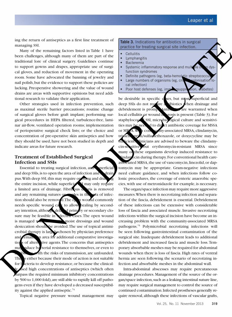
be desirable in specific cases, but most superficial and deep SSIs do not require antibiotics when drainage and debridement is prompt. Antibiotics are warranted when local cellulitis or wound necrosis is present (Table 3). For staphylococcal SSI, microbiological culture and sensitivi-ties may be needed to direct antibiotic coverage for MSSA or MRSA. With community-associated MRSA, clindamycin, trimethoprim/sulfamethoxazole, or doxycycline may be sufficient. Clinicians are advised to beware the clindamy-cin-sensitive but erythromycin-resistant MRSA since many of these organisms develop induced resistance to clindamycin during therapy. For conventional health care-associated MRSA, the use of vancomycin, linezolid, or dap-tomycin may be appropriate. Gram-negative infections need culture guidance, and when infections follow co-lonic procedures, the coverage of enteric anaerobic spe-cies, with use of metronidazole for example, is necessary.
The organ/space infection may require more aggressive measures. When there is necrotizing infection and separa-tion of the fascia, debridement is essential. Debridement of these infections can be extensive with considerable loss of fascia and associated muscle. Invasive necrotizing infections within the surgical incision have become an in-creasing problem with the community-associated MRSA pathogens.75 Polymicrobial necrotizing infections will be seen following gastrointestinal contamination of the surgical site. Inadequate debridement leads to additional debridement and increased fascia and muscle loss. Tem-porary absorbable meshes may be required for abdominal wounds when there is loss of fascia. High rates of ventral hernia are seen following the scenario of necrotizing in-fection and absorbable meshes in the abdominal wall.
Intra-abdominal abscesses may require percutaneous drainage procedures. Management of the source of the or-gan/space infection, such as a leaking intestinal suture line, may require surgical management to control the source of continued contamination. Infected prostheses generally re-quire removal, although these infections of vascular grafts,
Table 3. Indications for antibiotics in surgical practice for treating surgical site infection.
• Cellulitis• Lymphangitis• Bacteremia• Systemicinflammatoryresponseandmultipleorgandys-
function syndromes • Definitepathogens(eg,beta-hemolyticstreptococcus)• Largenumbersoforganisms(eg,critical-colonisationlo-
cal infection)• Poorhostdefenses(eg,immunosuppression,diabetes)
DO NOT D
UPLICATE

Leaper et al
320 WOUNDS® www.woundsresearch.com
heart valves, and prosthetic joints pose special problems. Antibiotics are almost always required for organ/space in-fections, the specific choice of which must be guided by culture and sensitivity data. Organ/space SSI pathogens are often staphylococcal but these infections are commonly associated with resistant organisms from the hospital envi-ronment. Antibiotic therapy again must be driven by spe-cific culture and sensitivity data.
Microbiological DiagnosisMicrobiological investigations may support the diag-
nosis of SSI and surgical infections, provided specimens are obtained appropriately. Thereby, microorganisms that are potentially causative for infection are yielded, and not colonized flora, which have no relevance for a suspected infection. Optimal results are achieved from specimens obtained directly from the infection site. Such material may include explanted grafts, extra- or intra-operatively obtained tissue biopsies from the infected area, and ma-terial aspirated from peri-graft fluid collection. Indirectly obtained material, such as blood cultures, may also yield important information. However, blood cultures may of-ten be negative, particularly in late-onset infection, but are more frequently positive in early-onset infection.76
Specimens may be processed using a number of tech-niques such as direct streaking swabs on agar plates, broth culture, homogenization of tissue specimens with serial dilution techniques, and sonication of the specimen to enhance the recovery of biofilm-forming organisms from graft or infected material.77 The report of processed micro-biological specimens is decisive for a valuable evaluation of therapy and clinical results.78,79 The demonstration of specimens’ relevance from the pre-, intra- and postopera-tive phases is considered most valuable, if feasible and pos-sible.80 Relevant preoperative samples include blood cul-tures taken from central and peripheral venous catheters or direct vein puncture, wound specimens, drainage fluid, nose and throat swabs in the case of MRSA colonization, and urinary samples. In any case, all responsible microor-ganisms should be classified according to their type (eg, Gram-positive or -negative, fungi, etc.) and specific ther-apy antibiograms offered for highly virulent microorgan-isms (eg, Pseudomonas aeruginosa). Careful assessment is needed for microorganisms isolated from overlying wounds or sinuses, as such microorganisms may represent colonizing flora, (eg, MRSA), and may be wrongly interpret-ed as causative agents.76 Relevant intraoperative samples are standard specimens obtained from the surgical site, the peri-graft fluid/pus, or explanted prosthetic material. Worth
noting is that a considerable number of specimens may be negative.81-83 A negative microbiological report, however, does not exclude an infection. Finally, relevant postopera-tive samples are blood cultures, drainage fluid, and wound specimens (eg, those usually taken when there is delayed wound healing). The development of polymerase chain reaction methodologies that will provide unique bacterial species and sensitivity signatures may greatly enhance the diagnosis of infection and will reduce the number of cir-cumstances where microbiological evaluation of wound and blood cultures are negative, but clinical infection ap-pears to be present.84
ConclusionSurgical site infection continues to be a complication
of surgical care. These infections span a continuum of se-verity with some being easy to manage while others are life-threatening. Considerable evidence provides direc-tion in the prevention of SSI such as systemic antibiotic prophylaxis, but many preventive strategies need better definition, with additional clinical studies. When SSI oc-curs, the clinician needs to be able to quickly recognize it and tailor management to the needs of the patient. In gen-eral, drainage, debridement, and specific antibiotics for the putative pathogen are the hallmarks of management.
References1. National Clinical Guideline Centre (UK). Infection: Pre-
vention and Control of Healthcare-Associated Infections
in Primary and Community Care: Partial Update of NICE
Clinical Guideline 2. London, UK: Royal College of Physi-
cians; 2012.
2. Johnson AP, Woodford N. Global spread of antibiotic re-
sistance: the example of New Delhi metallo-β-lactamase
(NDM)-mediated carbapenem resistance.J Med Microbi-
ol. 2013;62(Pt 4):499-513.
3. Nordmann P, Poirel L, Walsh TR, Livermore DM. The
emerging NDM carbapenemases. Trends Microbiol.
2011;19(12):588-595.
4. He M, Miyajima F, Roberts P, et al. Emergence and global
spread of epidemic healthcare-associated Clostridium dif-
ficile. Nat Genet. 2013;45(1):109-113.
5. World Health Organization. WHO Guidelines for Safe Sur-
gery 2009: Safe Surgery Saves Lives. http://whqlibdoc.
who.int/publications/2009/9789241598552_eng.pdf. Ac-
cesssed October 31, 2013.
6. Plowman R, Graves N, Griffin MA, et al. The rate and cost
of hospital-acquired infections occurring in patients ad-
mitted to selected specialities of a district general hospi-
DO NOT D
UPLICATE

Leaper et al
Vol. 25, No. 11 November 2013 321
tal in England and the national burden imposed. J Hosp
Infect. 2001;47(3):198-209.
7. McGarry SA, Engemann JJ, Schmader K, Sexton DJ, Kaye
KS. Surgical-site infection due to staphylococcus au-
reus among elderly patients: mortality, duration of hos-
pitalization and cost. Infect Control Hosp Epidemiol.
2004;25(6):461-467.
8. Smyth ET, McIlvenny G, Enstone JE, et al. Four country
healthcare associated infection prevalence survey 2006:
overview of the results. J Hosp Infect. 2008;69(3):230-
248.
9. Berwick DM, Calkins DR, McCannon CJ, Hackbarth AD.
The 100,000 lives campaign: setting a goal and a deadline
for improving health care quality. JAMA. 2006;295(3):324-
327.
10. McKibben L, Horan T, Tokars JI, et al. Guidance on public
reporting of healthcare-associated infections: recommen-
dations of the Healthcare Infection Control Practices Advi-
sory Committee. Am J Infect Control.2005;33(4):217-226.
11. Humphreys H, Cunney R. Performance indicators and the
public reporting of healthcare-associated infection rates.
Clin Microbiol Infect. 2008;14:892-894.
12. Astagneau P, Rioux C, Golliot F, Brucker G; INCISO Net-
work Study Group. Morbidity and mortality associated
with surgical site infections: results from the 1997–1999
INCISO surveillance. J Hosp Infect. 2001;48(4):267-274.
13. Bayat A, McGrouther DA, Ferguson MW. Skin scarring.
BMJ. 2003;326(7380):88-92.
14. Cruse PJ, Foord R. The epidemiology of wound infection.
A 10-year prospective study of 62,939 wounds. Surg Clin
North Am. 1980;60(1):27-40.
15. National Academy of Sciences: ad hoc committee of the
Committee on Trauma. Post-operative wound infections,
the incidence of ultraviolet light irradiation of the operat-
ing room and of various other factors. Annals of Surgery.
1964;169(supplement 2):1-92.
16. Horan TC, Gaynes RP, Martone WJ, Jarvis WR, Emori
TG. CDC definitions of nosocomial surgical site infec-
tions, 1992: a modification of CDC definitions of surgi-
cal wound infections. Infect Control Hosp Epidemiol.
1992;13(10):606-608.
17. Wilson AP, Treasure T, Sturridge MF, Gruneberg RN. A scor-
ing method (ASEPSIS) for postoperative wound infections
for use in clinical trials of antibiotic prophylaxis. Lancet.
1986;1(8476):311-313.
18. Williams N, Sweetland H, Goyal S, Ivins N, Leaper DJ. Ran-
domized clinical trial of antimicrobial-coated sutures to
prevent surgical site infection after breast cancer surgery.
Surg Infect. 2011;12(6):469-474.
19. Prospero E, Cavicchi A, Bacelli S, Barbadoro P, Tantucci L,
D’Errico MM. Surveillance for surgical site infection after
hospital discharge: a surgical procedure-specific perspec-
tive. Infect Control Hosp Epidemiol. 2006;27(12):1313-
1317.
20. Avato JL, Lai KK. Impact of postdischarge surveillance on
surgical-site infection rates for coronary artery bypass pro-
cedures. Infect Control Hosp Epidemiol. 2002;23(7):364-
367.
21. Reilly J, Allardice G, Bruce J, Hill R, McCoubrey J. Proce-
dure-specific surgical site infection rates and postdis-
charge surveillance in Scotland. Infection Control Hosp
Epidemiol. 2006;27(12):1318-1323.
22. Taylor EW, Byrne DJ, Leaper DJ, Karran SJ, Browne MK,
Mitchell KJ. Antibiotic prophylaxis and open groin hernia
repair. World J Surg. 1997;21(8):811-814.
23. Melling AG, Ali B, Scott EM, Leaper DJ. The effects of pre-
operative warming on the incidence of wound infection
after clean surgery: a randomised controlled trial. Lancet.
2001;358:876-880.
24. Platt R, Yokoe DS, Sands KE. Automated methods for sur-
veillance of surgical site infections. Emerg Infect Dis.
2001;7(2):212-216.
25. Tanner J, Padley W, Kiernan M, Leaper D, Norrie P, Bag-
gott R. A benchmark too far: findings from a national sur-
vey of surgical site infection surveillance. J Hosp Infect.
2013;83(2):1-3.
26. Leaper D, Tanner J, Kiernan M. Surveillance of surgical site
infection: more accurate definitions and intensive record-
ing needed. J Hosp Infect. 2013;83(2):83-86.
27. Li X, Zhang J, Sang L, et al. Laparoscopic versus conven-
tional appendectomy – a meta-analysis of randomized
controlled trials. BMC Gastroenterol. 2010;10:129.
28. Fariñas-Alvarez C, Fariñas MC, Prieto D, Delgado-Ro-
driquez M. Applicability of two surgical-site infection risk
indices to risk of sepsis in surgical patients. Infect Control
Hosp Epidemiol. 2000;2(10):633-638.
29. Delgado-Rodríguez M, Sillero-Arenas M, Medina-Cuadros
M, Martinez-Gallego G. Usefulness of intrinsic infection
risk indexes as predictors of in-hospital death. American
J Infect Control. 1997;25(5):365-370.
30. Leaper D, Nazir J, Roberts C, Searle R. Economic and clini-
cal contributions of an antimicrobial barrier dressing: a
strategy for the reduction of surgical site infections. J Med
Econ. 2010;13(3):447-452.
31. Strecker T, Rosch J, Horch RE, Weyand M, Kneser U. Sternal
wound infections following cardiac surgery: risk factor
analysis and interdisciplinary treatment. Heart Surg Fo-
rum. 2007;10(5):E366-371.
DO NOT D
UPLICATE

Leaper et al
322 WOUNDS® www.woundsresearch.com
32. Fry DE. The economic costs of surgical site infection.
Surg Infect (Larchmt). 2002;3(Suppl 1): S37-43.
33. National Institute of Health and Clinical Excellence. Sur-
gical site infection. Prevention and treatment of surgical
site infection. Clinical Guideline 74. www.nice.org.uk/
nicemedia/live/11743/42378/42378.pdf. Published Octo-
ber 2008. Accessed October 31, 2013.
34. Scottish Intercollegiate Guidelines Network. Antibiotic
prophylaxis in surgery: a national clinical guideline.
http://www.sign.ac.uk/pdf/sign104.pdf. Published July
2008. Accessed Octoner 31, 2013.
35. Fry DE. Surgical site infections and the surgical care im-
provement project (SCIP): evolution of national quality
measures. Surg Infect (Larchmt). 2008; 9: 579-584
36. Alexander JW, Solomkin JS, Edwards MJ. Updated recom-
mendations for control of surgical site infections. Ann
Surg. 2011;253(6);1082-1093.
37. Berenguer CM, Ochsner MG Jr, Lord SA, Senkowski CK.
Improving surgical site infections: using National Surgical
Quality Improvement Program data to institute Surgical
Care Improvement Project protocols in improving surgi-
cal outcomes. J Am Coll Surg. 2010;210:737-743.
38. Department of Health. HCAI Reducing healthcare asso-
ciated infections. High Impact Intervention. Care bundle
to prevent surgical site infection for prevention of SSI.
Available at: www.hcai.dh.gov.uk/files/2011/03/2011-
03-14-HII-Prevent-Surgical-Site-infection-FINAL.pdf.
39. Hawn MT, Vick CC, Richman J, et al. Surgical site infec-
tion prevention: time to move beyond the surgical care
improvement program. Ann Surg. 2011;254(3):494-501
40. Nicholas LH, Osborne NH, Birkmeyer JD, Dimick JB. Hos-
pital process compliance and surgical outcomes in Medi-
care beneficiaries. Arch Surg. 2010;145(10):999-1004.
41. Stulberg JJ, Delaney CP, Neuhauser DV, Aron DC, Fu P,
Koroukian SM. Adherence to surgical care improvement
project measures and the association with postoperative
infections. JAMA. 2010;303(24):2479-2485.
42. Fry DE. Fifty ways to cause surgical site infections. Surg
Infect (Larchmt). 2011;12(6):497-500.
43. Stewart AH, Eyers PS, Earnshaw JJ. Prevention of infection
in peripheral arterial reconstruction: a systematic review
and meta-analysis. J Vasc Surg. 2007;46(1):148-155.
44. Boyce JM. Methicillin-resistant Staphylococcus aureus in
hospitals and long-term care facilities: microbiology, epi-
demiology, and preventive measures. Infect Control Hosp
Epidemiol. 1992;13(12):725-737.
45. Boyce JM. Increasing prevalence of methicillin-resistant
Staphylococcus aureus in the United States. Infect Con-
trol Hosp Epidemiol. 1990;11(12):639-642.
46. Moran GJ, Krishnadasan A, Gorwitz RJ, et al. Methicillin-
resistant S. aureus infections among patients in the emer-
gency department. N Engl J Med. 2006;355(7):666-674.
47. Tanner J, Woodings D, Moncaster K. Preoperative hair re-
moval to reduce surgical site infection. Cochrane Data-
base Syst Rev. 2006;19(3):CD004122.
48. Kurz A, Sessler DI, Lenhardt R. Perioperative normother-
mia to reduce the incidence of surgical-wound infection
and shorten hospitalization. Study of Wound Infection and
Temperature Group. N Engl J Med. 1996;334(19):1209-
1215.
49. National Collaborating Centre for Nursing and Support-
ive Care.The management of inadvertent perioperative
hypothermia in adults. www.nice.org.uk/nicemedia/
live/11962/40429/40429.pdf. Published April 2008. Ac-
cessed October 31, 2013.
50. Kumar S, Wong PF, Melling AC, Leaper DJ. Effects of periop-
erative hypothermia and warming in surgical practice. Int
Wound J. 2005;2(3):193-204.
51. Furnary AP, Zerr KJ, Grunkemeier GL, Starr A. Continu-
ous intravenous insulin infusion reduces the incidence
of deep sternal wound infection in diabetic patients
after cardiac surgical procedures. Ann Thorac Surg.
1999;67(2):352-362.
52. Qadan M, Battista C, Gardner SA, Anderson G, Akca O,
Polk HC Jr. Oxygen and surgical site infection: a study of
underlying immunologic mechanisms. Anesthesiology.
2010;113(2):369-377.
53. Greif R, Akça O, Horn EP, Kurz A, Sessler DI; Outcomes
Research Group. Supplemental perioperative oxygen to
reduce the incidence of surgical-wound infection. N Engl
J Med. 2000;342(3):161-167.
54. Belda FJ, Aguilera L, García de la Asunción J, et al. Supple-
mental perioperative oxygen and the risk of surgical
wound infection: a randomized controlled trial. JAMA.
2005;294(16):2035-2042.
55. Pryor KO, Fahey TJ 3rd, Lien CA, Goldstein PA. Surgical
site infection and the routine use of perioperative hyper-
oxia in a general surgical population: a randomized con-
trolled trial. JAMA. 2004;291(1):79-87.
56. Franz MG, Robson MC, Steed DL, et al. Guidelines to aid
healing of acute wounds by decreasing impediments of
healing. Wound Repair Regen. 2008;16(6):723-748.
57. Leaper DJ. Risk factors for and epidemiology of surgical
site infections Surg Infect (Larchmt). 2010;11(3):283-287.
58. Kaye KS, Schmit K, Pieper C, et al. The effect of increas-
ing age on the risk of surgical site infection. J Infect Dis.
2005;191(7):1056-1062.
59. Bode LG, Kluytmans JA, Wertheim H, et al. Preventing sur-
DO NOT D
UPLICATE

Leaper et al
Vol. 25, No. 11 November 2013 323
gical site infection in nasal carriers of Staphylococcus au-
reus. N Engl J Med. 2010;362(1):9-17.
60. Guenaga KK, Matos D, Wille-Jørgensen P. Mechanical bow-
el preparation for elective colorectal surgery. Cochrane
Database Syst Rev. 2009;(1):CD001544.
61. Poth EJ. Historical development of intestinal antisepsis.
World J Surg. 1982;6(2):153-159.
62. Washington JA 2nd, Dearing WH, Judd ES, Elveback LR. Ef-
fect of perioperative antibiotic regimen on development
of infection after intestinal surgery; prospective, random-
ized, double- blind study. Ann Surg. 1974;180(4):567-572.
63. Clarke JS, Condon RE, Bartlett JG, Gorbach SL, Nichols
RL, Ochi S. Preoperative oral antibiotics reduce septic
complications of colon operations: results of prospec-
tive, randomized, double-blind clinical study. Ann Surg.
1977;186(3):251-259.
64. Lewis RT. Oral versus systemic antibiotic prophylaxis
in elective colon surgery: a randomized study and me-
ta-analysis send a message from the 1990s. Can J Surg.
2002;45(3):173-180.
65. Fry DE. Colon preparation and surgical site infection. Am
J Surg. 2011;202(2):225-232.
66. Darouiche RO, Wall MJ Jr, Itani KM, et al. Chlorhexidine-al-
cohol versus povidone-iodine for surgical site antisepsis.
N Engl J Med. 2010;362(1):18-26.
67. Leaper DJ. Surgical-site infection. Br J Surg.
2010;97(11):1610-1602.
68. Noorani A, Rabey N, Walsh SR, Davies RJ. Systematic re-
view and meta-analysis of preoperative antisepsis with
chlorhexidine versus povidone–iodine in clean-contami-
nated surgery. Br J Surg. 2010;97(11):1614-1620.
69. Fournel I, Tiv M, Soulias M, Hua C, Astruc K, Aho Glele
LS. Meta-analysis of intraoperative povidone–iodine ap-
plication to prevent surgical-site infection. Br J Surg.
2010;97(11):1603-1613.
70. Rozzelle CJ, Leonardo J, Li V. Antimicrobial suture wound
closure for cerebrospinal fluid shunt surgery: a prospec-
tive, double-blinded, randomized controlled trial. J Neuro-
surg Pediatr. 2008;2(2):111-117.
71. Justinger C, Moussavian MR, Schlueter C, Kopp B, Kollmar
O, Schilling MK. Antibacterial [corrected] coating of ab-
dominal closure sutures and wound infection. Surgery.
2009;145(3):330-334.
72. Galal I, El-Hindawy K. Impact of using triclosan-antibacte-
rial sutures on incidence of surgical site infection. Am J
Sur. 2011;202(2):133-138.
73. Fleck T, Moidl R, Blacky A, et al. Triclosan-coated sutures
for the reduction of sternal wound infections: economic
considerations. Ann Thorac Surg. 2007;84(1):232-236.
74. Wang ZX, Jiang CP, Cao Y, Ding YT. Systematic review and
meta-analysis of triclosan-coated sutures for the preven-
tion of surgical site infection. Br J Surg. 2013;100(4):465-
473.
75. Leaper DJ, Assadian O, Hubner NO, et al. Antimicrobial
sutures and prevention of surgical site infection: assess-
ment of the safety of the antiseptic triclosan. Int Wound J.
2011;8(6):556-566.
76. Miller LG, Perdreau-Remington F, Rieg G, et al. Necrotiz-
ing fasciitis caused by community-associated methicillin-
resistant Staphylococcus aureus in Los Angeles. N Engl J
Med. 2005;352(14):1445-1453.
77. FitzGerald SF, Kelly C, Humphreys H. Diagnosis and treat-
ment of prosthetic aortic graft infections: confusion and
inconsistency in the absence of evidence or consensus. J
Antimicrob Chemother. 2005;56(6):996-999.
78. Bergamini TM, Bandyk DF, Govostis D, Vetsch R, Towne
JB. Identification of Staphylococcus epidermidis vascu-
lar graft infections: a comparison of culture techniques. J
Vasc Surg. 1989;9(5):665-670.
79. Bisdas T, Wilhelmi M, Haverich A, Teebken OE. Cryopre-
served arterial homografts vs silver-coated Dacron grafts
for abdominal aortic infections with intraoperative evi-
dence of microorganisms. J Vasc Surg. 2011;53(5):1274-
1281.
80. Moll FL, Powell JT, Fraedrich G, et al. Management of ab-
dominal aortic aneurysms clinical practice guidelines of
the European Society for Vascular Surgery. Eur J Vasc En-
dovasc Surg. 2011;41:1-58.
81. Bisdas TE, Mattner F, Ella O, et al. Significance of infection
markers and microbiological findings during tissue pro-
cessing of cryopreserved arterial homografts for the early
postoperative course. Vasa. 2009;38(4):365-373.
82. Batt M, Jean-Baptiste E, O’Connor S, et al. In-situ revascu-
larization for patients with aortic graft infection: a single
centre experience with silver coated polyester grafts. Eur
J Vasc Endovasc Surg. 2008;36(2):182-188.
83. O´Connor S, Andrew P, Batt M, Becquemin JP. A systematic
review and meta-analysis of treatments for aortic graft in-
fection. J Vasc Surg. 2006;44(1):38-45.
84. Tran NK, Wisner DH, Albertson TE, et al. Multiplex poly-
merase chain reaction pathogen detection in patients
with suspected septicemia after trauma, emergency, and
burn surgery. Surgery. 2012;151(3):456-463.
85. Fry DE. Surgical site infections. In: Fry DE, ed. Surgical
Infections. London, UK: JP Medical Publishes Ltd;2013;49-
62.
DO NOT D
UPLICATE