Embed Size (px)
Citation preview
Mechanisms of Ageing and Development
122 (2001) 1–29
Perspective
Glutamatergic neurotransmission in aging: acritical perspective
G. Segovia, A. Porras 1, A. Del Arco, F. Mora *Department of Physiology, Faculty of Medicine, Complutense Uni6ersity of Madrid,
A6. Complutense s/n, 28040 Madrid, Spain
Received 23 March 2000; received in revised form 17 September 2000; accepted 6 October 2000
Abstract
The effects of aging on glutamate neurotransmission in the brain is reviewed andevaluated. Glutamate is the neurotransmitter in most of the excitatory synapses and appearsto be involved in functions such as motor behaviour, cognition and emotion, which alterwith age. However, relatively few studies have been conducted to study the relationshipbetween glutamate and aging of the brain. The studies presented here indicate the existenceof a number of changes in the glutamatergic system during the normal process of aging.First, an age-related decrease of glutamate content in tissue from cerebral cortex andhippocampus has been reported, although it may be mainly a consequence of changes inmetabolic activity rather than glutamatergic neurotransmission. On the other hand, studiesin vitro and in vivo have shown no changes in glutamate release during aging. Sinceglutamate sampled in most of these studies is the result of a balance between release anduptake processes, the lack of changes in glutamate release may be due to compensatorychanges in glutamate uptake. In fact, a reduced glutamate uptake capacity, as well as a lossin the number of high affinity glutamate transporters in glutamatergic terminals of aged rats,have been described. However, the most significant and consistent finding is the decrease inthe density of glutamatergic NMDA receptors with age. A new perspective, in whichglutamate interacts with other neurotransmitters to conform the substrates of specific circuitsof the brain and its relevance to aging, is included in this review. In particular, studies fromour laboratory suggest the existence of age-related changes in the interaction betweenglutamate and other neurotransmitters, e.g. dopamine and GABA, which are regionallyspecific. © 2001 Elsevier Science Ireland Ltd. All rights reserved.
Keywords: Brain aging; Glutamate; Neurotransmitters interaction
www.elsevier.com/locate/mechagedev
* Corresponding author. Tel.: +34-1-3941437; fax: 34-1-3941628.E-mail address: [email protected] (F. Mora).1 Present address: Medical Department, Pfizer, Spain.
0047-6374/01/$ - see front matter © 2001 Elsevier Science Ireland Ltd. All rights reserved.
PII: S0047 -6374 (00 )00225 -6

G. Sego6ia et al. / Mechanisms of Ageing and De6elopment 122 (2001) 1–292
1. Introduction
Aging could be defined as a process of progressive degeneration of an organism.Behaviourally, aged animals show impairments in cognitive (learning and memory),emotional (motivation) and motor functions (Jolles, 1986), alterations that are theconsequence of changes that occur in the brain with age. Changes in aged brainsinclude shrinkage and loss of neurons in several areas, such as the neocortex,hippocampus and substantia nigra (Coleman and Flood, 1987). Also, at thebeginning of the aging process, dendritic growth (perhaps as a compensatoryresponse to the degeneration of neighbouring neurons) has been described, followedby regression at older ages (Coleman and Flood, 1987). These morphologicalchanges, accompanied by changes in neurotransmission, may explain the alteredbrain function that occurs during aging. Neurochemical studies have shownchanges in cholinergic (Bartus et al., 1982), catecholaminergic (Roth et al., 1986;Morgan et al., 1987; Govoni et al., 1988) and peptidergic (Fliers and Swaab, 1986)neurotransmission with age. However, very few studies have investigated glutamateneurotransmission during aging. This review focuses on the effects of aging onglutamate content in brain tissue and glutamate neurotransmission in the brain,highlighting the age-related changes in glutamate interactions with otherneurotransmitters.
2. Glutamate neurotransmission
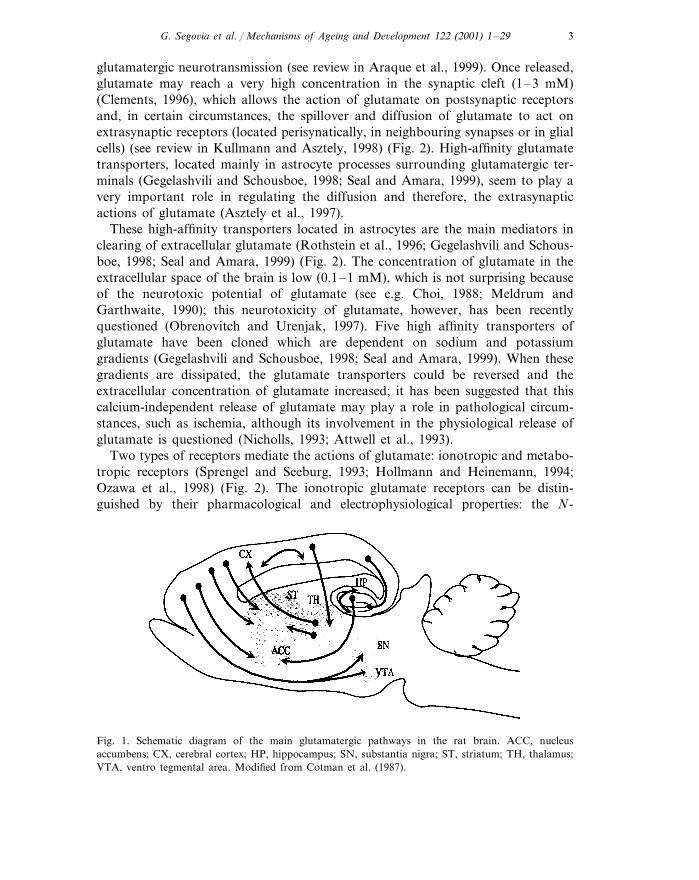
Glutamate is the neurotransmitter that acts in most of the excitatory synapses inthe mammalian central nervous system (CNS) (Fonnum, 1984; Orrego and Vil-lanueva, 1993). Glutamatergic neurons are widely distributed through the CNS,mainly in the forebrain, where most of the cortical projections contain glutamate(Fagg and Foster, 1983; Cotman et al., 1987). In particular, the cortico-striatal andthe cortico-cortical (hipoccampus, commisure) pathways have been extensivelystudied (Fagg and Foster, 1983; Peinado and Mora, 1986; Cotman et al., 1987)(Fig. 1).
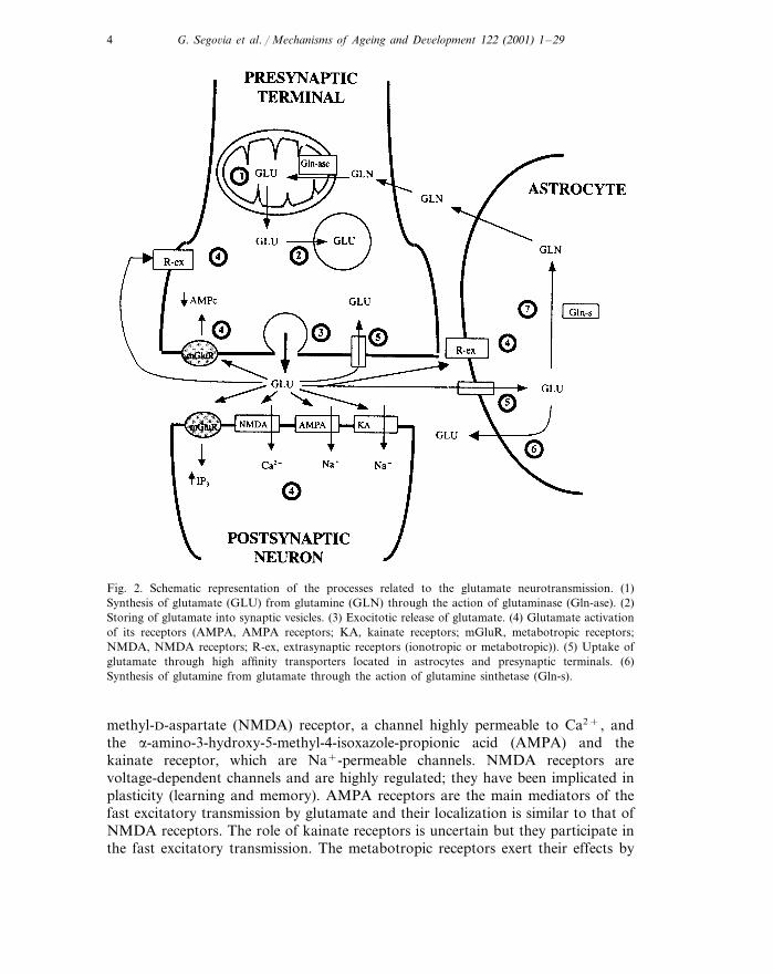
The metabolism of glutamate in the brain is separated into two compartments:neuronal and glial (Fig. 2). The synthesis of glutamate occurs mainly fromglutamine through the action of glutaminase, which is localized in the mitocondriaof glutamatergic terminals (Fonnum, 1993). Regarding the catabolism of glutamate,the glial enzyme glutamine synthetase converts glutamate to glutamine (Norenbergand Martınez-Hernandez, 1979; Fonnum, 1993). Thus, glutamate released fromneurons is transported to glial cells where it is converted into glutamine, which, inturn, diffuses through the extracellular space into neurons to be used for thesynthesis of glutamate.
Exocitotic calcium-dependent release of glutamate has been demonstrated in bothin vitro and in vivo preparations (Fonnum, 1984; Nicholls, 1993; Sanchez-Prieto etal., 1994; Zilkha et al., 1995) (Fig. 2). Recent studies have shown that, as well asneurons, astrocytes can release glutamate and therefore, be actively involved in
G. Sego6ia et al. / Mechanisms of Ageing and De6elopment 122 (2001) 1–29 3
glutamatergic neurotransmission (see review in Araque et al., 1999). Once released,glutamate may reach a very high concentration in the synaptic cleft (1–3 mM)(Clements, 1996), which allows the action of glutamate on postsynaptic receptorsand, in certain circumstances, the spillover and diffusion of glutamate to act onextrasynaptic receptors (located perisynatically, in neighbouring synapses or in glialcells) (see review in Kullmann and Asztely, 1998) (Fig. 2). High-affinity glutamatetransporters, located mainly in astrocyte processes surrounding glutamatergic ter-minals (Gegelashvili and Schousboe, 1998; Seal and Amara, 1999), seem to play avery important role in regulating the diffusion and therefore, the extrasynapticactions of glutamate (Asztely et al., 1997).
These high-affinity transporters located in astrocytes are the main mediators inclearing of extracellular glutamate (Rothstein et al., 1996; Gegelashvili and Schous-boe, 1998; Seal and Amara, 1999) (Fig. 2). The concentration of glutamate in theextracellular space of the brain is low (0.1–1 mM), which is not surprising becauseof the neurotoxic potential of glutamate (see e.g. Choi, 1988; Meldrum andGarthwaite, 1990); this neurotoxicity of glutamate, however, has been recentlyquestioned (Obrenovitch and Urenjak, 1997). Five high affinity transporters ofglutamate have been cloned which are dependent on sodium and potassiumgradients (Gegelashvili and Schousboe, 1998; Seal and Amara, 1999). When thesegradients are dissipated, the glutamate transporters could be reversed and theextracellular concentration of glutamate increased; it has been suggested that thiscalcium-independent release of glutamate may play a role in pathological circum-stances, such as ischemia, although its involvement in the physiological release ofglutamate is questioned (Nicholls, 1993; Attwell et al., 1993).
Two types of receptors mediate the actions of glutamate: ionotropic and metabo-tropic receptors (Sprengel and Seeburg, 1993; Hollmann and Heinemann, 1994;Ozawa et al., 1998) (Fig. 2). The ionotropic glutamate receptors can be distin-guished by their pharmacological and electrophysiological properties: the N-
Fig. 1. Schematic diagram of the main glutamatergic pathways in the rat brain. ACC, nucleusaccumbens; CX, cerebral cortex; HP, hippocampus; SN, substantia nigra; ST, striatum; TH, thalamus;VTA, ventro tegmental area. Modified from Cotman et al. (1987).
G. Sego6ia et al. / Mechanisms of Ageing and De6elopment 122 (2001) 1–294
Fig. 2. Schematic representation of the processes related to the glutamate neurotransmission. (1)Synthesis of glutamate (GLU) from glutamine (GLN) through the action of glutaminase (Gln-ase). (2)Storing of glutamate into synaptic vesicles. (3) Exocitotic release of glutamate. (4) Glutamate activationof its receptors (AMPA, AMPA receptors; KA, kainate receptors; mGluR, metabotropic receptors;NMDA, NMDA receptors; R-ex, extrasynaptic receptors (ionotropic or metabotropic)). (5) Uptake ofglutamate through high affinity transporters located in astrocytes and presynaptic terminals. (6)Synthesis of glutamine from glutamate through the action of glutamine sinthetase (Gln-s).
methyl-D-aspartate (NMDA) receptor, a channel highly permeable to Ca2+, andthe a-amino-3-hydroxy-5-methyl-4-isoxazole-propionic acid (AMPA) and thekainate receptor, which are Na+-permeable channels. NMDA receptors arevoltage-dependent channels and are highly regulated; they have been implicated inplasticity (learning and memory). AMPA receptors are the main mediators of thefast excitatory transmission by glutamate and their localization is similar to that ofNMDA receptors. The role of kainate receptors is uncertain but they participate inthe fast excitatory transmission. The metabotropic receptors exert their effects by
G. Sego6ia et al. / Mechanisms of Ageing and De6elopment 122 (2001) 1–29 5
means of G-protein-initiated biochemical events. Eight subtypes of metabotropicreceptors have been cloned, which mediate both excitatory and inhibitory actions ofglutamate (Ozawa et al., 1998).
3. Aging of brain glutamatergic system
It has been suggested that glutamate is involved in a number of cerebralfunctions which become altered with age, such as learning and memory (see e.g.Collindridge and Bliss, 1987; Cotman et al., 1988), emotion and motivation (Moraand Cobo, 1990; Cobo and Mora, 1991) and motor functions (Schmidt et al., 1992).Glutamate seems to be involved in the pathogenesis of several age-related neurode-generative disorders including Huntington’s, Parkinson’s and Alzheimer’s diseases(see Choi, 1988; Greenamyre and Young, 1989; Meldrum and Garthwaite, 1990;Obrenovitch and Urenjak, 1997 for reviews). An increased susceptibility to gluta-mate induced toxicity with age has also been described (Liu et al., 1996; Brewer,1998), which may be due in part to a reduction in the concentration of antioxidants(Vatassery et al., 1998; Lynch, 1998).
Studies on the aging of the brain are complicated by several confounding factors.The most evident are those related to species, strains and individual variability ofthe animals. For example, several reports have shown an age-related loss of neuronsin various cerebral areas in human and monkey, but not in the rodent (Colemanand Flood, 1987). Also, differences between strains in how a particular parameter,such as NMDA receptor density, undergoes a change with age have been described(Petersen and Cotman, 1989). This variability is mainly due to the fact thatchronological age is not the best indicator of the aging process and no biologicalmarkers are available to determine the onset of aging. So, in this review, to avoidinterspecies variability and since research on brain aging has been carried outmainly in rodents, the results reported in the scientific literature obtained withrodent brains are considered in the light of the limitations mentioned above.
Two methodological factors should be underlined. First, erroneous conclusionscould be drawn from studies that examined only two groups of age; studies onaging should be performed using animals of several ages (Coleman and Flood,1987). This recommendation circumvents the problem of differentiating maturationand aging processes. Secondly, the brain is a heterogeneous organ with anatomi-cally and physiologically different areas which may be affected in different mannersby the aging process. Studies examining the whole brain or large cerebral regions,which include a number of heterogeneous areas, can complicate the interpretationof data. All these factors are taken into account in the present review.
3.1. Brain glutamate content
Initial studies of the effects of aging on glutamatergic neurotransmission investi-gated the content of glutamate in cerebral tissue samples from both origin anddestination areas of the glutamatergic pathways. Table 1 summarizes available datafor changes in glutamate content in brain tissue during aging.
G. Sego6ia et al. / Mechanisms of Ageing and De6elopment 122 (2001) 1–296
Several reports have shown a decrease in glutamate concentration in tissuesamples taken from the whole cerebral cortex of rodents as a result of age (Davisand Himwich, 1975; Strolin-Benedetti et al., 1990, 1991; Saransaari and Oja, 1995).The cerebral cortex is a heterogeneous structure and age-related changes in gluta-mate content may occur at different rates in different areas. In fact, studiesperformed in tissue samples of discrete areas of the cerebral cortex of Fischer-344and Wistar rats show significant decreases in glutamate concentration in the frontalregion, by 15–20% at 21–24 months (Fornieles et al., 1986; Dawson et al., 1989;Wallace and Dawson, 1990) but not in other areas, such as the parietal andtemporal cortices (Fornieles et al., 1986; Banay-Schwartz et al., 1989). It isinteresting that differences seem to exist between frontal areas. For instance, anage-related decrease in glutamate has been reported in the medial prefrontal cortexbut not in the sulcal or dorsal prefrontal cortex (Fornieles et al., 1986). In contrast,no significant changes in frontal glutamate content have been reported in other ratstrains (e.g. Long–Evans rats) in this same area of the brain (Dawson and Wallace,1992). Since Long–Evans rats seem to have a longer life span (Coleman and Flood,1987), it is possible that decreases could be found in glutamate in the frontal cortexof this rat strain at older ages.
In the hippocampus of mice, Fisher-344 and Wistar rats, the glutamate contentin tissue samples seems to decrease with age (Banay-Schwartz et al., 1989; Strolin-Benedetti et al., 1990, 1991; Saransaari and Oja, 1995). It is interesting that thesedecreases in glutamate content have been consistently found at 12, 18, 21–24 and29 months, reaching 6–8% in 21 months rats (Strolin-Benedetti et al., 1990, 1991)and 23% in 29 months rats (Banay-Schwartz et al., 1989), whereas no changes inglutamate with age have been reported in the hippocampus of Long–Evans rats(Dawson and Wallace, 1992).
The decrease in glutamate content in the frontal cortex is in agreement with theneuronal loss described in this area of the brain (Mufson and Stein, 1980) as wellas in the cerebral cortex of aged rodents (Knox, 1982; Heumann and Leuba, 1983;Peters et al., 1987). The decrease in the glutamate content of the hippocampus alsoagrees well with the age-related decrease in neuronal density reported in the CA1and CA3 areas (Brizzee and Ordy, 1979; Landfield et al., 1981). Since glutamate isthe neurotransmitter of most cortical and hippocampal neurons (Fonnum, 1984),the glutamate decrease in tissue samples from these structures could be interpretedas a reflection of neuronal loss. Metabolic deficits in biochemical pathways thatgenerate or utilize glutamate as a substrate could also contribute to these decreases.For example, changes in the regulation of glutaminase, the enzyme responsible forthe hydrolysis of glutamine to form glutamate and ammonia, have been describedin certain areas of the aged brain (Wallace and Dawson, 1992, 1993).
In rodent striatum, controversial results have been reported: a decrease of 15%(Strolin-Benedetti et al., 1990, 1991), no changes (Wallace and Dawson, 1990;Dawson and Wallace, 1992; Saransaari and Oja, 1995) and even increases (Don-zanti and Ung, 1990) in glutamate content with age. Since the striatum is aheterogeneous structure, these results could be related to the anatomical andneurochemical differences within this structure. In fact, in aged rats, Donzanti and

G.
Sego6ia
etal./
Mechanism
sof
Ageing
andD
e6elopment
122(2001)
1–
297
Table 1Changes in glutamate content in brain tissue during aging
ReferencesAnimals Age (months) Changes UnitsBrain area
¡9%, PB0.05 mmoles/g wet tissue3 vs. 21–22 (Strolin-Benedetti et al., 1990)Wistar ratsWhole cerebral cortexmmoles/g wet tissue (Strolin-Benedetti et al., 1991)Whole cerebral cortex Wistar rats 3 vs. 21–22 ¡10%, PB0.05
Whole cerebral cortex (Saransaari and Oja, 1995)mmoles/kg proteinNMRI mice ¡:30% at 12 months3, 6, 12, 18, 24¡:30% at 18 months¡:45% at 24 months,PB0.01
¡16%, PB0.01Fischer-344 rats mmoles/g wet tissueFrontal cortex 6 vs. 24 (Dawson et al., 1989)Fischer-344 rats (Wallace and Dawson, 1990)6 vs. 24 ¡18%, PB0.05Frontal cortex mmoles/g wet tissue
ng/mg protein (Donzanti and Ung, 1990)Medial prefrontal cortex Fischer-344 rats 6 vs. 20 Not significantWistar rats mmoles/g wet tissue (Fornieles et al., 1986)Medial prefrontal cortex 2, 12, 21 ¡9% at 12 months,
PB0.05¡17% at 21 months,PB0.05mmoles/g wet tissue (Fornieles et al., 1986)Wistar ratsDorsal prefrontal cortex 2, 12, 21 Not significant
(Fornieles et al., 1986)mmoles/g wet tissueWistar rats Not significant2, 12, 21Sulcal prefrontal cortex
Wistar rats (Fornieles et al., 1986)2, 12, 21 Not significantTemporal cortex mmoles/g wet tissue
nmoles/mg protein (Banay-Schwartz et al., 1989)Occipital cortex Fischer-344 rats 3 vs. 29 Not significant
mmoles/g wet tissue (Strolin-Benedetti et al., 1990)3 vs. 21–22Hippocampus ¡6%, PB0.05Wistar ratsHippocampus (Strolin-Benedetti et al., 1991)mmoles/g wet tissueWistar rats ¡8%, PB0.053 vs. 21–22
(Saransaari and Oja, 1995)NMRI mice 3, 6, 12, 18, 24 ¡:20% at 12 months mmoles/kg proteinHippocampus¡:30% at 18 months¡:15% at 24 months
(Banay-Schwartz et al., 1989)Fischer-344 rats 3 vs. 29 ¡23%, PB0.02 nmoles/mg proteinHippocampusHippocampus Fischer-344 rats 6 vs. 24 Not significant mmoles/g wet tissue (Wallace and Dawson, 1990)
(Price et al., 1981)nmoles/mg protein¡17–20%, PB0.0013, 4, 6, 10, 19Striatum Sprague–Dawleyrats

G.
Sego6ia
etal./
Mechanism
sof
Ageing
andD
e6elopment
122(2001)
1–
298
Table 1 (Continued)
ReferencesAnimals Age (months) Changes UnitsBrain area
¡14%, PB0.01 mmoles/g wet tissue3 vs. 21–22 (Strolin-Benedetti et al., 1990)Wistar ratsStriatumWistar rats (Strolin-Benedetti et al., 1991)3 vs. 21–22 ¡15%, PB0.01Striatum mmoles/g wet tissueNMRI mice (Saransaari and Oja, 1995)3, 6, 12, 18, 24 Not significantStriatum mmoles/kg protein
(Wallace and Dawson, 1990)Fischer-344 rats 6 vs. 21 Not significant mmoles/g wet tissueStriatum
ng/mg protein (Donzanti and Ung, 1990)Fischer-344 ratsStriatum anterior dorso medial 6 vs. 20 Not significantng/mg protein (Donzanti and Ung, 1990)Striatum anterior dorso lateral 6 vs. 20Fischer-344 rats 40%, PB0.05
Striatum anterior ventro lateral (Donzanti and Ung, 1990)ng/mg proteinFischer-344 rats 21%, PB0.056 vs. 20(Donzanti and Ung, 1990)Fischer-344 rats 6 vs. 20 Not significant ng/mg proteinStriatum medial dorso medial(Donzanti and Ung, 1990)Fischer-344 rats 6 vs. 20 35%, PB0.05 ng/mg proteinStriatum medial dorso lateral
ng/mg protein (Donzanti and Ung, 1990) 24%, PB0.05Striatum medial ventro lateral 6 vs. 20Fischer-344 ratsNot significant ng/mg protein (Donzanti and Ung, 1990)Striatum posterior dorso Fischer-344 rats 6 vs. 20
medial(Donzanti and Ung, 1990)Fischer-344 rats ng/mg protein 18%, PB0.05Striatum posterior ventro 6 vs. 20
lateral
G. Sego6ia et al. / Mechanisms of Ageing and De6elopment 122 (2001) 1–29 9
Ung reported increases of 20–40% in the glutamate content in several striatalsubregions (dorsolateral and ventrolateral regions), but not in others (dorsomedialregions) (Donzanti and Ung, 1990).
Few studies have investigated the effects of age on glutamate content in othersubcortical structures. In the nucleus accumbens, both decreases and no changeshave been described (Donzanti and Ung, 1990; Strolin-Benedetti et al., 1990, 1991).In the substantia nigra, decreases (Strolin-Benedetti et al., 1990, 1991), no changes(Banay-Schwartz et al., 1989) and increases (Donzanti and Ung, 1990) in glutamatecontent have been reported.
In brief, the most consistent finding on the effects of aging on glutamate contentin tissue from different brain areas is a decrease in prefrontal cortex and inhippocampus. It has been suggested that these data are related to the cognitive andmemory deficits reported to occur with age and attributed, at least in part, tofunctions of the prefrontal cortex and hippocampus. However, since 70–80% oftissue glutamate is present in the metabolic pool and only 20–30% in glutamatergicnerve terminals (Fonnum, 1993), decreases in glutamate content may be mainly aconsequence of a change in metabolic activity. Whether or not these decreases inglutamate content in prefrontal cortex and hippocampus are involved in be-havioural deficits remains to be elucidated.
To investigate whether the effects of age on glutamatergic neurotransmissionoccur in a given area of the brain, it is necessary to study both presynaptic andpostsynaptic markers, such as release, number of high-affinity uptake sites, trans-port or receptor affinity, receptor density or postsynaptic effects, in cerebralstructures containing glutamatergic terminals. Studies of the effects of age on theseglutamatergic markers are reviewed below.
3.2. Glutamate release
Neurotransmitter release can be investigated using both in vitro and in vivotechniques. The in vitro studies utilize brain slices or isolated nerve terminals(synaptosomes) and analyze the content of glutamate in the superfusion medium.These techniques provide the simplest model to investigate neurotransmitter release(Nicholls, 1993; Sanchez-Prieto et al., 1994). In contrast, in vivo studies usingpush–pull or microdialysis techniques, analyze the concentration of glutamate insamples obtained from the extracellular space of a specific area of the brain(Benveniste, 1989; Westerink and Justice, 1991; Porras and Mora, 1995; Segovia etal., 1997). Data on changes in basal and induced glutamate release during aging aresummarized in Tables 2 and 3, respectively.
In the cerebral cortex, most of the in vitro and in vivo studies have shown nosignificant changes in basal extracellular concentrations of glutamate during aging.This remarkable stability of glutamate concentration seems to occur independentlyof the areas (frontal, parietal and occipital cortices), species (rat and mouse) andstrains (Wistar, Long–Evans and Fischer-344 rats) (Dawson et al., 1989; Mora andCobo, 1991; Cobo et al., 1992 Dawson and Wallace, 1992; Palmer et al., 1994;Saransaari and Oja, 1995; Porras et al., 1997). Also, the release of glutamate in the

G.
Sego6ia
etal./
Mechanism
sof
Ageing
andD
e6elopment
122(2001)
1–
2910
Table 2Changes in basal glutamate release during aging
ReferencesAnimals Age (months) Changes TechniqueBrain area
Not significant In vitro (slices)6 vs. 28 (Dawson and Wallace, 1992)Fisher-344 ratsWhole cerebral cortex(Saransaari and Oja, 1994)NMRI mice 3, 6, 12, 18, 24 Not significant In vitro (slices)Whole cerebral cortex
NMRI mice 3 vs. 24 (Saransaari and Oja, 1995)Not significant In vitro (slices)Whole cerebral cortex
(Palmer et al., 1994)Hybrid rats 3, 12, 24, 37 Not significant In vitro (slices)Neocortex
In vitro (slices) (Dawson et al., 1989)Fischer-344 ratsFrontal cortex 6 vs. 24 Not significant
In vivo (push–pull) (Cobo et al., 1992)Wistar ratsSulcal prefrontal cortex 3–4 vs. 24–26 Not significant
Wistar rats In vivo (push–pull)Not significantMedial prefrontal cortex 3–4 vs. 24–26 (Cobo et al., 1992)Wistar rats (Cobo et al., 1993)3–4 vs. 28–30 Not significantMedial prefrontal cortex In vivo (push–pull)
In vivo (push–pull) (Porras et al., 1997)Wistar ratsMedial prefrontal cortex 2–3, 11–14, 24–26 Not significant
(Cobo et al., 1992)Wistar rats 3–4 vs. 24–26 Not significant In vivo (push–pull)Parieto-temporal cortex
8 vs. 28–30 In vitro (slices) (Meldrum et al., 1992)Temporal cortex Fischer-344 rats Not significant
Wistar rats (Cobo et al., 1992)3–4 vs. 24–26 Not significantOccipital cortex In vivo (push–pull)
In vitro (slices) (Freeman and Gibson, 1987)Hippocampus Balb/c mice 3 vs. 30 94%, PB0.05In vitro (slices) (Saransaari and Oja, 1995)Hippocampus ¡:60%NMRI mice 3 vs. 24
(Meldrum et al., 1992)Fischer-344 rats 8 vs. 28–30 Not significant In vitro (slices)HippocampusHippocampus (Massieu and Tapia, 1997)3 vs. 22–24 :68%,Wistar rats In vivo (microdialysis)
PB0.05
77%, PB0.053 vs. 30 In vitro (slices)Balb/c miceStriatum (Freeman and Gibson, 1987)In vitro (slices) (Saransaari and Oja, 1995)Striatum NMRI mice 3 vs. 24 Not significant
Striatum (Porras and Mora, 1995)In vivo (push–pull)Wistar rats Not significant2–3, 12–13, 24–34(Corsi et al., 1997)Wistar rats 3, 12, 22 ¡40%, PB0.05 In vivo (microdialysis)Striatum
3 vs. 22–24 In vivo (microdialysis) (Massieu and Tapia, 1997)Striatum :190%,Wistar ratsPB0.05
3 vs. 22 In vivo (microdialysis)Wistar ratsStriatum Not significant (Corsi et al., 1999)(Segovia et al., 1999a)Wistar rats 2–3, 12–14, 27–32, 37 Not significant In vivo (microdialysis)Striatum
In vivo (microdialysis) (Donzanti et al., 1993)Lateral striatum Fischer-344 rats 4, 12, 18, 24–26 In vivo (microdialysis) (Donzanti et al., 1993)Medial striatum 4, 12, 18, 24–26Fischer-344 rats Not significant
Wistar rats (Segovia et al., 1999a)In vivo (microdialysis)Not significantNucleus accumbens 2–3, 12–14, 27–32, 37

G.
Sego6ia
etal./
Mechanism
sof
Ageing
andD
e6elopment
122(2001)
1–
2911
Table 3Changes in induced glutamate release during aging
StimulationAnimals ReferencesAge (months) Changes TechniqueBrain area
K+ 50 mM (Saransaari and Oja, 1995) :50% In vitro (slices)3 vs. 24NMRI miceWhole cerebralcortex
Not significant (Sanchez-Prieto et al., 1994)In vitro3 vs. 27–30Whole cerebral Wistar rats 4-aminopyridine 1cortex mM(synaptosomes)
(Palmer et al., 1994)K+ 50 mMIn vitro (slices)Neocortex Not significantHybrid rats 3, 12–24, 37
Fischer-344 rats K+ 56 mM (Dawson et al., 1989)6 vs. 24 Not significantFrontal cortex In vitro (slices)
Electrical (Cobo et al., 1993)In vivo (push–pull)¡:50%,Medial prefrontal 3–4 vs. 28–30Wistar ratscortex PB0.05
In vitro (slices)8 vs. 28–30 Electrical (Meldrum et al., 1992) :2-fold,Fischer-344 ratsTemporal cortexPB0.05
(Freeman and Gibson, 1987)In vitro (slices)¡:80%3 vs. 30 K+Hippocampus Balb/c miceK+ 50 mM (Saransaari and Oja, 1995)Hippocampus 3 vs. 24 :5-fold,NMRI mice In vitro (slices)
PB0.01Electrical (Meldrum et al., 1992) :2-fold, In vitro (slices)8 vs. 28–30Fischer-344 ratsHippocampus
PB0.05
(Freeman and Gibson, 1987)In vitro (slices)¡66%, PB0.05 K+3 vs. 30Balb/c miceStriatumNMRI mice In vitro (slices) K+ 50 mM (Saransaari and Oja, 1995)Striatum 3, 6, 12, 18, 24 ¡:30%,
PB0.014-Aminopyridine 1Striatum (Sanchez-Prieto et al., 1994)In vitroNot significant3 vs. 27–30Wistar rats
(synaptosomes) mMIn vivo3, 12, 22 K+ 100 mM (Corsi et al., 1997, 1999)Striatum Not significantWistar rats(microdialysis)

G. Sego6ia et al. / Mechanisms of Ageing and De6elopment 122 (2001) 1–2912
whole cerebral cortex induced by depolarizing agents, such as K+ or 4-aminopy-ridine, seems to be unchanged with age (Dawson et al., 1989; Palmer et al., 1994;Sanchez-Prieto et al., 1994). Only one study in vitro has reported an increase in therelease of glutamate induced by K+ up to 50% in aged versus young mice(Saransaari and Oja, 1995).
As shown above, extracellular levels of glutamate did not change in the frontalcortex during aging despite neuronal loss, which suggests that functional compensa-tions are made by surviving neurons (Dawson et al., 1989; Mora and Cobo, 1991;Cobo et al., 1992; Porras et al., 1997). However, a study from our laboratoryshowed that electrical stimulation at intensities that release glutamate in young ratsdoes not increase glutamate in aged rats although the basal levels were similar inboth age groups (Cobo et al., 1993). This change could be due to a shift in theexcitability of the prefrontal cortex in old animals that could make the stimulationintensity use a sub-threshold for influencing release. This is likely, since an increasein the release of glutamate in the prefrontal cortex of aged rats was obtained withan increase in the intensity of electrical stimulation. In any case, the study of Coboet al. suggests that neuronal changes, such as a decrease in excitability, do occurwith age in the prefrontal cortex of the rat (Cobo et al., 1993).
In the hippocampus, basal extracellular concentrations of glutamate in agedrodents have been reported to be 94% greater (Freeman and Gibson, 1987), equal(Meldrum et al., 1992) or 60% lower (Saransaari and Oja, 1995) compared withthose of adult rodents in in vitro studies. An in vivo study has reported an increaseof basal glutamate release in 24-month-old as compared with 3-month-old Wistarrats (Massieu and Tapia, 1997). In potassium induced release of glutamate in agedanimals there are reports of no change (Freeman and Gibson, 1987) and ofincreases by more than double (Meldrum et al., 1992; Saransaari and Oja, 1995).
In striatum, in vitro and in vivo studies have shown no changes in basal (Porrasand Mora, 1995; Saransaari and Oja, 1995; Corsi et al., 1999; Segovia et al., 1999a)or chemically induced (Donzanti et al., 1993; Sanchez-Prieto et al., 1994; Corsi etal., 1997, 1999) extracellular concentrations of glutamate in aged rats. In contrast,there have been reports of both increases and decreases in the basal release ofglutamate in the striatum (Freeman and Gibson, 1987; Massieu and Tapia, 1997;Corsi et al., 1997) and also a decrease in potassium induced relase of glutamate(Freeman and Gibson, 1987; Saransaari and Oja, 1995). Interestingly, one studydescribed regional differences in age-related changes in basal extracellular concen-trations of glutamate in striatum: an age-related increase in the extracellularconcentration of glutamate in the lateral, but not the medial striatum of rats(Donzanti et al., 1993). It is possible that an actual increase in glutamate concentra-tions in the lateral striatum could be hidden in the studies of the whole striatum.These considerations are reinforced by the recent data of dopamine and GABAobtained in the striatum using microdialysis, in which a clear regional differencebetween dorsal and ventral striatum was found (Segovia et al., 1999a; Segovia,1999).
To sum up, it might be said from the data revised above — in particular dataderived from synaptosomes studies — that no changes in glutamate release seem to
G. Sego6ia et al. / Mechanisms of Ageing and De6elopment 122 (2001) 1–29 13
occur during aging in cerebral cortex and striatum. However, the concentration ofglutamate sampled in most of these studies (slices, push–pull, microdialysis) is theresult of a balance between the release and uptake processes, so age-relateddecreases in presynaptic release of glutamate may be compensated by changes inuptake. On the other hand, push–pull and microdialysis techniques cannot differ-entiate between neuronal or glial sources of glutamate, so the cellular origin of theglutamate released is also uncertain (Timmerman and Westerink, 1997). Thus,decreases in glutamate release from the neuronal compartment could be masked byincreases of glutamate release from non-neuronal sources.
3.3. Glutamate uptake
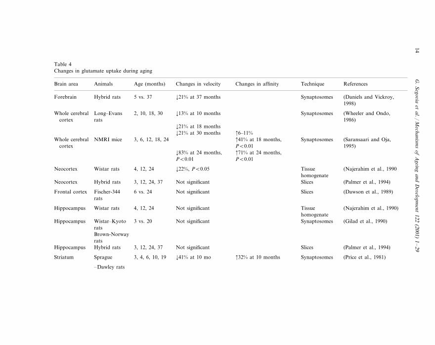
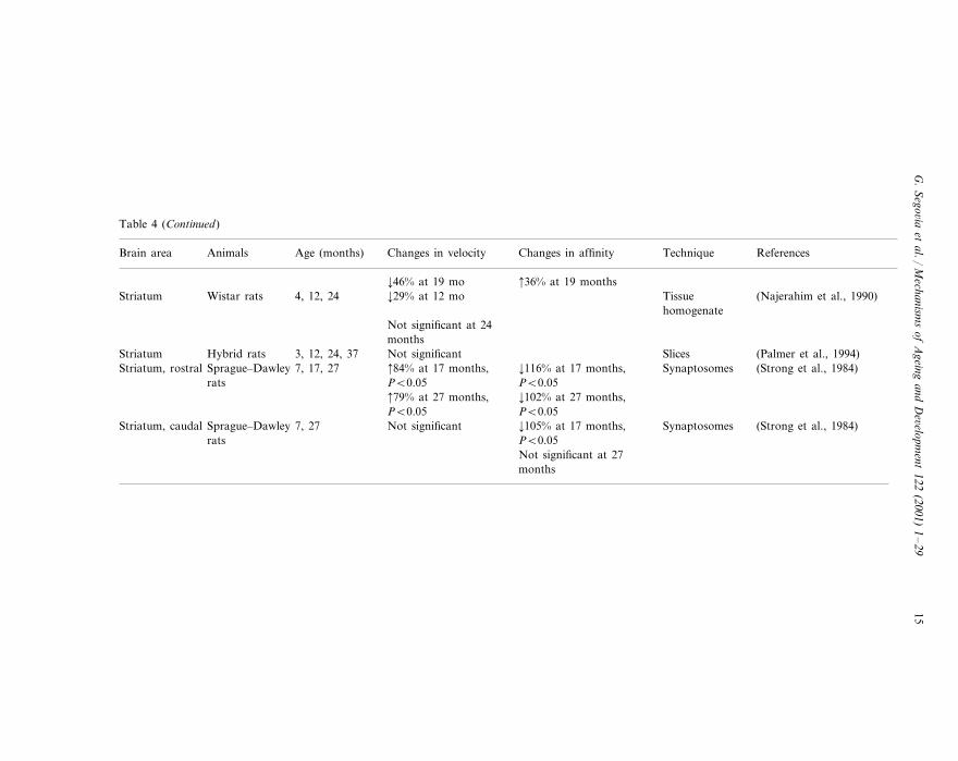
The main mechanism for clearing extracellular glutamate is the action of high-affinity transporters located mainly in astrocyte processes surrounding glutamater-gic terminals (Rothstein et al., 1996; Gegelashvili and Schousboe, 1998). Table 4summarizes data on changes in glutamate uptake during aging.
In the cerebral cortex and striatum, studies show a reduced ability to take upglutamate by aged rats (Wheeler and Ondo, 1986; Najerahim et al., 1990;Saransaari and Oja, 1995; Vatassery et al., 1998). The magnitude of this decrease inglutamate uptake with age has been averaged as 20–30% in rats at 24 months ofage. In mice, this decrease has been estimated at 70–80%. In agreement with thesefindings, changes in the modulation by protein kinase C of the uptake of glutamatehave been also reported (Daniels and Vickroy, 1998). In contrast, other studieshave reported increases (Strong et al., 1984) or no changes (Dawson et al., 1989;Palmer et al., 1994) in glutamate uptake.
Several reports indicate a decrease in the maximal velocity of glutamate uptake(Price et al., 1981; Wheeler and Ondo, 1986). This could be interpreted as a loss inthe number of high-affinity glutamate transport sites in these brain areas. Thehippocampus exhibits a consistently unaltered uptake of glutamate during aging(Gilad et al., 1990; Najerahim et al., 1990; Palmer et al., 1994).
As shown above, very few studies have addressed the effects of aging onglutamate uptake, but most reported a lower uptake capacity for glutamate as wellas a loss in the number of high affinity glutamate transport sites in the glutamater-gic terminals of aged rats in both striatum and prefrontal cortex (Price et al., 1981;Wheeler and Ondo, 1986; Wheeler and Ondo, 1991; Najerahim et al., 1990;Vatassery et al., 1998; Saransaari and Oja, 1995). This conclusion should be drawnwith caution when referred to its functional significance. That is, a decrease in thenumber of transport sites in nerve terminals could be compensated by increases inthe affinity of the remaining sites (Price et al., 1981; Saransaari and Oja, 1995).Moreover, glutamate is taken up mainly into glial cells (Rothstein et al., 1996;Gegelashvili and Schousboe, 1998) and age-related changes in the uptake ofglutamate seem to occur mainly in neurons (Daniels and Vickroy, 1998). Sinceastrocytes seem to increase in number and/or activity in the aging brain (Brizzee etal., 1983; Terry, 1986; Vazquez et al., 1992; David et al., 1997), this couldcompensate for decreases in neuronal glutamate uptake. In fact, one study using
G.
Sego6ia
etal./
Mechanism
sof
Ageing
andD
e6elopment
122(2001)
1–
2914
Table 4Changes in glutamate uptake during aging
Changes in affinity Technique ReferencesBrain area Age (months)Animals Changes in velocity
Synaptosomes (Daniels and Vickroy,5 vs. 37Forebrain ¡21% at 37 monthsHybrid rats1998)
Synaptosomes2, 10, 18, 30 (Wheeler and Ondo,¡13% at 10 monthsLong–EvansWhole cerebralcortex 1986)rats
¡21% at 18 months 6–11%¡21% at 30 months 41% at 18 months,NMRI mice 3, 6, 12, 18, 24Whole cerebral Synaptosomes (Saransaari and Oja,
1995)PB0.01cortex 71% at 24 months,¡83% at 24 months,
PB0.01 PB0.01
TissueNeocortex (Najerahim et al., 1990Wistar rats 4, 12, 24 ¡22%, PB0.05homogenate
(Palmer et al., 1994)Not significant Slices3, 12, 24, 37Neocortex Hybrid rats
Fischer-344 Slices (Dawson et al., 1989)6 vs. 24 Not significantFrontal cortexrats
Wistar rats Tissue4, 12, 24 (Najerahim et al., 1990)Not significantHippocampushomogenateSynaptosomes (Gilad et al., 1990)Not significant3 vs. 20Hippocampus Wistar–Kyoto
ratsBrown-Norwayrats
(Palmer et al., 1994)Not significant Slices3, 12, 24, 37Hippocampus Hybrid rats
32% at 10 monthsSprague Synaptosomes (Price et al., 1981)3, 4, 6, 10, 19 ¡41% at 10 moStriatum
–Dawley rats
G.
Sego6ia
etal./
Mechanism
sof
Ageing
andD
e6elopment
122(2001)
1–
2915
Table 4 (Continued)
Changes in affinity Technique ReferencesBrain area Age (months)Animals Changes in velocity
¡46% at 19 mo 36% at 19 months¡29% at 12 mo (Najerahim et al., 1990)TissueStriatum 4, 12, 24Wistar rats
homogenateNot significant at 24months
SlicesHybrid rats (Palmer et al., 1994)3, 12, 24, 37 Not significantStriatum¡116% at 17 months, 84% at 17 months, Synaptosomes (Strong et al., 1984)Striatum, rostral Sprague–Dawley 7, 17, 27PB0.05PB0.05rats¡102% at 27 months, 79% at 27 months,PB0.05PB0.05¡105% at 17 months,Sprague–Dawley 7, 27 Synaptosomes (Strong et al., 1984)Not significantStriatum, caudal
rats PB0.05Not significant at 27months
G. Sego6ia et al. / Mechanisms of Ageing and De6elopment 122 (2001) 1–2916
cortical slices, which include glial elements, reported no change in glutamate uptakewith age (Dawson et al., 1989).
3.4. Glutamate receptors
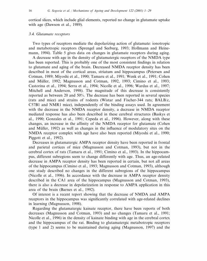
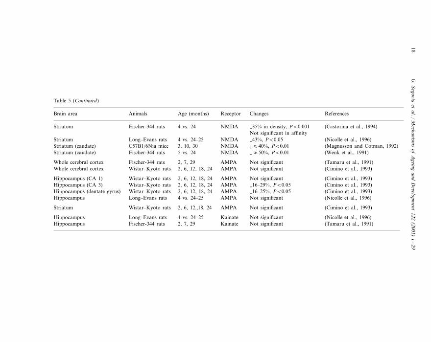
Two types of receptors mediate the depolarizing action of glutamate: ionotropicand metabotropic receptors (Sprengel and Seeburg, 1993; Hollmann and Heine-mann, 1994). Table 5 gives data on changes in glutamate receptors during aging.
A decrease with age in the density of glutamatergic receptors of the NMDA typehas been reported. This is probably one of the most consistent findings in relationto glutamate and aging of the brain. Decreased NMDA receptor density has beendescribed in most of the cortical areas, striatum and hippocampus (Petersen andCotman, 1989; Miyoshi et al., 1990; Tamaru et al., 1991; Wenk et al., 1991; Cohenand Muller, 1992; Magnusson and Cotman, 1992, 1993; Cimino et al., 1993;Castorina et al., 1994; Serra et al., 1994; Nicolle et al., 1996; Wardas et al., 1997;Mitchell and Anderson, 1998). The magnitude of this decrease is consistentlyreported as between 20 and 50%. The decrease has been reported in several species(rats and mice) and strains of rodents (Wistar and Fischer-344 rats; BALB/c,C57B1 and NMR1 mice), independently of the binding assays used. In agreementwith the decrease in the NMDA receptor density, a decrease in NMDA receptormediated response has also been described in these cerebral structures (Baskys etal., 1990; Gonzales et al., 1991; Cepeda et al., 1996). However, along with thesechanges, an increase in the affinity of the NMDA receptor for glutamate (Cohenand Muller, 1992) as well as changes in the influence of modulatory sites on theNMDA receptor complex with age have also been reported (Miyoshi et al., 1990;Piggott et al., 1992).
Decreases in glutamatergic AMPA receptor density have been reported in frontaland parietal cortices of mice (Magnusson and Cotman, 1993), but not in thecerebral cortex of rats (Tamaru et al., 1991; Cimino et al., 1993). In the hippocam-pus, different subregions seem to change differently with age. Thus, an age-relateddecrease in AMPA receptor density has been reported in certain, but not all areasof the hippocampus (Cimino et al., 1993; Magnusson and Cotman, 1993), althoughone study described no changes in the different subregions of the hippocampus(Nicolle et al., 1996). In accordance with the decrease in AMPA receptor densitydescribed in the CA1 area of the hippocampus (Magnusson and Cotman, 1993),there is also a decrease in depolarization in response to AMPA application in thisarea of the brain (Barnes et al., 1992).
Of interest is a recent report showing that the decrease of NMDA and AMPAreceptors in the hippocampus was significantly correlated with age-related declinesin learning (Magnusson, 1998).
Regarding the glutamatergic kainate receptor, there have been reports of bothdecreases (Magnusson and Cotman, 1993) and no changes (Tamaru et al., 1991;Nicolle et al., 1996) in the density of kainate binding with age in the cerebral cortexand the hippocampus of the rat. Binding to glutamatergic metabotropic receptors(type 1 and 2) seems to be maintained during aging (Magnusson, 1997) and the
G.
Sego6ia
etal./
Mechanism
sof
Ageing
andD
e6elopment
122(2001)
1–
2917
Table 5Changes in glutamate receptors during aging
Animals ReferencesAge (months) ReceptorBrain area Changes
NMDA ¡29% in density, PB0.012, 7, 29 (Tamaru et al., 1991)Fischer-344 ratsWhole cerebral cortexNot significant in affinity¡13–23%, PB0.052, 6, 12, 18, 24 (Cimino et al., 1993)Whole cerebral cortex NMDAWistar–Kyoto rats
NMR1 mice (Cohen and Muller, 1992)3 vs. 20 NMDAForebrain ¡33% in density, PB0.001 4-fold in affinity, PB0.01
¡:40%, PB0.01 (Wenk et al., 1991)Anterior cortex Fischer-344 rats 5 vs. 24 NMDA
(Wenk et al., 1991)¡:60%, PB0.015 vs. 24Fischer-344 rats NMDAPosterior cortex
Fischer-344 rats (Miyoshi et al., 1990)2, 5, 13, 21 NMDAFrontal cortex ¡60%, PB0.01¡:30%, PB0.01 (Magnusson and Cotman, 1992)Frontal cortex C57131/6Nia mice 3, 10, 30 NMDA
Balb/c mice (Magnusson and Cotman, 1993)3 vs. 30 NMDAFrontal cortex ¡Frontal cortex (Castorina et al., 1994)¡39% in density, PB0.01Fischer-344 rats NMDA4 vs. 24
Not significant in affinity
¡51%, PB0.01 (Miyoshi et al., 1990)Parietal cortex NMDAFischer-344 rats 2, 5, 13, 21(Magnusson and Cotman, 1992)C57B1/6Nia mice 3, 10, 30 NMDA ¡:35%, PB0.01Parietal cortex
Parietal cortex Balb/c mice 3 vs. 30 NMDA ¡ (Magnusson and Cotman, 1993)
(Magnusson and Cotman, 1992)Not significantNMDAOccipital-temporal cortex 3, 10, 30C57B1/6Nia mice
¡47–51% (Miyoshi et al., 1990)Hippocampus Fischer-344 rats 2, 5, 13, 21 NMDAHippocampus (Castorina et al., 1994)¡26% in density, PB0.001Fischer-344 rats NMDA4 vs. 24
Not significant in affinity(Magnusson and Cotman, 1992)C57B1/6Nia mice 3, 10, 30 NMDA ¡:10–20%, PB0.01Hippocampus
NMDA (Tamaru et al., 1991)¡24% in density, PB0.012, 7, 29Hippocampus Fischer-344 ratsNot significant in affinity¡:50%, PB0.01 (Wenk et al., 1991)Fischer-344 ratsHippocampus 5 vs. 24 NMDA
Hippocampus Long–Evans rats 4 vs. 24–25 NMDA Not significant (Nicolle et al., 1996)
(Miyoshi et al., 1990)¡67%, PB0.01Striatum Fischer-344 rats NMDA2, 5, 13, 21(Cimino et al., 1993)Wistar–Kyoto rats 2, 6, 12, 18, 24 NMDA ¡21–35%, PB0.01Striatum
G.
Sego6ia
etal./
Mechanism
sof
Ageing
andD
e6elopment
122(2001)
1–
2918
Table 5 (Continued)
Changes ReferencesBrain area Age (months)Animals Receptor
¡35% in density, PB0.001 (Castorina et al., 1994)Fischer-344 ratsStriatum 4 vs. 24 NMDANot significant in affinity¡43%, PB0.05 (Nicolle et al., 1996)Long–Evans ratsStriatum 4 vs. 24–25 NMDA
C57B1/6Nia mice (Magnusson and Cotman, 1992)3, 10, 30 NMDAStriatum (caudate) ¡:40%, PB0.01(Wenk et al., 1991)Fischer-344 rats 5 vs. 24 NMDA ¡:50%, PB0.01Striatum (caudate)
Not significant (Tamaru et al., 1991)Fischer-344 ratsWhole cerebral cortex 2, 7, 29 AMPANot significant (Cimino et al., 1993)2, 6, 12, 18, 24Whole cerebral cortex AMPAWistar–Kyoto rats
(Cimino et al., 1993)Not significantAMPAHippocampus (CA 1) 2, 6, 12, 18, 24Wistar–Kyoto ratsWistar–Kyoto rats (Cimino et al., 1993)2, 6, 12, 18, 24 AMPAHippocampus (CA 3) ¡16–29%, PB0.05
(Cimino et al., 1993)Wistar–Kyoto rats 2, 6, 12, 18, 24 AMPA ¡16–25%, PB0.05Hippocampus (dentate gyrus)Long–Evans rats Not significant (Nicolle et al., 1996)AMPAHippocampus 4 vs. 24–25
Not significant (Cimino et al., 1993)Striatum 2, 6, 12.,18, 24Wistar–Kyoto rats AMPA
(Nicolle et al., 1996)Not significantHippocampus 4 vs. 24–25Long–Evans rats Kainate(Tamaru et al., 1991)Fischer-344 rats 2, 7, 29 Kainate Not significantHippocampus
G. Sego6ia et al. / Mechanisms of Ageing and De6elopment 122 (2001) 1–29 19
responses mediated by these receptors remained unaltered in the hippocampus ofaged animals (Jouvenceau et al., 1997).
In contrast to the reported decrease in the density of NMDA and AMPAreceptors in cortex and hippocampus, several electrophysiological studies havedescribed no changes in neuronal sensitivity to iontophoretic applications ofglutamate with age in these areas of the brain (Lippa et al., 1981; Rao et al., 1993;Abdulla et al., 1995). This could be due to an increase of the affinity of glutamatefor its receptors that compensates for the decrease in the number of receptors. Instriatum, however, there are reports of a decrease of the neuronal responsiveness toglutamate (Cepeda and Levine, 1991; Cepeda et al., 1996).
3.5. Summary of the effects of aging on brain glutamatergic system
Several conclusions could be drawn from the data reviewed here on the effects ofaging on brain glutamatergic system. First, most of the in vitro and in vivo studieson basal and stimulated release of glutamate in the cerebral cortex, hippocampusand striatum show no changes during aging, which suggests that the potentiality ofglutamatergic terminals to release glutamate is unaltered. However, the stability ofthe release of glutamate should be interpreted with caution, since the concentrationof glutamate sampled in most of the studies (slices, push–pull, microdialysis) is theresult of a balance between the release and uptake processes. Thus, the reportedage-related decrease in the capacity of glutamate uptake in glutamatergic terminalsmay compensate for changes in glutamate release. Moreover, the increase in thenumber and/or activity of astrocytes during aging is a confounding factor whichcould obscure the significance of the changes in glutamate release and uptake.Second, a consistent decrease in the number of glutamatergic receptors, mainly ofthe NMDA type, occurs in most of the cerebral areas of aged rodents, themagnitude of this decrease being estimated at 20–50%. This effect of aging onglutamatergic receptors would suggest that changes occur in glutamate neurotrans-mission in the brain.
4. Interactions of glutamate with other neurotransmitter systems
Over 30 years ago, clinical data suggested the existence of a balance or interac-tion between dopamine and acetylcholine as the basis of striatal functions (Barbeau,1962), but this type of interaction was revised after the finding that glutamate alsointeracts with dopamine in this area of the brain (see e.g. Giorguieff et al., 1997).Since then, the concept of a complex reciprocal modulation of neural transmissionamong a number of neurotransmitters, including glutamate, dopamine, acetyl-choline, GABA and neuropeptides, has emerged to give a better understanding ofthe physiology of specific circuits of the brain (Di Chiara et al. 1994; Mora et al.,1999). Although the nature of these reciprocal interactions among neurotransmit-ters in areas of the brain, such as prefrontral cortex and striatum, is not wellunderstood, its dysfunction has been implied in drug addiction and in several
G. Sego6ia et al. / Mechanisms of Ageing and De6elopment 122 (2001) 1–2920
neurological disorders such as Parkinson’s disease and schizophrenia (Carlssonand Carlsson, 1990; Grace, 1991; Greenamyre, 1993; Del Arco et al., 1998,1999). Moreover, it has been suggested that a disruption in the fine adjustmentof these neurotransmitter interactions could be at the basis of the functionaldecreases found during aging of the brain (Mora, 1991).
In recent years, several in vitro and in vivo studies have reported a decreaseor lack of the modulation by different neurotransmitters of stimulated glutamaterelease in different areas of the brain during aging (Donzanti et al., 1993; Mur-ray et al., 1997; Corsi et al., 1997). Thus, Donzanti et al. reported that activa-tion of dopamine D2-receptor inhibits glutamate release evoked by potassium instriatal synaptosomes of young Fisher-344 rats. In contrast, this inhibition ofglutamate release was not evoked in 24 to 26-month-old rats (Donzanti et al.,1993). In line with these results are those reported by Murray et al., who alsoshowed than in old rats (22 months of age) there is a lack of inhibition byinterleukin-1 of potassium-evoked release of glutamate in hippocampal synapto-somes of young rats (Murray et al., 1997).
In an in vivo study in young Wistar rats, Corsi et al. (1997) reported thatpotassium-evoked glutamate release in the striatum was potentiated by anadenosine antagonist, effects which were no longer produced in old rats (22months) (Corsi et al., 1997). In contrast, these same authors showed that theeffects of an adenosine agonist on basal and potassium-evoked glutamate releasedid not change during aging (Corsi et al., 1999). An in vivo study in ourlaboratory reported the loss in old (24 months) Wistar rats of the ability ofmelatonin to evoke glutamate release when the dopamine system is activated(Exposito et al., 1995).
Other studies have evaluated the effects of aging on the responses induced byglutamate (Cepeda and Levine, 1991; Gonzales et al., 1991; Pazzagli et al., 1995;Cepeda et al., 1996). A reduction was reported in aged Fischer-344 rats of theability of dopamine to modulate striatal neuron responses induced by corticalstimulation in vivo (Cepeda and Levine, 1991) or activation of glutamatergicreceptors by NMDA or glutamate itself in vitro (Cepeda et al., 1996). Gonzaleset al. reported that dopamine release from striatal slices evoked by NMDA islower in aged (24–28 months) than in young (3–5 months) Fischer-344 rats(Gonzales et al., 1991). In contrast, activation of metabotropic receptors in vivoincreased dopamine release in prefrontal cortex of aged rats (24 months) but hadno effect on young rats (3 months) (Pintor et al., 1998). An in vivo study alsoreported that glutamate is responsible for much of the basal striatal adenosine inold (20–22 months) but not in young (3 months) Wistar rats (Pazzagli et al.,1995).
In a recent series of in vivo experiments, we investigated the effects of agingon the glutamate–dopamine interaction in striatum and prefrontal cortex ofWistar rats (Porras and Mora, 1995; Porras et al., 1997). Interaction of neuro-transmitters in striatum and prefrontal cortex has been the focus of intensiveresearch (see e.g. Exposito et al., 1994; Sanz et al., 1997; Segovia et al., 1997;Segovia and Mora, 1998, for reviews see Di Chiara et al., 1994; Lannes and

G. Sego6ia et al. / Mechanisms of Ageing and De6elopment 122 (2001) 1–29 21
Micheletti, 1994; Mora and Porras, 1994; Mora et al., 1999). These areas are ofrelevance for studies of brain aging due to their involvement in motor functionsand in cognition, emotion and motivation, all of which deteriorate with age (seee.g. Ingram, 1985; Murray and Waddington, 1991; Deptula et al., 1993). In ourstudies, the intracerebral perfusion of a dopamine agonist, apomorphine, pro-duced an increase of extracellular glutamate in striatum and prefrontal cortex ofyoung rats (2–3 months), whereas a decreased response of glutamate was ob-tained in middle-aged (12–14 months) and aged (24–26 months) rats, whichsuggests a deterioration in glutamate–dopamine interaction with age (Porras andMora, 1995; Porras et al., 1997). These data may be explained by the decrease inthe number of dopamine receptors or the alterations in dopamine receptor-medi-ated responses (Joyce et al., 1986; Hyttel, 1987; Han et al., 1989; Morelli et al.,1990; Murray and Waddington, 1991).
We have recently used an original approach to study in vivo the endogenousinteractions of glutamate with other neurotrasmitter systems in several areas ofthe brain (Segovia et al., 1997, 1999a,b; Del Arco and Mora, 1999; Mora et al.,1999, 2000). In young rats, intracerebral perfusion (microdialysis) of an inhibitorof glutamate uptake increases the extracellular concentrations of endogenousglutamate in a range that allowed a study of the synaptic and extrasynapticinteractions of glutamate with dopamine and GABA. In striatum and nucleusaccumbens, the increase in endogenous glutamate produced a release of do-pamine and GABA that was attenuated by antagonists of NMDA and AMPA/kainate receptors (Segovia et al., 1997, 1999a; Segovia, 1999). Moreover, theincreases of glutamate were correlated with the increases of dopamine andGABA (Segovia et al., 1997, 1999a). When these studies were extended to aging(Segovia, 1999; Segovia et al., 1999a Mora et al., 2000), we found that theeffects of endogenous glutamate on dopamine and GABA in striatum did notchange during aging. On the contrary, in the nucleus accumbens, there was anage-related reduction of the increases of dopamine produced by glutamate. Theeffects of glutamate on GABA tended to be higher in the nucleus accumbens.These findings suggest that the changes in glutamate/dopamine and glutamate/GABA interaction during the normal process of aging show a dorso-ventralpattern in the basal ganglia, with changes in the ventral (nucleus accumbens) butnot in the dorsal striatum (Segovia, 1999; Segovia et al., 1999a).
As shown above, our studies of glutamate interactions during aging suggestthat although the basal release of glutamate is maintained, the effects of gluta-mate on other neurotransmitter systems may change as a consequence of aging(Fig. 3). These findings would be in agreement with studies reporting a decreasein the number of glutamate receptors (see above). Thus, this new perspective ofan interaction among different neurotransmitters provides a better understandingof the neurochemical substrates of functional deficits during aging. The approachdeveloped in our laboratory to study in vivo endogenous interactions amongseveral neurotransmitters is proving to be a powerful tool to investigate thechanges of these interactions in specific circuits of the brain during aging.

G. Sego6ia et al. / Mechanisms of Ageing and De6elopment 122 (2001) 1–2922
5. Future perspectives
Studies on the interaction between different neurotransmitter systems (i.e. gluta-mate and dopamine) in aging are providing new clues to understand the age-relatedchanges in specific circuits of the brain. Specifically the interaction between gluta-mate, dopamine and GABA could be of interest to understand the functionalneurochemical substrates of aging as it has been useful to understand the neu-ropathology of Parkinson’s disease. In fact, this type of interaction correlated withbehavioral parameters could provide significant advances in our understanding ofaging. This approach might also be the basis for the development of therapeutical
Fig. 3. Summary of the effects of aging on glutamate interactions in the nucleus accumbens: (A)Glutamate released from glutamate terminals could act extrasynaptically on dopamine terminalsproducing a release of dopamine that in turn could act on GABA neurons. (B) In aged rats, unalteredglutamate release produces less dopamine and GABA responses (see Segovia, 1999 for details).

G. Sego6ia et al. / Mechanisms of Ageing and De6elopment 122 (2001) 1–29 23
tools aimed not only to compensate deficits of just one single neurotransmitter, butto rebalance the possible deficits in the interaction of multiple neurotransmitters ina specific circuit of the brain.
Acknowledgements
This work was supported by grants DGICYT Nos. PB93-0075 and PM96-0046.The authors thank Pat Lambert (Department of Exercise Science, University ofIowa) and Inmaculada Exposito (Medical Department, Pfizer, Spain) for theircomments.
References
Abdulla, F.A., Abu-Bakra, M.A., Calamicini, M.R., Stephenson, J.D., Sinden, L.D., 1995. Importanceof forebrain cholinergic and GABAergic systems to the age-related deficits in water maze perfor-mance of rats. Neurobiol. Aging 16, 41–52.
Araque, A., Parpura, V., Sanzgiri, R.P., Haydon, P.G., 1999. Tripartite synapses: glia, the unacknowl-edged partner. Trends Neurosci. 22, 208–215.
Asztely, F., Erdemli, G., Kullmann, D.M., 1997. Extrasynaptic glutamate spillover in the hippocampus:dependence on temperature and the role of active glutameate uptake. Neuron 18, 281–293.
Attwell, D., Barbour, B., Szatkowski, M., 1993. Nonvesicular release of neurotransmitter. Neuron 11,401–407.
Banay-Schwartz, M., Lajtha, A., Palkovits, M., 1989. Changes with aging in the levels of amino acidsin rat CNS structural elements. 1. Glutamate and related amino acids. Neurochem. Res. 14,555–562.
Barbeau, A., 1962. The pathogenesis of Parkinson’s disease: a new hypothesis. Can. Med. Assoc. 87,802–807.
Barnes, C.A., Rao, G., Foster, T., McNaughton, B.L., 1992. Region-specific age effects on AMPAsensitivity: electrophysiological evidence for loss of synaptic contacts in hippocampal field CA1.Hippocampus 2, 457–468.
Bartus, R.T., Dean, R.L., Beer, B., Lippa, A.S., 1982. The cholinergic hypothesis of geriatric memorydysfunctions. Science 217, 408–417.
Baskys, A., Reynolds, J.N., Carlen, P.L., 1990. NMDA depolarizations and long-term potentiation arereduced in the aged rat neocortex. Brain Res. 530, 142–146.
Benveniste, H., 1989. Brain microdialysis. J. Neurochem. 52, 1667–1679.Brewer, G.J., 1998. Age-related toxicity to lactate, glutamate, and beta-amyloid in cultured adult
neurons. Neurobiol. Aging 19, 561–568.Brizzee, K.R., Ordy, J.M., 1979. Age pigments, cell loss and hippocampal function. Mech. Aging Dev.
9, 143–162.Brizzee, K.R., Samorajski, T., Brizze, D.L., Ordy, J.M., Dunlap, W., Smith, R., 1983. Age pigments and
cell loss in the mammalian nervous system: functional implications. In: Cervos Navarro, J.,Sarkander, H.I. (Eds.), Brain Aging: Neuropathology and Neuropharmacology. Raven Press, NewYork, pp. 211–229.
Carlsson, M., Carlsson, A., 1990. Interaction between glutamatergic and monoaminergic systems withinthe basal ganglia — implications for schizophrenia and Parkinson’s disease. Trends Neurosci. 13,272–276.
Castorina, M., Ambrosini, A.M., Pacifici, L., Ramacci, M.T., Angelucci, L., 1994. Age-dependent lossof NMDA receptors in hippocampus, striatum, and frontal cortex of the rat: prevention byacetyl-L-carnitine. Neurochem. Res. 19, 795–798.

G. Sego6ia et al. / Mechanisms of Ageing and De6elopment 122 (2001) 1–2924
Cepeda, C., Levine, M.S., 1991. Altered responsiveness of caudate neurons to amino acids anddopamine in aged cats. Brain Dysfunc. 4, 17–27.
Cepeda, C., Li, Z., Levine, M.S., 1996. Aging reduces neostriatal responsiveness to N-methyl-D-aspar-tate and dopamine: an in vitro electrophysiological study. Neuroscience 73, 733–750.
Choi, D.W., 1988. Glutamate neurotoxicity and disease of the nervous system. Neuron 1, 623–634.Cimino, M., Marini, P., Cattabeni, F., Meldolesi, J., 1993. [3H]-CGP 39653 mapping of glutamatergic
N-methyl-D-aspartate receptors in the brain of aged rats. Neurosci. Res. Comm. 12, 31–39.Clements, J.D., 1996. Transmitter timecourse in the synaptic cleft: its role in central synaptic function.
Trends Neurosci. 19, 163–171.Cobo, M., Mora, F., 1991. Acidic amino acids and self-stimulation of the prefrontal cortex in the rat.
Eur. J. Neurosci. 3, 531–538.Cobo, M., Exposito, I., Porras, A., Mora, F., 1992. Release of amino acid neurotransmitters in different
cortical areas of conscious adult and aged rats. Neurobiol. Aging 13, 705–709.Cobo, M., Exposito, I., Mora, F., 1993. Aging, prefrontal cortex, and amino acid neurotransmitters:
differential effects produced by electrical stimulation. Neurobiol. Aging 14, 187–190.Cohen, S.A., Muller, W.E., 1992. Age-related alterations of NMDA-receptor properties in the mouse
forebrain: partial restoration by chronic phophatidylserine treatment. Brain Res. 584, 174–180.Coleman, P.D., Flood, D.G., 1987. Neuron numbers and dendritic extent in normal aging and
Alzheimer’s disease. Neurobiol. Aging 8, 521–545.Collindridge, G.L., Bliss, T.V.P., 1987. NMDA receptors — their role in long term potentiation. Trends
Neurosci. 10, 288–293.Corsi, C., Pazzagli, M., Bianchi, L., Della Corte, L., Pepeu, G., Pedata, F., 1997. In vivo amino acid
release from the striatum of aging rats: adenosine modulation. Neurobiol. Aging 18, 243–250.Corsi, C., Melani, A., Bianchi, L., Pepeu, G., Pedata, F., 1999. Striatal A2A adenosine receptors
differentially regulate spontaneous and K+-evoked glutamate release in vivo in young and aged rats.NeuroReport 10, 687–691.
Cotman, C.W., Monaghan, D.T., Ottersen, O.P., Storm-Mathisen, J., 1987. Anatomical organization ofexcitatory amino acid receptors and their pathways. Trends Neurosci. 10, 273–280.
Cotman, C.W., Monaghan, D.T., Ganong, A.H., 1988. Excitatory amino acid neurotransmission:NMDA receptors and Hebb-type synaptic plasticity. Ann. Rev. Neurosci. 11, 61–80.
Daniels, K.K., Vickroy, T.W., 1998. Selective loss of phorbol-12,13-dibutyrate-facilitated L-glutamatetransport in forebrain neurons of aged rats. J. Gerontol. A. Biol. Sci. Med. Sci. 53, B449–451.
David, J.F., Ghozali, F., Bianco, C.F., Wattez, A., Delaine, S., Boniface, B., Di Menza, C., Delacourte,A., 1997. Glial reaction in the hippocampal formation is highly correlated with aging in humanbrain. Neurosci. Lett. 235, 53–56.
Davis, I.M., Himwich, W.A., 1975. Neurochemistry of the developing and aging mammalian brain. In:Ordy, J.M., Brizzee, K.R. (Eds.), Neurobiology of Aging. Plenum, New York, pp. 329–357.
Dawson, R., Jr, Wallace, D.R., 1992. Kainic acid-induced seizures in aged rats: neurochemicalcorrelates. Brain Res. Bull. 29, 459–468.
Dawson, R., Jr, Wallace, D.R., Meldrum, M.J., 1989. Endogenous glutamate release from frontal cortexof adult and aged rats. Neurobiol. Aging 10, 665–668.
Del Arco, A., Mora, F., 1999. Effects of endogenous glutamate on extracellular concentrations ofGABA, dopamine and dopamine metabolites in the prefrontal cortex of the freely moving rat:involvement of NMDA and AMPA/kainate receptors. Neurochem. Res. 24, 1027–1035.
Del Arco, A., Castaneda, T.R., Mora, F., 1998. Amphetamine releases GABA in striatum of the freelymoving rat: involvement of calcium and high affinity transporter mechanisms. Neuropharmacology37, 199–205.
Del Arco, A., Gonzalez-Mora, J.L., Armas, V.R., Mora, F., 1999. Amphetamine increases extracellularconcentrations of glutamate in striatum of the awake rat: involvement of high affinity transportermechanisms. Neuropharmacology 38, 943–954.
Deptula, D., Singh, R., Pomara, N., 1993. Aging, emotional states, and memory. Am. J. Psychiatry 150,429–434.
Di Chiara, G., Morelli, M., Consolo, S., 1994. Modulatory functions of neurotransmitters in thestriatum: ACh/dopamine/NMDA interactions. Trends Neurosci. 17, 228–233.
G. Sego6ia et al. / Mechanisms of Ageing and De6elopment 122 (2001) 1–29 25
Donzanti, B.A., Ung, A.K., 1990. Alterations in neurotransmitter amino acid content in the aging ratstriatum are subregion dependent. Neurobiol. Aging 11, 159–162.
Donzanti, B.A., Hite, J.F., Yamamoto, B.K., 1993. Extracellular glutamate levels increase with age inthe lateral striatum-potential involvement of presynaptic D-2 receptors. Synapse 13, 378–382.
Exposito, I., Porras, A., Sanz, B., Mora, F., 1994. Effects of apomorphine and L-methionine sulfoximineon the release of excitatory amino acid neurotransmitters and glutamine in the neostriatum of theconscious rat. Eur. J. Neurosci. 6, 287–291.
Exposito, I., Mora, F., Zisapel, N., Oaknin, S., 1995. The modulatory effect of melatonin on thedopamine–glutamate interaction in the anterior hypothalamus during ageing. NeuroReport 6,2399–2403.
Fagg, G.E., Foster, A.C., 1983. Amino acid neurotransmitters and their pathways in the mammaliancentral nervous system. Neuroscience 9, 701–719.
Fliers, E., Swaab, D.F., 1986. Neuropeptide changes in aging and Alzheimer’s disease. In: Swaab, D.,Fliers, E., Mirmiran, M., Van Gool, W.A., Van Haaren, F. (Eds.), Aging of the Brain andAlzheimer’s Disease — Progress in Brain Research. Elsevier, Amsterdam, pp. 141–152.
Fonnum, F., 1984. Glutamate: a neurotransmitter in mammalian brain. J. Neurochem. 42, 1–11.Fonnum, F., 1993. Regulation of the synthesis of the transmitter glutamate pool. Prog. Biophys. Mol.
Biol. 60, 47–57.Fornieles, F., Peinado, J.M., Mora, F., 1986. Endogenous levels of amino acid neurotransmitters in
different regions of frontal and temporal cortex of the rat during the normal process of aging.Neurosci. Lett. Suppl. 26, 150.
Freeman, G.B., Gibson, G.E., 1987. Selective alteration of mouse brain neurotransmitter release withage. Neurobiol. Aging 8, 147–152.
Gegelashvili, G., Schousboe, A., 1998. Cellular distribution and kinetic properties of high-affinityglutamate transporters. Brain Res. Bull. 45, 233–238.
Gilad, G.M., Gilad, V.H., Tizabi, Y., 1990. Aging and stress-induced changes in choline and glutamateuptake in hippocampus and septum of two rat strains differing in longevity and reactivity tostressors. Int. J. Dev. Neurosci. 8, 709–713.
Giorguieff, M.F., Kemel, M.L., Glowinski, I., 1997. Presynaptic effect of L-glutamic acid on the releaseof dopamine in rat striatal slices. Neurosci. Lett. 6, 73–77.
Gonzales, R.A., Brown, L.M., Jones, T.W., Trent, R.D., Westbrook, S., Leslie, S.W., 1991. N-methyl-D-aspartate mediated responses decrease with age in Fischer 344 rat brain. Neurobiol. Aging 12,219–225.
Govoni, S., Rius, R.A., Battaini, F., Magnoni, M.S., Lucchi, L., Trabucchi, M., 1988. The centraldopaminergic system: susceptibility to risk factors for accelerated aging. Gerontology 34, 29–34.
Grace, A.A., 1991. Phasic versus tonic dopamine release and the modulation of dopamine systemresponsivity: a hypothesis for the etiology of schizophrenia. Neuroscience 41, 1–24.
Greenamyre, J.T., 1993. Glutamate–dopamine interactions in the basal ganglia: relationship to Parkin-son’s disease. J. Neural Transm. 91, 255–269.
Greenamyre, J.T., Young, A.B., 1989. Excitatory amino acids and Alzheimer’s disease. Neurobiol.Aging 10, 593–602.
Han, Z., Kuyatt, B.L., Kochman, K.A., DeSouza, E.B., Roth, G.S., 1989. Effect of aging onconcentrations of D2-receptor containing neurons in rat striatum. Brain Res. 488, 299–307.
Heumann, D., Leuba, G., 1983. Neuronal death in the development and aging of the cerebral cortex ofthe mouse. Neuropathol. Appl. Neurobiol. 9, 297–311.
Hollmann, D., Heinemann, S., 1994. Cloned glutamate receptors. Ann. Rev. Neurosci. 17, 31–108.Hyttel, J., 1987. Age related decrease in the density of dopamine D1 and D2 receptors in corpus striatum
of rats. Pharmacol. Toxicol. 61, 126–129.Ingram, D.K., 1985. Analysis of age-related impairments in learning and memory in rodent models.
Ann. N.Y. Acad. Sci. 444, 312–331.Jolles, J., 1986. Cognitive, emotional and behavioral dysfunctions in aging and dementia. In: Swaab,
D.F., Fliers, E., Mirmiran, M., Van Gool, W.A., Van Haaren, F. (Eds.), Aging of the Brain andAlzheimer’s Disease — Progress of Brain Research, vol. 70. Elsevier, Amsterdam, pp. 15–39.

G. Sego6ia et al. / Mechanisms of Ageing and De6elopment 122 (2001) 1–2926
Jouvenceau, A., Dutar, P., Billard, J.M., 1997. Is the activation of the metabotropic glutamate receptorsimpaired in the hippocampal CA1 area of the aged rat. Hippocampus 7, 455–459.
Joyce, J.N., Loeshen, S.K., Sapp, D.W., Marshall, J.F., 1986. Age-related regional loss of caudate-puta-men dopamine receptors revealed by quantitative autoradiography. Brain Res. 378, 209–218.
Knox, C.A., 1982. Effects of aging and chronic arterial hypertension on the cell populations in theneocortex and archicortex of the rat. Acta Neuropathol. 56, 139–145.
Kullmann, D.M., Asztely, F., 1998. Extrasynaptic glutamate spillover in the hippocampus: evidence andimplications. Trends Neurosci. 21, 8–14.
Landfield, P.W., Braun, L.D., Pitler, T.A., Lindsey, J.D., Lynch, G., 1981. Hippocampal aging in rats:A morphometric study of multiple variables in semithin sections. Neurobiol. Aging 2, 265–275.
Lannes, B., Micheletti, G., 1994. Glutamate–dopamine balance in the striatum: pre- and post-synapticinteractions. In: Percheron, G., McKenzie, J.S., Feger, J. (Eds.), The Basal Ganglia IV: New Ideasand Data on Structure and Function. Plenum, New York, pp. 475–491.
Lippa, A.S., Critchett, D.J., Ehlert, U., Yamamura, H.I., Enna, S.I., Bartus, R.T., 1981. Age-relatedalterations in neurotransmitter receptors: an electrophysiological and biochemical analysis. Neuro-biol. Aging 2, 3–8.
Liu, Z., Stafstrom, C.E., Sarkisian, M., Tandon, P., Yang, Y., Hori, A., Holmes, G.L., 1996.Age-dependent effects of glutamate toxicity in the hippocampus. Brain Res. Dev. Brain Res. 23,178–184.
Lynch, M.A., 1998. Age-related impairment in long-term potentiation in hippocampus: a role for thecytokine, interleukin-1 beta? Prog. Neurobiol. 56, 571–589.
Magnusson, K.R., 1997. The effects of age and dietary restriction on metabotropic glutamate receptorsin C57B1 mice. J. Gerontol. A. Biol. Sci. Med. Sci. 52, B291–299.
Magnusson, K.R., 1998. Aging of glutamate receptors: correlations between binding and spatial memoryperformance in mice. Mech. Ageing Dev. 104, 227–248.
Magnusson, K.R., Cotman, C.W., 1992. Effects of aging on NMDA and MK-801 binding sites in mice.Brain Res. 604, 334–337.
Magnusson, K.R., Cotman, C.W., 1993. Age-related changes in excitatory amino acid receptors in twomouse strains. Neurobiol. Aging 14, 197–206.
Massieu, L., Tapia, R., 1997. Glutamate uptake impairment and neuronal damage in young and agedrats in vivo. J. Neurochem. 69, 1151–1160.
Meldrum, B.S., Garthwaite, J., 1990. Excitatory amino acid neurotoxicity and neurodegenerative.Trends Pharmacol. Sci. 11, 379–387.
Meldrum, M.J., Glenton, P., Dawson, R., Jr, 1992. [3H]D-aspartic acid release in brain slices of adultand aged Fischer 344 rats. Neurochem. Res. 7, 151–156.
Mitchell, J.J., Anderson, K.J., 1998. Age-related changes in [3H]MK-801 binding in the Fischer 344 ratbrain. Neurobiol. Aging 19, 259–265.
Miyoshi, R., Kito, S., Doudou, N., Nomoto, T., 1990. Age-related changes of strychnine-insensitiveglycine receptors in rat brain as studied by in vitro autoradiography. Synapse 6, 338–343.
Mora, F., 1991. Interaction of neurotransmitters and cerebral aging. An. Psiquiatrico 2, 57–62.Mora, F., Cobo, M., 1990. The neurobiological basis of prefrontal cortex self-stimulation: a review and
an integrative hypothesis. In: Uylings, H.B.M., Van Eden, C.G., De Bruin, J.P.C., Comer, M.A.,Feenstra, M.G.P. (Eds.), The Prefrontal Cortex: Its Function, Structure and Pathology — Progressin Brain Research, vol. 85. Elsevier, Amsterdam, pp. 409–431.
Mora, F., Cobo, M., 1991. Are there plastic and compensatory mechanisms in EAA neurotransmissionduring aging of the cerebral cortex? In: Gangliosides: The Pharmacology of Neuronal Plasticity.Fidia Research Foundation, Rome, p. 43.
Mora, F., Porras, A., 1994. Interaction of dopamine, excitatory amino acids, and inhibitory amino acidsin the basal ganglia of the conscious rat. In: Percheron, G., McKenzie, J.S., Feger, J. (Eds.), TheBasal Ganglia IV: New Ideas and Data on Structure and Function. Plenum, New York, pp.441–447.
Mora, F., Segovia, G., Del Arco, A., 1999. Endogenous glutamate–dopamine-GABA interactions inspecific circuits of the brain of the awake animal. In: Pandalai, S.G. (Ed.), Recent ResearchDevelopments in Neurochemistry. Research Signpost, Trivandrum, pp. 171–178.

G. Sego6ia et al. / Mechanisms of Ageing and De6elopment 122 (2001) 1–29 27
Mora, F., Del Arco, A., Segovia, G., 2000. Endogenous interaction of glutamate and dopamine in thebasal ganglia of the awake rat during aging. In: DeLong, M., Kitai, S., Graybiel, A.M. (Eds.), BasalGanglia VI, Kluwer Academic/Plenum, (in press).
Morelli, M., Mennini, T., Cagnotto, A., Toffano, G., Di Chiara, G., 1990. Quantitative autoradiograph-ical analysis of the age-related modulation of central dopamine D1 and D2 receptors. Neuroscience36, 403–410.
Morgan, D.G., May, P.C., Finch, C.E., 1987. Dopamine and serotonine systems in human and rodentbrain: effects of age and neurodegenerative diseases. J. Am. Geriatr. Soc. 35, 335–342.
Mufson, E.J., Stein, D.G., 1980. Behavioral and morphological aspect of aging: an analysis of rat frontalcortex. In: Stein, D.G. (Ed.), The Psychobiology of Aging: Problems and Perspectives. Elsevier, NewYork, pp. 99–125.
Murray, A.M., Waddington, J.L., 1991. Age-related changes in the regulation of behaviour by D1:D2dopamine receptor interactions. Neurobiol. Aging 12, 431–435.
Murray, C.A., McGahon, B., McBennect, S., Lynch, M.A., 1997. Interleukin-1 beta inhibits glutamaterelease in hippocampus of young, but not aged rats. Neurobiol. Aging 18, 343–348.
Najerahim, A., Francis, P.T., Bowen, D.M., 1990. Age-related alteration in excitatory amino acidneurotransmission in rat brain. Neurobiol. Aging 11, 155–158.
Nicholls, D.G., 1993. The glutamatergic nerve terminal. Eur. J. Biochem. 212, 613–631.Nicolle, M.M., Bizon, J.L., Gallagher, M., 1996. In vitro autoradiography of ionotropic glutamate
receptors in hippocampus and striatum of aged Long–Evans rats: relationship to spatial learning.Neuroscience 74, 741–756.
Norenberg, M.D., Martınez-Hernandez, A., 1979. Fine structural localization of glutamine synthetase inastrocytes of rat brain. Brain Res. 161, 303–310.
Obrenovitch, T.P., Urenjak, J., 1997. Altered glutamatergic transmission in neurological disorders: fromhigh extracellular glutamate to excessive synaptic efficacy. Prog. Neurobiol. 51, 39–87.
Orrego, F., Villanueva, S., 1993. The chemical nature of the main central excitatory transmitter: acritical appraisal based upon release studies and synaptic vesicle localization. Neuroscience 56,539–555.
Ozawa, S.O., Kamiya, H., Tsuzuki, K., 1998. Glutamate receptors in the mammalian central nervoussystem. Prog. Neurobiol. 54, 581–618.
Palmer, A.M., Robichaud, P.J., Reiter, C.T., 1994. The release and uptake of excitatory amino acids inrat brain: effect of aging and oxidative stress. Neurobiol. Aging 15, 103–111.
Pazzagli, M., Corsi, C., Fratti, S., Pedata, F., Pepeu, G., 1995. Regulation of extracellular adenosinelevels in the striatum of aging rats. Brain Res. 684, 103–106.
Peinado, J.M., Mora, F., 1986. Glutamic acid as a putative transmitter of the interhemisphericcorticocortical connections in the rat. J. Neurochem. 47, 15498–16003.
Peters, A., Harriman, K.M., West, C.D., 1987. The effect of increased longevity, produced by dietaryrestriction, on the neuronal population of area 17 in rat cerebral cortex. Neurobiol. Aging 8, 7–20.
Petersen, C., Cotman, C.W., 1989. Strain-dependent decrease in glutamate binding to the N-methyl-D-aspartic acid receptor during aging. Neurosci. Lett. 104, 309–313.
Piggott, M.A., Perry, E.K., Perry, R.H., Court, J.A., 1992. [3H]MK-801 binding to the NMDA receptorcomplex, and its modulation in human frontal cortex during development and aging. Brain Res. 588,277–286.
Pintor, A., Tiburzi, F., Pezzola, A., Volpe, M.T., 1998. Metabotropic glutamate receptor agonist(1S,3R-ACPD) increased frontal cortex dopamine in aged but not in young rats. Eur. J. Pharmacol.359, 139–142.
Porras, A., Mora, F., 1995. Dopamine–glutamate–GABA interactions and aging: studies in striatum ofthe conscious rat. Eur. J. Neurosci. 7, 2183–2188.
Porras, A., Sanz, B., Mora, F., 1997. Dopamine–glutamate interactions in the prefrontal cortex of theconscious rat: studies on aging. Mech. Aging Dev. 99, 9–17.
Price, M.T., Olney, J.W., Haft, R., 1981. Age-related changes in glutamate concentration and synapto-somal glutamate uptake in adult rat striatum. Life Sci. 28, 1365–1370.
Rao, G., Barnes, C.A., McNaughton, B.L., 1993. Effects of age on L-glutamate-induced depolarizationin three hippocampal subfields. Neurobiol. Aging 14, 27–33.
G. Sego6ia et al. / Mechanisms of Ageing and De6elopment 122 (2001) 1–2928
Roth, G.S., Henry, J.M., Joseph, J.A., 1986. The striatal–dopaminergic system as a model formodulation of altered neurotransmitter action during aging: effects of dietary and neuroendocimemanipulations. In: Swaab, D.F., Fliers, E., Mirmiran, M., Van Gool, W.A., Van Haaren, F. (Eds.),Aging of the Brain and Alzheimer’s Disease — Progress in Brain Research, vol. 70. Elsevier,Amsterdam, pp. 473–484.
Rothstein, J.D., Dykes-Hoberg, M., Pardo, C.A., Bristol, L.A., Jin, L., Kunci, R.W., Kanai, Y.,Hediger, M.A., Wang, Y., Schielke, J.P., Welty, D.F., 1996. Knockout of glutamate transportersreveals a major role for astroglial transport in excitotoxicity and clearance of glutamate. Neuron 16,675–688.
Sanchez-Prieto, J., Herrero, I., Miras-Portugal, M.T., Mora, F., 1994. Unchanged exocytotic release ofglutamic acid in cortex and neostriatum of the rat during aging. Brain Res. Bull. 33, 357–359.
Sanz, B., Exposito, I., Mora, F., 1997. M1 acetylcholine receptor stimulation increases the extracellularconcentrations of glutamate and GABA in the medial prefrontal cortex of the rat. Neurochem. Res.22, 281–286.
Saransaari, P., Oja, S.S., 1995. Age-related changes in the uptake and release of glutamate and aspartatein the mouse brain. Mech. Ageing Dev. 81, 61–71.
Schmidt, W.J., Bubser, M., Hauber, W., 1992. Behavioural pharmacology of glutamate in the basalganglia. J. Neural Transm. 38, 65–89.
Seal, R.P., Amara, S.G., 1999. Excitatory amino acid transporters: a family in flux. Annu. Rev.Pharmacol. Toxicol. 39, 431–456.
Segovia, G., 1999. Neurotransmitters and aging of the brain: studies on the interaction betweenglutamate, dopamine and GABA in striatum and nucleus accumbens of the awake rat. DoctoralThesis. Universidad Complutense, Madrid, Spain.
Segovia, G., Mora, F., 1998. Role of nitric oxide in modulating the release of dopamine, glutamate andGABA in striatum of the freely moving rat. Brain Res. Bull. 45, 275–279.
Segovia, G., Del Arco, A., Mora, F., 1997. Endogenous glutamate increases extracellular concentrationsof dopamine, GABA, and taurine through NMDA and AMPA/kainate receptors in striatum of thefreely moving rat: a microdialysis study. J. Neurochem. 69, 1476–1483.
Segovia, G., Del Arco, A., Mora, F., 1999a. Effects of aging on the interaction between glutamate,dopamine and GABA in striatum and nucleus accumbens of the awake rat. J. Neurochem. 73,2063–2072.
Segovia, G., Del Arco, A., Mora, F., 1999b. Role of glutamate receptors and glutamate transporters inthe regulation of the glutamate–glutamine cycle in the awake rat. Neurochem. Res. 24, 779–783.
Serra, M., Ghiani, C.A., Foddi, M.C., Motzo, C., Biggio, G., 1994. NMDA receptor functions isenhanced in the hippocampus of aged rats. Neurochem. Res. 19, 483–487.
Sprengel, R., Seeburg, P.H., 1993. The unique properties of glutamate receptor channels. FEBS Lett.325, 90–94.
Strong, R., Samorajski, T., Gottesteld, Z., 1984. High-affinity uptake of neurotransmitters in ratneostriatum: effects of aging. J. Neurochem. 43, 1766–1768.
Strolin-Benedetti, M., Cini, M., Fusi, R., Marrari, P., Dostert, P., 1990. The effects of aging on MAOactivity and amino acid levels in rat brain. J. Neural Transm. 29, 259–268.
Strolin-Benedetti, M., Ruso, A., Marrari, P., Dostert, P., 1991. Effects of aging on the content insulfur-containing amino acids in rat brain. J. Neural Transm. Gen. Sect. 86, 191–203.
Tamaru, M., Yoneda, Y., Ogita, K., Shimizu, J., Nagata, Y., 1991. Age-related decreases of theN-methyl-D-aspartate receptor complex in the rat cerebral cortex and hippocampus. Brain Res. 542,83–90.
Terry, R.D., 1986. Interrelations among the lesions of normal and abnormal aging of the brain. In:Swaab, D.F., Fliers, E., Mirmiran, M., Van Gool, W.A., Haaren, F. (Eds.), Aging of the Brain andAlzheimer’s Disease. Elsevier, Amsterdam, pp. 41–48.
Timmerman, W., Westerink, B.H.C., 1997. Brain microdialysis of GABA and glutamate: What does itsignify? Synapse 27, 242–261.
Vatassery, G.T., Lai, J.C.K., Smith, W., Quach, H.T., 1998. Aging is associated with a decrease insynaptosomal glutamate uptake and an increase in the susceptibility of synaptosomal vitamin E tooxidative stress. Neurochem. Res. 23, 121–125.
G. Sego6ia et al. / Mechanisms of Ageing and De6elopment 122 (2001) 1–29 29
Vazquez, M.T., Prados, J., Puerta, A.J., Mora, F., 1992. Increase of astrocyte population in the medialprefrontal cortex of the rat during aging. Eur. J. Neurosci. Suppl. 5, 100.
Wallace, D.R., Dawson, R., Jr, 1990. Effect of age and monosodium-L-glutamate (MSG) treatment onneurotransmitter content in brain regions from male Fischer-344 rats. Neurochem. Res. 5, 889–898.
Wallace, D.R., Dawson, R., Jr, 1992. Ammonia regulation of phosphate-activated glutaminase displaysregional variation and impairment in the brain of aged rat. Neurochem. Res. 17, 1113–1122.
Wallace, D.R., Dawson, R., Jr, 1993. Regional differences in glutaminase activation by phosphate andcalcium in rat brain: impairment in aged rats and implications for regional glutaminase isozymes.Neurochem. Res. 18, 1271–1279.
Wardas, J., Pietraszek, M., Schulze, G., Ossowska, K., Wolfarth, S., 1997. Age-related changes inglutamate receptors: an autoradiographic analysis. Pol. J. Pharmacol. 49, 401–410.
Wheeler, D.D., Ondo, I.G., 1986. Time course of the aging of the high affinity L-glutamate transporterin rat cortical synaptosomes. Exp. Gerontol. 21, 159–168.
Wenk, G., Walker, L.C., Price, D.L., Cork, L.C., 1991. Loss of NMDA, but not GABA-A, binding inthe brains of aged rats and monkeys. Neurobiol. Aging 12, 93–98.
Westerink, B.H.C., Justice, J.B., 1991. Microcialysis compared with other in vivo release models. In:Robinson, T.E., Justice, J.B. (Eds.), Microdialysis in the Neurosciences. Elsevier, Amsterdam, pp.23–43.
Zilkha, E., Obrenovitch, T.P., Koshy, A., Kusakabe, H., Bennetto, H.P., 1995. Extracellular glutamate:on-line monitoring using microdialysis coupled to enzyme-amperometric analysis. J. Neurosci.Methods 60, 1–9.
.