Embed Size (px)
Citation preview
Mike D’Orazio, ET, BA, M.Mgt.
Peristomal (Skin) Dermatites
Dedicated to my DVET Colleagues
By Mike D’Orazio, ET
Published in UOA Ostomy Quarterly
Spring & Summer 2003
Mike D’Orazio, ET, BA, M.Mgt.
1
Abstract
The peristomal skin presents as a chronic management concern. Whether using
adhesive based pouches or wafers or pressure based devices, the skin zone against
which these agents are in contact is under constant threat from contact or irritant
sensitivity. Complicating the picture is the considerable interindividual variation
in susceptibility to irritant dermatitis along with the risk factors of age, race, and
genetic background.1
The peristomal skin plane presents its own unique microclimate, influenced, in
part, by the potentially occlusive and irritant properties of the ostomy pouching
system. The stomal outputs threatening the skin, the physical stresses of wafer or
pouch interfaces along the skin surface, the opportunistic environment for
infectious overgrowth, the adverse effects of medications and treatments, such as
radiation therapy, as well as the clothing normally worn to accomplish a
fashionable appearance can all contribute to peristomal skin dermatites. All these
variables interact to threaten a normally benign abdominal skin zone. Adding to
the microclimate analogy is the issue of humidification, either excessive or
lacking, as an additional skin risk factor – either extreme of skin moisture level
induces skin breakdown or discomfort.
1 Wilhelm, K. P., Maibach, H. I. 1990. “Factors predisposing to cutaneous irritation.” Dermatology Clinics 8
(1): 17-22.

Mike D’Orazio, ET, BA, M.Mgt.
2
Introduction: ASS (Appliance Skin Stoma) - expanded
As I had written in the Spring 2001 issue of the OQ, you can not apply an
appliance or pouching system without the skin upon which it rests. Nor can you
have a stoma without the skin surface upon which it is placed. Given these facts,
the issue of skin integrity gives rise to the current discussion of peristomal skin
dermatites that threaten the well being of ostomates.
Dermatological Termsi
1. Skin. Organ system which covers and protects internal body organs. The skin
is synonymous with the integumentary or cutaneous system. It is the largest
Mike D’Orazio, ET, BA, M.Mgt.
3
organ of the human organ systems, and is directly exposed to external stimuli
and potential irritants. The skin has five major functions: protection,
temperature regulation, sensory perception, excretion, and vitamin
production.ii For the ostomate the skin accomplishes an additional critical
function of anchor for pouching strategies. For our purposes of discussion,
the skin has two main layers, epidermis, and dermis and each of these layers
has their respective sublayers and critical components.
2. Epidermis. The first line of defense. This outermost or topmost layer, also
called the horny layer, is the most crucial for its protective role as well as for
its ability, in its unbroken state, to prevent the entrance of many disease-
causing microorganisms. It also shields the underlying tissues against
excessive water loss and harmful chemicals. Housed within the sublayers of
the epidermis are those structures which contribute to skin color and
replacement skin cells that continually migrate outward to the top horny layer,
comprised of dead but still useful cells, that we see and touch and upon which
rests the ostomy pouch system.
3. Dermis. Below the epidermis is this thicker layer which contains the blood
vessels, nerve endings, hair shafts, sweat and sebaceous glands, muscle fibers
and the assorted fibrous and elastic tissues that give our skin its toughness and
elasticity.
4. Acid mantle of the skin. The normal pH environment of healthy skin is more
acid than alkaline. Certain factors can alter the pH from more acidic,
secondary to increased sweating, to more alkaline, secondary to cleansing and
topical agents, injury or effluent contact. Changes in the pH and the organic
factors influencing it appear to play a role in the pathogenesis, prevention, and
treatment of irritant contact dermatitis.iii In general, as the pH rises the threat
of adverse skin effects increase. The paradox, of course, is that acid

Mike D’Orazio, ET, BA, M.Mgt.
4
environments can also leach out chemicals from artifacts in contact with the
skin and cause dermatites.
5. Erythema. Sheet-like redness of the skin. Erythema, due to an increased
amount of blood in the cutaneous blood vessels, accompanies an
inflammatory response that may arise from either an allergic or an irritant
reaction, and often accompanies infectious responses.
6. Inflammation. “Inflammation is a defense reaction caused by tissue damage
or injury, characterized by redness, heat, swelling, and pain. The primary
objective of inflammation is to localize and eradicate the irritant and repair the
surrounding tissue. For the survival of the host, inflammation is a necessary
and beneficial process. The inflammatory response involves three major
stages: first, dilation of capillaries to increase blood flow; second,
microvascular structural changes and escape of plasma proteins from the
bloodstream; and third, leukocyte transmigration through endothelium and
accumulation at the site of injury.” iv
7. Dermatitis / Eczema. A catch all word for inflammation of the skin. There
are numerous types of dermatites with many causes. For our purposes here
we will be focusing upon the more common dermatites occurring around the
peristomal skin: enzymatic maceration, urine maceration (pseudoverrucous
lesions), excoriation or physical trauma, irritant contact dermatitis (ICD),
allergic contact dermatitis (ACD), microorganism infections (candida,
staphylococcus, pseudomonas, impetigo), etc.
8. ICD versus ACD.v “Irritant contact dermatitis is caused by a chemical
irritant; allergic contact dermatitis by an antigen (allergen) that elicits a type
IV (cell-mediated or delayed) hypersensitivity reaction.”vi Clinical features of
chronic ICD include redness, lichenification, excoriations, scaling, and
hyperkeratosis. Lichenification results in thick leathery skin arising from
constant scratching and rubbing. Hyperkeratosis is a thickening of the

Mike D’Orazio, ET, BA, M.Mgt.
5
outermost or horny layer of the skin. Despite their different pathogenesis
(development of morbid conditions or of disease), ICD and ACD, especially
in their chronic forms, show a remarkable similarity with respect to clinical
appearance, histology (tissue structure & function), and immunohistology (the
immune response mechanism). Frequently, even their therapy is similar. The
mainstay of the symptomatic treatment of chronic contact dermatitis is still
topical steroids.
9. Excoriation. Mechanical trauma or pressure injury to skin surface, appearing
like an abrasion or scraping. Excoriation often arises from aggressive
scratching, but can arise from ostomy equipment trauma.
10. Enzymatic maceration. Synonymous with erythematous-erosive. Used to be
mistakenly called excoriation. Arises from the irritant effects of stoma
contents, especially fecal ileostomy drainage, bathing the skin and causing
inflammation and itching or burning. The enzymes found in the liquid stool
literally begin to digest the skin, which is composed of protein! The
maceration component refers to the saturated condition of the skin that arises
when moisture is trapped for prolonged periods against the skin surface.
11. Pseudoverrucous.vii Urine-logged skin lesion. A grayish wart-like skin lesion
found around urostomies. Typically arises when faceplate / wafer opening is
too large and permits continuous exposure of urine onto peristomal skin
surface. Earlier, was mistakenly called acanthosis (thickening of the
epidermis) or pseudoepitheliomatous hyperplasia (PEH).
12. Xerosis. Dry skin.
13. Miliaria.viii Heat rash or prickly heat. Acute inflammation of the skin
associated with excessive sweating (hyperhidrosis) and blocked sweat ducts,
which is often mischaracterized as an allergic or irritant reaction. The
moisture of sweat, with its pH concentration ranging from 4.2 to 7.5, and the
chemicals in the sweat are contributing factors in skin breakdown. Exercise
Mike D’Orazio, ET, BA, M.Mgt.
6
and warm weather increase the acidity of sweat thus permitting chemicals and
dyes found in ostomy products to leach out onto the skin in greater quantity,
contributing to irritant dermatitis.
14. Pruritis / itch.ix Itching of the skin with or without evidence of a lesion or
injury. Can arise from a physical or psychological cause. It is among the
most well known and common symptoms of many skin diseases. Some
people itch moreso than others even in response to minimal stimulus. This
may explain the differences in the presence or lack and degree of itching in
some skin disorders. Throughout the skin surface are located numerous
discrete itch receptors that are separate from pain receptors. Thus, the skin
contains both itch and pain receptors.
15. Macule. A flat colored lesion not raised above the skin surface and less than
one centimeter in diameter. A freckle is an example of a macule.
16. Patch. Similar to a macule except it is more than a centimeter in diameter.
17. Papule. A small solid mass raised above skin level and less than a centimeter
in diameter. The whitehead of a pimple is an example of a papule.
18. Plaque. A flat topped, raised lesion with or without distinct edges. Psoriasis
includes plaque like characteristics.
19. Psoriasis. Can consist of papules and patches. Is a genetic skin disease that
can be made worse or flare in the presence of trauma or irritants (Koebner’s
Phenomenon).x xi The presence of an ostomy and associated artifacts and
treatment routines can cause a flare up of psoriasis around a stoma site.
20. Vesicle. Small blister. A clear fluid-filled skin lesion typically less than a
centimeter in diameter. Vesicles appear after one experiences an irritant
reaction to plants such as poison ivy, oak or sumac.
21. Bulla. Large blister. Similar to a vesicle but larger than a centimeter in
diameter.

Mike D’Orazio, ET, BA, M.Mgt.
7
22. Wheals / hives / urticaria. Are raised, erythematous (reddened) plaques with
associated swelling and itching.
23. Scratch.xii The response to an itch. The act of satisfying an itch. Itching
triggers scratching which results in temporary relief.
24. Lewis Triple Response or Wheal Effect.xiii When the itch/scratch cycle
persists, a vicious cycle of skin trauma ensues that causes further chemical
irritation from injured blood vessels and histamine leakage into surrounding
tissues. The subsequent inflammation further heightens the itch distress,
inviting more aggressive scratching. What once may have been a small
discrete itchy zone now enlarges to a widely disseminated region of “angry”
skin. Often, what once started out as a local contact irritant effect will now
appear as an allergic irritant effect, confusing both patient and health care
provider.
About Fungal Infections (since much self diagnosis is made by patients and care givers)
Monilia, candida, and candidiasis are terms commonly used to identify yeast
infections. There are many genera of fungi. Perhaps the most common species to
affect human skin are the candida species, with Candida albicans or C albicans the
most familiar. Under normal conditions, the yeast fungi live in peaceful harmony
with the many other microorganisms that inhabit the human body. However,
whenever the balance of “good and bad” microorganisms becomes distorted or
tilted, then one will become opportunistic and proliferate at the expense of the
other. Candidal infections are an example of this opportunistic imbalance.
So often, the diagnosis of yeast infection is made when one presents with an itchy,
reddened and scaly lesion. Yeast lesions may present with erythema, cracking,
and maceration with soreness and itching and typically have an irregular margin
Mike D’Orazio, ET, BA, M.Mgt.
8
with surrounding satellite papules and pustules. One of the problems with
accurately diagnosing a peristomal skin condition is that there are times when
confounding circumstances make it quite difficult to accurately diagnosis. Prickly
heat rash, early stages of psoriasis or an episode of irritant contact dermatitis can
mimic a topical yeast infection in appearance and symptoms. Often, one can
make a summary diagnosis and then treat with topical antifungal powders, with
relief and improvement. In so doing, one may become lulled into believing that
repeat episodes of skin irritations are all yeast related. This practice then leads
not only to overuse of antifungal medicines, and its attendant problems with
microorganism resistance to the self-prescribed therapy, but also to the real threat
of misdiagnosis of a completely different disorder. The definitive proof of a yeast
infection is determined by appropriate lab studies such as microscopic
examination of skin scrapings prepared with calcofluor white stain or use of a
KOH preparation, and in concert with a careful history and physical.xiv
While it is true that humans do have yeast fungi throughout their intestinal tract,
and not uncommonly within the vagina, the skin does not normally harbor these
microorganisms.xv However, yeast colonies may colonize peristomal zones and
fingers or body folds. Additionally, local irritation of the skin by friction,
ammonia from bacterial breakdown of urea, detergents, disinfectants, and
maceration predispose to yeast infections. However, it is important to realize that
the organism is usually transported to these zones by either contaminated fluids or
the fingers. It is not sufficient to say that wet zones, per se, cause yeast infections.
Poor hygiene or other well-defined opportunistic circumstances are contributory
to a yeast or fugal infection.
Mike D’Orazio, ET, BA, M.Mgt.
9
Case histories as teaching aids
Because skin conditions comprise visible phenomena, it may be best to discuss
the various dermatites that can and do arise around ostomy sites by using case
history files and associated photographs of actual ostomates. Some of the
graphical slides were provided by Hollister and ConvaTec, however, all the
patient slides were from the author’s limited collection.
Conventional wisdom held that colostomies suffered more from mechanical
trauma and ileostomies suffered more from chemical injuries and in between
these extremes were other causative factors for skin irritation indigenous to each
ostomy type. In general, this holds true, however, this should not be relied upon
as gospel since much overlap can and does occur in real life situations.
Ileostomy Colostomy
EFFLUENT SKIN STRIPPING
Allergy / Irritant Allergy / Irritant
Skin stripping effluent
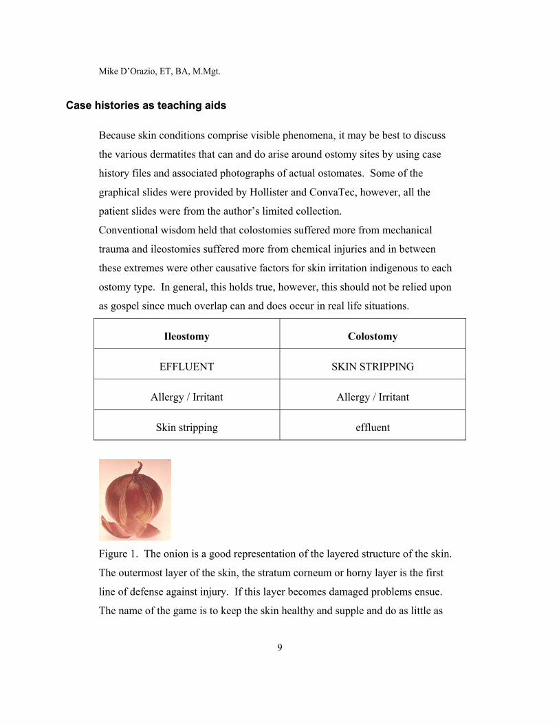
Figure 1. The onion is a good representation of the layered structure of the skin.
The outermost layer of the skin, the stratum corneum or horny layer is the first
line of defense against injury. If this layer becomes damaged problems ensue.
The name of the game is to keep the skin healthy and supple and do as little as
Mike D’Orazio, ET, BA, M.Mgt.
10
possible to the skin to achieve ostomy success. In this setting less can truly
become more. Tenderness is the approach we should employ when applying and
removing ostomy products to and from the skin.
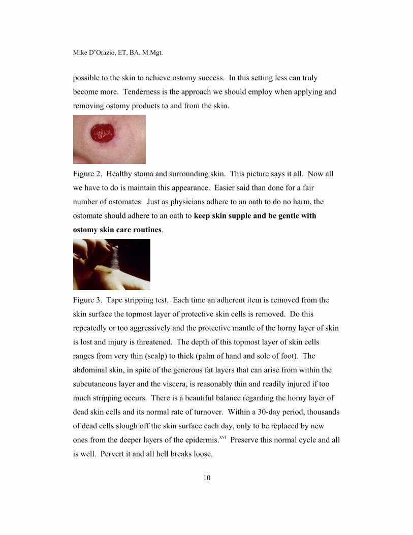
Figure 2. Healthy stoma and surrounding skin. This picture says it all. Now all
we have to do is maintain this appearance. Easier said than done for a fair
number of ostomates. Just as physicians adhere to an oath to do no harm, the
ostomate should adhere to an oath to keep skin supple and be gentle with
ostomy skin care routines.
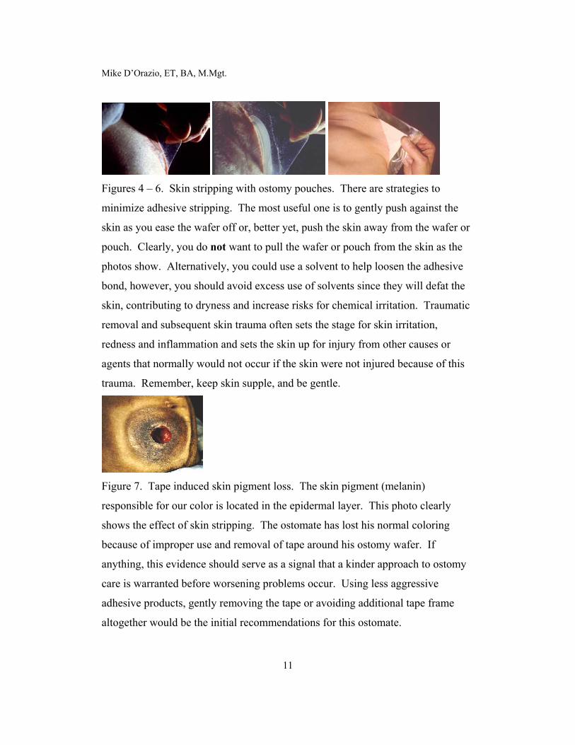
Figure 3. Tape stripping test. Each time an adherent item is removed from the
skin surface the topmost layer of protective skin cells is removed. Do this
repeatedly or too aggressively and the protective mantle of the horny layer of skin
is lost and injury is threatened. The depth of this topmost layer of skin cells
ranges from very thin (scalp) to thick (palm of hand and sole of foot). The
abdominal skin, in spite of the generous fat layers that can arise from within the
subcutaneous layer and the viscera, is reasonably thin and readily injured if too
much stripping occurs. There is a beautiful balance regarding the horny layer of
dead skin cells and its normal rate of turnover. Within a 30-day period, thousands
of dead cells slough off the skin surface each day, only to be replaced by new
ones from the deeper layers of the epidermis.xvi Preserve this normal cycle and all
is well. Pervert it and all hell breaks loose.
Mike D’Orazio, ET, BA, M.Mgt.
11
Figures 4 – 6. Skin stripping with ostomy pouches. There are strategies to
minimize adhesive stripping. The most useful one is to gently push against the
skin as you ease the wafer off or, better yet, push the skin away from the wafer or
pouch. Clearly, you do not want to pull the wafer or pouch from the skin as the
photos show. Alternatively, you could use a solvent to help loosen the adhesive
bond, however, you should avoid excess use of solvents since they will defat the
skin, contributing to dryness and increase risks for chemical irritation. Traumatic
removal and subsequent skin trauma often sets the stage for skin irritation,
redness and inflammation and sets the skin up for injury from other causes or
agents that normally would not occur if the skin were not injured because of this
trauma. Remember, keep skin supple, and be gentle.
Figure 7. Tape induced skin pigment loss. The skin pigment (melanin)
responsible for our color is located in the epidermal layer. This photo clearly
shows the effect of skin stripping. The ostomate has lost his normal coloring
because of improper use and removal of tape around his ostomy wafer. If
anything, this evidence should serve as a signal that a kinder approach to ostomy
care is warranted before worsening problems occur. Using less aggressive
adhesive products, gently removing the tape or avoiding additional tape frame
altogether would be the initial recommendations for this ostomate.
Mike D’Orazio, ET, BA, M.Mgt.
12
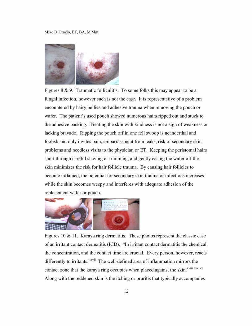
Figures 8 & 9. Traumatic folliculitis. To some folks this may appear to be a
fungal infection, however such is not the case. It is representative of a problem
encountered by hairy bellies and adhesive trauma when removing the pouch or
wafer. The patient’s used pouch showed numerous hairs ripped out and stuck to
the adhesive backing. Treating the skin with kindness is not a sign of weakness or
lacking bravado. Ripping the pouch off in one fell swoop is neanderthal and
foolish and only invites pain, embarrassment from leaks, risk of secondary skin
problems and needless visits to the physician or ET. Keeping the peristomal hairs
short through careful shaving or trimming, and gently easing the wafer off the
skin minimizes the risk for hair follicle trauma. By causing hair follicles to
become inflamed, the potential for secondary skin trauma or infections increases
while the skin becomes weepy and interferes with adequate adhesion of the
replacement wafer or pouch.
Figures 10 & 11. Karaya ring dermatitis. These photos represent the classic case
of an irritant contact dermatitis (ICD). “In irritant contact dermatitis the chemical,
the concentration, and the contact time are crucial. Every person, however, reacts
differently to irritants.”xvii The well-defined area of inflammation mirrors the
contact zone that the karaya ring occupies when placed against the skin.xviii xix xx
Along with the reddened skin is the itching or pruritis that typically accompanies

Mike D’Orazio, ET, BA, M.Mgt.
13
this type of dermatitis. The ultimate solution here is to avoid the offending
material, in this case karaya, and use a non-karaya pouching alternative.
Fortunately, there are adequate substitutes available. To help reduce the
inflammation and itching, topical steroid spray applications are most effective and
will not interfere with pouch or wafer adherence.
Figures 12 – 15. Irritant or allergic dermatitis from Stomahesive® and its
offspring DuoDerm E® are not common, yet it has been reported in the
professional medical literature.xxi xxii Note, however, that not all Stomahesive-
type (hydrocolloid) barriers are the same, even if they do look remarkably similar.
In the case of Stomahesive® paste, one of the suspected ingredients has been
identified as Gantrez.xxiii xxiv In addition, for the solid barrier the suspect agent was
a derivative of adhesive rosin (colophony).
In an attempt to accurately assess the risk factors presenting whenever one
suspects an allergic or irritant reaction to a product one must rule out other
culprits, especially those techniques or agents used to prepare the ostomy site. It
is tempting to jump to conclusions, especially when the visual evidence seems so
clear cut, but a careful history of pouching behaviors is always in order. For
example, adhesive remover wipesxxv have been implicated in topical irritations, as
have other cleansing and moisturizing agents. “Soaps, detergents and solvents are
examples of mild irritants which produce dryness, fissuring and dermatitis in most
individuals sufficiently exposed. After an erythematous eruption, the pH of the
skin tends to shift to the alkaline side with resulting impairment of alkali
neutralization, which in turn, enhances the damaging effect of acanthosis (skin
thickening) which increases skin susceptibility to primary irritants.”xxvi

Mike D’Orazio, ET, BA, M.Mgt.
14
As in the case of karaya irritations, the most useful approach to treatment is to
avoid repeat contact or use of Stomahesive and replace with other barriers and
topical steroid spray to help resolve the acute inflammation.
Figures 16 – 19. The yeast series, but with a wrinkle in photo 19. Photos 16 &
17-show pouch induced yeast infection of the groin area. Sweat buildup and poor
pouch hygiene have contributed to an opportunistic environment for fungal
overgrowth. A straightforward treatment recommendation consists of using a
pouch cover to keep the plastic pouch off the skin, thereby eliminating the sweat
problem. Improving overall pouch emptying and closure hygiene, to avoid undue
contamination of skin with pouch contents, will further aid recovery. Use of a
short-term course of topical anti-fungal nystatin powder or absorbable azole or
imidazole cream until the lesion heals fully and drying the skin well after
showering or bathing are the final recommendations here. Tinactin® (Tolnaftate)
is not effective for candidal infections!
Photo 18 reveals a yeast infection close to the stoma edge. Often times, and in
this case, stool leakage onto the skin will inoculate the skin with the offending
organism. Add to this the occlusive nature of the wafer against the skin and the
yeast organisms now have a happy environment in which to grow and romp
Mike D’Orazio, ET, BA, M.Mgt.
15
about. Of course, one needs to rule out other systemic or internal factors that may
predispose one to a yeast overgrowth, such as antibiotics, chemotherapy or an
immunocompromised condition, etc. Straightforward treatment
recommendations, after culturing the lesion, are to treat with topical antifungal
powder, improve skin–wafer interface to avoid repeat soiling, readjust wear time
to permit more frequent monitoring of skin while treating and gradually
reestablish “normal” wear time that avoids reinfection.
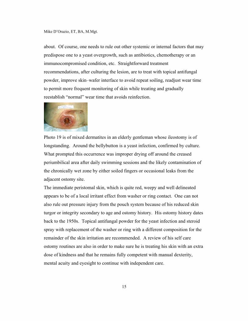
Photo 19 is of mixed dermatites in an elderly gentleman whose ileostomy is of
longstanding. Around the bellybutton is a yeast infection, confirmed by culture.
What prompted this occurrence was improper drying off around the creased
periumbilical area after daily swimming sessions and the likely contamination of
the chronically wet zone by either soiled fingers or occasional leaks from the
adjacent ostomy site.
The immediate peristomal skin, which is quite red, weepy and well delineated
appears to be of a local irritant effect from washer or ring contact. One can not
also rule out pressure injury from the pouch system because of his reduced skin
turgor or integrity secondary to age and ostomy history. His ostomy history dates
back to the 1950s. Topical antifungal powder for the yeast infection and steroid
spray with replacement of the washer or ring with a different composition for the
remainder of the skin irritation are recommended. A review of his self care
ostomy routines are also in order to make sure he is treating his skin with an extra
dose of kindness and that he remains fully competent with manual dexterity,
mental acuity and eyesight to continue with independent care.

Mike D’Orazio, ET, BA, M.Mgt.
16
Figure 20. Deep peristomal ulcerations of unknown origin. This retired
pharmacist had been treating his ileostomy site with his own concoctions for
sometime before he sought professional intervention. It is surprising how much
some patients are willing to tolerate before they throw in the towel and seek help.
Careful history and exam revealed a cycle of recurrence with periods of slight
improvement but never completely healed states. The initial suspicion was
pyoderma gangrenosum since he had a history of ulcerative colitis and had a
subtotal colectomy at the time of his ileostomy many years ago. This
undiagnosed peristomal lesion was referred to a dermatologist for definitive
diagnosis and treatment. Unfortunately, he was lost to follow up by distance and
eventual death, and his dermatitis and its resolution remained unknown.
Figure 21. Blenderm® tape reaction and Crohn’s disease at skin level. This
elderly gentleman with a loop colostomy presents with a classic reaction to a
plastic-backed or occlusive adhesive tape frame attached to his ostomy pouch.
This particular tape is not so common today but it was on the market for many
years throughout the 60s through the 80s. The easiest solution for the plastic-
backed tape irritation of the skin was to treat topically with steroid spray and use
the same manufacturer’s paper-backed alternative pouch system as a substitute.
The small fistula at the top edge of the stoma fortunately did not actively drain

Mike D’Orazio, ET, BA, M.Mgt.
17
and only needed the tape frame to be cut away and an absorbent dressing placed
over it until it finally closed.
Figure 22 –24. These photos represent the maceration problem when a wet
surface is allowed to contact the skin; in this case, a long stoma lies partly against
the skin close to the stoma edge. The mucous secretions of a stoma can be quite
irritating to the skin, even with out the stool component, and cause enzymatic
maceration. Bulbous, mushroom shaped or long stomas are more difficult to
position into the wafer opening. Often, some of the slippery mucous film from
the surface will slip behind the wafer as it is being guided over the stoma and
become trapped between the skin and wafer. Mucous drainage may also become
trapped if the skin planes at the stoma junction are not even or recessed. In this
case using protective caulks or powders may be sufficient to protect the skin from
any gaposis that allows mucus to pool between skin and wafer. Differing tricks of
the trade can be employed to prevent soiling of the wafer surface as it is being
slipped over the stoma.
Figures 25 & 26. Rubber allergy affecting stoma. These two photos are of
historical importance if for no other reason than they clearly reveal the effect of
rubber upon the stoma. While rubber pouches are no longer in vogue, some folks
still use them. In this case, the rubber pouch was of the Davol vintage, circa
Mike D’Orazio, ET, BA, M.Mgt.
18
1960s. Today’s plastic pouches can cause similar irritant responses to stomas and
adjacent skin planes as reported in the medical literature.xxvii Remedies in these
situations require non-irritating substitutes.
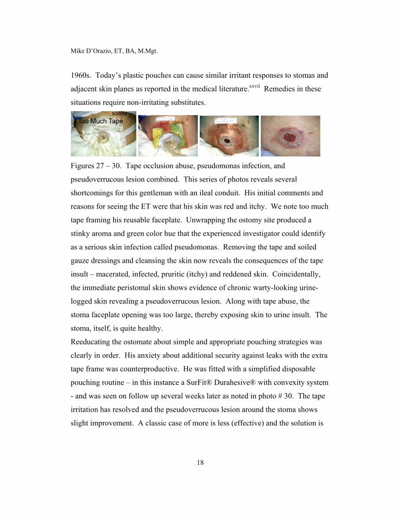
Figures 27 – 30. Tape occlusion abuse, pseudomonas infection, and
pseudoverrucous lesion combined. This series of photos reveals several
shortcomings for this gentleman with an ileal conduit. His initial comments and
reasons for seeing the ET were that his skin was red and itchy. We note too much
tape framing his reusable faceplate. Unwrapping the ostomy site produced a
stinky aroma and green color hue that the experienced investigator could identify
as a serious skin infection called pseudomonas. Removing the tape and soiled
gauze dressings and cleansing the skin now reveals the consequences of the tape
insult – macerated, infected, pruritic (itchy) and reddened skin. Coincidentally,
the immediate peristomal skin shows evidence of chronic warty-looking urine-
logged skin revealing a pseudoverrucous lesion. Along with tape abuse, the
stoma faceplate opening was too large, thereby exposing skin to urine insult. The
stoma, itself, is quite healthy.
Reeducating the ostomate about simple and appropriate pouching strategies was
clearly in order. His anxiety about additional security against leaks with the extra
tape frame was counterproductive. He was fitted with a simplified disposable
pouching routine – in this instance a SurFit® Durahesive® with convexity system
- and was seen on follow up several weeks later as noted in photo # 30. The tape
irritation has resolved and the pseudoverrucous lesion around the stoma shows
slight improvement. A classic case of more is less (effective) and the solution is

Mike D’Orazio, ET, BA, M.Mgt.
19
less is more (effective)! There is ample evidence about the effect of prolonged
occlusion on the microbial flora, pH, and moisture loss on the skin.xxviii
Figures 31 & 32. Psoriasis involving the peristomal planes. This elderly lady had
an emergency temporary colostomy for diverticulitis. She also had a seven-year
history of psoriasis prior to surgery that also involved her hands and groin zones,
and unfortunately, developed a psoriatic flare (Koebner Phenomenon) around her
stoma site. Management of her psoriasis required a simple, non-traumatic
pouching strategy that would allow easy daily pouch removal to permit skin
treatments. This was accomplished using karaya seal pouches and belt without
any additional tapeframe.
Figures 33 & 34. Tape frame induced contact dermatitis. Young man with a
staging temporary loop ileostomy for eventual J-pouch presents with a classic
case of tape-induced irritant contact dermatitis; severe itching complaint, and
aggressive scratching response; pouch-induced maceration or sweating and
secondary yeast infection. Finally, throw in the use of a Skin Prep® film wipe, as
recommended by his well-intentioned health care provider and you have a vicious
cycle of unrelenting skin trauma.
Let me sketch this out: tape > irritant dermatitis > severe itching > aggressive
scratching + pouch > maceration > contributes to yeast overgrowth + Skin Prep®

Mike D’Orazio, ET, BA, M.Mgt.
20
wipes > increased chemical sensitization > vicious cycle of trauma and insult
(Lewis Wheal effect)!!!
Effective intervention for this poor chap included the following: stop use of tape
framed wafer and Skin Prep® wipes; short-term use of topical steroid spray to
treat acute inflammation of skin and reduce itch stress; short-term use of topical
nystatin anti-fungal powder; pouch cover or cloth baby bib between skin and
pouch to minimize occlusivexxix effects of maceration or sweating. No wonder
this chap was anxious to have the stoma reversed ASAP!
Figures 35 – 37. Tape occlusion and mechanical trauma. Occlusion, as seen in
previous photos, increases the penetration of chemicals and antigens (irritants),
exacerbates sweat phenomena, and tends to exacerbate irritant and allergic contact
dermatitis.xxx Tape trauma caused by distortion of the skin surface under the tape
also contributes to irritant dermatitisxxxi and leaves a well-delineated imprint on
the skin localized to the site of contact, unlike allergic reactions which tend to
spread beyond the areas of actual contact.
Not only is this ostomate suffering from tape occlusion caused by the relatively
stiff plastic-backed tape but as photos #35 & #36 show his skin is wrinkled up
under the tape. This sets up a mechanical shear force that literally disrupts the
cells of the skin from their anchor and induces a traumatic result with
inflammation and discomfort. Simple solution offered here was to replace the
pouch with the stiff plastic-backed tape with one that had a more conformable
porous, paper-backed one. The other important recommendation was to instruct
the patient to not allow his skin to be wrinkled or unduly stretched when he
applied his pouch.
Mike D’Orazio, ET, BA, M.Mgt.
21
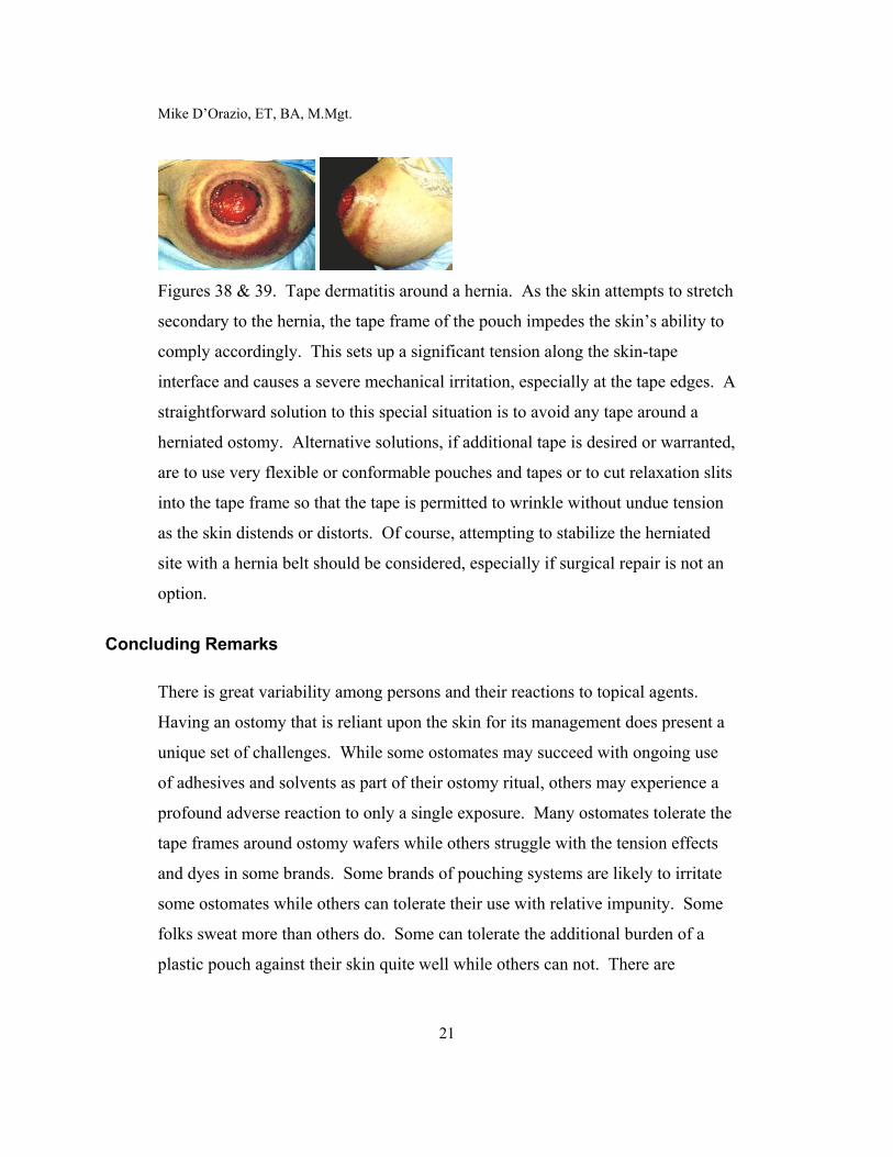
Figures 38 & 39. Tape dermatitis around a hernia. As the skin attempts to stretch
secondary to the hernia, the tape frame of the pouch impedes the skin’s ability to
comply accordingly. This sets up a significant tension along the skin-tape
interface and causes a severe mechanical irritation, especially at the tape edges. A
straightforward solution to this special situation is to avoid any tape around a
herniated ostomy. Alternative solutions, if additional tape is desired or warranted,
are to use very flexible or conformable pouches and tapes or to cut relaxation slits
into the tape frame so that the tape is permitted to wrinkle without undue tension
as the skin distends or distorts. Of course, attempting to stabilize the herniated
site with a hernia belt should be considered, especially if surgical repair is not an
option.
Concluding Remarks
There is great variability among persons and their reactions to topical agents.
Having an ostomy that is reliant upon the skin for its management does present a
unique set of challenges. While some ostomates may succeed with ongoing use
of adhesives and solvents as part of their ostomy ritual, others may experience a
profound adverse reaction to only a single exposure. Many ostomates tolerate the
tape frames around ostomy wafers while others struggle with the tension effects
and dyes in some brands. Some brands of pouching systems are likely to irritate
some ostomates while others can tolerate their use with relative impunity. Some
folks sweat more than others do. Some can tolerate the additional burden of a
plastic pouch against their skin quite well while others can not. There are
Mike D’Orazio, ET, BA, M.Mgt.
22
ostomates who never experience a yeast infection while others will flare after
minimal provocation or a single predisposing event. Many ostomates scrub the
hell out of their skin with any soap or detergent at hand as part of their preparation
ritual while others can tolerate only the mildest formulations. Some ostomates
can wear a wafer or pouch for very long periods while others are lucky to get a
day or two before they experience skin discomfort. Some folks are itchers and
others are practically immune to any itch provocation. Some folks are susceptible
to dry skin while others are susceptible to hyperhidrosis (increased sweating).
Some folks subscribe to the belief that they have or should strive for tough skin
while others pamper their skin at every turn.
In spite of all these apparent differences and contradictions there are some very
basic facts that all should note. The skin is an enormously complex organ subject
to many internal and external threats throughout its lifespan. It does not remain in
a static state. As Fisher so elegantly stated in 1967: “Excessive humidity, friction,
pressure, hyperhidrosis, and maceration may allow many non-irritating substances
to produce inflammation. Repeat exposures to irritants may be cumulative
resulting in a state of skin fatigue characterized by hyperirritability or, on the
other hand, to thickening and a state of hardening. The greatest number of skin
reactions which are observed in relation to the wearing of adhesive tape [or
adhesive products] are of a mechanical nature.”xxxii
The accumulating medical evidence informs us that the skin should not be taken
for granted or abused out of ignorance, arrogance, or neglect.
Mike D’Orazio, ET, BA, M.Mgt.
23
Recommended Readings
The following book is highly recommended reading, especially for ET nurses, in
spite of its 1983 publication date. It is chock full of excellent photographs and
illustrations that reinforce the dictum that a picture is truly worth a thousand
words.
Franchini, A., B. Cola and P. J. D’E. Stevens. 1983. Atlas of Stomal Pathology.
New York: Raven Press
Another and newer book on peristomal skin problems is the following:
Lyon, Calum C and Smith, Amanda J. 2001. Abdominal Stomas and their Skin
Disorders. London: Martin Dunitz
Mike D’Orazio, ET, BA, M.Mgt.
24
References
i Steigleder, G. K., and H.I. Maibach. 1984. Pocket Atlas of Dermatology. New York: George Thieme Verlag. ii The Skin: Anatomy and Physiology. http://www.caretechlabs.com/skin1.htm iii Rippke, F., Schreiner, V., Schwanitz, H. J. 2002. “The acidic milieu of the horny layer: new findings on the
physiology and pathophysiology of skin pH.” American Journal of Clinical Dermatology 3 (4): 261-72. iv Inflammation: The Leukocyte Adhesion Cascade. http://hsc.virginia.edu/medicine/basic-sci/biomed/ley/ last
updated June 22, 2000. v Gebhardt, M., P. Elsner, and J. G. Marks, Jr. 2000. Handbook of Contact Dermatitis. London: Martin Dunitz. vi Fitzpatrick, T. B., R. A. Johnson, K. Wolff, and D. Suurmond. 2001. Color Atlas and Synopsis of Clinical
Dermatology. New York: McGraw-Hill. vii Borglund, E., Nordstrom, G., Nyman, C. R.. 1988. “Classification of peristomal skin changes in patients with
urostomy.” Journal American Academy of Dermatology 19 (4): 623-8. viii Fisher, A. A. 1968. Contact Dermatitis. Philadelphia: Lea & Febiger. ix Panconesi, E., ed. 1984. Clinics in Dermatology: Stress and Skin Diseases. Philadelphia: Lippincott. x Broadwell, D. C. and B. S. Jackson, ed. 1982. Principles of Ostomy Care. St. Louis: C. V. Mosby. xi Steigleder, G. K., and H.I. Maibach (ibid.) xii IASP Newsletter Technical Corner: Neural Mechanisms of Itch Sensation.
http://www.halcyon.com/iasp/TC96SeptOct.html xiii Changes in Vessel Calibre. http://medweb.bham.ac.uk/http/mod/3/1/a/calibre.html xiv Fitzpatrick, T. B., R. A. Johnson, K. Wolff, and D. Suurmond. (ibid.) xv Candidiasis, Cutaneous. http://www.emedicine.com/DERM/topic67.htm. Last Updated: January 18, 2002 xvi Solomon, E. P., Schmidt, R. R., and Adragna, P. J. 1990. Human Anatomy & Physiology, Second Edition.
Philadelphia: Saunders College Publishing. xvii Steigleder, G. K., and H.I. Maibach (ibid.) xviii Burt-McAliley, D., Eberhardt, D, van Rijswijk, L. 1994. “Clinical study: peristomal skin irritation in
colostomy patients.” Ostomy Wound Management 40 (6): 28-30, 32-4, and 36-7. xix Ronnen, M., Suster, S., Kahana, M., Schewach-Millet, M. 1986. “Contact dermatitis due to karaya gum and
induced by the application of electrodes.” International Journal of Dermatology 25 (3): 189-90. xx Camarasa, J., M., Alomar, A. 1980. “Contact dermatitis from a karaya seal ring.” Contact Dermatitis 6 (2):
139-40. xxi Sasseville, D., Tennstedt, D., Lachapelle, J. M. 1997. “Allergic contact dermatitis from hydrocolloid
dressings.” American Journal of Contact Dermatitis 8 (4): 236-8.
Mike D’Orazio, ET, BA, M.Mgt.
25
xxii Schliz, M., Rauterberg, A., Weiss, J. 1996. “Allergic contact dermatitis from hydrocolloid dressings.”
Contact Dermatitis 34 (2): 146-7. xxiii Heskel, N. S. 1987. “Allergic contact dermatitis from Stomahesive paste.” Contact Dermatitis 16 (3): 119-
21. xxiv Scalf, L. A., Fowler, J.F., Jr. 2000. “Peristomal allergic contact dermatitis due to Gantrez in Stomahesive
paste.” Journal American Academy of Dermatology 42 (2 Pt 2): 355-6. xxv Lazarov, A., Trattner, A. 1998. “Allergic contact dermatitis from the adhesive remover wipe of stoma bags.” Contact Dermatitis 39 (1): 48-9. xxvi Fisher, A. A. 1968 (ibid.) xxvii van Ketel, W. G., van de Burg, C. K., de Haan, P. 1983. “Sensitization to epoxy resin from an ileostomy
bag.” Contact Dermatitis 9 (6): 516
Beck, M. H., Burrows, D., Fregert, S., Mendelsohn, S. 1985. “Allergic contact dermatitis to epoxy resin in ostomy bag.” British Journal of Surgery 72 (3): 202-3.
Van Hecke, E., Vossaert, K. 1988. “Allergic contact dermatitis from an ostomy bag.” Contact Dermatitis 18 (2): 121-2.
de Pablo, P., Ortiz, J., Borrego, L., Romero, G., Iglesias, L. 1992. “Allergic contact dermatitis from diaminodiphenylmethane in an ostomy bag.” Contact Dermatitis 27 (4): 260-1.
Parslew, R., Evans, S., King, C. M. 1996. “Allergic contact dermatitis from polyisobutylene in stoma bags.” Contact Dermatitis 35 (3): 178-9.
xxviii Aly, R., Shirley, C., Cunico, B., Maibach, H. I. 1978. “Effect of prolonged occlusion on the microbial flora, pH, carbon dioxide and transepidermal water loss on human skin.” Journal of Investigative Dermatology 71 (6): 378-81.
xxix Matsumura H, Oka K, Umekage K, Akita H, Kawai J, Kitazawa Y, Suda S, Tsubota K, Ninomiya Y, Hirai H, et al. 1995. “Effect of occlusion on human skin.” Contact Dermatitis 33 (4): 231-5.
xxx Zhai H, Maibach H I. 2001. “Skin occlusion and irritant and allergic contact dermatitis.” Contact Dermatitis 44 (4): 201-6.
xxxi Tokumura F, Ohyama K, Fujisawa H, Matsuda T, Kitazaki Y. 1997. “Conformability and irritancy of adhesive tapes on the ski.” Contact dermatitis 37 (4): 193-8.
xxxii Fisher, A. A. 1968 (ibid.)
MLD, ET