Embed Size (px)
Citation preview

CONSENSUS Open Access
Perioperative Quality Initiative (POQI)consensus statement on fundamentalconcepts in perioperative fluidmanagement: fluid responsiveness andvenous capacitanceGreg S. Martin1, David A. Kaufman2, Paul E. Marik3, Nathan I. Shapiro4, Denny Z. H. Levett5,17, John Whittle6,David B. MacLeod6, Desiree Chappell7,18, Jonathan Lacey8, Tom Woodcock9, Kay Mitchell10,Manu L. N. G. Malbrain11 , Tom M. Woodcock12, Daniel Martin13, Chris H. E. Imray14, Michael W. Manning6,Henry Howe7, Michael P. W. Grocott5,17, Monty G. Mythen15, Tong J. Gan16 and Timothy E. Miller6*
Abstract
Background: Optimal fluid therapy in the perioperative and critical care settings depends on understanding theunderlying cardiovascular physiology and individualizing assessment of the dynamic patient state.
Methods: The Perioperative Quality Initiative (POQI-5) consensus conference brought together an internationalteam of multidisciplinary experts to survey and evaluate the literature on the physiology of volume responsivenessand perioperative fluid management. The group used a modified Delphi method to develop consensus statementsapplicable to the physiologically based management of intravenous fluid therapy in the perioperative setting.
Discussion: We discussed the clinical and physiological evidence underlying fluid responsiveness and venouscapacitance as relevant factors in fluid management and developed consensus statements with clinical implicationsfor a broad group of clinicians involved in intravenous fluid therapy. Two key concepts emerged as follows: (1) Theultimate goal of fluid therapy and hemodynamic management is to support the conditions that enable normalcellular metabolic function in order to produce optimal patient outcomes, and (2) optimal fluid and hemodynamicmanagement is dependent on an understanding of the relationship between pressure, volume, and flow in adynamic system which is distensible with variable elastance and capacitance properties.
Keywords: Perioperative fluid management, Physiology, Fluid responsiveness, Venous capacitance, Goal-directed fluid therapy
© The Author(s). 2020 Open Access This article is licensed under a Creative Commons Attribution 4.0 International License,which permits use, sharing, adaptation, distribution and reproduction in any medium or format, as long as you giveappropriate credit to the original author(s) and the source, provide a link to the Creative Commons licence, and indicate ifchanges were made. The images or other third party material in this article are included in the article's Creative Commonslicence, unless indicated otherwise in a credit line to the material. If material is not included in the article's Creative Commonslicence and your intended use is not permitted by statutory regulation or exceeds the permitted use, you will need to obtainpermission directly from the copyright holder. To view a copy of this licence, visit http://creativecommons.org/licenses/by/4.0/.The Creative Commons Public Domain Dedication waiver (http://creativecommons.org/publicdomain/zero/1.0/) applies to thedata made available in this article, unless otherwise stated in a credit line to the data.
* Correspondence: [email protected] of Anesthesiology, Division of General, Vascular and TransplantAnesthesia, Duke University School of Medicine, Duke University MedicalCenter, Durham, NC, USAFull list of author information is available at the end of the article
Martin et al. Perioperative Medicine (2020) 9:12 https://doi.org/10.1186/s13741-020-00142-8

Consensus statementsPhysiological principles of fluid resuscitation
1. The ultimate goal of fluid and hemodynamicmanagement is to support normal cellularmetabolic function.
2. Achieving normal cellular metabolic functionrequires maintenance or restoration of effectivecoordinated function of the macrocirculation andthe microcirculation, as well as intact cellularmetabolism.
3. Practically speaking, most clinical management iscurrently targeted at macrocirculatory variables andsurrogates of cellular metabolism (e.g., lactate, baseexcess).
4. The therapeutic rationale of intravenous fluidadministration is to optimize macrocirculatoryfunction in order to improve or optimizemicrocirculatory and cellular function.
5. There is a minimal intravascular volume required tomaintain cardiac output and stroke volume andnormal tissue perfusion. Below this volume, cardiacoutput and blood pressure may be maintained atthe expense of microcirculatory blood flow andcellular function.
6. The majority of intravascular volume is in thevenous circulation; therefore, the venouscapacitance is a critical determinant of effectivemacrocirculatory function.
Physiology of fluid responsiveness
1. There is no readily available method of measuringintravascular volume and it is uncertain if knowingthis static value would have clinical utility.
2. Optimal intravascular volume can only becharacterized through dynamic evaluation.
3. Administration of a fluid bolus as part of a fluidchallenge is a means of increasing intravascularvolume to evaluate the effect on stroke volume.
4. Fluid responsiveness is defined as a state ofrecruitable stroke volume in response tointravascular fluid administration.
Venous capacitance
1. The venous circulation is comprised of stressed andunstressed volumes.
2. Stressed volume is the (theoretically measurable)volume of blood that exerts distending pressureagainst the venous wall. In contrast, unstressedvolume is the volume of blood up to the point offilling the veins but without exerting any pressureon the vessel walls.
3. Stressed volume determines the mean systemicfilling pressure (MSFP, the pressure of venousreturn when cardiac activity is absent), related tothe elastic recoil of the venous system. Thedifference between the MSFP and right atrialpressure is the major factor determining venousreturn to the heart. The MSFP provides drivingpressure against right atrial pressure which createsa gradient promoting forward flow.
Practical implications
1. Full characterization of fluid responsivenessrequires consideration of the type, amount andtiming of fluid as well as the expected change instroke volume.
2. The best method of measuring fluid responsivenessis a continuous or rapidly repeatable measure ofstroke volume.
3. A common approach to test fluid responsiveness isthe administration of 250-500 mL bulos in < 15 minwith a positive response defined by a 10-15%increase in stroke volume.
4. The passive leg-raise maneuver replicates atransient fluid bolus and predicts fluidresponsiveness without administration ofintravenous (IV) fluids (positive response defintedas > 10% increase in stroke volume), therebymitigating the risks of excess IV fluidadministration. This maneuver has limited utility inthe intraoperative setting.
5. Alternative methods for predicting fluidresponsiveness include stroke volume variation(SVV), pulse pressure variation (PPV), systolicpressure variation (SPV), and (in certainmechanically ventilated patients) end-expiratoryocclusion test and respiratory systolic variation test.All have limitations (Table 1).
6. Sonographic evaluation of IVC size, distensibility, orcollapsibility has limited and unproven utility at thepresent time.
7. The actions of vasoactive drugs are typicallyconsidered in relation to the arterial circulation butmany have significant effects on the venouscirculation.
8. Venoconstrictors (e.g., alpha agonists) increasevenous tone and thereby reduce venouscapacitance, thus increasing the stressed volume atthe expense of the unstressed volume. In ahypovolemic patient, this reduction in unstressedvolume may in turn reduce microcirculatory bloodflow and thereby compromise cellular metabolism,due to reduced perfusion despite maintenance ofnormal blood pressure.
Martin et al. Perioperative Medicine (2020) 9:12 Page 2 of 12
9. Venodilators (e.g., nitroglycerin) reduce venoustone and thereby increase venous capacitance anddecrease the stressed volume. This typicallydecreases venous return and left ventricular end-diastolic volume.
10. Intra-abdominal hypertension (e.g.,pneumoperitoneum) may reduce venous return orvenous capacitance.
BackgroundMore than 230 million major surgical procedures areundertaken worldwide each year (Weiser et al., 2008).Data from the USA and Europe suggests that approxi-mately 18% of patients undergoing surgery will developa major postoperative complication and 3 to 5% will diebefore hospital discharge (Weiser et al., 2008; Khuriet al., 2005; Ghaferi et al., 2009; Pearse et al., 2012).Those patients who develop a postoperative complica-tion and survive to hospital discharge have diminishedfunctional independence and reduced long-term survivalup to 8 years after major surgery (Khuri et al., 2005). In-terventions that reduce the risks of postoperative deathand complications, particularly in high risk patients havebecome a priority in perioperative medicine (Jacobs,2009). Perioperative goal-directed therapy (GDT), basedon the titration of fluids and vasoactive drugs to achievephysiological, flow-related end points, is a promising ap-proach to reduce postoperative complications and deaths(Bednarczyk et al., 2017; Pearse et al., 2014; Hamiltonet al., 2011).Optimal fluid therapy in the perioperative and critical
care settings depends on understanding the underlyingcardiovascular physiology and individualizing assessmentof the dynamic patient (Malbrain et al., 2018). Historicalapproaches to fluid administration based on clinicalexamination (e.g., “volume status”) or static measures ofcardiovascular function (e.g., central venous pressure orpulmonary artery occlusion pressure) do not adequately
determine fluid needs or to predict response to fluidadministration (Van der Mullen et al., 2018). Dynamicindices, such as SVV, utilize the concepts of the Frank-Starling relationship that implicitly incorporate venouscapacitance and mean systemic filling pressure to predictfluid responsiveness with good accuracy. However, inthe context of optimal fluid administration, knowledgegaps remain for several key physiologic concepts andclinical scenarios. This manuscript from the 5th Periopera-tive Quality Initiative (POQI) group addresses fundamentalconcepts in fluid responsiveness and venous capacitance inorder to provide an educational update and clinical guid-ance related to perioperative fluid management.
MethodsPOQI is an international, multidisciplinary non-profitorganization that organizes consensus conferences onclinical topics related to perioperative medicine (Milleret al., 2016). Each conference assembles a collaborativegroup of diverse international experts from multiplehealthcare disciplines to develop consensus-based rec-ommendations in perioperative medicine.Applying a modified Delphi method, designed to use
the collective expertise of a diverse group of experts toanswer clinically important questions, we achieved con-sensus on several topics related to fluid responsivenessand fluid management.
Expert groupAn international group of authorities, with specific con-tent area expertise (based on the conduct of researchand education in this area), was invited to participate. Intotal, 21 experts from around North America andEurope met in Durham, NC, on June 16-17, 2018, to it-eratively discuss the clinical and physiological evidenceof fluid responsiveness and venous capacitance as rele-vant factors in fluid management, in order to developconsensus statements with practical recommendations
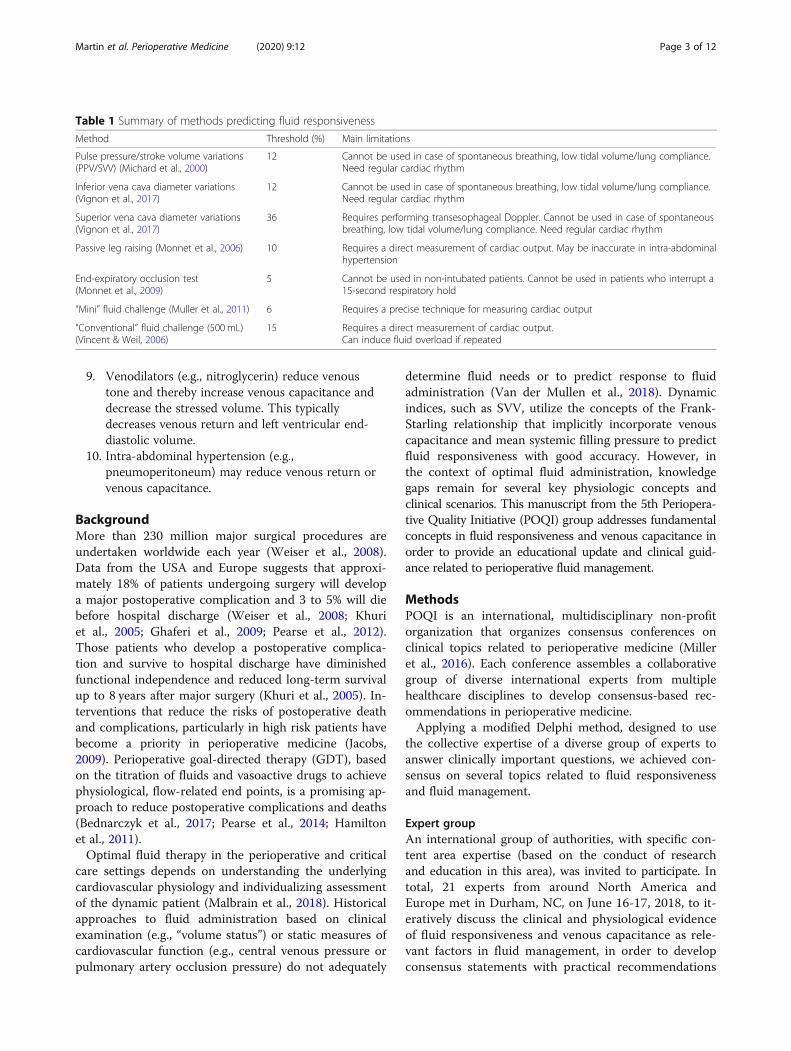
Table 1 Summary of methods predicting fluid responsiveness
Method Threshold (%) Main limitations
Pulse pressure/stroke volume variations(PPV/SVV) (Michard et al., 2000)
12 Cannot be used in case of spontaneous breathing, low tidal volume/lung compliance.Need regular cardiac rhythm
Inferior vena cava diameter variations(Vignon et al., 2017)
12 Cannot be used in case of spontaneous breathing, low tidal volume/lung compliance.Need regular cardiac rhythm
Superior vena cava diameter variations(Vignon et al., 2017)
36 Requires performing transesophageal Doppler. Cannot be used in case of spontaneousbreathing, low tidal volume/lung compliance. Need regular cardiac rhythm
Passive leg raising (Monnet et al., 2006) 10 Requires a direct measurement of cardiac output. May be inaccurate in intra-abdominalhypertension
End-expiratory occlusion test(Monnet et al., 2009)
5 Cannot be used in non-intubated patients. Cannot be used in patients who interrupt a15-second respiratory hold
“Mini” fluid challenge (Muller et al., 2011) 6 Requires a precise technique for measuring cardiac output
“Conventional” fluid challenge (500mL)(Vincent & Weil, 2006)
15 Requires a direct measurement of cardiac output.Can induce fluid overload if repeated
Martin et al. Perioperative Medicine (2020) 9:12 Page 3 of 12
for a broad group of clinicians involved in intravenousfluid therapy.
ProcessBased on literature searches performed by POQI groupmembers, a list of relevant questions was collectively for-mulated and circulated electronically prior to the meeting.In the first plenary session, these questions were presentedto receive feedback and assistance in refining the questions.There were then at least two Delphi rounds to develop thestatements before final agreement. This manuscript is basedon these multiple rounds of feedback from all the expertspresent at the POQI meeting.
Results/discussionKey physiologic and clinical terminology are shown inTables 2 and 3. Based on the literature identified by theparticipants and discussions held both prior to the confer-ence and during the iterative consensus-building process,the following key concepts and core questions were consid-ered most relevant to perioperative fluid management withrespect to fluid responsiveness and venous capacitance:
Key concepts
I. The ultimate goal of fluid therapy andhemodynamic management is to provide theconditions that enable normal homeostasis andcellular metabolic function in order to produceoptimal patient outcomes.
II. Fluid and hemodynamic management is dependenton the relationship between pressure, volume, and
flow in a dynamic system which is distensible withvariable elastance and capacitance properties.
Core questionsWhat are the physiologic and clinical goals of therapeuticfluid (hemodynamic) resuscitation?The ultimate goal of fluid therapy and hemodynamicmanagement is to provide the conditions that enablenormal cellular metabolic function in order to produceoptimal patient outcomes. The discrete goals of fluidtherapy exist at several levels: at the level of the macro-circulation, the microcirculation, and at the cellular level(Fig. 1). It is a limitation of medical science that cellularmetabolic function cannot be discretely and specificallymeasured in the clinical context, particularly locally foreach of the wide variety of organ systems, and in con-tinuous or repeatable series that permit clinical decision-making. Because of this limitation, for clinical care, wetarget intermediate variables of varying sensitivity, such ascardiac output (CO), stroke volume (SV), mean arterialpressure (MAP), central venous pressure (CVP), mixedvenous saturation (SvO2) and central venous oxygen satur-ation (ScvO2), heart rate (HR), and urine output. Whilethese parameters may indicate the presence of, or the riskfor, cellular metabolic dysfunction, they are insensitive inthis regard and do not differentiate cellular dysfunction dueto macrocirculatory versus microcirculatory abnormalities.Although restoration of the macrocirculation provides
the basis for normal microcirculatory and cellular meta-bolic function, it does not guarantee them. Microcircula-tory and cellular metabolic dysfunction may develop orpersist despite establishing normal microcirculatoryparameters, such as MAP and CO. However, becausedysfunction of the macrocirculation (e.g., hypotension)often produces dysfunction at the microcirculatory andcellular levels, the first step in therapeutic fluid resusci-tation is restoration of the macrocirculation. It is worthnoting that restoration of the macrocirculation is notachieved by reaching the same target in every patient.Macrocirculatory parameters such as MAP and COshould be personalized both for the individual and forthe patients’ current condition.Recently, several investigators introduced the term
“hemodynamic coherence” to describe the physiologicstate in which improved macrocirculatory function re-sults in improvements in the microcirculation (Ince,2015; Morelli & Passariello, 2016). In contrast, the term“hemodynamic incoherence” describes a physiologicalstate in which resuscitation to adequate macrocircula-tory parameters does not result in an improved micro-circulation. This state appears to happen frequently inpatients with sepsis. Four subsets of hemodynamic inco-herence are proposed. In the first, obstruction of somesmall blood vessels results in heterogeneous perfusion of
Table 2 Physiologic terminology
Term Definition
Arterial elastance The ratio of left ventricular end-systolic pressureand stroke volume
Intravascularvolume
The blood volume within the vascular system(arteries, capillaries, veins)
Mean systemicfilling pressure
The pressure of venous return when cardiac activityis absent
Preload Volume defined by the distending pressure itgenerates. In the heart, preload is LV wall stress atend of diastole (= EDV)
Stressed volume The (theoretically measurable) volume of blood thatexerts distending pressure against the vascular wall
Total body water The amount of sodium-free water in the whole body,commonly divided into the extracellular fluid spaceand the intracellular fluid space
Unstressedvolume
The volume of blood just to the point of filling theblood vessels but without exerting any pressure onthe vessel walls
Vascularcapacitance
The change in volume divided by the change inpressure (i.e., the inverse of elastance)
Martin et al. Perioperative Medicine (2020) 9:12 Page 4 of 12

Table 3 Clinical terminology
Term Definition
Fluid bolus The rapid administration of intravenous fluid with therapeutic intent, most often to rapidly replace intravascularvolume in patients who are presumed to be fluid responsive.
Fluid challenge The rapid administration of intravenous fluid with diagnostic intent, most often to determine whether a patientwith hemodynamic compromise will benefit from further fluid administration.
Fluid overload(overhydration)
Increased total body fluid volume (intravascular, interstitial, and intracellular). Fluid overload may be defined byat least 10% increase in total body fluid volume. Sometimes referred to as “overhydration” or “hyperhydration.”Fluid overload is the opposite of dehydration.
Fluid underload(dehydrataion)
Decreased total body fluid volume. The percentage of fluid loss is defined by dividing the cumulative fluidbalance in liters by the patient’s baseline body weight and multiplying by 100%. Dehydration is defined by aminimum value of 5% fluid loss. Dehydration is considered mild (5-7.5%), moderate (7.5-10%), while loss ofover 10% is considered severe. Sometimes referred to as “fluid underload.” Dehydration is the opposite offluid overload.
Fluid responsiveness An increase in stroke volume in response to an increase in intravascular volume. Also referred to as “volumeresponsiveness.”
Hypovolemia Reduced intravascular volume and marked by increases in stroke volume when intravenous fluid is given(i.e., the state of being fluid responsive). Clinical “hypovolemia” may exist, for example, from loss ofintravascular volume (e.g., hemorrhage) or from reductions in intravascular volume due to increases in venouscapacitance. Sometimes referred to as “fluid underload.”
Hypervolemia Hypervolemia is above normal or increased intravascular volume. Hypervolemia is the opposite of hypovolemia.
Passive leg raise A diagnostic postural maneuver raising the lower extremities up to 45 degrees from the recumbent position, totransiently increase venous return from the lower extremities in order to measure the hemodynamic effect andthus determine if a patient is fluid responsive.
Fig. 1 The macrocirculation, microcirculation, and the cellular level relevant for fluid therapy. Figure reused with the permission of thePerioperative Quality Initiative (POQI). For permission requests, contact [email protected]
Martin et al. Perioperative Medicine (2020) 9:12 Page 5 of 12

the microcirculation. In the second, hemodilution resultsin perfusion of capillaries with blood that has a low oxy-gen carrying capacity. In the third, increased arterial re-sistance and increased venous pressures lead to capillarystasis due to low arterial-venous pressure gradients (seebelow). In the fourth, edema causes large distances be-tween capillaries and target tissues across which oxygenand other energy substrates must diffuse to reach theirtargets. Notably, administration of IV fluid may lead tohemodilution, increased venous pressures, and edema for-mation, thus contributing to hemodynamic incoherence.The first goal of therapeutic fluid and hemodynamic
resuscitation is targeted at macrocirculatory parameters(usually MAP) because they are measurable and theyrepresent the most apparent clinical markers of organ ortissue perfusion, particularly in combination with otherclinical and laboratory assessments. When the MAP fallsbelow an organ’s autoregulatory range, there is an al-most linear decrease in organ blood flow (Ackland et al.,2019). The fall in blood flow is likely to occur at a higherMAP in patients with long-standing hypertension due toa shift in the autoregulatory range. Furthermore, differ-ent vascular beds will lose autoregulation at differentMAPs. For example, the mammalian kidney loses auto-regulation at a MAP of about 70 mmHg, the brain be-tween 60-70 mm Hg, while the coronary circulationloses autoregulation at a MAP of about 50-55 mmHg(Drummond, 1997; Drummond, 2019; Paulson et al.,1990; Bellomo & Di Giantomasso, 2001; Meng, 2019).Therefore, the first hemodynamic goal is to achieve aMAP > 65-70 mmHg to preserve organ perfusion (Sess-ler et al., 2019).The second and third goals of fluid and hemodynamic
resuscitation are targeted at the microcirculation and thecellular levels. As noted above, dysfunction at theselevels may develop or persist despite the appearance ofnormal macrocirculatory parameters, and devices tocharacterize the microcirculation and cellular functionare not routinely available for clinical use. The microcircu-lation may be assessed directly using tools such as intravi-tal microscopy or laser Doppler flowmetry, or indirectlyusing tissue oximetry and near infrared spectroscopy(Tafner et al., 2017). Clinical examination (e.g., examin-ation of capillary refill time or assessing for skin mottling)may also give important clues about the adequacy ofperfusion in the microcirculation (Hernandez et al., 2019).Efforts to realize the potential for microcirculatory moni-toring for point-of-care diagnosis in real-time at the bed-side are currently underway (Naumann et al., 2016).In the clinical setting, the only reason to give any pa-
tient a fluid bolus is to increase the SV. In the absenceof an increase in SV, giving a fluid challenge serves nouseful purpose and is likely to be harmful. An increasein SV will only occur if two conditions are met: (1) that
the fluid bolus increases the stressed blood volume caus-ing an increase in MSFP, thereby increasing venous re-turn, and (2) that both ventricles are functioning on theascending limb of the Frank-Starling curve.Organ blood flow is driven by the difference in the
pressure between the arterial and venous circulation. Forexample, the MAP minus the CVP is the driving forcefor organ blood flow while the difference between post-arteriolar and venular pressure determines microcircula-tory flow. In circumstances of increased venous pressure,such as high right atrial pressure, the backwards trans-mission of pressure may impede microcirculatory flowin the tissues and organs.
What is the role of venous capacitance in fluid andhemodynamic therapy?Fluid optimization must consider each of the fluid com-partments in the body: total body water divided into theintracellular and extracellular spaces, and more discretelythe plasma volume and blood volume, the interstitial fluidvolume, and the tissue-bound water volume (Fig. 2).The left ventricle can only pump into the arterial circula-
tion; the volume of blood that it receives from venous return(Funk et al., 2013). Because approximately two-thirds of thetotal blood volume is in the venous system, the roles of thevenous system in general and venous capacitance specificallyare important for fluid therapy and hemodynamic manage-ment. Through their capacitance function, veins and venulesregulate both regional and central blood volume and there-fore cardiac preload: changes in venous tone directly influ-ence SV and CO via the Frank-Starling mechanism. It isimportant for anesthesia providers to understanding venousreturn, venous capacitance, and their role in determiningCO.With two-thirds of the blood volume within the
venous system, changes in venous blood volume playa major role in determining venous return and CO(Fig. 3). The venous system can be divided into twotheoretical compartments, the unstressed and thestressed volume (Gelman, 2008). The intravascularvolume that fills the venous system to the point justbefore intravascular pressure starts to rise is calledunstressed volume, whereas the volume that stretchesthe veins and causes intravascular pressure to rise iscalled the stressed volume (Figs. 4, 5). Another way tothink of stressed volume is the (theoretically measur-able) portion of blood that exerts distending pressureagainst the vein wall. Differentiating between thesetwo volumes is important because stressed volumedetermines the MSFP, which is the pressure of venousreturn when cardiac activity is absent, representingthe elastic recoil of the venous system. ThroughMSFP, the stressed blood volume is a major contribu-tor of venous pressure and therefore venous return.
Martin et al. Perioperative Medicine (2020) 9:12 Page 6 of 12

Fig. 3 Pressure and volume in the venous system. Figure reused with the permission of the Perioperative Quality Initiative (POQI). For permissionrequests, contact [email protected].
Fig. 2 Fluid compartments in adult humans. Figure reused with the permission of the Perioperative Quality Initiative (POQI). For permissionrequests, contact [email protected]
Martin et al. Perioperative Medicine (2020) 9:12 Page 7 of 12
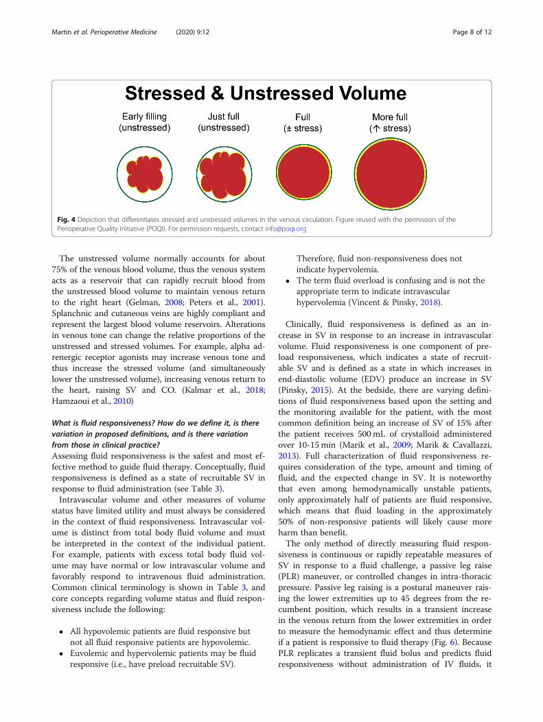
The unstressed volume normally accounts for about75% of the venous blood volume, thus the venous systemacts as a reservoir that can rapidly recruit blood fromthe unstressed blood volume to maintain venous returnto the right heart (Gelman, 2008; Peters et al., 2001).Splanchnic and cutaneous veins are highly compliant andrepresent the largest blood volume reservoirs. Alterationsin venous tone can change the relative proportions of theunstressed and stressed volumes. For example, alpha ad-renergic receptor agonists may increase venous tone andthus increase the stressed volume (and simultaneouslylower the unstressed volume), increasing venous return tothe heart, raising SV and CO. (Kalmar et al., 2018;Hamzaoui et al., 2010)
What is fluid responsiveness? How do we define it, is therevariation in proposed definitions, and is there variationfrom those in clinical practice?Assessing fluid responsiveness is the safest and most ef-fective method to guide fluid therapy. Conceptually, fluidresponsiveness is defined as a state of recruitable SV inresponse to fluid administration (see Table 3).Intravascular volume and other measures of volume
status have limited utility and must always be consideredin the context of fluid responsiveness. Intravascular vol-ume is distinct from total body fluid volume and mustbe interpreted in the context of the individual patient.For example, patients with excess total body fluid vol-ume may have normal or low intravascular volume andfavorably respond to intravenous fluid administration.Common clinical terminology is shown in Table 3, andcore concepts regarding volume status and fluid respon-siveness include the following:
� All hypovolemic patients are fluid responsive butnot all fluid responsive patients are hypovolemic.
� Euvolemic and hypervolemic patients may be fluidresponsive (i.e., have preload recruitable SV).
Therefore, fluid non-responsiveness does notindicate hypervolemia.
� The term fluid overload is confusing and is not theappropriate term to indicate intravascularhypervolemia (Vincent & Pinsky, 2018).
Clinically, fluid responsiveness is defined as an in-crease in SV in response to an increase in intravascularvolume. Fluid responsiveness is one component of pre-load responsiveness, which indicates a state of recruit-able SV and is defined as a state in which increases inend-diastolic volume (EDV) produce an increase in SV(Pinsky, 2015). At the bedside, there are varying defini-tions of fluid responsiveness based upon the setting andthe monitoring available for the patient, with the mostcommon definition being an increase of SV of 15% afterthe patient receives 500 mL of crystalloid administeredover 10-15 min (Marik et al., 2009; Marik & Cavallazzi,2013). Full characterization of fluid responsiveness re-quires consideration of the type, amount and timing offluid, and the expected change in SV. It is noteworthythat even among hemodynamically unstable patients,only approximately half of patients are fluid responsive,which means that fluid loading in the approximately50% of non-responsive patients will likely cause moreharm than benefit.The only method of directly measuring fluid respon-
siveness is continuous or rapidly repeatable measures ofSV in response to a fluid challenge, a passive leg raise(PLR) maneuver, or controlled changes in intra-thoracicpressure. Passive leg raising is a postural maneuver rais-ing the lower extremities up to 45 degrees from the re-cumbent position, which results in a transient increasein the venous return from the lower extremities in orderto measure the hemodynamic effect and thus determineif a patient is responsive to fluid therapy (Fig. 6). BecausePLR replicates a transient fluid bolus and predicts fluidresponsiveness without administration of IV fluids, it
Fig. 4 Depiction that differentiates stressed and unstressed volumes in the venous circulation. Figure reused with the permission of thePerioperative Quality Initiative (POQI). For permission requests, contact [email protected]
Martin et al. Perioperative Medicine (2020) 9:12 Page 8 of 12

mitigates the risk of excessive IV fluids that may be par-ticularly deleterious in patients at greater risk for or poortolerance of hypervolemia (e.g., heart failure, chronic kid-ney disease, chronic lung disease). Alternative methods forpredicting fluid responsiveness include SVV, PPV, SPV,and (in certain mechanically ventilated patients) the end-expiratory occlusion test and respiratory systolic variationtest (see Table 1).A common approach to test fluid responsiveness is the
administration of a 500 mL fluid challenge over < 15 minwith a positive response defined by a ≥ 15% increase inSV, or a 250 mL fluid challenge over < 15 min with apositive response defined by a ≥ 10% increase in SV.However:
� Investigations into fluid responsiveness vary in fluidtype, volume, infusion time, and consequent changein SV.
� Patients at risk for adverse effects from fluidadministration, such as CHF and ESRD, may receivelower volume fluid challenges (e.g., 100 mL),although the accuracy of the test to predict fluidresponsiveness is reduced (i.e., greater risk of falsenegative test).
� The volume of fluid challenge depends on the typeof fluid given: crystalloid, colloid, or blood.
Recognizing that SV cannot be measured in all set-tings, MAP or HR may be used as crude surrogate indi-cators, recognizing they have limited predictive value.Although the existing literature has examined the effectof a fluid challenge on SV, a fluid challenge or PLRcould be useful in detecting whether inadequate preloadis contributing to hypotension. If the fluid challenge/
Fig. 5 Effects of fluid and vasoactive agents on cardiovascularperformance and the venous system. Figure reused with thepermission of the Perioperative Quality Initiative (POQI). Forpermission requests, contact [email protected]. I Effect of volumeloading on mean systemic filling pressure (Pmsf) and (un)stressedvolume. Administration of a fluid bolus increases Pmsf (from Pmsf1to Pmsf2, indicated respectively by position A (red dot) to B (greendot) on the pressure/volume curve). Unstressed volume remainsconstant while stressed volume increases. Total volume = unstressed+ stressed increases, carrying a risk for fluid overload. See text forexplanation. II Effect of venoconstriction and venodilation on meansystemic filling pressure (Pmsf) and (un)stressed volume.Venoconstriction increases Pmsf (from Pmsf1 to Pmsf2, indicatedrespectively by position A (red dot) to B (green dot) on thepressure/volume curve). Unstressed volume decreases while stressedvolume increases. Total volume = unstressed + stressed remainsconstant, resulting in an auto-transfusion effect. Venodilation as seenin sepsis (vasoplegia) decreases Pmsf (from Pmsf1 to Pmsf3,indicated respectively by position A (red dot) to C (blue dot) on thepressure/volume curve). Unstressed volume increases while stressedvolume decreases. Total volume = unstressed + stressed remainsconstant, resulting in an intravascular underfilling effect
Martin et al. Perioperative Medicine (2020) 9:12 Page 9 of 12

PLR does not correct hypotension, additional monitoringmay be appropriate and further management shouldfocus on vascular tone and chronotropy/inotropy (McE-voy et al., 2019).
Under what situations can fluid responsiveness be usedclinically to decide when to give, and when to stop givingfluid?In the perioperative period, when fluid losses may besubstantial, fluid responsiveness is generally an indica-tion for fluid administration but should be interpreted inthe clinical context of the patient. In patients who arepredicted to be non-responsive to fluid administration,fluids should not be given unless other clinical indicatorssuggest net benefit (e.g., need for water). The same as-sessments apply to the decision to stop giving intraven-ous fluids, which may be based upon the parametersjudging fluid responsiveness and the clinical context ofthe patient for a global assessment or benefit and risk.A key clinical question to ask when assessing a pa-
tient’s fluid responsiveness is whether increasing the SVand CO is beneficial. For example, a patient might havean adequate or high SV index and cardiac index and stillshow evidence of fluid responsiveness. Giving fluidmight not be indicated since achieving a still higher SVindex and cardiac index might not yield important clin-ical benefits.All tests for fluid responsiveness have limitations
(Table 1). For example, SVV testing requires a regularcardiac rhythm, the lack of patient inspiratory efforts,and consistent changes in intrathoracic pressure pro-duced by mechanical ventilation with tidal volumes of atleast 8 mL/kg predicted body weight. PLR testing requiresmeasurement of SV or CO and may produce false nega-tive results in the setting of intra-abdominal hypertension.
While ultrasonographic evaluation of vena cava diam-eter, distensibility, and collapsibility are increasinglyused in emergency medicine and critical care, its usehas numerous confounders (right heart dysfunction,obstructive cardiac physiology, transpulmonary andintraabdominal pressure, mode of ventilatory support)and is not currently supported by evidence in the peri-operative setting (Millington, 2019; Via et al., 2016). Fi-nally, it is important to note that fluid bolus therapyrapidly impacts the macrocirculation but does not ne-cessarily alter the microcirculation or cellular function,especially during the short time frame used to assessfluid responsiveness. Further, the effects of crystalloidboluses on SV and CO are often short-lived, as thecrystalloid fluid redistributes into the extravascularextracellular space (Nunes et al., 2014; Aya et al., 2016).Despite these limitations the assessment of fluid re-
sponsiveness probably leads to improved patient out-comes among patients who undergo high-risk andcomplex surgery (Bednarczyk et al., 2017).
What is the research agenda?Our increasing understanding of the physiological andclinical consequences of intravenous fluid therapy hasled to new and important questions that must be an-swered in order to further refine our clinical use ofintravenous fluids. Future research should focus on thefollowing key areas:
� As the fundamental therapeutic goal of intravenousfluid administration into the macrocirculation is tooptimize microcirculatory and cellular function, weneed better tools to assess both of those criticalfeatures at the bedside.
Fig. 6 Stylized depiction of the passive leg raise (PLR) maneuver. Figure reused with the permission of the Perioperative Quality Initiative (POQI).For permission requests, contact [email protected]
Martin et al. Perioperative Medicine (2020) 9:12 Page 10 of 12

� We need to identify or create methods witheveryday clinical utility to measure intravascularvolume, and for monitoring the benefits/harm offluid therapy.
� We need to better define exactly how to perform afluid challenge and a fluid responsisve patient.
� Emerging evidence suggests that fluid managementshould always be individualized based on eachpatients unique hemodynamics and cardiac funcion,underlying disease process and co-morbidities. Wemust recognize the dynamic nature of patienttrajectories throughout the perioperative period tobetter define optimal fluid administration andremoval strategies.
Further research in these key areas will lay the founda-tion for moving from group-targeted fluid therapy totruly individualized fluid therapy.
ConclusionsIn the POQI-5 consensus conference, we discussed theclinical and physiological evidence of fluid responsivenessand venous capacitance as relevant factors in fluid man-agement and developed consensus statements with clinicalimplications for a broad group of clinicians involved inintravenous fluid therapy. Two key concepts emerged: (1)The ultimate goal of fluid therapy and hemodynamicmanagement is to provide the conditions that enable nor-mal cellular metabolic function in order to produce opti-mal patient outcomes, and (2) fluid and hemodynamicmanagement is dependent on the relationship betweenpressure, volume, and flow in a dynamic system which isdistensible and has variable elastance and capacitance.
AbbreviationsCVP: Central venous pressure; CO: Cardiac output; EDV: End-diastolic volume;GDT: Goal-directed therapy; HR: Heart rate; IV: Intravenous; MAP: Meanarterial pressure; MSFP: Mean systemic filling pressure; PLR: Passive leg raise;POQI: Perioperative Quality Initiative; PPV: Pulse pressure variation;SPV: Systolic pressure variation; ScvO2: Mixed central venous oxygensaturation; SvO2: Mixed venous oxygen saturation; SV: Stroke volume;SVV: Stroke volume variation
AcknowledgementsNot applicable
Authors’ contributionsAll authors contributed to the development of the manuscript, agree to itscontents, and approved its final version.
FundingThe Perioperative Quality Initiative-5 consensus conference was supportedby an unrestricted educational grant from the Perioperative Quality Initiative,which has received grants from Baxter, Bev MD, Cadence, Cheetah Medical,Edwards, Heron Pharmaceutical, Mallinckrodt, Masimo, Medtronic, Merck,Trevena, and Pacira.
Availability of data and materialsNot applicable
Ethics approval and consent to participateNot applicable
Consent for publicationNot applicable
Competing interestsGSM - none; DAK - grant funding and travel reimbursement for CheetahMedical, Advisory Board and speaker honoraria for Pulsion Medical Systems;PEM - advisory board and research funding from Baxter; NS - none ; DL -none; JW – none; DBM – none; DC - Speakers Bureau for EdwardsLifesciences, Medtronic; JL – none; TW – none; KM – none; MLNGM – xxx;TMW – none; DM – consultant for Edwards Lifesciences and SiemensHealthineers; CHEI – none; MWM – none; HH – none; MPWG – researchfunding from Sphere Medical Ltd (UK) and Pharmacosmos Ltd (UK), advisoryboard of Sphere Medical Ltd.; MGM - University Chair Sponsor at UCL bySmiths Medical, Director Evidence Based Perioperative Medicine (EBPOM)Community Interest Company (CiC), Director Medinspire Ltd (Patent holder“QUENCH”), Paid consultant for Edwards Lifesciences and Baxter; TJG -Consultant for Acacia, Edwards Lifesciences, Mallinckrodt, Medtronic andMerck; TEM – research funding and consultant for Edwards Lifesciences.
Author details1Division of Pulmonary, Allergy, Critical Care and Sleep Medicine, EmoryCritical Care Center, Emory University School of Medicine, Grady HealthSystem, Atlanta, GA, USA. 2Division of Pulmonary, Critical Care, and SleepMedicine, NYU School of Medicine, New York, NY, USA. 3Division ofPulmonary and Critical Care Medicine, Eastern Virginia Medical School,Norfolk, VA, USA. 4Department of Emergency Medicine, Beth IsraelDeaconess Medical Center, Boston, MA, USA. 5Critical Care Research Group,NIHR Biomedical Research Centre, University Hospital Southampton NHSFoundation Trust/University of Southampton, Southampton, UK.6Department of Anesthesiology, Division of General, Vascular and TransplantAnesthesia, Duke University School of Medicine, Duke University MedicalCenter, Durham, NC, USA. 7TopMedTalk, London, UK. 8Institute of SportExercise & Health, University College London, London, UK. 9UniversityHospitals Southampton, Southampton, UK. 10Respiratory Biomedical ResearchUnit, University of Southampton, Southampton, England. 11Department ofIntensive Care, University Hospital Brussels, Jette, Belgium and FacultyofMedicine and Pharmacy, Vrije Universiteit Brussels, Brussels, Belgium.12Elsevier R&D Solutions, 1600 JFK Blvd, Philadelphia, PA 19103, USA.13Intensive Care Unit and Division of Surgery and Interventional Science,Royal Free Hospital, London, UK. 14Vascular and Renal Tranplant Surgeon,National Institute of Health Research Clinical Research Facility, Coventry, UK.15UCL/UCLH National Institute of Health Research Biomedical ResearchCentre, London, UK. 16Department of Anesthesiology, Stony Brook University,Stony Brook, NY, USA. 17Department of Anesthesiology and Critical Care,Stony Brook University, Stony Brook, New York, USA. 18Private address:Louisville, Kentucky, USA.
Received: 30 September 2019 Accepted: 18 March 2020
ReferencesAckland GL, Brudney CS, Cecconi M, et al. Perioperative Quality Initiative
consensus statement on the physiology of arterial blood pressure control inperioperative medicine. Br J Anaesth. 2019;122:542–51.
Aya HD, Ster IC, Fletcher N, Grounds RM, Rhodes A, Cecconi M.Pharmacodynamic analysis of a fluid challenge. Crit Care Med. 2016;44:880–91.
Bednarczyk JM, Fridfinnson JA, Kumar A, et al. Incorporating dynamic assessmentof fluid responsiveness into goal-directed therapy: a systematic review andmeta-analysis. Crit Care Med. 2017;45:1538–45.
Bellomo R, Di Giantomasso D. Noradrenaline and the kidney: friends or foes? CritCare. 2001;5:294–8.
Drummond JC. The lower limit of autoregulation: time to revise our thinking?Anesthesiol. 1997;86:1411–33.
Drummond JC. Blood pressure and the brain: how low can you go? AnesthAnalg. 2019;128:759–71.
Funk DJ, Jacobsohn E, Kumar A. Role of the venous return in critical illness andshock: part II-shock and mechanical ventilation. Crit Care Med. 2013;41:573–9.
Martin et al. Perioperative Medicine (2020) 9:12 Page 11 of 12
Gelman S. Venous function and central venous pressure: a physiologic story.Anesthesiology. 2008;108:735–48.
Ghaferi AA, Birkmeyer JD, Dimick JB. Variation in hospital mortality associatedwith inpatient surgery. N Engl J Med. 2009;361:1368–75.
Hamilton MA, Cecconi M, Rhodes A. A systematic review and meta-analysis onthe use of preemptive hemodynamic intervention to improve postoperativeoutcomes in moderate and high-risk surgical patients. Anesth Analg. 2011;112:1392–402.
Hamzaoui O, Georger JF, Monnet X, et al. Early administration of norepinephrineincreases cardiac preload and cardiac output in septic patients with life-threatening hypotension. Crit Care. 2010;14:R142.
Hernandez G, Ospina-Tascon GA, Damiani LP, et al. Effect of a resuscitationstrategy targeting peripheral perfusion status vs serum lactate levels on 28-day mortality among patients with septic shock: the ANDROMEDA-SHOCKrandomized clinical trial. JAMA. 2019;321:654–64.
Ince C. Hemodynamic coherence and the rationale for monitoring themicrocirculation. Crit Care 2015;19 Suppl 3:S8.
Jacobs DO. Variation in hospital mortality associated with inpatient surgery--an S.O.S. N Engl J Med. 2009;361:1398–400.
Kalmar AF, Allaert S, Pletinckx P, et al. Phenylephrine increases cardiac output byraising cardiac preload in patients with anesthesia induced hypotension. JClin Monit Comput. 2018;32:969–76.
Khuri SF, Henderson WG, DePalma RG, et al. Determinants of long-term survivalafter major surgery and the adverse effect of postoperative complications.Ann Surg. 2005;242:326–41.
Malbrain M, Van Regenmortel N, Saugel B, et al. Principles of fluid managementand stewardship in septic shock: it is time to consider the four D’s and thefour phases of fluid therapy. Ann Intensive Care. 2018;8:66.
Marik PE, Cavallazzi R. Does the central venous pressure predict fluidresponsiveness? An updated meta-analysis and a plea for some commonsense. Crit Care Med. 2013;41:1774–81.
Marik PE, Cavallazzi R, Vasu T, Hirani A. Dynamic changes in arterial waveformderived variables and fluid responsiveness in mechanically ventilatedpatients: a systematic review of the literature. Crit Care Med. 2009;37:2642–7.
McEvoy MD, Gupta R, Koepke EJ, et al. Perioperative Quality Initiative consensusstatement on postoperative blood pressure, risk and outcomes for electivesurgery. Br J Anaesth. 2019;122:575–86.
Meng L. Heterogeneity and variability in pressure autoregulation of organ bloodflow: lessons learned over 100+ years. Crit Care Med. 2019;47:436–48.
Michard F, Boussat S, Chemla D, et al. Relation between respiratory changes inarterial pulse pressure and fluid responsiveness in septic patients with acutecirculatory failure. Am J Respir Crit Care Med. 2000;162:134–8.
Miller TE, Shaw AD, Mythen MG, Gan TJ. Perioperative Quality Initiative IW.Evidence-based perioperative medicine comes of age: the PerioperativeQuality Initiative (POQI): the 1st consensus conference of the PerioperativeQuality Initiative (POQI). Perioper Med (Lond). 2016;5:26.
Millington SJ. Ultrasound assessment of the inferior vena cava for fluidresponsiveness: easy, fun, but unlikely to be helpful. Can J Anaesth. 2019;66:633–8.
Monnet X, Osman D, Ridel C, Lamia B, Richard C, Teboul JL. Predicting volumeresponsiveness by using the end-expiratory occlusion in mechanicallyventilated intensive care unit patients. Crit Care Med. 2009;37:951–6.
Monnet X, Rienzo M, Osman D, et al. Passive leg raising predicts fluidresponsiveness in the critically ill. Crit Care Med. 2006;34:1402–7.
Morelli A, Passariello M. Hemodynamic coherence in sepsis. Best Pract Res ClinAnaesthesiol. 2016;30:453–63.
Muller L, Toumi M, Bousquet PJ, et al. An increase in aortic blood flow after aninfusion of 100 ml colloid over 1 minute can predict fluid responsiveness: themini-fluid challenge study. Anesthesiology. 2011;115:541–7.
Naumann DN, Mellis C, Husheer SL, et al. Real-time point of care microcirculatoryassessment of shock: design, rationale and application of the point of caremicrocirculation (POEM) tool. Crit Care. 2016;20:310.
Nunes TS, Ladeira RT, Bafi AT, de Azevedo LC, Machado FR, Freitas FG. Durationof hemodynamic effects of crystalloids in patients with circulatory shock afterinitial resuscitation. Ann Intensive Care. 2014;4:25.
Paulson OB, Strandgaard S, Edvinsson L. Cerebral autoregulation. CerebrovascBrain Metab Rev. 1990;2:161–92.
Pearse RM, Harrison DA, MacDonald N, et al. Effect of a perioperative, cardiacoutput-guided hemodynamic therapy algorithm on outcomes followingmajor gastrointestinal surgery: a randomized clinical trial and systematicreview. JAMA. 2014;311:2181–90.
Pearse RM, Moreno RP, Bauer P, et al. Mortality after surgery in Europe: a 7 daycohort study. Lancet. 2012;380:1059–65.
Peters J, Mack GW, Lister G. The importance of the peripheral circulation incritical illnesses. Intensive Care Med. 2001;27:1446–58.
Pinsky MR. Defining the boundaries of preload responsiveness at the bedside.Pediatr Crit Care Med. 2015;16:82–3.
Sessler DI, Bloomstone JA, Aronson S, et al. Perioperative Quality Initiativeconsensus statement on intraoperative blood pressure, risk and outcomes forelective surgery. Br J Anaesth. 2019;122:563–74.
Tafner P, Chen FK, Rabello RF, Correa TD, Chaves RCF, Serpa AN. Recent advancesin bedside microcirculation assessment in critically ill patients. Rev Bras TerIntensiva. 2017;29:238–47.
Van der Mullen J, Wise R, Vermeulen G, Moonen PJ, Malbrain M. Assessment ofhypovolaemia in the critically ill. Anaesthesiol Intensive Ther. 2018;50:141–9.
Via G, Tavazzi G, Price S. Ten situations where inferior vena cava ultrasound mayfail to accurately predict fluid responsiveness: a physiologically based pointof view. Intensive Care Med. 2016;42:1164–7.
Vignon P, Repesse X, Begot E, et al. Comparison of echocardiographic indicesused to predict fluid responsiveness in ventilated patients. Am J Respir CritCare Med. 2017;195:1022–32.
Vincent JL, Pinsky MR. We should avoid the term “fluid overload”. Crit Care. 2018;22:214.
Vincent JL, Weil MH. Fluid challenge revisited. Crit Care Med. 2006;34:1333–7.Weiser TG, Regenbogen SE, Thompson KD, et al. An estimation of the global
volume of surgery: a modelling strategy based on available data. Lancet.2008;372:139–44.
Publisher’s NoteSpringer Nature remains neutral with regard to jurisdictional claims inpublished maps and institutional affiliations.
Martin et al. Perioperative Medicine (2020) 9:12 Page 12 of 12