Embed Size (px)
Citation preview
Periodontal
ligament

The periodontium
The periodontium includes:
The gingiva
Cementum
Periodontal ligament
Alveolar bone

Def: The periodontal
ligament is the
dense fibrous
connective tissue
that occupies the
periodontal space
between the root of
the tooth and the
alveolus.

The periodontal ligament characterized by:
1- The highest rate of turnover in the body.
2- Large volume of ground substance.
3- The presence of oxytalen fibers.
4- High cellularity.

Width of the periodontal ligament: It ranges from 0.15-0.21 mm.
The region at the alveolar crest is the widest followed by the (apical
region) and the narrowest area is at Fulcrum (midroot)
Dentin Bone
The space is reduced in
non-functional and
unerupted teeth,
while is increased in teeth
subjected to heavy occlusal
stress and in deciduous teeth

Development of periodontal ligament
•The periodontal ligament originates from the
progenitor cells of the dental sac surround the
enamel organ just after root formation.
•The innermost cells differentiate into
cementoblasts and lay down cementum.
•The outermost cells differentiate into
osteoblasts and lay down bone.
•The centrally located cells differentiate into
fibroblast that produce collagen fibers of the
P.L that their ends embedded inside the
cementum & bone .

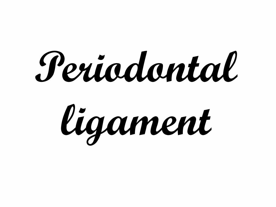
1- Cells
3-Fibers,
2- Intercellular substances
Synthetic
Resorpative
Progenitor
Defensive
4- ground substances
5- blood vessels,
nerves & lymphatic.
Histological structure
The periodontal ligament is formed of:
Epithelial

epithelial cells
remnants of the epithelial
root sheath of Hertwig
The cells Synthetic
cells
Resorptive cells
Progenitor cells
Defensive cells
fibroblasts, osteoblasts ,cementoblasts.
cementoclasts , osteoclasts, fibroblasts.
undifferentiated mesenchymal cells (Stem Cells)
macrophage, lymphocytes and mast cells

- Epithelial rests of
Malassez:
**deeply stained
cuboidal cells
surrounded by basal
lamina.
** They may
proliferate to form
cysts or tumors.

II- The fibers
*The fibers of the periodontal ligament are mainly collagen.
They are divided into:
A) The principal fibers.
B) The accessory fibers.
C) The oxytalan fibers.
*Elastic fibers are restricted almost entirely to the
walls of blood vessels.

A- The principal fibers of periodontal ligament are
formed of collagen bundles, which are wavy in course
and are arranged mainly in three ligaments.
a) Gingival ligament.
b) Transseptal or interdental ligament.
c) Alveolodental ligament which is subdivided into the following five groups:
1- Alveolar crest group.
2- Horizontal group.
3- Oblique group.
4-Apical group.
5- Interradicular group.

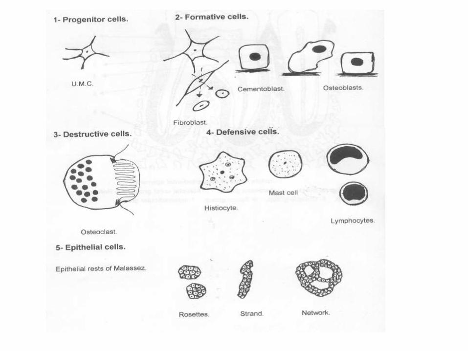
1- The principal fibers: a- The gingival ligament:
1- Gingival fibers: extend from the cervical cementum into the lamina propria of the gingiva.
2- Alveologingival group: extends from
the alveolar crest into the lamina propria.
3- Circular group: a small group of
fibers that encircles the tooth and interlaces with the outer fibers & bone.
4- Dentoperiosteal fibers: they extend
from the cementum direct over the crest of the alveolar bone then inserted in periosteum of the labial surface of the alveolar bone.
Alveolo-gingival Dento-
gingival
Dento-periosteal
Circular fibers
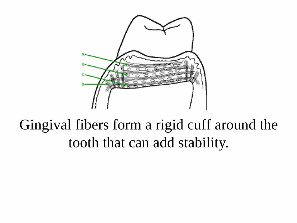
Gingival fibers form a rigid cuff around the
tooth that can add stability.

b- The Transseptal fibers: *It connects two adjacent teeth
so it is called interdental group of fibers.
*The ligament runs from the cementum of one tooth over the crest of the alveolus to the cementum of the adjacent tooth.
* Responsible for the mesial shift of the teeth.
Dentin
Dentin
Bone

Transseptal Fibers

c- The alveolodental fibers:
1-Alveolar crest group:
radiate from the crest of the
alveolar process and attach
themselves to the cervical part
of the cementum.
2-Horizontal group:
The fiber bundles run from the
cementum to the bone at right
angle to the long axis of the
tooth.
Bone Dentin

3- Oblique group:
The fiber bundles run obliquely.
Their attachment in the bone is somewhat coronal than the attachment in the cementum.
It is the greatest number of fiber bundles found in this group.
They perform the main support of the tooth against masticatory force.
bone
dentin

4- Apical group:
The bundles radiate from the
apical region of the root to
the surrounding bone.
5- Interradicular group:
The bundles radiate from the
interradicular septum to the
furcation of the multirooted
tooth.
dentin
bone
dentin
bone

a
l
v
e
o
l
o
d
e
n
t
a
l
P
r
i
n
c
i
p
l
e
f
i
b
e
r
s

B- Accessory fibers:
It is collagenous in nature and
run from bone to cementum
in different planes, more
tangentially to prevent
rotation of the tooth and
found in the region of the
horizontal group.

C- Oxytalan fibers
These are immature elastic (pre-elastic) fibers.
They need special stains to be demonstrated.
They tend to run in an axial direction, one end being embedded in bone or cementum and the other in the wall of blood vessels.

The function of the oxytalan fibers has been
suggested that they play a part in supporting the
blood vessels of the periodontal ligament during
mastication i.e., it prevents the sudden closure of the
blood vessels under masticatory forces.

Interstitial tissue
It is found between the fibers
of the periodontal ligament.
They are areas containing
some of the blood vessels,
lymphatics and nervs and
surrounded by loose
connective tissue.

Blood supply
The arterial blood supply of the periodontal ligament is derived from 3 sources:
3- Branches from the apical vessels that supply the dental pulp.
2- Branches from the intra-alveolar
vessels, these branches run horizontally
and these constitutes the main blood
supply.
1- Branches from the gingival vessels.
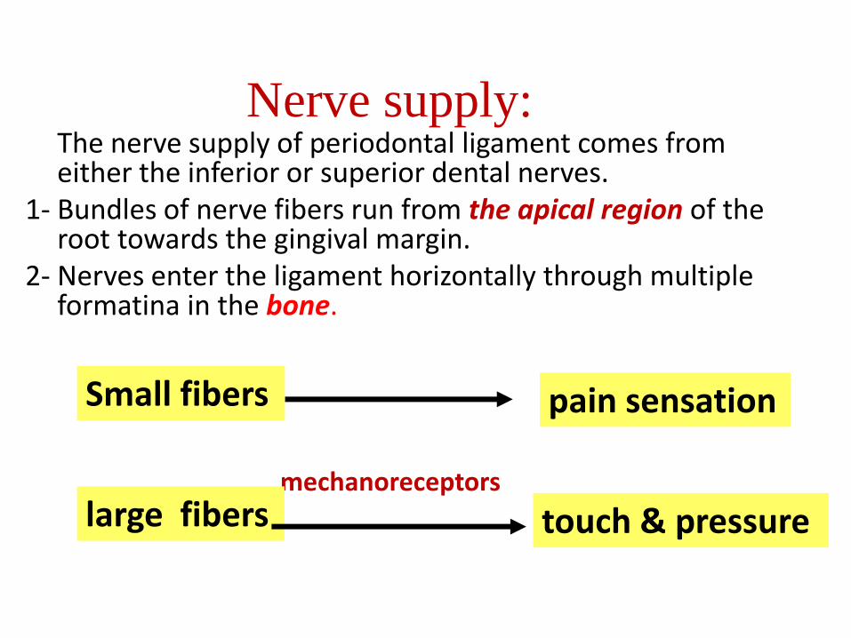
Nerve supply: The nerve supply of periodontal ligament comes from
either the inferior or superior dental nerves. 1- Bundles of nerve fibers run from the apical region of the
root towards the gingival margin. 2- Nerves enter the ligament horizontally through multiple
formatina in the bone.
mechanoreceptors
large fibers
Small fibers pain sensation
touch & pressure

Functions of the periodontal ligament:
1- Supportive: *periodontal ligament permits the teeth to withstand the
considerable forces of mastication.
*As the force is applied on the teeth, the wavy course
collagen fibers are transmitting tension to the wall of the
alveolus instead of pressure.
*Also periodontal fibers being non elastic to prevent the
tooth from being moved too far.

2- Sensory:
• The periodontal ligament having the
mechanoreceptor contributes to the
sensation of touch and pressure on the
teeth.
sudden overload proprioceptive reflex inhibition of the activity
of the masticatory muscles
Opening the mouth

3- Nutritive:
The blood vessels in the periodontal ligament provide nutrient supply required by the cells of the ligament and to the cementocytes and the most superficial osteocytes.
4- Formative: The fibroblasts are responsible for the formation of new
periodontal ligament fibers and dissolution of the old fibers
Cementoblasts and osteoblasts are essential in building up cementum and bone.

5- Protective:
The protective function of the periodontal ligament is achieved by:
a- The principal fibers.
b- The blood vessels.
c- The nerves.
a- The principal fibers:
The arrangement of the fiber bundles in the different groups is well adapted to fulfill the functions of the periodontal ligament.
The alveolodental ligament transforms the masticatory pressure exerted on the tooth into tension or traction on the cementum and bone.
If the exerted force on a tooth is transmitted as pressure this will lead to differentiation of osteoclasts in the pressure area and resorption of bone.

b- The blood vessels:
The capillaries form a rich network, they are arranged in the form of a coil and attached to bone and cementum through the oxytalen fibers.
This arrangement makes it possible when pressure is exerted on the tooth, the blood does not escape immediately from the capillaries and thus buffering the pressure action before it reaches the bone.
The behavior of the blood in the capillaries may be simulated to a hydraulic brake.
c- The nerves:
By its mechanoreceptors nerves.

Age Changes of periodontal ligament
The periodontal ligament through aging shows
• Vascularity
• Cellularity
• Thickness
*It may contain cementicles.

The cementicles appear near the surface of cementum may be free , attached or embedded in the cementum.
They have nidus favoring the deposition of concentric layers of calcospherite as degenerated cells, area of hemorrhage and epithelial rest's of Malassez.
Cementicles are usually seen in periodontal ligament by aging but in some cases they may be seen in a younger person after local trauma.

Thank you