Embed Size (px)
Citation preview
1
Periodic reversals in Paenibacillus dendritiformis swarming 1
2 Avraham Be'er#1,2, Shinji K. Strain*2, Roberto A. Hernández2, Eshel Ben-Jacob3,4, and E.-L. Florin2 3
4 5 1The Jacob Blaustein Institutes for Desert Research, Zuckerberg Institute for Water Research, Ben-Gurion University of the 6
Negev, Sede Boqer 84990, Israel; 2Center for Nonlinear Dynamics and Department of Physics, University of Texas, Austin, 7
TX 78712; 3School of Physics and Astronomy, Raymond and Beverly Sackler Faculty of Exact Sciences, Tel Aviv University, 8
Tel Aviv 69978, Israel; 4Center for Theoretical Biological Physics, Rice University, Houston TX 77025 9
10
# Corresponding Author: Tel 011-972-544-337478; Fax 011-972-8-6563503; Email [email protected] 11
* Currently at the Department of Bioengineering, Rice University, Houston TX 12 13 Running title: Swarming of long bacteria 14 15 Keywords: Bacterial swarming; Paenibacillus dendritiformis; periodic reversals; cell motility; long bacteria; 16 17 18 ABSTRACT 19 20 Bacterial swarming is a type of motility characterized by a rapid and collective migration of bacteria on 21 surfaces. Most swarming species form densely packed dynamic clusters in the form of whirls and jets 22 where hundreds of rod-shaped rigid cells move in circular and straight patterns, respectively. Recent 23 studies have suggested that short-range steric interactions may dominate hydrodynamic interactions, 24 and that geometrical factors, such as cell’s aspect ratio, play an important role in bacterial swarming. 25 Typically, the aspect ratio for most swarming species is only up to 5, and detailed understanding of the 26 role of much larger aspect ratios remains an open challenge. Here we study the dynamics of 27 Paenibacillus dendritiformis C morphotype, a very long hyperflagellated straight (rigid) rod-shaped 28 bacterium with an aspect ratio of ~20. We find that instead of swarming in whirls and jets as observed 29 in most species, including the shorter T-morphotype of P. dendritiformis, the C-morphotype moves in 30 densely packed straight, but thin, long lines. Within these lines, all bacteria show periodic reversals, 31 with a typical reversal time of 20 s, which is independent of their neighbors, the initial nutrient level, 32 agar rigidity, surfactant addition, humidity level, temperature, nutrient chemotaxis, oxygen level, 33 illumination intensity or gradient, and cell length. The evolutionary advantage of this unique back-and-34 forth surface translocation remains unclear. 35 36 37 38
39
Copyright © 2013, American Society for Microbiology. All Rights Reserved.J. Bacteriol. doi:10.1128/JB.00080-13 JB Accepts, published online ahead of print on 19 April 2013

2
INTRODUCTION 40
Motile bacteria are able to colonize surfaces using various motility mechanisms (1). One efficient 41
method includes flagellated-based cell motion in conjunction with collective lubrication (typically by 42
secretion of surfactants) to enable fast expansion on hard surfaces. This mode of ‘bacterial swarming’ 43
that has been studied extensively for many species (1-15), enables rapid colony expansion (up to 44
centimeters per hour). Swarming is often marked by hundreds of cells moving in a coordinated fashion 45
while generating whirl and jet patterns. 46
47
Studies of the collective dynamics of swarming have examined multiple aspects of motility. On the 48
macroscopic level it was discovered that swarming colonies show an advantage over liquid cultures in 49
that they exhibit an increased resistance to antimicrobials (1, 4-5, 15-20). Studies of collective 50
secretions of signaling and quorum sensing molecules have shown how interactions between cells in 51
swarming colonies are controlled (11), and exposed the identification of associated genetic 52
manipulations and upregulated proteins that control biosurfactant secretions and flagellar behavior. On 53
the single cell level, attention was given to swarm-cell trajectories and the ways in which these 54
trajectories are determined by flagellar motion (8, 21-23). A combination of experiments (2-3, 14-15, 55
24-38) and theory (39-43) suggests that hydrodynamic interactions play a significant role in this social 56
form of migration. 57
58
Hydrodynamic interactions may not always be the dominant physical mechanism controlling bacterial 59
motion. During differentiation to the swarming state, the cells of most species elongate and are thus 60
subjected to strong steric and excluded volume interactions (29, 44-45). The cells are typically 61
considered self propelled rods, having a straight rigid body; i.e., no cell bending is observed by light 62
microscopy (Fig. S1). For very long cells this may affect the self-organization, and change the dynamic 63
patterns. The result is that the very long straight bacteria may no longer swarm in whirls and jets (45). 64
In a recent study on collective motion of Bacillus subtilis in liquids (29), clusters of bacteria with high 65
orientational order have locally high swimming speeds, while orientationally disordered regions have 66
lower speeds. The role of length/width body-ratio plays an important role in the explanations offered 67
for the mode of collective motion (45), but since most swarmers in nature are self propelled straight 68
rods and have length/width body ratio of up to ~5 (around ~10 for two daughter cells before 69
reproduction is completed), swarming of longer (e.g., length/width >20 of a single cell) straight rods 70
was never studied experimentally. [Recently, Tuson et al. (46) studied the dynamics of Proteus 71
mirabilis, a long (length/width >20) swarming species, and observed that they move in whirls and jets 72

3
similarly to short (aspect ratio~5) and straight species. However, because these cells are curved and 73
bent during swarming, (typically look like boiled spaghetti (46)) they do not fall under the category of 74
self-propelled rods, i.e. straight (rigid) bacterial cells (45). In our study we see that the rods do not bend 75
even during collisions (Fig. S1)] 76
77
In this study, we quantitatively examine the swarming dynamics of very long rods using P. 78
dendritiformis morphotype C (47). Surprisingly, we find that instead of the standard dynamic patterns 79
of whirls and jets, observed in shorter species (e.g. B. subtilis (2)), and in particular the shorter 80
morphotype of P. dendritiformis (morphotype T) (3, 14), P. dendritiformis morphotype C forms long 81
tracks in which individual bacteria repeatedly move back-and-forth along moderately curved lines. 82
Direction switching is periodic, and each cell reverses (backs up) on average approximately every 20 s 83
independently of its neighbors. The time between switching was found to be independent of initial 84
nutrient level, agar rigidity, surfactant additions, cell-length (a broad length distribution of ~17±12 μm, 85
and a fixed width of ~1 μm are always present in a normal culture), humidity level, temperature, food 86
chemotaxis and oxygen level. This independence of reversal times suggests an extraordinary robust 87
internal clock for reversal events. The observed periodic reversals are different from those observed for 88
other species such as Myxococcus xanthus (48-52), Halobacterium salinarium (53-54) and Acetobacter 89
xylinum (55), in that the motive organelles are different. We thus report a new behavior for swarming 90
motility: periodic reversals in very long and rigid hyperflagellated bacteria. This unique behavior is so 91
far limited to P. dendritiformis. The generality of this type of swarming is yet to be known and so is the 92
general correlation to cell aspect ratio. 93
94
MATERIALS AND METHODS 95
Strain and growth media 96
Paenibacillus dendritiformis (morphotype C; aka chiral morphotype) is a spore-forming, motile 97
bacterial species (56). Each bacterium is an elongated (filamentous) rigid (seldom bends by collisions) 98
rod-shaped cell, with a fixed thickness of ~1 μm and a very broad length distribution of ~ 1217 ± μm. 99
The bacteria were maintained at -80 ºC in Luria Broth (LB) (Sigma, St. Louis, MO) with 25% [wt/vol] 100
glycerol. Luria Broth was inoculated with the frozen stock and grown for 24 hours at 30 ºC while 101
shaking; it was subsequently grown to an OD650 of 0.8, corresponding to approximately 7101× 102
bacteria/ml (calibrated by counting colonies on agar after appropriate dilution). 103
104

4
The agar plates were prepared as follows: peptone medium contained NaCl (5 g/l), K2HPO4 (5 g/l), and 105
Bacto Peptone (Becton Dickinson) in the range of 0.5 to 8 g/l. Either Eiken (Tokyo, Japan) or Difco 106
(Becton Dickinson) agar was added at concentration 0.7 to 1.5% [wt/vol]. Twelve ml of molten agar 107
was poured into 8.8 cm diameter Petri plates, which were dried for 4 days at 25 ºC in 50% relative 108
humidity (RH) until the weight decreased by 1 g. Similar plates were prepared with addition of 109
0.0006% [wt/vol] Brij 35. At these concentrations Brij 35 was found to not affect bacterial metabolism; 110
however, it does reduce the surface tension of the colony enabling faster colonial expansion. Nutrient-111
rich plates were prepared by mixing 2.5% [wt/vol] Luria Broth with agar in the concentrations noted; 112
25 ml of molten agar was poured into similar plates and dried for 24 h. 113
114
Some peptone plates were used to grow colonies at higher and lower oxygen levels. The plates were 115
inserted, after inoculation, into plastic bags filled with pure oxygen or with pure nitrogen. In other 116
cases the colonies were exposed to oxygen or nitrogen only at the time of observation by bounding the 117
microscope-stage area with a cell to which the gases were streamed. This was done as the bacteria can 118
adapt to a certain oxygen level; however, a rapid change of oxygen in the course of observation might 119
produce a strong effect on the reversal rates and on the speed (57). Nutrient chemotaxis experiments 120
were performed on plates that were kept slightly tilted while the agar was cooling. 121
122
A motility buffer was used to move cells from the agar into liquid. The motility buffer was made of 123
0.067 M sodium chloride, 0.01 M potassium phosphate (pH 7.0), 0.01 M sodium lactate, 10-4 M EDTA 124
and 10-6 M L-Methionine. 125
126
Colonial expansion 127
The agar plates were inoculated by placing 5 µl droplets of the culture at the center of the plate. The 128
plates were mounted on a rotating stage inside a 1 m3 chamber typically maintained at 5.00.30 ± ºC 129
and %290 ± (RH) (58). Some experiments were performed at other temperatures (25-40 ºC) and 130
humidities (35-90% RH). The rotating stage system enabled us to monitor growth development of ten 131
plates simultaneously; the rotation has no influence on any of the bacterial properties. The stage was 132
controlled by a stepper motor that stops sequentially for each bacterial colony to be imaged. A rotation 133
period of 1 h was sufficiently short to capture the growth of the colony. The reproducibility of 134
positioning of the agar plates was 15± µm, allowing successive images of a given colony to be 135
subtracted to determine growth patterns. Images were obtained with a 10 Megapixel Nikon D200 136
camera with a 60 mm lens. The camera was placed above the rotating stage and was programmed to 137

5
take an image and store the data when a sample was stationed below it. For plates imaged only at a 138
single point in time, colonies were stained with 0.1% [wt/vol] Coomassie Brilliant Blue to obtain 139
higher contrast images as compared to those obtained in the sequences of images. 140
141
Microscopic measurements 142
An optical microscope (Olympus IX50) equipped with a LD 60X Phase contrast (PH2) and LD 20X 143
Phase contrast (PHC) objective lenses was used to follow the microscopic motion. The microscope was 144
placed in a temperature and humidity controlled environment. A digital camera captured the 145
microscopic motion at a rate of 60 frames per second and a spatial resolution of 10001000× pixels. 146
Images were taken for 5 min periods, resulting in 18,000 images in a sequence. Tracking bacterial 147
motion was done manually, using a program called Tracker, by Douglas Brown 148
(http://www.cabrillo.edu/~dbrown/tracker/). 149
150
Electron Microscopy 151
We used an FEI Tecnai transmission electron microscope (TEM) operated at 80 kV. The long rod-152
shaped cells were collected from the agar using different methods and placed on 400-mesh copper 153
carbon grids (from Electron Microscopy Sciences). Flagella were lost in most cases. The best results 154
were obtained by gently stamping the grids on the live colony at the region of interest for 1 s and then 155
lifting the grid. The sample was immediately stained with 0.5% [wt/vol] uranyl acetate for 10 s which 156
fixes the cells and flagella in the state they were at the time of contact with the grid. 157
158
RESULTS 159
Growth at canonical conditions 160
In this section we describe the colony developments at intermediate conditions (2 g/l peptone, 1% 161
[wt/vol] agar, 30 ºC and 90% RH) which are considered to be the canonical or typical for the 162
development of P. dendritiformis chiral morphotype C colonies. 163
164
Colony expansion: Colonial development involves the following stages: 1. An incubation lag time of 165
11 h, during which bacteria reproduce in the center of the inoculation producing a “mother colony” 166
(Fig. 1A-B). 2. Sprouting of leading (pioneering) branches from the mother colony (Fig. 1B). 3. 167
Colony expansion. The colony expands on the surface by sending out thin curved branches (curly 168
branches with well defined handedness), forming intricate branched patterns within a well-defined 169
circular envelope (Fig. 1C-E). Several thin branches, on the rim of the circle of inoculation, start to 170

6
stem perpendicularly to the rim, and expand outward. For several hours the branches continuously curl 171
clockwise (colony faces up, agar faces down) and with the same speed grow back towards the circle of 172
the inoculation, ending at (or crossing) the neighboring curly branch. During this growth, the branches 173
become wider, and newly formed curly branches stem from both the circle of inoculation and from the 174
older branches forming a complex pattern (Fig. 1E). Expansion rate was estimated by measuring the 175
speed of an imaginary circular envelope surrounding the colony. The speed of the growing envelope, 176
0.48 mm/h, is isotropic and constant as illustrated by the straight line in Fig 1F. The time development 177
of such a colony is presented in Movie S1. 178
179
Cellular motility: A closer look at the tips of the growing branches reveals that the long bacteria 180
exhibit a back-and-forth swarming (moving back and forth), mostly aligned parallel to their neighbors 181
due to orientation interactions (47) (Movie S2). However, instead of the classical pattern of clusters of 182
cells, here each cell was found to move solitarily. At the outer parts of the colony they form a 183
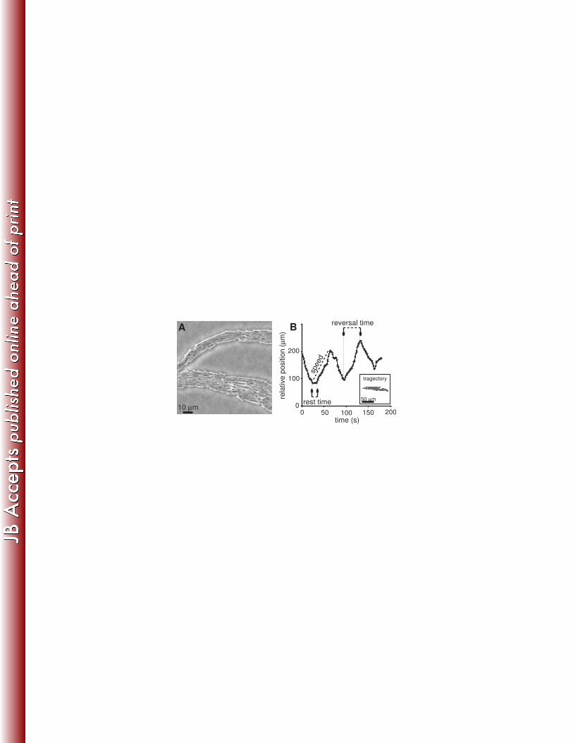
monolayer where roughly 10 bacteria lie next to one another (Fig. 2A). A typical trajectory of a single 184
bacterium among its neighbors is shown in Fig. 2B. Data was collected for single bacteria, located at 185
the middle of the branch, until they exited the field of view. Between reversal events, a bacterium’s 186
microscopic speed is fairly constant and remains the same after it reverses direction. We define the 187
reversal time as the time between switching events; set between the point in times at which the 188
bacterium begins moving and the point in time at which it stops and changes direction. Reversal times 189
for a single bacterium are typically the same but statistics for the same bacterium is limited as they tend 190
to leave the field of view after less than about 6 reversal events. Cells tend to exit the field of view in 191
the direction of colonial spreading; see moderate tendency in Fig. 2B. The cells typically rest for few 192
seconds (“rest time”) before they start moving in the opposite direction (Fig. 2B). 193
194
Speed and reversal time: To gain a better understanding of what determines the reversal times, we 195
analyzed 45 bacterial cells at the same growth conditions (canonical growth conditions of 2 g/l peptone 196
and 1% [wt/vol] agar, 30 ºC and 90% RH). About 15 cells were tracked in a field of view and the 197
experiment was repeated 3 times. All tracked cells were located at the middle of the branch. A total of 198
150 reversal events were observed. The average value for the bacterial microscopic speed was 2.1±0.7 199
µm/s and the average reversal time was 20.0±11.1 s with a positive skew. Figures 3A-B show 200
distribution for microscopic bacterial speeds and reversal times for 150 data points. A plot of reversal 201
time versus microscopic speed (Fig. 3C) shows no obvious correlation (p-value=0.004) and we 202
conclude that the reversal time is independent of bacterial microscopic speed. We also found that the 203

7
values for both the microscopic speed and the reversal time are constrained within some fixed 204
boundaries. For instance, at canonical growth conditions, bacteria never travel slower than 0.8 μm/s or 205
faster than 4.0 μm/s, and never switch direction sooner than 4.0 s after previous reversal. 206
207
Effect of growth conditions on the reversal time 208
By varying the nutrition level and agar concentration, we showed in our earlier work for morphotype T 209
(14) that the colonial expansion rate is independent of parameters of microscopic motion. Here, for 210
morphotype C, we grew bacteria at various peptone levels (1 g/l, 2g/l, 4 g/l, 8 g/l), and in LB, while 211
keeping the agar concentration (1% [wt/vol]), the temperature (30° C) and the relative humidity (90%) 212
unchanged. As expected, the expansion rate of the colony increases monotonically with increasing 213
nutrition levels (Fig. 4A). However, the microscopic speed as well as the reversal time seems to be 214
essentially independent of the nutrition level (Figs. 4 B-C) in contrast to the morphotype T for which 215
we observed a dependence of microscopic speed and nutrition level. 216
217
In the next step we investigated whether the reversal time could be affected by a gradient of nutrients, 218
namely whether food chemotaxis could affect the reversal time. Colonies were inoculated on agar 219
plates that were kept slightly tilted while the agar was cooling, enabling the colony to sense different 220
food levels in different directions. The colonies spread faster towards the richer (thicker agar) regions; 221
however, the microscopic motion, namely the bacterial speed and the reversal times were not affected. 222
223
To further check factors which might affect the motility and reversal time, we tested the effect of 224
surface tension. For this we have added to the agar, prior to autoclaving, very low concentrations 225
(0.0006% [wt/vol]) of Brij 35, which is a nonionic detergent solution that reduces the surface tension of 226
the medium. At these concentrations Brij 35 does not affect bacterial metabolism. As seen in Fig. 4, for 227
a constant nutrient level (2 g/l), colonial expansion was much faster due to lower surface tension. 228
However, the speed and reversal time remained unchanged. 229
230
Next we tested the effect of the surface hardness by varying the agar concentration (0.7-1.5% [wt/vol]) 231
while keeping the nutrient level constant (Fig. S2). On hard (1.3-1.4% [wt/vol]) Difco agar the bacteria 232
grew thick branches, forming multiple layers. Tracking individual cells was limited to the branches 233
tips, and statistics was poor. On Eiken agar, associated with high wetability, the colonies formed 234
monolayers at concentrations 0.7-1.3% [wt/vol]. Macroscopic expansion was affected by agar 235

8
concentration in a nonmonotonic way: fast expansion at intermediate levels of 0.9-1.1% [wt/vol] with a 236
maximum at 1.0% [wt/vol], and slow expansion at high (1.2-1.3% [wt/vol]) and low (0.7-0.8% 237
[wt/vol]) concentrations. The microscopic speed and reversal times were not affected and were similar 238
to those obtained on Difco agar. These results again suggest that the reversal time is a robust parameter, 239
essentially independent of any external factor. 240
241
Effect of environmental conditions 242
To further explore other possible factors which might affect the reversal time, we have performed four 243
additional experiments in which we changed environmental conditions that affected both the colonial 244
expansion and the motility (temperature, humidity, oxygen concentration and illumination). 245
246
Temperature effect: We have performed experiments at different temperatures in the range of 25-40 247
ºC (Fig. S3). At low temperatures (< 28 ºC) the colony expanded slowly, and the microscopic bacterial 248
speed was slow as well. At high temperatures (>37 ºC) the colony did not expand at all and no 249
microscopic motion was detected; loss of motility was observed at these temperatures in liquid cultures 250
too, but bacteria were alive. Between 28-37 ºC, the microscopic bacterial speed changed in a 251
nonmonotonic way: fastest motion at 32 ºC and slowest at 28 ºC and 36 ºC. In the regimes that motion 252
was detected (28-37 ºC), the reversal time was not affected. 253
254
Humidity effect: We have changed the humidity between 35-90% RH; below 35% no growth was 255
observed. The drier the air, the slower the colonies expanded and the slower the bacteria moved at the 256
microscopic level (Fig. S4). For instance, at intermediate dry growth conditions (2 g/l peptone, 1% 257
[wt/vol] agar, 30 ºC and 40% RH), the average colonial expansion rate was 0.5 mm/day (~20 times 258
slower than at 90% RH), and the average microscopic speed was 0.5 μm/s (4 times slower than at 90% 259
RH). But again, the reversal time was not affected and bacteria changed direction every ~20 s as 260
observed for all other cases. 261
262
Oxygen effect: We have grown the colonies at high, medium and low oxygen levels. At very low 263
oxygen levels (plastic bags filled with nitrogen), the expansion rate, the microscopic speed and the 264
reversal time were not affected (similar to ambient conditions). At very high oxygen levels (plastic 265
bags filled with oxygen), no growth was detected (plates were later placed at ambient conditions for 266
weeks with no recovery; we could not recover cells from the colony by reinoculating on fresh LB 267
substrates). At medium oxygen levels (plastic bags filled with a mixture of oxygen and ambient air 268

9
with no real control of oxygen percentage), the colonies expanded slower, the microscopic motion was 269
slower but the reversal time remained 20 s on average. In other cases pure oxygen was added at the 270
time of observation only. In these cases a gradual reduction in each bacterial speed was observed, 271
typically reducing their speeds from ~2 μm/s to ~0.5 μm/s, until an abrupt stop. Most cells stopped 272
almost simultaneously after 20 min, and no motion was detected after 30 min. However, the reversal 273
time remained ~20 s as long as the cells moved. 274
275
Illumination effect: Some phototactic bacteria such as H. salinarium show reversals due to gradients 276
of light. Therefore, we tested whether P. dendritiformis C-morphotype will react to changes in 277
illumination. Different band pass filters (blue ~480 nm, green ~508 nm, yellow ~560 nm and red ~650 278
nm) were applied successively; the microscope light source (Olympus IX50 – standard 30 W halogen 279
light bulb) was turned on and off for different time lengths (between 0.5 s and 1 h) and the motion of 280
the cells was monitored. No changes in bacterial speed or in the reversal time were detected. We have 281
repeated these experiments by illuminating colonies from the side. Side illumination was done at 282
different angles (10º, 20º, and 30º with respect to the surface – this indirectly created a dark-field 283
effect) by using a goose neck fiber optic illuminator (150H, Schölly Fiberoptik GmbH, Germany), 284
creating a gradient in intensity along the xy plane. Illumination intensity at the sample was ~2 W/cm2. 285
Again, no response to light was detected suggesting that phototaxis is not a mechanism dictating the 286
reversal events in P. dendritiformis C-morphotype bacteria. 287
288
Effect of neighboring cells 289
We found that cells reversed their direction of motion independently of collisions with neighboring 290
cells. For instance, neighboring cells were tracked to see if they tended to move in the same or opposite 291
directions, or whether they tended to switch directions upon contact with cells that were moving in the 292
same or opposite direction. Almost 60% of tracked cells (1240 cells) were moving to the direction their 293
close (in contact) neighbors were moving, indicating hydrodynamic interaction. However, cells were 294
switching direction independently of the direction of motion of their neighbors. Out of 380 tracked 295
cells, 184 (48%) switched direction while close neighbors moved to opposite direction. Cells that 296
moved to one direction having 2 neighboring cells, at both sides, moving to the opposite direction, 297
showed same results. This demonstrates that the tendency of switching direction is intrinsic, and is 298
independent of hydrodynamics. 299
300

10
Additionally, cells located at the outer part of the branches (having neighbors on one side only) were 301
tracked too and found to reverse their direction every 20 s as well. However, they moved slightly 302
slower (10% speed reduction) probably due to a shallower liquid film. 303
304
Many one-dimensional head-on collisions, with bacteria moving in opposite directions, were observed 305
but these incidents did not lead to reversals. Thus, this back-and-forth swarming differs from the 306
reversal behavior of swarming E. coli and other bacteria during collisions. To further verify that 307
collisions did not play a role in direction switching, cells grown on agar were moved into a motility 308
liquid-medium by placing a small drop of the motility buffer on the colony, letting bacteria migrate to 309
the drop, then moving it to a glass slide. In the drop, the bacterial concentration was much smaller, thus 310
no collisions were observed. However, the cells swam in the drop similarly to what was observed on 311
the agar in that they typically reversed their directions every 20 s. We also observed that cells which 312
were initially grown in standard liquid media, LB, (not transferred from the agar) exhibited an entirely 313
different motion: they swam along nearly straight trajectories with no back-and-forth reversals or run-314
and-tumble movements. Such cells were monitored for more than 15 minutes. 315
316
Does the C-morphotype swarm? 317
To obtain a better understanding of the motility of the C-morphotype on an agar surface and clearly 318
define its motion as “swarming”, we have performed several experiments. First, we have used 319
transmission electron microscopy (TEM) to find the motive organelle of P. dendritiformis morphotype 320
C. Possible mechanisms are the S motility of type-IV pulling pili, a.k.a. twitching (e.g., A. xylinum or 321
Pseudomonas aeruginosa), a combination of S motility at one pole and an A motility engine that works 322
in the opposed direction (e.g., M. xanthus), a single flagellum that can switch rotational direction from 323
clockwise (CW) to counterclockwise (CCW) (e.g., H. salinarium), and motility due to multiple flagella 324
as often seen in swarming cells (e.g., E. coli)). All of these have been previously associated with run-325
and-tumble or back-and-forth periodic events. Amphitrichous, i.e., two polar flagella, each located at a 326
different pole (e.g., Spirillum), is another potential configuration that bacteria can utilize in back-and-327
forth reversals. 328
329
Independent of growth conditions, multiple flagella were observed for cells harvested from agar plates, 330
but bacteria grown on Eiken agar gave better images (the cells are slightly shorter on the Eiken agar). 331
The many flagella (~100 for each cell) are peritrichous - they are uniformly distributed all over the cell 332
(Fig. 5A-B). A closer look shows that each flagellum is solitarily connected to the membrane through 333

11
the basal body (Fig. 5C), and in most cases, a few flagella formed bundles (Fig. 5D). Many of these 334
flagellar bundles were found to be distributed in various places around the cell. All of the flagella have 335
similar physical features; they are 10-20 μm in length and 17±1 nm in width (Fig. 5E). No pili were 336
observed, suggesting that twitching is not the motility mode of P. dendritiformis morphotype C in these 337
growth conditions. The images were compared to those obtained for cells grown in liquid media where 338
a single bundle of 4 flagella, located at one pole, was observed (Fig. 5F). This suggests that 339
extracellular conditions had a very large influence on the number and position of flagella on the cells. 340
341
Secondly, we have used standard flagellar shearing protocols to shear flagella off of the cells. As 342
described above, bacteria were collected from agar plates by placing a small drop of the motility buffer 343
on the colony, allowing bacteria to migrate to the drop. Cells that were not sheared moved back-and-344
forth in the liquid, indicating that their motion on the agar was flagellar driven. Shearing was achieved 345
by repeated pipetting through a narrow (0.6 mm in diameter) straw. Sheared cells did not swim in the 346
liquid bulk and flagella were not observed in TEM. Thirdly, a suspension of sheared bacteria was 347
placed on an untreated standard glass slide. After bacteria attached to the surface they started to rotate 348
either CW or CCW. Independent of whether they rotated CW or CCW, they reversed their direction 349
every few seconds, indicating that the flagellar rotors are still present and active (Movie S3). 350
351
All three results together strongly suggest that flagella are the motive organelles in the C-morphotype 352
and that the spreading of the C-morphotype fulfills the more stringent definition of swarming as a 353
flagellar driven motility on surfaces (1) (the less stringent definition of swarming is any type of 354
collective bacterial motion even if not powered by flagella). 355
356
Effect of bacterial length 357
Our results that the reversal time is essentially independent of all parameters tested point towards the 358
existence of a robust internal clock for the timing of reversal events. It is very likely that the flagella 359
need to be synchronized to reverse the direction. The way flagella are structured on the cells raises the 360
possibility that, similarly to other peritrichous-flagellated species, large bundles are formed at the poles 361
during motion. For a reversal event to happen, the signal has to travel through the cell from one pole to 362
the other turning on and off the rotation of the flagellar bundles or switching their direction. If the 363
signal propagation is the limiting factor for the speed of reversals, one expects a significant rest time 364
when bacteria change their direction and that this rest time depends on the length of the bacteria. 365
However, Figure 6 shows that the reversal time (Fig. 6A) as well as the rest time (Fig. 6B) is 366

12
independent of bacterial length (p-values are 0.076 and 0.068 respectively). The histogram of rest times 367
(Fig. 6C) is strongly asymmetric with a maximum around 3 s, with many shorter events, and a 368
minimum rest time of approximately 0.4 s. Also, the minimum rest time seems to be independent of 369
the bacterial length as suggested by Fig. 6B which points towards another origin. Similar rest times 370
have been described for B. subtilis (29) and attributed to flagellar bundle kinetics. 371
372
DISCUSSION 373
Hydrodynamic interaction has been considered as the key player in many cases of collective motion 374
observed in a number of bacterial species. However, it has been recently suggested that short-range 375
steric interactions might dominate over hydrodynamic interaction in swarming bacteria. For elongated 376
rigid self-propelled particles, collisions (i.e. short-range steric interactions) seem to result in the 377
alignment of motion that leads to a particular length and time correlation, and to the formation of the 378
classical swarming patterns of whirls and jets (2, 3). It is expected that geometrical factors of cell 379
structure, such as the aspect ratio (length/width), are important for the type of collective motion. The 380
pioneering work on P. dendritiformis (47, 59-60) has shown that morphotype C colonies exhibit chiral 381
morphology with twisted branches, as a combined result of flagellar chirality and cell-cell orientation 382
interactions. The theoretical models showed that the length of these motile bacteria plays a major role 383
in the resultant pattern which limits the average rotation, reducing the chance to form whirls and jets. 384
385
Wensink et al. (45) have recently calculated a non-equilibrium phase diagram for various aspect ratios 386
and volume fractions of self-propelled rods (bacteria), taking into account only steric interaction 387
between the rods; hydrodynamic interaction was neglected. For very long (rigid) rods (aspect ratio>13) 388
and at sufficiently high volume fractions (>0.3), they found that “bacteria” tend to assemble in 389
homogeneous long lanes corresponding to quasi-smectic regions of polar order. Our results are in 390
agreement with this prediction. 391
392
Besides being assembled in homogeneous long lanes, we found that all cells show periodic reversals 393
with a reversal time that was robust under all conditions tested. This phenomenon was not included in 394
recent models (45). Periodic reversals have been observed experimentally in other species too, but their 395
mechanism, properties or their function seem to be different. Starting with peritrichously flagellated 396
bacteria, it was recently shown that CW flagellar rotation in swarming E. coli cells results in reversal 397
events (21). These reversals are different from the run and tumble events of swimming bacteria and 398
were only found in a small fraction at the edges of the colonies. The distribution of reversal times was 399

13
approximately exponential, corresponding to a constant probability of changing direction, in contrast to 400
the distribution of reversal times observed here. Instead of an exponential decay, we found a broad 401
distribution and a clear maximum at about 12 s indicating that the mechanism that drives the reversals 402
might be different. 403
404
Reversals were also observed in B. subtilis. Cisneros et al. (61) observed reversals of cells in liquid 405
media (swimming cells) upon collisions with obstacles, indicating some mechanical sensing. One 406
suggested function of these reversals is that the cells try to avoid jammed areas. As discussed earlier, 407
reversals in P. dendritiformis C-morphotype seem to be spontaneous and independent of any 408
mechanical interaction with neighboring cells or any other object. 409
410
Reversals found in the archaeon H. salinarium (53-54), which grow in high salt environments, are a 411
behavioral response to gradients of light or oxygen (phototaxis and aerotaxis). No response to food or 412
light gradients was found for the P. dendritiformis C-morphotype. Finally, reversals were found in 413
pilus-driven species. For example, A. xylinum bacteria (55) are known for prolific synthesis of 414
cellulose, where the motion is pilus-driven and the microscopic back-and-forth motion is achieved by 415
attaching to the extruded cellulose ribbon produced. In M. xanthus (48-52), the cell movement is 416
propelled by a polar S engine (pulling type IV pili) at the leading end of the cell, and by an A gliding 417
engine that generates motion in the opposed direction. Measurements of the expansion rates of M. 418
xanthus mutants have revealed that the reversal period of wild-type strains is optimized for fast 419
expansion. It was suggested that the back-and-forth motion in this case increases the local alignment of 420
cells and reduces the interference between cellular motions. M. xanthus moves much slower, switches 421
direction at a much slower rate and uses a different motive organelle than the P. dendritiformis C-422
morphotype; however, it is still possible that the reversals fulfill a similar function in P. dendritiformis. 423
424
To reverse the direction of motion, flagella at different locations along the cell body need to be 425
synchronized which requires a signal to travel along the cell body. The speed of the signal and the body 426
length should set a minimum rest time during reversals. Surprisingly, we observed no dependence of 427
the minimal rest time (0.35 s) on body length which indicates that signaling along the cell body is not 428
the limiting factor for flagellar synchronization. Assuming a rest time of 0.50 s and a cell length of 40 429
µm, we can calculate the lower bound for the speed of the signal to be approximately 60 µm/s. This 430
speed seems to be too fast even for fast calcium waves (62) and it remains unclear how periodic 431
reversals are orchestrated. 432

14
433
P. dendritiformis has two motile morphotypes; the C-morphotype discussed here and the T-morphotype 434
which is a shorter (aspect ratio~5) rod-shaped motile strain that swarms in whirls and jets (14). Both 435
have the same 16S rRNA. Colonies of either morphotype, grown from a single cell are likely to exhibit 436
spontaneous transitions where small regions of a colony might grow branches containing cells of the 437
other morphotype. When this happens, the dynamics of swarming of the bacteria at the transitioned 438
regions is changed as well (see arrow in Fig. 1E). Our experiments suggest that the dramatic change in 439
aspect ratio, resulted by the spontaneous morphological transition, might be correlated with the 440
different type of collective motion. By now it is known that one major difference between morphotypes 441
T and C is a topological defect in cell division which makes the C much longer. However, the study of 442
the differences between the two morphotypes is not completed, and concrete conclusions about the role 443
of aspect ratio in changing the swarming pattern must be postponed. Patrick et al. (63) showed that 444
topological defects (ΔminJ) in cell division (i.e. the formation of longer cells) create swarming motility 445
defects in B. subtilis. The long hyperflagellated and surfactant producer cells form spiraling whorls on 446
the surface of the media instead of the classic spreading pattern formed by the wild type. 447
448
To conclude we have found the periodic reversals to be independent of environmental conditions such 449
as initial nutrient level, agar rigidity, surfactant addition, humidity level, temperature, nutrient 450
chemotaxis, oxygen level, illumination intensity or gradient and additionally independent of cell length 451
and cell density (collisions with other cells). Together with the fact that the reversal periodicity is also 452
not correlated with the colony expansion (in contrast to other bacteria), the evolutionary advantage of 453
this unique back-and-forth swarming remains a mystery. 454
455
ACKNOWLEDGMENTS 456
We thank Inna Brainis for providing the bacterial strain and the growth protocol. We are grateful to 457
Rasika M. Harshey, Harry L. Swinney, and Shelley M. Payne for many useful discussions and 458
guidance. 459
460
E.B-J. acknowledges support from the Tauber Family Funds and the Maguy-Glass Chair in Physics of 461
Complex Systems at Tel Aviv University, and partial support from NSF Grant PHY-0822283 and 462
Cancer Prevention and Research Institute of Texas (CPRIT) at Rice University. E.L.F. acknowledges 463
support from the Robert A. Welch Foundation under Grant F-1573. 464
465
15
466 REFERENCES 467 468 1. Harshey RM. 2003. Bacterial motility on a surface: Many ways to a common goal. Annual Review 469 of Microbiology 57:249-273. 470 471 2. Zhang HP, Be’er A, Florin E-L and Swinney HL. 2010. Collective motion and density 472 fluctuations in bacterial colonies. PNAS 107:13626-13630. 473 474 3. Zhang HP, Be’er A, Smith RS, Florin E-L and Swinney HL. 2010. Swarming dynamics in 475 bacterial colonies. EPL 87:48011. 476 477 4. Kearns DB. 2010. A field guide to bacterial swarming motility. Nature Reviews Microbiology 478 8:634-644. 479 480 5. Butler MT, Wang Q, and Harshey RM. 2010. Cell density and mobility protect swarming bacteria 481 against antibiotics. PNAS 107:3776-3781. 482 483 6. Matsuyama T, Kaneda K, Nakagawa Y, Isa K, Hara-Hotta H, and Yano I. 1992. A novel 484 extracellular cyclic lipopeptide which promotes flagellum-dependent and -independent spreading 485 growth of Serratia marcescens. Journal of Bacteriology 174:1769-1776. 486 487 7. Harshey RM, Matsuyama T. 1994. Dimorphic transition in Escherichia coli and Salmonella 488 typhimurium: Surface-induced differentiation into hyperflagellate swarmer cells. PNAS 91:8631-8635. 489 490 8. Wu Y, Hosua BG, and Berg HC. 2011. Microbubbles reveal chiral fluid flows in bacterial swarms. 491 PNAS 108: 4147-4151. 492 493 9. Kearns DB, Losick R. 2003. Swarming motility in undomesticated Bacillus subtilis. Molecular 494 Microbiology 49:581-590. 495 496 10. Wang Q, Frye JG, McClelland M, Harshey RM. 2004. Gene expression patterns during 497 swarming in Salmonella typhimurium: Genes specific to surface growth and putative new motility and 498 pathogenicity genes. Molecular Microbiology 52:169-187. 499 500 11. Daniels R, Reynaert S, Hoekstra H, Verreth C, Janssens J, Braeken K, Fauvart M, Beullens 501 S, Heusdens C, Lambrichts I, De Vos DE, Vanderleyden J, Vermant J, and Michiels J. 2006. 502 Quorum signal molecules as biosurfactants affecting swarming in Rhizobium etli. PNAS 103:14965-503 14970. 504 505 12. Ben-Jacob E, Becker I, Shapira Y, and Levine H. 2004. Bacterial linguistic communication and 506 social intelligence. Trends in Microbiology 12:366-372. 507 508 13. Ben-Jacob E, Cohen I, and Gutnick DL. 1998. Cooperative organization of bacterial colonies: 509 from genotype to morphotype. Annual Review of Microbiology 52:779-806. 510 511 14. Be’er A, Smith RS, Zhang HP, Florin E-L, Payne SM, and Swinney HL. 2009. Paenibacillus 512 dendritiformis bacterial colony growth depends on surfactant but not on bacterial motion. Journal of 513 Bacteriology 191:5758-5764. 514

16
515 15. Ingham CJ and Ben-Jacob E. 2008. Swarming and complex pattern formation in Paenibacillus 516 vortex studied by imaging and tracking cells. BMC Microbiology 8:36. 517 518 16. Henrichsen, J. 1972. Bacterial surface translocation: a survey and a classification. Bacteriological 519 Reviews 36:478-503. 520 521 17. Copeland MF, Weibel DB. 2009. Bacterial swarming: A model system for studying dynamic self-522 assembly. Soft Matter 5:1174-1187. 523 524 18. Kim W, Killam T, Surette MG. 2003. Swarm-cell differentiation in Salmonella enterica serovar 525 typhimurium results in elevated resistance to multiple antibiotics. Journal of Bacteriology 185:3111-526 3117. 527 528 19. Lai S, Tremblay J, and De´ziel E. 2009. Swarming motility: a multicellular behaviour conferring 529 antimicrobial resistance. Environmental Microbiology 11:126-136. 530 531 20. Ben-Jacob E. 2003. Bacterial self-organization: co-enhancement of complexification and 532 adaptability in a dynamic environment. Phil. Trans. R. Soc. London. A. 361:1283-1312. 533 534 21. Turner L, Zhang R, Darnton NC, and Berg HC. 2010. Visualization of flagella during bacterial 535 swarming. Journal of Bacteriology 192:3259-3267. 536 537 22. Zhang R, Turner L, and Berg HC. 2010. The upper surface of an Escherichia coli swarm is 538 stationary. PNAS 107:288-290. 539 540 23. Darnton NC, Turner L, Rojevsky S, and Berg HC. 2010. Dynamics of bacterial swarming. 541 Biophysical Journal 98:2082-2090. 542 543 24. Sokolov A and Aranson IS. 2009. Reduction of viscosity in suspension of swimming bacteria. 544 Phys Rev Lett 103:148101. 545 546 25. Dombrowski C, Cisneros L, Chatkaew S, Goldstein RE, Kessler JO. 2004. Self-concentration 547 and large-scale coherence in bacterial dynamics. Phys Rev Lett 93:098103. 548 549 26. Sokolov A, Aranson IS, Kessler JO and Goldstein RE. 2007. Concentration dependence of the 550 collective dynamics of swimming bacteria. Phys Rev Lett 98:158102. 551 552 27. Tuval I, Cisneros L, Dombrowsky C, Wolgemuth CW, Kessler JO, Goldstein RE. 2005. 553 Bacterial swimming and oxygen transport near contact lines. PNAS 102:2277-2282. 554 555 28. Cisneros LH, Cortez R, Dombrowski C, Goldstein RE, Kessler JO. 2007. Fluid dynamics of 556 self-propelled microorganisms, from individuals to concentrated populations. Experiments in Fluids 557 43:737-753. 558 559 29. Cisneros LH, Kessler JO, Ganguly S, and Goldstein RE. 2011. Dynamics of swimming bacteria: 560 Transition to directional order at high concentration. Phys Rev E 83:061907. 561 562
17
30. Be’er A, Harshey RM. 2011. Collective motion of surfactant-producing bacteria imparts 563 superdiffusivity to their upper surface. Biophysical Journal 101:1017-1024. 564 565 31. Sokolov A, Apodaca MM, Grzybowski BA, and Aranson IS. 2010. Swimming bacteria power 566 microscopic gears. PNAS 107:969-974. 567 568 32. Narayan V, Ramaswamy S, Menon N. 2007. Long-lived giant number fluctuations in a swarming 569 granular nematic. Science 317:105-108. 570 571 33. Aranson IS, Snezhko A, Olafsen JS and Urbach JS. 2008. Comment on “long-lived giant 572 number fluctuations in a swarming granular nematic”. Science 320:612. 573 574 34. Parrish JK, and Edelstein-Keshet L. 1999. Complexity, pattern, and evolutionary trade-offs in 575 animal aggregation. Science 284:99-101. 576 577 35. Makris NC, Ratilal P, Jagannathan S, Gong Z, Andrews M, Bertsatos I, Godø OR, Nero RW, 578 Jech JM. 2009. Critical Population Density Triggers Rapid Formation of Vast Oceanic Fish Shoals. 579 Science 323:1734-1737. 580 581 36. Ballerini M, Cabibbo N, Candelier R, Cavagna A, Cisbani E, Giardina I, Lecomte V, Orlandi 582 A, Parisi G, Procaccini A, Viale M, and Zdravkovic V. 2008. Interaction ruling animal collective 583 behavior depends on topological rather than metric distance: Evidence from a field study. PNAS 584 105:1232-1237. 585 586 37. Nathan R, Getz WM, Revilla E, Holyoak M, Kadmon R, Saltz D, and Smouse PE. 2008. A 587 movement ecology paradigm for unifying organismal movement research. PNAS 105:19052-19059. 588 589 38. Aranson IS, Sokolov A, Kessler JO and Goldstein RE. 2007. Model for dynamical coherence in 590 thin films of self-propelled microorganisms. Phys Rev E 75:040901R. 591 592 39. Simha RA, Ramaswamy S. 2002. Hydrodynamic fluctuations and instabilities in ordered 593 suspensions of self-propelled particles. Phys Rev Lett 89:058101. 594 595 40. Ramaswamy S, Simha RA, Toner J. 2003. Active nematics on a substrate: Giant number 596 fluctuations and long-time tails. EPL 62:196-202. 597 598 41. Toner J, Tu YH, Ramaswamy S. 2005. Hydrodynamics and phases of flocks. Annals of Physics 599 (New York) 318:170-244. 600 601 42. Dreschera K, Dunkela J, Cisneros LH, Gangulya S, and Goldstein RE. 2011. Fluid dynamics 602 and noise in bacterial cell–cell and cell–surface scattering. PNAS 108:10940-10945. 603 604 43. Gyrya V, Aranson IS, Berlyand LV, Karpeev D. 2010. A model of hydrodynamic interaction 605 between swimming bacteria. Bulletin of Mathematical Biology 72:148-183. 606 607 44. Peruani F, Deutsch A, and Bar M. 2006. Nonequilibrium clustering of self-propelled rods. Phys 608 Rev E. 74:030904R. 609 610

18
45. Wensink HH, Dunkel J, Heidenreich S, Drescher K, Goldstein RE, Löwen H, and Yeomans 611 JM. 2012. Meso-scale turbulence in living fluids. PNAS 109:14308-14313. 612 613 46. Tuson HH, Copeland MF, Carey S, Sacotte R, and Weibel DB. 2012. Flagella density regulates 614 Proteus mirabilis swarm cell motility in viscous environments. Journal of Bacteriology (in press); 615 personal communication at http://www.biochem.wisc.edu/faculty/weibel/lab/. 616 617 47. Ben-Jacob E, Cohen I, Shochet O, Tenenbaum A, Czirok A and Tamas Vicsek. 1995. 618 Cooperative Formation of Chiral Patterns during Growth of Bacterial Colonies. Phys Rev Lett 619 75:2899-2902. 620 621 48. Wu Y, Kaiser AD, Jiang Y and Alber SA. 2009. Periodic reversal of direction allows 622 Myxobacteria to swarm. PNAS 106:1222-1227. 623 624 49. Yu R and Kaiser D. 2007. Gliding motility and polarized slime secretion. Molecular Microbiology 625 63:454-467. 626 627 50. Blackhart BD and Zusman DR. 1985. “Frizzy” genes of Myxococcus xanthus are involved in 628 control of frequency of reversal of gliding motility. PNAS 82:8767-8770. 629 630 51. Igoshin OA, Welch R, Kaiser D, and Oster G. 2004. Waves and aggregation patterns in 631 myxobacteria. PNAS 101:4256-4261. 632 633 52. Mauriello EMF, Mignot T, Yang Z, and Zusman DR. 2010. Gliding Motility Revisited: How do 634 the myxobacteria move without flagella? Microbiol. and Molec. Biol. Revs, 74:229-249. 635 636 53. Krosh U. 1995. Damped Oscillations in Photosensory Transduction of Halobacterium salinarium 637 Induced by Repellent Light Stimuli. Journal of Bacteriology 177:3067-3070. 638 639 54. Lindbeck JC, Goulbourne EA Jr, Johnson MS, and Barry L. Taylor. 1995. Aerotaxis in 640 Halobacterium salinarium is methylation-dependent. Microbiology 141:2945-2953. 641 642 55. T Kondo, M Nojiri, Y Hishikawa, E Togawa, D Romanovicz, and RM Brown Jr. 2002. 643 Biodirected epitaxial nanodeposition of polymers on oriented macromolecular templates. PNAS 644 99:14008-14013. 645 646 56. Sirota-Madi A, Olender T, Helman Y, Brainis I, Finkelshtein A, Roth D, Hagai E, Leshkowitz 647 D, Brodsky L, Galatenko V, Nikolaev V, Gutnick DL, Lancet D, and Ben-Jacob E. 2012. Genome 648 Sequence of the Pattern-Forming Social Bacterium Paenibacillus dendritiformis C454 Chiral 649 Morphotype. Journal of Bacteriology 194:2127-2128. 650 651 57. Sokolov A and Aranson IS. 2012. Physical Properties of Collective Motion in Suspensions of 652 Bacteria. Phys Rev Lett 109:248109. 653 654 58. Be’er A, Zhang HP, Florin E-L, Payne SM, Ben-Jacob E, and Swinney HL. 2009. Deadly 655 competition between sibling bacterial colonies. PNAS 106:428-433. 656 657 59. Cohen I and Ben-Jacob E. 2000. Orientation field model for chiral branching growth of bacterial 658 colonies. arXiv:cond-mat/0008446v1. 659

19
660 60. Ben-Jacob E, Cohen I, Golding I and Kozlovsky Y. 2001. Modeling Branching and Chiral 661 Colonial Patterning of Lubricating Bacteria. The IMA Volumes in Mathematics and its Applications 662 121:211-253. 663 664 61. Cisneros L, Dombrowski C, Goldstein RE, Kessler JO. 2006. Reversal of bacterial locomotion 665 at an obstacle. Phys Rev E 73:030901R. 666 667 62. Jaffe LF. 2010. Fast calcium waves. Cell Calcium 48:102-113. 668 669 63. Patrick JE and Kearns DB. 2008. MinJ (YvjD) is a topological determinant of cell division in 670 Bacillus subtilis. Mol Microbiol 70:1166-1179. 671 672 673 FIGURE LEGENDS 674 675 FIG. 1 Macroscopic colonial expansion of P. dendritiformis morphotype C bacteria. (A-E) Different 676
stages of growth; A, at t=0 h, initial inoculation, B, at t=13 h, the spot inoculation is brighter and a few 677
branches have stemmed from the spot rim, C, t=17, the curly branches grow back towards the circle of 678
the inoculation, ending at (or crossing) the neighbor curly branch, D, t=23 h, the branches become 679
wider, and newly born curly branches stem from both the circle of inoculation and from the older 680
branches, E, t=28 h, the complex growth continues. The arrow indicates a small region of morphotype 681
T cells (that spontaneously switched from the C morphotype) that exhibit whirl and jet swarming 682
patterns at the microscopic level. (F) The macroscopic expansion speed (0.48 mm/h) is indicated by the 683
slope of a plot of the position of the imaginary envelope (x) as a function of time. The expending 684
envelope has concentric circles at time intervals for each colony. The radii are determined by following 685
the farther tip of the colony at each time step (see circle in D). The error bars indicate the standard 686
deviations for measurements for 10 colonies. Growth conditions are 2 g/l peptone, 1% [wt/vol] Difco 687
agar, 30 ºC and 90% RH. 688
689
FIG. 2 Microscopic reversals of a single cell. (A) A microscopic picture of the outer parts of a colony. 690
The cells form a monolayer where roughly 10 bacteria lay one next to the other. Bacterial length 691
distribution is broad; some cells are very long (~ 40 μm) and some are much shorter (~5 μm). (B) The 692
relative position (with respect to some arbitrary fixed point on the agar) of a single cell in a colony as a 693
function of time. Reversal time is defined as the time between the point at which the bacterium begins 694
moving and the point at which it stops changing direction. A trajectory of the bacterium is shown in the 695
inset. Growth conditions are 2 g/l peptone, 1% [wt/vol] Difco agar, 30 ºC and 90% RH. 696
697

20
FIG. 3 Microscopic speeds and reversal times of many bacteria at the same growth conditions (2 g/l 698
peptone, 1% [wt/vol] Difco agar, 30 ºC and 90% RH). (A) Microscopic speed distribution for 150 data 699
points; a quite large range of speeds is observed with an average of 2±0.7 μm/s. (B) Reversal time 700
distribution for 150 data points; a quite large range of reversal times is observed with an average of 701
20±11 s. (C) The reversal time as a function of microscopic speed shows no correlation. All values for 702
both the microscopic speeds and the reversal times are constrained within some fixed values. For 703
instance, at these growth conditions bacteria never travel slower than 0.8 μm/s or faster than 4 μm/s, 704
and never switch direction sooner than 4 s after previous switch. 705
706
FIG. 4 Nutrient dependence. (A) The expansion rate plotted for different nutrients levels: 1 g/l, 2 g/l, 4 707
g/l and 8 g/l peptone, and for LB (rich medium). Data was plotted for the case of 4 g/l with added Brij 708
35 (a lower surface tension). Increasing food level resulted in a faster colonial expansion. The data 709
points represent a constant speed in all cases. Data was collected for 1% [wt/vol] agar concentration at 710
30 ºC and 90% RH). (B) The microscopic bacterial speed and (C) the reversal time for the same 711
conditions as in A. The results show that the microscopic speed and the reversals are largely 712
independent of nutrient levels, and that the standard deviation for each condition is larger than the 713
standard deviation between different conditions. 714
715
FIG. 5 TEM images of P. dendritiformis morphotype C bacteria. (A-B) Low, and high magnifications, 716
of cells harvested from Eiken agar plates expose peritrichous flagella; they are uniformly distributed 717
over the cell. Cells grown on Difco agar look similar but the image quality is poorer due to harvesting 718
problems. (C-D) A closer look on cells harvested from Eiken agar plates shows the basal bodies 719
(marked with white arrows in C); each flagellum is solitarily connected to the membrane through the 720
basal body. In most cases groups of few flagella form bundles (the base of the bundle is marked with a 721
black arrow in D), so that many flagellar bundles are distributed all over the cell as seen in B. (E) All 722
flagella look similar; they are 10-20 μm in length and 17 nm in width. (F) Cells grown in LB liquid 723
media exhibit a single bundle made of 4 flagella, located at one pole. 724
725
FIG. 6 Effect of Bacterial length. (A) Reversal time as a function of bacterial length (2 g/l peptone, 1% 726
[wt/vol] Difco agar, 30 ºC and 90% RH). No correlation was observed. (B) Rest time as a function of 727
bacterial length (same growth conditions). No correlation was observed. (C) Probability of rest time. 728
729 730

10
20
30
20 40 60time (h)
x (
mm
)
slop
e=0.
48 m
m/h
A
5 mm t=0 h
B
C D
E
t=13 h
t=17 h t=23 h
t=28 h
F
00 80
50 µm
tragectory
0
100
200
0 50 100 150 200
time (s)
rela
tive
po
sitio
n (
µm
)
10 µm
reversal timeA B
speed
rest time

0
10
counts
0
20
40
60
counts
0.5 1.0 1.5 2.0 2.5 3.0 3.5 4.00
40
30
20
8 16 24 32 40 48 560
reversal time (s)microscopic speed (µm/s)
0
20
40
60
0 1 2 3 4 5
revers
al tim
e (
s)
microscopic speed (µm/s)
A B C

0
1
2
3
0
10
20
30
0 1 2 4 8
mic
rosco
pic
spe
ed µ
m/s
reve
rsal tim
e (
s)
0 1 2 4 8
B C
peptone (g/l) peptone (g/l)4+Brij 4+BrijLB LB
4 40
0 1 2 4 8 4+Brij
A
LB
peptone (g/l)
expansio
n s
peed (
mm
/h)
1
1.5
0.5
0
5

1 µm
17 nm
E
5 µm
A
1 µm
B
100 nm
C D
100 nm
F
1µm

0
20
40
reve
rsa
l tim
e (
s)
0 15 30 45bacterial length (µm)
60
bacterial length (µm)
rest tim
e (
s)
0 10 20 30 40 500
2
4
6
rest time (s)
eve
nts
0
15
30
45
0 1 62 3 4 5
A B C