Embed Size (px)
Citation preview

RESEARCH ARTICLE
Perfusion of surgical cavity wall enhancement
in early post-treatment MR imaging may
stratify the time-to-progression in
glioblastoma
Ji Eun Park1☯, Kyoung Hwa Ryu2☯, Ho Sung Kim1*, Hyo Won Kim1, Woo Hyun Shim1,
Seung Chai Jung1, Choong Gon Choi1, Sang Joon Kim1, Jeong Hoon Kim3
1 Department of Radiology and Research Institute of Radiology, University of Ulsan College of Medicine,
Asan Medical Center, Seoul, Korea, 2 Department of Radiology, Gyeongsang National University School of
Medicine, Gyeongsang National University Changwon Hospital, Changwon, Republic of Korea,
3 Department of Neurosurgery, University of Ulsan College of Medicine, Asan Medical Center, Seoul, Korea
☯ These authors contributed equally to this work.
Abstract
Objective
To determine if perfusion in surgical cavity wall enhancement (SCWE) obtained in early
post-treatment MR imaging can stratify time-to-progression (TTP) in glioblastoma.
Materials and methods
This study enrolled 60 glioblastoma patients with more than 5-mm-thick SCWEs as
detected on contrast-enhanced MR imaging after concurrent chemoradiation therapy.
Two independent readers categorized the shape and perfusion state of SCWEs as
nodular or non-nodular and as having positive or negative perfusion compared with the
contralateral grey matter on arterial spin labeling (ASL). The perfusion fraction on ASL
within the contrast-enhancing lesion was calculated. The independent predictability
of TTP was analyzed using the Kaplan-Meier method and Cox proportional hazards
modelling.
Results
The perfusion fraction was higher in the non-progression group, significantly for reader 2 (P
= 0.03) and borderline significantly for reader 1 (P = 0.08). A positive perfusion state and (P
= 0.02) a higher perfusion fraction of the SCWE were found to become an independent pre-
dictor of longer TTP (P = 0.001 for reader 1 and P < 0.001 for reader 2). The contrast
enhancement pattern did not become a TTP predictor.
PLOS ONE | https://doi.org/10.1371/journal.pone.0181933 July 21, 2017 1 / 13
a1111111111
a1111111111
a1111111111
a1111111111
a1111111111
OPENACCESS
Citation: Park JE, Ryu KH, Kim HS, Kim HW, Shim
WH, Jung SC, et al. (2017) Perfusion of surgical
cavity wall enhancement in early post-treatment
MR imaging may stratify the time-to-progression
in glioblastoma. PLoS ONE 12(7): e0181933.
https://doi.org/10.1371/journal.pone.0181933
Editor: Giovanni Grasso, Universita degli Studi di
Palermo, ITALY
Received: March 12, 2017
Accepted: July 10, 2017
Published: July 21, 2017
Copyright: © 2017 Park et al. This is an open
access article distributed under the terms of the
Creative Commons Attribution License, which
permits unrestricted use, distribution, and
reproduction in any medium, provided the original
author and source are credited.
Data Availability Statement: All relevant data are
within the paper.
Funding: This research was supported by the Basic
Science Research Program through the National
Research Foundation of Korea (NRF) funded by the
Ministry of Education, Science and Technology
(grant number: NRF-2017R1A2A2A05001217) and
by through the Korea Health Industry Development
Institute (KHIDI), funded by the Ministry of Health
& Welfare, Republic of Korea (grant number:
HI14C1090). The funders had no role in study

Conclusion
Assessment of perfusion in early post-treatment MR imaging can stratify TTP in patients
with glioblastoma for adjuvant temozolomide therapy. Positive perfusion in SCWEs can
become a predictor of a longer TTP.
Introduction
Surgical cavity wall enhancement (SCWE) indicates a nonmeasurable lesion according to the
Response Assessment in Neuro-Oncology (RANO) Working Group [1], unless any measur-
able criteria exist. Previous assessments of nonmeasurable and measurable SCWEs using early
postoperative MR imaging to evaluate remnant tumor and prognosis in patients with glioblas-
toma [2–4] revealed that thick, nodular SCWEs are associated with a poorer prognosis com-
pared with lesions showing thin or linear enhancement [2, 4]. However, because of the narrow
time window of MR imaging, no clinical study to date has reported on the significance of
SCWEs on MR imaging obtained after concurrent chemoradiation therapy (CCRT) but before
adjuvant temozolomide (TMZ) therapy (early post-CCRT MR imaging).
From early post-CCRT MR imaging analysis, the microenvironment of SCWEs has been
shown to be complex, containing a mixture of radiation necrosis, recurrent tumor, parenchy-
mal gliosis, and ‘inactive’ neoplasm [5]. Arterial spin labeling (ASL) appears to be a promising
tool for perfusion evaluation [6–8], offering a strong radiology–pathology correlation [9] and
the advantage of being a completely noninvasive method that uses an endogenous tracer from
inflowing arterial blood. ASL imaging has been used to differentiate pseudoprogression and
recurrent tumor in the early post-CCRT state [10, 11], with pseudoprogression or radiation
necrosis showing decreased perfusion and recurrent tumor showing increased perfusion.
Although increased perfusion is generally associated with increased tumor vascularity [6, 9,
12], recent translational research has proposed that increased perfusion may be an indicator of
normalized tumor vessels that can alleviate hypoxia and improve drug delivery to tumors [13–
16]. On the other hand, immature tumor vessels can lead to a heterogeneous pattern of tumor
perfusion [17], ineffective tumor blood supply, and reduced effective drug delivery [15]. We
hypothesized that the perfusion status of SCWEs in the early post-CCRT state determined
using ASL may be predictive of the time-to-progression (TTP) because low perfusion would
reduce the effectiveness of subsequent adjuvant chemotherapy treatment. In addition, the
enhancement pattern of SCWEs found on early post-CCRT MR imaging would help to evalu-
ate the clinical significance of SCWEs. Thus, the purpose of our study was to determine if per-
fusion in surgical cavity wall enhancement (SCWE) obtained with early post-treatment MR
imaging could be used to stratify the time-to-progression (TTP) in glioblastoma.
Methods
Patient selection
Our institutional review board approved this retrospective study and waived the need for
informed consent. Two hundred and forty-one consecutive patients with newly diagnosed
glioblastoma who had undergone surgical resection or stereotactic biopsy from August 2010 to
July 2016 at our institution were identified from our neuro-oncologic database. The inclusion
criteria were as follows: (a) histopathologically proven newly diagnosed glioblastoma; (b)
12-week treatment with the standard CCRT ([60 Gy administered as 2-Gy fractions 5 days per
Surgical cavity wall enhancement in early post-treatment MR imaging may stratify progression in glioblastoma
PLOS ONE | https://doi.org/10.1371/journal.pone.0181933 July 21, 2017 2 / 13
design, data collection and analysis, decision to
publish, or preparation of the manuscript.
Competing interests: The authors have declared
that no competing interests exist.
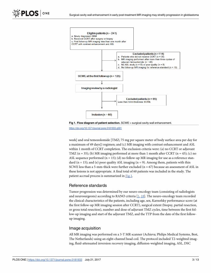
week] and oral temozolomide [TMZ; 75 mg per square meter of body surface area per day for
a maximum of 49 days]) regimen; and (c) MR imaging with contrast enhancement and ASL
within 1 month of CCRT completion. The exclusion criteria were: (a) no CCRT or adjuvant
TMZ (n = 35); (b) MR imaging performed at more than 1 month after CCRT (n = 45); (c) no
ASL sequence performed (n = 15); (d) no follow-up MR imaging for use as a reference stan-
dard (n = 15); and (e) poor quality ASL imaging (n = 9). Among them, patients with thin
SCWE less than a 5-mm-thick were further excluded (n = 67) because an assessment of ASL in
these lesions is not appropriate. A final total of 60 patients was included in the study. The
patient accrual process is summarized in Fig 1.
Reference standards
Tumor progression was determined by our neuro-oncology team (consisting of radiologists
and neurosurgeons) according to RANO criteria [1, 18]. The neuro-oncology team recorded
the clinical characteristics of the patients, including age, sex, Karnofsky performance score (at
the first follow-up MR imaging session after CCRT), surgical extent (biopsy, partial resection,
or gross total resection), number and dose of adjuvant TMZ cycles, time between the first fol-
low-up imaging and start of the adjuvant TMZ, and the TTP from the date of the first follow-
up imaging.
Image acquisition
All MR imaging was performed on a 3-T MR scanner (Achieva; Philips Medical Systems, Best,
The Netherlands) using an eight-channel head coil. The protocol included T2-weighted imag-
ing, fluid-attenuated inversion recovery imaging, diffusion-weighted imaging, ASL, DSC
Fig 1. Flow diagram of patient selection. SCWE = surgical cavity wall enhancement.
https://doi.org/10.1371/journal.pone.0181933.g001
Surgical cavity wall enhancement in early post-treatment MR imaging may stratify progression in glioblastoma
PLOS ONE | https://doi.org/10.1371/journal.pone.0181933 July 21, 2017 3 / 13
perfusion MR imaging, and precontrast T1-weighted imaging (T1WI). Contrast enhancement
was achieved with 0.1 mmol/kg gadoterate meglumine (Dotarem; Guerbet, Paris, France) after
a fat-suppression pulse.
Acquisition of two-dimensional pseudo-continuous ASL was performed using multi-shot
spin-echo echo-planar imaging. Imaging parameters were as follows: labeling time, 1800 ms;
labeling width, 130 mm; repetition time, 3000 ms; echo time, 13 ms; no vascular crushing;
acquisition matrix, 64 × 59; acquisition voxel size, 3.44 × 3.67 mm; reconstruction matrix,
128 × 128; reconstruction voxel size, 1.72 × 1.72 × 6 mm; field of view, 220 × 220 × 104 mm; 15
slices of 6-mm thickness with a 1-mm slice gap; SENSE factor, 2.3; whole brain coverage; and a
total scan time of 5 min. After motion correction of the ASL images, ASL imaging data were
digitally transferred from the PACS workstation to a personal computer.
Qualitative analysis of SCWEs
SCWEs on post-CCRT MR imaging were evaluated by two radiologists (H.S.K. and J.E.P. with
17 years and 5 years of experience in neuro-oncology MRI, respectively) who were blind to the
patient histories. The readers were asked to independently evaluate each SCWE for the follow-
ing: (1) the contrast enhancement pattern (nodular or non-nodular); (2) the presence of mea-
surable contrast-enhancing lesions at two maximal perpendicular diameters of the SCWE in
the axial plane; and (3) positive or negative perfusion at the SCWE.
The enhancement pattern of the SCWE was categorized as either non-nodular, when the
wall enhancement was thickened but without definite nodular enhancement, or nodular,
when there was a nodular enhancement of� 5 mm at two maximal perpendicular diameters.
The presence of measurable contrast-enhancing lesion was recorded when the SCWE included
a bidimensional contrast-enhancing lesion with two perpendicular diameters of at least 10
mm [1].
The perfusion state was assessed in patients with a� 5-mm-thick contrast-enhanced
SCWE because the acquisition voxel size of ASL was 3.44 × 3.67 mm and the minimum size
for perfusion evaluation was at least 4.00 mm. For perfusion assessment, the two readers visu-
ally assessed using ASL whether there was positive or negative perfusion in the SCWE com-
pared with normal-appearing contralateral cortical grey matter. Positive perfusion was defined
when as the tumor showing a similar to increased perfusion compared with normal-appearing
cortical grey matter [6, 12, 19].
Each reader recorded the SCWE category but the final enhancement pattern was deter-
mined by consensus to resolve disagreements and to improve reproducibility, as suggested in a
previous study [4]. Before consensus, the kappa value for each category was as follows: (1)
enhancement pattern 0.84 (95% confidence interval [CI] = 0.70–0.99), (2) presence of measur-
able contrast-enhancing lesions 1.00, and (3) determination of positive or negative perfusion
0.84 (95% CI = 0.70–0.99).
Calculation of perfusion fraction in SCWEs
The two readers independently drew regions of interest (ROIs) encompassing the entire
SCWE on the postcontrast T1WI. A second set of ROIs were then drawn within the tumor on
the cerebral blood flow (CBF) map to mark areas showing similar to increased perfusion com-
pared with normal-appearing cortical grey matter. This was accomplished using a matching
slice in the postcontrast T1WI MR imaging, and only areas of increased perfusion within the
contrast-enhancing tumor were included. The perfusion fraction was calculated by dividing
Surgical cavity wall enhancement in early post-treatment MR imaging may stratify progression in glioblastoma
PLOS ONE | https://doi.org/10.1371/journal.pone.0181933 July 21, 2017 4 / 13
the area of the high perfusion by the area of contrast enhancement.
Perfusion fraction ð%Þ ¼High perfusion areaROI on CBF map
Enhancing areaROI on post � contrast T1WI� 100
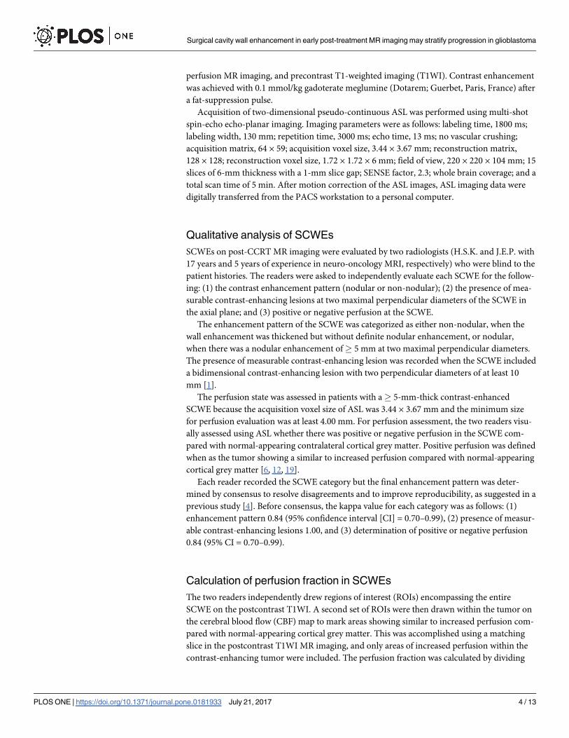
The accrual process for volume fraction calculation is summarized in Fig 2.
Statistical analysis
All continuous variables were initially assessed for normality using the Kolmogorov-Smirnov
test. Imaging characteristics of SCWEs were compared between progression and non-progres-
sion group patients using Fisher’s exact test or the chi-square test for categorical data, with the
Student’s independent t test used for non-categorical data.
Univariate and multivariate analyses of TTP were performed using a Cox proportional haz-
ards model. These statistical analyses were used to evaluate the association between clinical
outcomes and covariates of age, surgical extent, enhancement pattern, presence of measurable
contrast enhancement, and perfusion status. Backwards elimination with a 0.10 significance
level was used to develop a multivariate model. In terms of TTP related to the perfusion status
of the SCWE, survival curves were created using Kaplan-Meier analysis, and a log-rank test
was used to compare differences.
For the perfusion fraction of SCWEs, univariate and multivariate analyses of TTP were per-
formed. Additionally, standard linear regression analysis was employed to evaluate the rela-
tionship between perfusion fraction and TTP.
The inter-reader agreement of the perfusion fraction was assessed using the intraclass cor-
relation coefficient (ICC) using a two-way random effect model with consistency assumption
and classified according to common criteria as excellent (ICC > 0.75), fair to good
(ICC = 0.40–0.75), or poor (ICC� 0.40) [20]. P< 0.05 was considered statistically significant.
Statistical analyses were performed using MedCalc 15.6.1 (MedCalc Software, Mariakerke,
Belgium).
Fig 2. Accrual process for determining the perfusion status of surgical cavity wall enhancements. The
perfusion fraction was calculated by dividing the area of the high perfusion on ASL MR imaging by the area of
contrast-enhancement on postcontrast T1-weighted imaging. The perfusion fraction was 58.8% for reader 1
and 69% for reader 2.
https://doi.org/10.1371/journal.pone.0181933.g002
Surgical cavity wall enhancement in early post-treatment MR imaging may stratify progression in glioblastoma
PLOS ONE | https://doi.org/10.1371/journal.pone.0181933 July 21, 2017 5 / 13

Results
Clinical characteristics of the study patients
Table 1 summarizes the clinical characteristics of the study patients. The mean age of these 60
enrolled patients (33 men, 27 women) at the initial diagnosis was 58.5 ± 9.8 years. The Kar-
nofsky Performance Scale score was dichotomized as< 70 or� 70 (the ability of a person to
perform usual activities) [21]. The mean number of cycles of adjuvant TMZ therapy after ASL
MR imaging was 5.9. By the time of the last assessment (February 2, 2017), 42 of the 60 patients
(70%) had tumor progression.
Image analysis of SCWEs
The imaging characteristics of the SCWEs are summarized in Table 2.
Neither the contrast enhancement patterns, nor the presence of a measurable enhancing
lesion at the SCWEs were significantly different between the two groups. On visual analysis of
ASL images, increased perfusion at SCWEs was more commonly observed in the non-progres-
sion group than in the progression group, but this difference did not reach statistical signifi-
cance. The perfusion fraction was higher in the non-progression group, which was significant
for reader 2 (P = 0.03) and borderline significant for reader 1 (P = 0.08). The reproducibility of
the perfusion fraction between the two readers was excellent (ICC = 0.76, 95% CI = 0.61–0.86).
Table 1. Clinical characteristics and outcome of the study patients.
Characteristics N = 60
Age (years, mean ± SD) 58.5 ± 9.8
Sex
Male 33
Female 27
Karnofsky performance score
<70 8
�70 52
Surgery
Partial resection 44
Gross total resection 16
Number of adjuvant TMZ cycles after MR imaging 5.9 ± 2.4
Dose of adjuvant TMZ (mg) 334 ± 34
Mean size of the surgical cavity (bidimensional, mm2) 296 ± 329
Pattern of contrast enhancing lesion
Non-nodular 17
Nodular 43
Presence of measurable enhancing lesion 20
Clinical outcome
Median TTP (months)* 10 (6–22)
Mean TTP (months) 16 ± 15
No progression during follow up (censored) 18
Key: SD, standard deviation; TMZ, temozolomide; TTM, time-to-progression.
*Data are median values, with interquartile range shown in parentheses. Unless otherwise indicated, data
are expressed as a mean ± standard deviation.
https://doi.org/10.1371/journal.pone.0181933.t001
Surgical cavity wall enhancement in early post-treatment MR imaging may stratify progression in glioblastoma
PLOS ONE | https://doi.org/10.1371/journal.pone.0181933 July 21, 2017 6 / 13
TTP according to the imaging characteristics of SCWEs
The median observation period was 16 months (interquartile range, 10–30 months). The
median TTP was 10 months (interquartile range, 6–22 months). Analysis using the univariate
Cox model revealed that positive perfusion on visual analysis was significantly associated with
a longer TTP (hazard ratio [HR] = 0.49, P = 0.04). A perfusion fraction with a higher volume
fraction was found to be associated with a longer TTP for both readers (HR = 0.98, P = 0.021
for reader 1; HR = 0.98, P = 0.005 for reader 2). The patients’ characteristics, enhancement pat-
tern, and presence of a measurable enhancing lesion were not predictive for TTP.
The multivariate Cox model showed that a younger age (P = 0.03), gross total resection
(P = 0.013), positive perfusion status (P = 0.02), and high perfusion fraction (P = 0.001 for
reader 1 and < .001 for reader 2) remained an independent predictor of a longer TTP
(Table 3).
Table 2. Comparison of SCWE imaging characteristics.
Characteristics Total (n = 60) Progression group (n = 42) Non-progression group (n = 18) P value
Pattern of SCWE 0.23
Non-nodular 17 10 7
Nodular 43 32 11
Presence of measurable enhancing lesion 20 16 4 0.23
Perfusion status 0.25
Increased 44 29 15
Decreased 16 13 3
Perfusion fraction (%)
Reader 1 38.9 ± 33 34.1 ± 32 50.1 ± 33 0.08
Reader 2 35.4 ± 35 28.9 ± 24 50.6 ± 36 0.03
SCWE, surgical cavity contrast enhancement.
https://doi.org/10.1371/journal.pone.0181933.t002
Table 3. Cox proportional model analysis of time-to-progression.
Variable Univariate analysis Multivariate analysis
HR (95% CI) P value HR (95% CI) P value
Age (years) 1.02 (0.99–1.04) 0.13 1.02 (0.99–1.05) 0.06
Male sex 0.95 (0.52–1.76) 0.88
KPS (binary) 0.97 (0.41–2.30) 0.94
Surgical method
Partial resection 0.81(0.41–1.63) 0.56
Gross total resection 0.54 (0.24–1.19) 0.12 0.72 (0.48–1.07) 0.11
Dose × number of adjuvant TMZ 1.00 (0.99–1.00) 0.96
SCWE pattern
Non-nodular enhancement 0.51 (0.27–1.13) 0.10
Presence of measurable-enhancing lesion 1.47 (0.78–2.78) 0.23
SCWE Perfusion using
positive perfusion state 0.49 (0.25–0.96) 0.04 0.33 (0.15–0.68) 0.02
Perfusion fraction (reader 1) 0.98 (0.98–0.99) 0.021 0.97 (0.97–0.99) 0.001
Perfusion fraction (reader 2) 0.98 (0.98–0.99) 0.005 0.98 (0.97–0.99) < 0.001
Note: KPS was either < 70 or� 70. Key: TMZ = temozolomide; TBF = tumour blood flow.
https://doi.org/10.1371/journal.pone.0181933.t003
Surgical cavity wall enhancement in early post-treatment MR imaging may stratify progression in glioblastoma
PLOS ONE | https://doi.org/10.1371/journal.pone.0181933 July 21, 2017 7 / 13
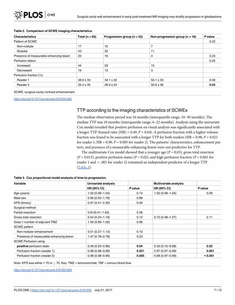
The Kaplan-Meier method with log-rank testing indicated the same trend, namely, that the
increased-perfusion group was associated with a longer TTP compared with the decreased-
perfusion group (median TTP = 13 vs. 29 months, indicating a maximum 2.2-fold increase in
the median TTP; P = 0.036; Fig 3). A representative case is shown in Figs 4 and 5.
Standard linear regression coefficients were calculated to determine the association between
the perfusion fraction and the TTP. Linear regression analysis revealed an R2 = 0.18 (P< .001;
slope = 19.3; 95% CI: 8.44–30.06) for reader 1 and an R2 = 0.17 (P< .001; slope = 17.5; 95% CI:
7.43–27.61). The results are presented in Table 4.
Discussion
Our present study suggests that the perfusion status of SCWEs on post-CCRT MR imaging
could be a significant predictor of the TTP in glioblastoma patients. In particular, the patients
with a SCWE with positive perfusion on ASL showed a longer TTP. Semi-quantitative analysis
also indicated a longer TTP in patients with a higher perfusion fraction. The enhancement pat-
tern or presence of a measurable enhancing lesion was not found to be significant TTP predic-
tors. Based on our results, we suggest that an increased perfusion may be associated with a
longer TTP in glioblastoma and ASL MR imaging may be used as a predictive imaging bio-
marker for the post-CCRT status- in these patients before adjuvant TMZ therapy.
We hypothesized that ASL would become a predictive biomarker for subsequent TMZ ther-
apy, and identify individuals who are more likely to respond to TMZ. Our patients received
Fig 3. Kaplan-Meier survival curves showing clinical outcome comparisons in the increased- and
decreased-perfusion groups (median time-to-progression, 13 vs. 29 months, P = 0.036).
https://doi.org/10.1371/journal.pone.0181933.g003
Surgical cavity wall enhancement in early post-treatment MR imaging may stratify progression in glioblastoma
PLOS ONE | https://doi.org/10.1371/journal.pone.0181933 July 21, 2017 8 / 13
adjuvant TMZ after ASL MR imaging (mean number of adjuvant TMZ cycles after MR imag-
ing = 5.9). Based on our results, increased perfusion may be associated with a longer TTP for
glioblastoma. We speculate that an area with increased perfusion could possibly reflect
enhanced TMZ delivery, whereas impaired perfusion would severely compromise the delivery
of TMZ. It may seem paradoxical to expect a favourable outcome in the positive-perfusion
group, but observations from preclinical data have indicated that vascular normalization [14,
15] of an abnormal tumor vasculature results in increased perfusion, which allows more effi-
cient delivery of combined therapeutic agents [13, 16]. This observation is further strength-
ened by the findings of recent clinical trials that combination therapy achieving vascular
normalization is associated with a favourable outcome in head and neck cancer [22] and in
metastatic colorectal, renal, and lung cancer [23–25]. In the present study, we speculated that
areas of increased perfusion compared with contralateral grey matter may reflect a region of
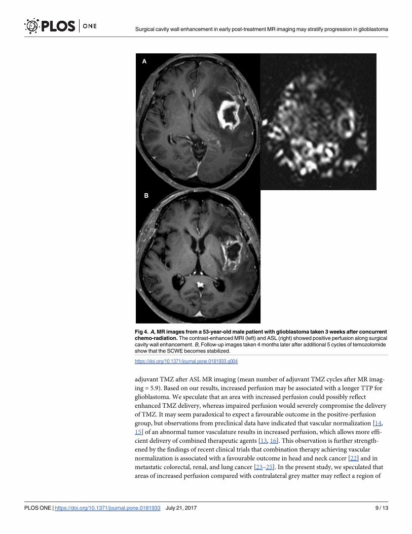
Fig 4. A, MR images from a 53-year-old male patient with glioblastoma taken 3 weeks after concurrent
chemo-radiation. The contrast-enhanced MRI (left) and ASL (right) showed positive perfusion along surgical
cavity wall enhancement. B, Follow-up images taken 4 months later after additional 5 cycles of temozolomide
show that the SCWE becomes stabilized.
https://doi.org/10.1371/journal.pone.0181933.g004
Surgical cavity wall enhancement in early post-treatment MR imaging may stratify progression in glioblastoma
PLOS ONE | https://doi.org/10.1371/journal.pone.0181933 July 21, 2017 9 / 13
enhanced perfusion of a viable tumor or a post-treatment tissue change. Although the mecha-
nism underlying this is not clear, we assume that an area with increased perfusion could possi-
bly reflect enhanced TMZ delivery, whereas low and impaired perfusion from excessive
damage to the vasculature would severely compromise the delivery of chemotherapeutics.
We used semi-quantitative calculations of increased perfusion areas by comparison with
normal grey matter on ASL MR imaging. More quantitative methods of analysis, such as abso-
lute CBF calculation [26] is available, but quantitative studies have limited applicability in a
clinical setting due to their low precision; previous studies have reported low intra- and inter-
session reproducibility indexes for quantitative measurement as low as 18–25% [27]. Our
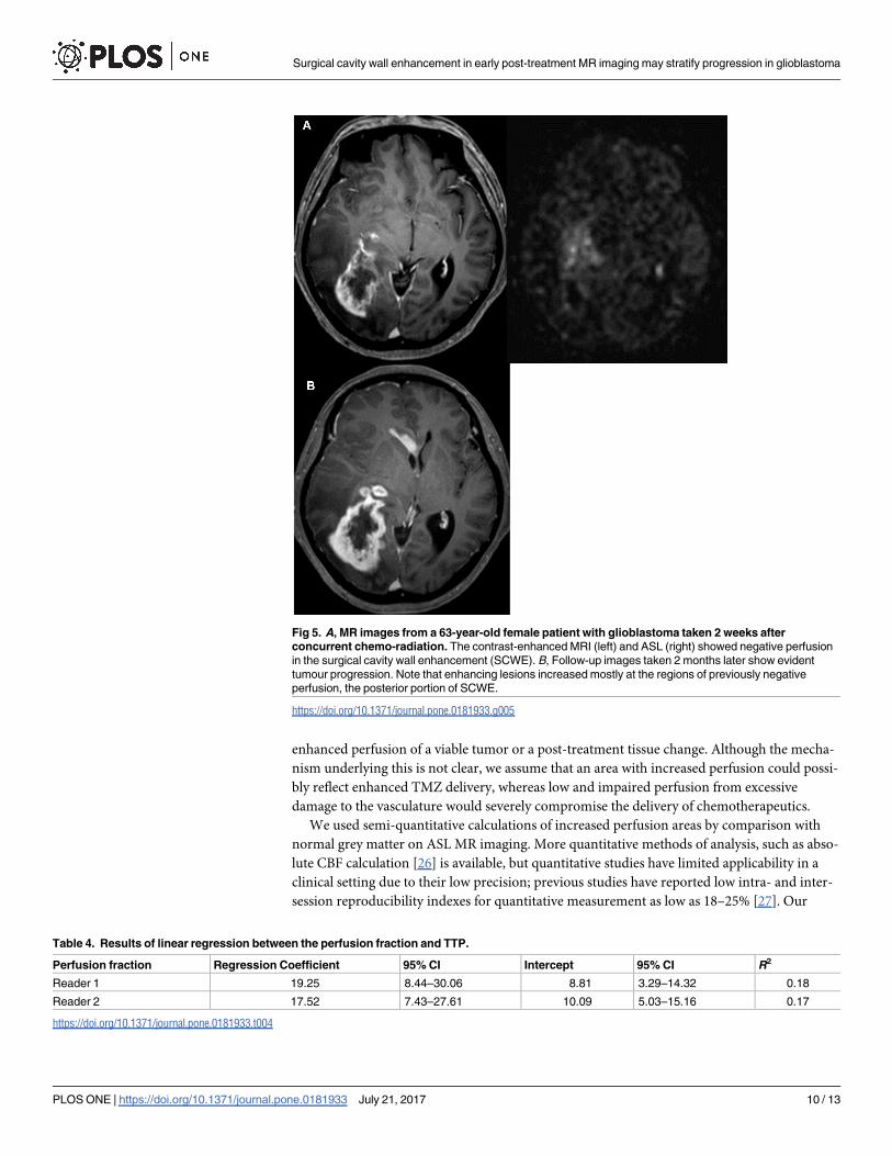
Fig 5. A, MR images from a 63-year-old female patient with glioblastoma taken 2 weeks after
concurrent chemo-radiation. The contrast-enhanced MRI (left) and ASL (right) showed negative perfusion
in the surgical cavity wall enhancement (SCWE). B, Follow-up images taken 2 months later show evident
tumour progression. Note that enhancing lesions increased mostly at the regions of previously negative
perfusion, the posterior portion of SCWE.
https://doi.org/10.1371/journal.pone.0181933.g005
Table 4. Results of linear regression between the perfusion fraction and TTP.
Perfusion fraction Regression Coefficient 95% CI Intercept 95% CI R2
Reader 1 19.25 8.44–30.06 8.81 3.29–14.32 0.18
Reader 2 17.52 7.43–27.61 10.09 5.03–15.16 0.17
https://doi.org/10.1371/journal.pone.0181933.t004
Surgical cavity wall enhancement in early post-treatment MR imaging may stratify progression in glioblastoma
PLOS ONE | https://doi.org/10.1371/journal.pone.0181933 July 21, 2017 10 / 13
current results demonstrate an improve robustness in the the dichotomization of increased-
and decreased-perfusion groups, with an agreement of visual analysis of kappa 0.84. In addi-
tion, semiquantitative analysis of perfusion does not require for excessive post-processing and
shows sufficient reproducibility (ICC = 0.76) to be adopted in clinical practice.
The enhancement pattern or presence of a measurable enhancement was not found to be a
significant predictor of the TTP. Previous studies using postoperative MR imaging have
reported that thick or nodular enhancement patterns had a poorer prognosis than thin
enhancements, according to a thickness criterion of 5 mm [2–4]. In addition, a recent study by
Kim et al. [28] of newly developed non-measurable enhancing lesions after CCRT reported a
similar result, namely that the progression group showed frequent thick (� 3 mm) or nodular
(� 5 mm) enhancements. Our current study differs from those of previous studies however
because only thick wall enhancement (� 5 mm) was included in our present analysis to assess
the significance of perfusion at SCWEs. In addition, unlike other reports, we analyzed a data
set within a narrow time period post-CCRT but prior to adjuvant TMZ therapy.
Pseudoprogression can be a confounder in the assessment of an adjuvant TMZ treatment
response. Pseudoprogression is diagnosed when a post-radiation MRI indicates an increase in
contrast enhancement that subsides with time, without any change in therapy, and may there-
fore have represented radiation change [1, 29]. However, pseudoprogression has also shown
decreased perfusion on ASL in previous studies [10, 11] and its possible confounding effect of
pseudoprogression is unlikely to have caused the association between decreased perfusion and
shorter TTP in our current study.
The present study had several limitations of note, beyond those associated with retrospec-
tive analyses. First, the study population was relatively small. Although we detected statistically
significant results for the perfusion status of SCWEs, larger populations are needed to
strengthen this statistical power, particularly with regard to the enhancement pattern. How-
ever, our patient group was unique in that we performed ASL imaging within 1 month of
CCRT completion, providing a relatively homogenous group with a narrow time period for
the determination of TTP. Second, we did not include the MGMT promoter methylation sta-
tus in the analysis, which may synergistically improve clinical outcomes in TMZ therapy. The
MGMT promoter methylation status is a determinant of chemosensitivity, whereas increased
tumor perfusion is related to efficient drug delivery. Hence, the potential predictive value of
combining ASL imaging and MGMT promoter methylation status should be a future study
topic.
In conclusion, an assessment of perfusion in early post-treatment MR imaging can stratify
the TTP in patients with glioblastoma. ASL MR imaging may be used as a predictive imaging
biomarker for the post-CCRT status- in these patients before adjuvant TMZ therapy, and nota-
bly, increased perfusion in SCWEs can become a predictor of a longer TTP.
Acknowledgments
The Institutional Review Board of Asan Medical Center approved this study ([http://eirb.amc.
seoul.kr]: S2015-1270) and wavied the requirement for written informed consent. This
research was supported by the Basic Science Research Program through the National Research
Foundation of Korea (NRF) funded by the Ministry of Education, Science and Technology
(grant number: NRF-2017R1A2A2A05001217) and by through the Korea Health Industry
Development Institute (KHIDI), funded by the Ministry of Health & Welfare, Republic of
Korea (grant number: HI14C1090).
Surgical cavity wall enhancement in early post-treatment MR imaging may stratify progression in glioblastoma
PLOS ONE | https://doi.org/10.1371/journal.pone.0181933 July 21, 2017 11 / 13
Author Contributions
Conceptualization: Ho Sung Kim, Jeong Hoon Kim.
Data curation: Ji Eun Park, Kyoung Hwa Ryu, Hyo Won Kim.
Formal analysis: Ji Eun Park, Kyoung Hwa Ryu.
Funding acquisition: Ho Sung Kim.
Investigation: Seung Chai Jung, Choong Gon Choi, Sang Joon Kim.
Methodology: Ji Eun Park, Ho Sung Kim.
Project administration: Jeong Hoon Kim.
Software: Woo Hyun Shim.
Supervision: Ho Sung Kim.
Validation: Ho Sung Kim, Jeong Hoon Kim.
Visualization: Ji Eun Park, Ho Sung Kim.
Writing – original draft: Ji Eun Park.
Writing – review & editing: Ji Eun Park, Kyoung Hwa Ryu, Ho Sung Kim.
References
1. Wen PY, Macdonald DR, Reardon DA, Cloughesy TF, Sorensen AG, Galanis E, et al. Updated
response assessment criteria for high-grade gliomas: response assessment in neuro-oncology working
group. J Clin Oncol. 2010; 28: 1963–1972. https://doi.org/10.1200/JCO.2009.26.3541 PMID:
20231676
2. Ekinci G, Akpinar IN, Baltacioglu F, Erzen C, Kilic T, Elmaci I, et al. Early-postoperative magnetic reso-
nance imaging in glial tumors: prediction of tumor regrowth and recurrence. Eur J Radiol. 2003; 45: 99–
107. PMID: 12536087
3. Farace P, Amelio D, Ricciardi GK, Zoccatelli G, Magon S, Pizzini F, et al. Early MRI changes in glioblas-
toma in the period between surgery and adjuvant therapy. Journal of Neuro-Oncology. 2013; 111: 177–
185. https://doi.org/10.1007/s11060-012-0997-y PMID: 23264191
4. Majos C, Cos M, Castaner S, Gil M, Plans G, Lucas A, et al. Early post-operative magnetic resonance
imaging in glioblastoma: correlation among radiological findings and overall survival in 60 patients. Eur
Radiol. 2016; 26: 1048–1055. https://doi.org/10.1007/s00330-015-3914-x PMID: 26188660
5. Kelly PJ, Daumas-Duport C, Scheithauer BW, Kall BA, Kispert DB. Stereotactic histologic correlations
of computed tomography- and magnetic resonance imaging-defined abnormalities in patients with glial
neoplasms. Mayo Clin Proc. 1987; 62: 450–459. PMID: 3553757
6. Dangouloff-Ros V, Deroulers C, Foissac F, Badoual M, Shotar E, Grevent D, et al. Arterial Spin Labeling
to Predict Brain Tumor Grading in Children: Correlations between Histopathologic Vascular Density and
Perfusion MR Imaging. Radiology. 2016: 152228. https://doi.org/10.1148/radiol.2016152228 PMID:
27257950
7. Jiang J, Zhao L, Zhang Y, Zhang S, Yao Y, Qin Y, et al. Comparative analysis of arterial spin labeling
and dynamic susceptibility contrast perfusion imaging for quantitative perfusion measurements of brain
tumors. Int J Clin Exp Pathol. 2014; 7: 2790–2799. PMID: 25031698
8. Qiao XJ, Ellingson BM, Kim HJ, Wang DJ, Salamon N, Linetsky M, et al. Arterial spin-labeling perfusion
MRI stratifies progression-free survival and correlates with epidermal growth factor receptor status in
glioblastoma. AJNR Am J Neuroradiol. 2015; 36: 672–677. https://doi.org/10.3174/ajnr.A4196 PMID:
25542879
9. Schor-Bardach R, Alsop DC, Pedrosa I, Solazzo SA, Wang X, Marquis RP, et al. Does arterial spin-
labeling MR imaging-measured tumor perfusion correlate with renal cell cancer response to antiangio-
genic therapy in a mouse model? Radiology. 2009; 251: 731–742. https://doi.org/10.1148/radiol.
2521081059 PMID: 19474376
10. Ye J, Bhagat SK, Li HM, Luo XF, Wang BH, Liu LQ, et al. Differentiation between recurrent gliomas and
radiation necrosis using arterial spin labeling perfusion imaging. Experimental and Therapeutic Medi-
cine. 2016; 11: 2432–2436. https://doi.org/10.3892/etm.2016.3225 PMID: 27284331
Surgical cavity wall enhancement in early post-treatment MR imaging may stratify progression in glioblastoma
PLOS ONE | https://doi.org/10.1371/journal.pone.0181933 July 21, 2017 12 / 13
11. Choi YJ, Kim HS, Jahng GH, Kim SJ, Suh DC. Pseudoprogression in patients with glioblastoma: added
value of arterial spin labeling to dynamic susceptibility contrast perfusion MR imaging. Acta Radiol.
2013; 54: 448–454. https://doi.org/10.1177/0284185112474916 PMID: 23592805
12. Noguchi T, Yoshiura T, Hiwatashi A, Togao O, Yamashita K, Nagao E, et al. Perfusion imaging of brain
tumors using arterial spin-labeling: correlation with histopathologic vascular density. AJNR Am J Neu-
roradiol. 2008; 29: 688–693. https://doi.org/10.3174/ajnr.A0903 PMID: 18184842
13. Goel S, Duda DG, Xu L, Munn LL, Boucher Y, Fukumura D, et al. Normalization of the vasculature for
treatment of cancer and other diseases. Physiol Rev. 2011; 91: 1071–1121. https://doi.org/10.1152/
physrev.00038.2010 PMID: 21742796
14. Jain RK. Normalization of tumor vasculature: an emerging concept in antiangiogenic therapy. Science.
2005; 307: 58–62. https://doi.org/10.1126/science.1104819 PMID: 15637262
15. Jain RK. Normalizing tumor microenvironment to treat cancer: bench to bedside to biomarkers. J Clin
Oncol. 2013; 31: 2205–2218. https://doi.org/10.1200/JCO.2012.46.3653 PMID: 23669226
16. Stylianopoulos T, Jain RK. Combining two strategies to improve perfusion and drug delivery in solid
tumors. Proc Natl Acad Sci U S A. 2013; 110: 18632–18637. https://doi.org/10.1073/pnas.1318415110
PMID: 24167277
17. Jain RK. Determinants of tumor blood flow: a review. Cancer Res. 1988; 48: 2641–2658. PMID:
3282647
18. Pope WB, Young JR, Ellingson BM. Advances in MRI assessment of gliomas and response to anti-
VEGF therapy. Curr Neurol Neurosci Rep. 2011; 11: 336–344. https://doi.org/10.1007/s11910-011-
0179-x PMID: 21234719
19. Kim C, Kim HS, Shim WH, Choi CG, Kim SJ, Kim JH. Recurrent Glioblastoma: Combination of High
Cerebral Blood Flow with MGMT Promoter Methylation Is Associated with Benefit from Low-Dose
Temozolomide Rechallenge at First Recurrence. Radiology. 2017; 282: 212–221. https://doi.org/10.
1148/radiol.2016152152 PMID: 27428890
20. Bartko JJ. The intraclass correlation coefficient as a measure of reliability. Psychol Rep. 1966; 19: 3–
11. https://doi.org/10.2466/pr0.1966.19.1.3 PMID: 5942109
21. Peus D, Newcomb N, Hofer S. Appraisal of the Karnofsky Performance Status and proposal of a simple
algorithmic system for its evaluation. Bmc Medical Informatics and Decision Making. 2013; 13. Artn 72
https://doi.org/10.1186/1472-6947-13-72 PMID: 23870327
22. Schmitt P, Kotas M, Tobermann A, Haase A, Flentje M. Quantitative tissue perfusion measurements in
head and neck carcinoma patients before and during radiation therapy with a non-invasive MR imaging
spin-labeling technique. Radiotherapy and Oncology. 2003; 67: 27–34. https://doi.org/10.1016/S0167-
8140(03)00024-0 PMID: 12758237
23. Hurwitz H, Fehrenbacher L, Novotny W, Cartwright T, Hainsworth J, Heim W, et al. Bevacizumab plus
irinotecan, fluorouracil, and leucovorin for metastatic colorectal cancer. New England Journal of Medi-
cine. 2004; 350: 2335–2342. https://doi.org/10.1056/NEJMoa032691 PMID: 15175435
24. Rini BI, Halabi S, Rosenberg JE, Stadler WM, Vaena DA, Archer L, et al. Phase III Trial of Bevacizumab
Plus Interferon Alfa Versus Interferon Alfa Monotherapy in Patients With Metastatic Renal Cell Carci-
noma: Final Results of CALGB 90206. Journal of Clinical Oncology. 2010; 28: 2137–2143. https://doi.
org/10.1200/JCO.2009.26.5561 PMID: 20368558
25. Sandler A, Gray R, Perry MC, Brahmer J, Schiller JH, Dowlati A, et al. Paclitaxel-carboplatin alone or
with bevacizumab for non-small-cell lung cancer. New England Journal of Medicine. 2006; 355: 2542–
2550. https://doi.org/10.1056/NEJMoa061884 PMID: 17167137
26. Wang J, Alsop DC, Li L, Listerud J, Gonzalez-At JB, Schnall MD, et al. Comparison of quantitative per-
fusion imaging using arterial spin labeling at 1.5 and 4.0 Tesla. Magn Reson Med. 2002; 48: 242–254.
https://doi.org/10.1002/mrm.10211 PMID: 12210932
27. Heijtel DF, Mutsaerts HJ, Bakker E, Schober P, Stevens MF, Petersen ET, et al. Accuracy and precision
of pseudo-continuous arterial spin labeling perfusion during baseline and hypercapnia: a head-to-head
comparison with (1)(5)O H(2)O positron emission tomography. Neuroimage. 2014; 92: 182–192.
https://doi.org/10.1016/j.neuroimage.2014.02.011 PMID: 24531046
28. Kim BR, Choi SH, Yun TJ, Lee ST, Park CK, Kim TM, et al. MR Imaging Analysis of Non-Measurable
Enhancing Lesions Newly Appearing after Concomitant Chemoradiotherapy in Glioblastoma Patients
for Prognosis Prediction. PLoS One. 2016; 11: e0166096. https://doi.org/10.1371/journal.pone.
0166096 PMID: 27835666
29. Hygino da Cruz LC Jr., Rodriguez I, Domingues RC, Gasparetto EL, Sorensen AG. Pseudoprogression
and pseudoresponse: imaging challenges in the assessment of posttreatment glioma. AJNR Am J Neu-
roradiol. 2011; 32: 1978–1985. https://doi.org/10.3174/ajnr.A2397 PMID: 21393407
Surgical cavity wall enhancement in early post-treatment MR imaging may stratify progression in glioblastoma
PLOS ONE | https://doi.org/10.1371/journal.pone.0181933 July 21, 2017 13 / 13

![arXiv:1610.01240v2 [physics.optics] 14 Oct 2016 · Spontaneous Emission and Light Extraction Enhancement of Light Emitting Diode Using Partially-Reflecting Metasurface Cavity (PRMC)](https://img.dokumen.tips/doc/110x75/5aef60547f8b9a8b4c8c3782/arxiv161001240v2-14-oct-2016-emission-and-light-extraction-enhancement-of.jpg)