Embed Size (px)
Citation preview
92 Chapter 4
42. M. Kidowaki, T. Nakajima, J. Araki, A. Inomata, H. Ishibashi and K. Ito, Macromolecules, 2007, 40, 6859.
43. J. Araki, T. Kataoka and K. Ito, Soft Matter, 2008, 4, 245. 44. P. C. Nicolson and J. Vogt, Biomaterials. 2001, 22, 3273. 45. T. Ogoshi and Y. Chujo, Compos. Interfaces, 2005. II, 539. 46. R. Tamaki, K. Naka andY. Chujo, Polym. J., 1998, 30, 60. 47. K. Kato. K. Inoue, M. Kidowaki and K. Ito, Macromolecules, 2009, 42,
7129. 48. H. Okumura, Y. Kawaguchi and A. Harada, Macromolecules, 2001, 34,
6338. 49. J. Araki and K. Ito, Macromolecules. 2005, 38, 7524. 50. S. Brochsztain and M. J. Politi, Lanwnuir, 1999, 15, 4486. 51. K. Kato, H. Komatsu and K. Ito, Macromolecules, 2010.43. 8799. 52. G. Wenz, B. H. Han and A. Muller, Chun. Rev., 2006, 106, 782. 53. K. Ito, Polym. J., 2012, 44, 38. 54. A. Banda, K. Kato, Y. Sakai. H. Yokoyama and K. Ito, to be submitted. 55. K. Kato and K. Ito, Soft Maller, 2011, 7, 8737. 56. M. Rubinstein and R. Colby, Polymer Physics, Oxford University Press.
New York, 2003.
CHAPTER 5
Peptide and Protein Hydrogels
LAWRENCE J. DOOLING AND DAVID A. TIRRELL*
California Institute of Technology, Division of Chemistry and Chemical Engineering, 1200 East California Boulevard, MC 210-41, Pasadena, CA 91125, USA *Email: tirrell(o caltech.edu
5.1 Introduction Proteins are a fascinating class of macromolecules from both functional and structural perspectives. They catalyze the reactions that sustain life. bind ligands with h1gh affinity and specificity, and mediate interactions among biomolccules in complex cellular milieux. Proteins also assemble into higherorder structures that arc responsible for the mechanical integrity of cells and tissues. Their diverse functional and structural properties have made proteins important building blocks in the development of new biomaterials. l-4
Hydrogels are physically or chemically cross-linked polymer networks with high water content. 5
·6 Their formation requires a balance between the forces
driving the association of polymer chains and those mediating solvation of the network. Given that proteins have evolved to fold and function in aqueous environments and that many proteins self-assemble into larger structures, they would seem to be ideal candidates for use as hydrogel precursors. Indeed, proteins, and more broadly peptides of all sizes, are widely used for this purpose.
The development of peptide and protein hydrogels has been a crossdisciplinary effort combining the knowledge of protein structure and function from biology with the synthesis and characterization tools of macromolecular
Monograph~ in Supramolecular Chemistry No. II Polymeric and Self Assembled HyJrogels: From Fundamental Underqanding to Applications Edited by Xian Jun Loh and Oren A. Scherman :1" The Royal Society of Chemistry 20 l ~ Published hy the Royal Society of Chcmi~try. www.rsc.org
93
94 Chapter 5
chemistry and materials science. The primary motivation driving this field is the need for implantable scaffolds for soft-tissue enginecring,7
·H benign methods for encapsulation of cells and biomolccules,9
·10 matrices for in vitro cell culture, 11
and injectable delivery vehicles for therapcutics. 12.1
3 Moreover, peptide hydrogels show promise as templates for biomineralization 14 and inorganic nanostructures15 The ability of proteins to undergo biochemical and structural changes in response to pH and temperature changes, ligand binding, light, and mechanical force also suggests potential applications for peptide hydrogels as biosensors and stimulus-responsive materials. 16
·17 Finally, peptide hydrogels
provide simplified systems for studying and engineering the assembly of biological molecules as well as inspiration for the development of self-assembling synthetic structures.
We identify four classes of peptide hydrogels: (!) hydrogels from selfassembling oligopeptides, (2) hydrogels from recombinant proteins, (3) hydrogels that are hybrid materials combining peptides and proteins with synthetic polymers, and ( 4) hydrogels from naturally sourced proteins and proteoglycans such as collagen. Despite the importance of this last class of hydrogels in cell culture and tissue engineering applications, there are only limited opportunities for engineering and rational design of its macromolecular components, and detailed characterization can be challenging because of material heterogeneity. Therefore, after a brief background on peptide and protein structure, we will limit our discussion to examples from the first three classes of peptide hydrogels before concluding with future directions and challenges in this field.
5.2 Peptide and Protein Structure Peptides are polymers formed by the condensation of amino acids. Short polymer chains with degrees of polymerization less than approximately 25 amino acids are generally described as oligopeptides, while longer chains are known as polypeptides. The term protein will be reserved for polypeptides that have been synthesized by ribosomal translation.
5.2.1 Peptide and Protein Synthesis
The most basic level of protein structure is the linear sequence of amino acids, also known as the primary structure. The protein sequence is genetically encoded in DNA. or more specifically in the portion of the DNA that is transcribed into messenger RNA. Protein synthesis occurs on the ribosome. which translates the messenger RNA and catalyzes the formation of amide bonds between amino acids and the growing polypeptide chain. Through genetic engineering, it is possible to produce natural or artificial proteins in a wide variety of host organisms, typically Escherichia coli but occasionally yeast and higher eukaryotes. Recombinant protein production yields monodisperse polymers with precise sequence control that is not possible with synthetic
Peptide and Protein Hydrof?els· 95
polymerization methods. Oligopeptides and short polypeptides arc synthesized in vitro using solid-phase peptide synthesis (SPPS), with an upper limit of approximately 50 amino acids. 18 In this method, the peptide chain remains tethered to a resin support as each residue is added by amide coupling of a protected amino acid and its subsequent deprotection. Longer sequences can be synthesized by combining oligopeptides using native chemical ligation. 19
With a few important exceptions,20•21 nature incorporates only 20 different
amino acids into proteins. These are known as the canonical or natural amino acids. While the number of ways in which these monomers can be combined is nearly infinite, the diversity of the canonical amino acid side chains is somewhat limited from the viewpoint of synthetic chemistry. From the perspective of investigators who wish to design new peptide and protein biomaterials. it is advantageous to augment the canonical amino acids with new monomers that carry more diverse functionality, including reactive groups for bioorthogonal chemistry,22
·23 halogens,24 26 and photo-reactive moieties. 27 Furthermore.
natural proteins contain exclusively L-amina acids. which can influence protein structure and biomaterial assembly. 2x Non-canonical amino acids can be incorporated into synthetic oligopeptides, assuming that their side chains do not interfere with the coupling and deprotection reactions or that suitable protecting groups arc available. Numerous strategies have also been described for the co-translational incorporation of non-canonical amino acids in a residuc-specific29 or site-specific manner.-' 0
5.2.2 Higher-Order Structure and Hydrogel Assembly
Protein secondary structure describes the local conformation of the peptide backbone. Different conformations arise from the rotational degrees of freedom of the N--C"l and Ca--C bonds. The most common secondary structures are the cr-hclix and ~-sheet, which arc characterized by extensive hydrogen bonding between amine and carbonyl groups. Other secondary structures include turns and loops, ~-hairpins, ~-spirals, and polyproline helices. As discussed in the next section, regular secondary structure is a common feature in peptide and protein hydrogels and provides a basis for classifying these materials.
Tertiary structure is the spatial arrangement of all of the atoms in a protein. It may also be referred to as the folded state of a protein and is closely related to function. Many proteins also have a quaternary structure that describes the assembly of multiple polypeptide subunits. Typically these subunits associate noncovalently through hydrogen bonding, hydrophobic interactions, and salt bridges. Alternatively they may be bound covalently through disulfide bond formation or enzymatic cross-linking. The intermolecular interactions that arc responsible for protein quaternary structure closely resemble those that facilitate the self-assembly of peptide and protein hydrogels.
In addition to classification based on the type of peptide precursor (i.e. oligopeptides, recombinant proteins, etc.) or secondary structures (i.e. ~-sheets. ex-helices. etc.), peptide hydrogels can also be described by the mechanism of
96 Chapter 5
(a) Nanofibrous Hydrogels Parallel fibers
I ;~ Perpendicular fibers
Intramolecular loop
(c) Chemically cross-linked Hydrogels I
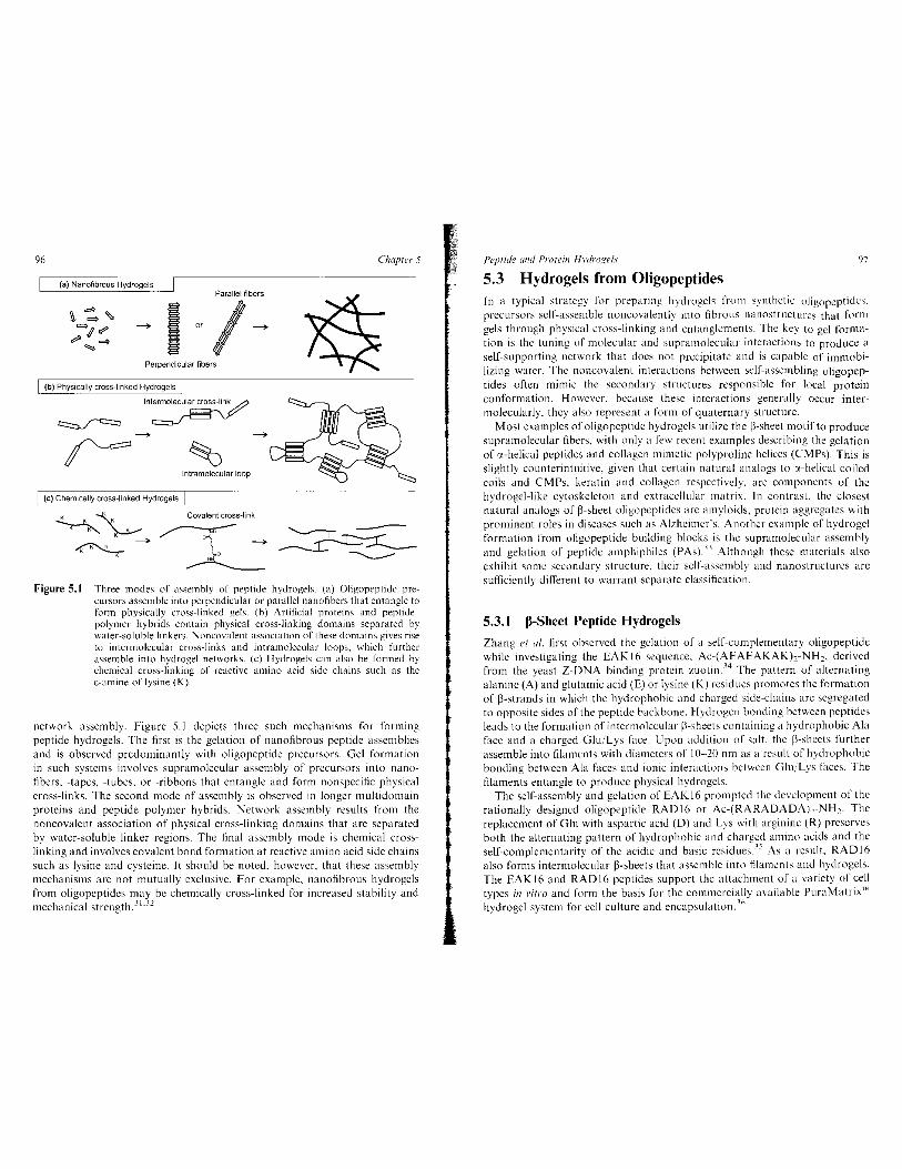
Figure 5.1 Three modes of a~semhly of peptide hydrogels. (a) Oligopeptidc precursors assemble into perpendicular or parallel nanofibers that entangle to form physically cross-linked gels. (b) Artificial proteins and peptide polymer hybrids contain physical cross-linking domains separated by water-soluble linkers. Noncovalent association of these domains gives rise to intermolecular cross-links and intramolecular loops, which further assemble into hydrogel netw·orks. (c) Hydrogels can also be formed by chemical cross-linking of reactive amino acid side chains such as the 0-amine of lysine (K).
network assembly. Figure 5.1 depicts three such mechanisms for forming peptide hydrogels. The first is the gelation of nanofibrous peptide assemblies and is observed predominantly with oligopeptide precursors. Gel formation in such systems involves supramolecular assembly of precursors into nanofibers, -tapes, -tubes. or -ribbons that entangle and form nonspecific physical cross-links. The second mode of assembly is observed in longer multidomain proteins and peptide-polymer hybrids. Network assembly results from the noncovalent association of physical cross-linking domains that are separated by water-soluble linker regions. The flnal assembly mode is chemical crosslinking and involves covalent bond formation at reactive amino acid side chains such as lysine and cysteine. It should be noted, however. that these assembly mechanisms are not mutually exclusive. For example, nanofibrous hydrogcls from oligopcptidcs may be chemically cross-linked for increased stability and mechanical strength. 31
·32
Peptide and Protein Hydrogel.\· 97
5.3 Hydrogels from Oligopeptides In a typical strategy' for preparing hydrogels from synthetic oligopeptidc'l. precursors self-assemble noncovalently into fibrous nanostructures that form gels through physical cross-linking and entanglements. The key to gel formation is the tuning of molecular and supramolecular interactions to produce a self-supporting network that docs not precipitate and is capable of immobilizing water. The noncovalent interactions between self-assembling oligopeptides often mimic the secondary structures responsible for local protein conformation. However, because these interactions generally occur intermolecularly, they also represent a form of quaternary structure.
Most examples of oligopeptidc hydrogcls utilize the ~-sheet motif to produce supramolecular fibers, with only a few recent examples describing the gelation of ex-helical pep tides and collagen mimetic polyproline helices (CMPs). This is slightly counterintuitive, given that certain natural analogs to :x-helical coiled coils and CMPs, keratin and collagen respectively, arc components of the hydrogel-like cytoskeleton and extracellular matrix. In contrast the closest natural analogs of ~-sheet oligopeptides are amyloids. protein aggregates with prominent roles in diseases such as Alzheimer's. Another example of hydrogel formation from oligopeptide building blocks is the supramolecular assembly and gelation of peptide amphiphiles (PAs). 13 Although these materials also exhibit some secondary structure, their self-assembly and nanostructurcs arc sufficiently different to warrant separate classification.
5.3.1 ~-Sheet Peptide Hydrogels
Zhang et a/. first observed the gelation of a self-complementary oligopeptide while investigating the EAK 16 sequence, Ac-(AEAEAKAK),-NH2• derived from the yeast Z-ONA binding protein zuotin. 34 The pattern of alternating alanine (A) and glutamic acid (E) or lysine (K) residues promotes the formation of ~-strands in which the hydrophobic and charged side-chains arc segregated to opposite sides of the peptide backbone. Hydrogen bonding between peptidcs leads to the formation of intermolecular ~~-sheets containing a hydrophobic Ala face and a charged Glu/Lys face. Upon addition of salt, the ~-sheets further assemble into filaments with diameters of I 0-20 nm as a result of hydrophobic bonding between Ala faces and ionic interactions between Glu,iLys faces. The filaments entangle to produce physical hydrogels.
The self-assembly and gelation of EAK16 prompted the development of the rationally designed oligopeptide RAD16 or Ac-(RARADADA),-NH,. The replacement of Glu with aspartic acid (D) and Ly's with arginine (R) preserves both the alternating pattern of hydrophobic and charged amino acids and the self-complementarity of the acidic and basic rcsiducs.:1 5 As a result. RAD16 also forms intermolecular ~-sheets that assemble into filaments and hydrogels. The EAK 16 and RAD16 pep tides support the attachment of a variety of cell types in J'itro and form the basis for the commercially available PuraMatrixiM hydrogel system for cell culture and encapsulation. :1o
98 Chapter 5
Several addi tional peptide hydrogels are also based on sequences that form 13-sheets. For example, Aggeli et a/. have investigated the secondary structure and self-assembly of two peptides derived from lysozyme and the transmembrane protein lsK as a functio n of the solvent dielectric constant and hydrogen bonding ability37 The resulting phase diagram led to the de novo design of DN I (Ac-QQRFQWQFEQQ-N H2), a n 11-residue peptide that forms anti parallel, intermolecular 13-sheets referred to as 13-tapes (Q =glutamine, F = phenylalanine, W = tryptophan). Hydrophobic and 1!- 1! in teractions between aromatic side chains may cause two 13-tapes to associate along their hydrophobic faces to form a 13-ribbon. TEM reveals helical and twisting behavior in 13-tapes and ribbons as well as higher-o rder self-assembly into fibrils and fibers 38
·39
An important consideration in many hydrogel applications is the abi lity to control the self-assembly process.40 For example, the EAK16 and RADI 6 family of peptides requires physiological salt concentrations to form hydrogels. In water, electrostatic repulsions between like charges on opposing peptides result in a significant energy barrier for self-assembly. Ions lower this barrier and promote peptide gelation by forming an electric double layer that screens the repulsive charges and permits 13-sheet assembly4
1.42 Using the
self-complementary oligopeptide FEK16 or (FEFEFKFK)z, Messersmith and co-workers have demonstrated controlled assembly of hydrogels by photochemical and therma l release of CaCI2 from light- and temperature-sensitive liposomes43 Yu and co-workers have described an a lternative approach for controlling self-assembly that involves two complementary peptide sequences, Ac-WKVKVK VK VK-NH2 and Ac-EWEVEVEVEV-NH2 (V =valine). Under appropriate conditions, the individual peptides remain in solution because of ionic self-repulsion but assemble into nanofibers and hydrogels upon mixing due to the complementa ry acidic and basic residues.44
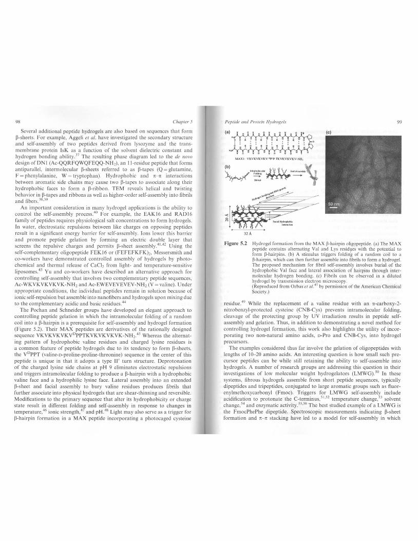
The Pochan and Schneider groups have developed an elegant approach to controlling peptide gelation in which the intramolecular folding of a random coil into a 13-hairpin is a prerequisite for self-assembly and hydrogel formation (Figure 5.2). Their MAX peptides are derivatives of the rationally designed sequence VKVKVKVKV0 PPTKVKVKVKYK-NH2.
45 Whereas the a lternating pattern o f hydrophobic valine res idues and charged lysine residues is a common feature of peptide hydrogels due to its tendency to form 13-sheets, the V0 PPT (valine-o-proline-proline-threonine) sequence in the center of this peptide is unique in that it adopts a type II ' turn structure. Deprotonation of the charged lysine side chains a t pH 9 eliminates electrostatic repulsions and triggers intramolecular folding to produce a 13-hairpin with a hydrophobic valine face and a hydrophilic lysine face. Lateral assembly into an extended 13-sheet and facial assembly to bury valine residues produces fibrils that further associate into physical hydrogels that are shear-thinning and reversible. Modifications to the primary sequence that a lter its hydrophobicity or charge state result in different folding and self-assembly in response to changes in temperature,46 ionic strength,47 and pH4 8 Light may a lso serve as a trigger for 13-hairpin formation in a MAX peptide incorporating a photocaged cysteine
Peptide and Protein Hydrogels 99
(a) • .1.-Y-/-rY-tr:Y./r:Y.;;.o 110111 o111,0111 o • o):.
.fr~~~~~~))-~~ Y O W O yO YO TO
(c)
MAXI : VKVKVKVKV Dplp TKVKVKVKV-NHJ
Figure 5.2 Hydrogel formation from the MAX ~-hairpin oligopeptide. (a) The MAX peptide contains alternating Val and Lys residues with the potential to form ~-hairpi ns . (b) A stimulus triggers foldin~ of a random coil to a ~-hairpin, which can then further assemble into fibrils to form a hydrogel. The proposed mechanism for fibril self-assembly involves burial of the hydrophobic Val face and lateral association of hairpins through intermolecular hydrogen bonding. (c) Fibrils can be observed in a diluted hydrogel by transmission electron microscopy. (Reproduced from Ozbas et al. 41 by permission of the American Chemical Society.)
residue.49 While the replacement of a valine residue with an <X-carboxy-2-nitrobenzyl-protected cysteine (CNB-Cys) prevents intramolecula r folding, cleavage of the protecting group by UV irradiation results in peptide selfassembly and gelation. Thus, in addition to demonstrating a novel method for controlling hydrogel formation, this work also highlights the utility of incorporating two non-natural amino acids, o-Pro and CNB-Cys, into hydrogel precursors.
The examples considered thus far involve the gelation of oligopeptides with lengths of 10-20 amino acids. An interesting question is how small such precursor peptides can be while still retaining the ability to self-assemble into hydrogels. A number of research groups are addressing this question in their investigations of low molecular weight hydrogelators (LMWG).50 In these systems, fibrous hydrogels assemble from short peptide sequences, typically dipeptides and tripeptides, conjugated to large aromatic groups such as tluorenylmethoxycarbonyl (Fmoc). Triggers for LMWG self-assembly include acidification to protonate the C-terminus/1
•52 temperature change,53 solvent
change, 54 and enzymatic activity.s5•56 The best studied example of a LMWG is
the FmocPhePhe dipeptide. Spectroscopic measurements indicating 13-sheet formation and 1!- 1! stacking have led to a model for self-assembly in which
100 Chapter 5
anti-parallel FmocFF 13-sheets form nanotubcs 3 nm in diameter. 57 The keys to nanotube formation appear to be the rr-rr interactions between Fmoc groups on adjacent 13-sheets and the twisting of 13-sheets that leads to tube closure. Functionalization of LMWG systems with the FmocRGD peptide results in hydrogel matrices that support cell attachment ;n vitro. 58 These systems offer the potential to form hydrogels from relatively inexpensive and easily synthesized starting materials.
5.3.2 0!-Helical Coiled-Coil Oligopeptide Hydrogels
When compared to 13-sheets, ct-helical structures have played a less prominent role in the development of hydro gels from synthetic oligopcptides. While there are numerous examples of gelating j3-sheet peptides, similar a-helical or coiledcoil systems have required more careful design to assemble into hydrogcls. As a result, the earliest examples of coiled-coil hydrogels were self-assembling recombinant proteins 59 and protein-polymer hybrids,60 and only recently have research groups reported the gelation of synthetic oligopeptide coiled coils. nl--('
3
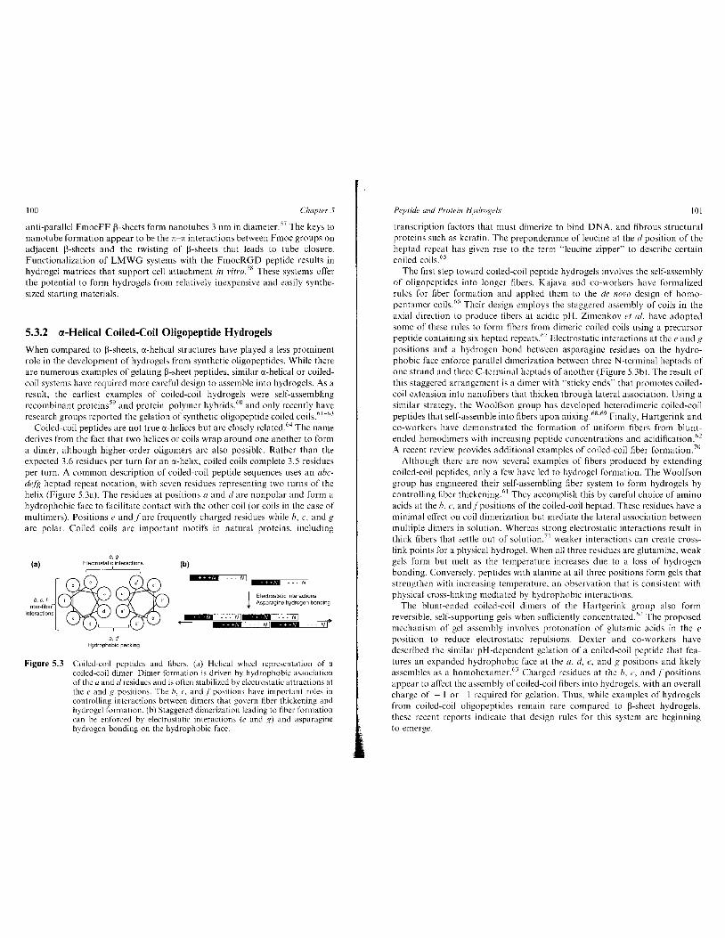
Coiled-coil peptides are not true a-helices but are closely related.64 The name derives from the fact that two helices or coils wrap around one another to form a dimer, although higher-order oligomers arc also possible. Rather than the expected 3.6 residues per turn for an a-helix, coiled coils complete 3.5 residues per turn. A common description of coiled-coil peptide sequences uses an abcdefg heptad repeat notation, with seven residues representing two turns of the helix (Figure 5.3a). The residues at positions a and dare nonpolar and form a hydrophobic face to facilitate contact with the other coil (or coils in the case of multimcrs). Positions e and fare frequently charged residues while h, c, and g are polar. Coiled coils are important motifs in natural proteins, including
(a) e.g
Electrostatic interactions (b)
--~~==· R]N I Milllf - Nl
1 Electrostatic 1nteract1ons Asparagine hydrogen bondmg
*' --- Nl M --- Ni *88 & --- N'*ii& --""'N'f'
Figure 5.3 Coiled-coil pcptidcs and fibers. (a) Helical \vheel representation of a coiled-coil dimcr. Dimer formation is driven by hydrophobic association of the a and d residues and is often stabilized by electrostati\.: attractions at the c and g positions. The h, c. and f positions have important roles in controlling: interactions between dimers that govern fiber thickening and hydrogel formation. (b) Staggered dimerization leading to fiber formation can be enforced by electrostatic interactions (e and g) and asparagine hydrogen bonding on the hydrophobic face.
Peptide and Protein Hydro!{cls 101
transcription factors that must dimerize to bind DNA, and fibrous structural proteins such as keratin. The preponderance of leucine at the d position of the heptad repeat has given rise to the term "leucine zipper" to describe certain coiled coils. 65
The first step toward coiled-coil peptide hydrogels involves the self-assembly of oligopeptides into longer fibers. Kajava and co-workers have formalized rules for fiber formation and applied them to the de nora design of homopentamer coils. 66 Their design employs the staggered assembly of coils in the axial direction to produce fibers at acidic pH. Zimenkov et a/. have adopted some of these rules to form fibers from dimeric coiled coils using a precursor peptide containing six heptad repeats. 67 Electrostatic interactions at thee and!? positions and a hydrogen bond between asparagine residues on the hydrophobic face enforce parallel dimerization between three N-terminal heptads of one strand and three C-tenninal heptads of another (Figure 5.3b). The result of this staggered arrangement is a dimer with "sticky ends"' that promotes coiledcoil extension into nanofibers that thicken through lateral association. Using a similar strategy, the Woolfson group has developed heterodimeric coiled-coil peptides that self-assemble into fibers upon mixing 68
·69 Finally, Hartgerink and
co-workers have demonstrated the formation of uniform fibers from bluntended homodimers with increasing peptide concentratiOns and acidification.02
A recent review provides additional examples of coiled-coil fiber formation. 70
Although there arc now several examples of fibers produced by extending coiled-coil peptides, only a few have led to hydrogel formation. The Woolfson group has engineered their self-assembling fiber system to form hydrogels by controlling fiber thickening61 They accomplish this by careful choice of amino acids at the h, c, and.fpositions of the coiled-coil heptad. These residues have a minimal effect on coil dimerization but mediate the lateral association between multiple dimers in solution. Whereas strong electrostatic interactions result in thick fibers that settle out of solution,71 weaker interactions can create crosslink points for a physical hydrogel. When all three residues are glutamine, weak gels form but melt as the temperature increases due to a loss of hydrogen bonding. Conversely, pcptides with alanine at all three positions form gels that strengthen with increasing temperature, an observation that is consistent with physical cross-linking mediated by hydrophobic interactions.
The blunt-ended coiled-coil dimers of the Hartgerink group also form reversible, self-supporting gels when sufficiently concentrated. 62 The proposed mechanism of gel assembly involves protonation of glutamic acids in the e position to reduce electrostatic repulsions. Dexter and co-workers have described the similar pH-dependent gelation of a coiled-coil peptide that features an expanded hydrophobic face at the a, d. e, and g positions and likely assembles as a homohexamer. 63 Charged residues at the h, c, and f positions appear to a !Teet the assembly of coiled-coil fibers into hydrogels, with an overall charge of + I or I required for gelation. Thus, while examples of hydro gels from coiled-coil oligopeptides remain rare compared to 13-sheet hydrogels. these recent reports indicate that design rules for this system are beginning to emerge.
102 Chapter 5
5.3.3 Collagen Mimetic Peptide Hydrogels
An emerging class of peptide hydrogels is based on the triple-helical structure of collagen and its assembly into higher-order fibrous structures. With at least 28 isoforms, collagens are the most abundant proteins in vertebrates and are responsible for much of the mechanical integrity of the extracellular matrix72
While naturally derived sources of collagens are common materials for cell culture and tissue engineering, a synthetic substitute would be more homogeneous and eliminate the potential for contamination from animal tissues. To realize this goal, several research groups have explored the molecular and supramolecular structures of collagen mimetic peptides, or CMPs.73
·74
Early efforts in this field sought to analyze the assembly and stability of triple-helical CMPs as a model system for collagen. The most common CMP sequences are repeats of the Gly-Pro-Pro (GPP) or Gly-Pro-Hyp (GPO) tripeptides, where Hyp (0) is hydroxyproline. These primary sequences adopt a left-handed polyproline type II helical structure, and three such helices associate as a right-handed superhelix. The requirement for Gly in CMPs stems from the steric hindrance encountered by larger side chains in the center of the helix, while Pro residues promote the preorganization of helical strands and Hyp contributes to stability through solvation or stereoelectronic effects.72
While researchers have gained important structural information from these first-generation CMPs, evidence for their higher-order assembly into fibers and gels has been limited75
As with coiled-coil peptides, progress toward higher-order CMP structures requires the extension of triple helices in the axial direction followed by their lateral and head-to-tail association into fibers. Kotch and Raines have accomplished axial extension using disulfide bonds to create cysteine-knotted trimers with single- and double-stranded overhangs at the termini76 The overhangs associate with the appropriate strands on opposing molecules to extend the triple helix into nanofibers. The Koide group has also developed a cysteine knot method for producing long triple-helical CMPs that form gels77
•78 Chaikof, Conticello and co-workers have described the formation of
the most collagen-like synthetic fibers to date using a zwitterionic CMP, (PRG)4(POG)4(EOGk79 Electrostatic interactions between Arg and Glu side chains produce triple helices that further assemble upon thermal annealing into 70 nm wide fibers with periodic banding patterns similar to those in native collagen. Alternative strategies to produce long triple helices involve chain extension of telechelic CMPs functionalized with thiols and thioesters for native chemicalligation,80 ligands for metal-ion coordination,8 1
•82 and aromatic rings
for n- n stacking and cation- It interactions.83·84
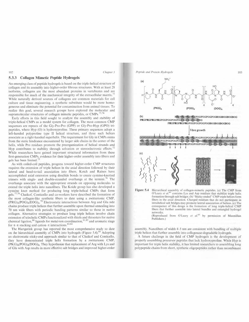
The Hartgerink group has reported the most comprehensive study to date on the hierarchical assembly of CMPs into hydrogels (Figure 5.4)85 Adopting an electrostatic sticky-end approach similar to that of Chaikof and Conticello, they have demonstrated triple helix formation by a zwitterionic CMP, (PKG)4(POG)4(DOGk They hypothesize that replacement of Arg with Lys and of Glu with Asp results in more effective salt bridges and improved higher-order
Peptide and Protein Hydrogels 103
(PKG)4 (POG)4 (DOG)4
(b) ........ !@!_1@2_POGPOGPOGPOGOOGDOGDOGDOG PKGPKGPKGPKGPOGPOGPOG~
PKGPKGPKGPKGPOGPOGPOGPOGDOGI)()GI)()
j Fibre &rowth
OCPOO~~POGPOG~ ~~PKGPKGPOCPOCPOC~
Figure 5.4
~ l l ~ l I
!(.( ~
Hierarchical assembly of collagen-mimetic peptides. (a) The CMP from O'Leary et a/.85 contains Lys and Asp residues that stabilize triple helix formation through salt bridges. (b) "Sticky-ended" CMP triple helices form fibers in the axial direction. Charged residue that do not participate in intra helical salt bridges may promote lateral association of helices. (c) The consequence of this design is the formation of long triple-helical CMP fibers that further assemble into lateral bundles and entangled hydrogel networks. (Reproduced from O'Leary et a/.85 by permission of Macmillan Publishers.)
assembly. Nanofibers of width 4-5 nm are consistent with bundling of multiple triple helices that further assemble into collagenase-degradable hydrogels.
A future challenge in the field of CMP hydrogels is the development of properly assembling precursor peptides that lack hydroxyproline. While Hyp is important for triple helix stabili ty, it has limited researchers to assembling long polypeptide chains from short, synthetic oligopeptides rather than recombinant

104 Chapter 5
proteins. Using a combination of electrostatic stabilization and cysteine knots, Krishna and Kiick have demonstrated the formation of triple helices and fibrillar structures from CMPs lacking Hyp 86 Engineering recombinant organisms capable of incorporating Hyp during protein synthesisH7 or modifying proline residues post-translationall/'s would offer alternative solutions.
5.3.4 Peptide Amphiphile Hydrogels
Hydrogel formation in many P-shcet and coiled-coil systems is due in part to the amphiphilic nature of the precursor peptides. Self-assembly in such materials results from the burial of apolar regions and a combination of electrostatic attractions, hydrogen bonding, and solvation in polar regions. These characteristics are also specific design features of a class of materials known as peptide amphiphilcs (PAs). For example, Tirrell and co-workers have synthesized oligopeptides conjugated to long alkyl chains at the N-terminus and demonstrated that these hydrophobic tails produce monolayers at an air/water interface." The PA monolayers are useful for displaying cell adhesion molecules and orienting peptides to promote intermolecular interactions such as triple helix formation in CMPs 90
·91 The Zhang group has also developed
amphiphilic molecules composed completely of amino acids. These PAs associate in water to form nanovesicles and nanotubes by burying hydrophobic rcsiducs.92
The Stupp group has thoroughly investigated the selt~asscmbly and gelation of peptide amphiphiles, as well as numerous applications for these materials. They have synthesized PAs containing a 16-carbon alkyl tail covalently bonded to the N-terminus of a hydrophilic peptide.n94 An example of this type of molecule is their phosphoserine (S'P04')-containing PA, CHJ(CH2)"0-CCCCGGGstP04'RGD (C =cysteine). Since the hydrophobic tail is slightly narrower than the peptide head, PAs tend to form cylindrical micelles in water. Upon slow acidification or addition of calcium ions. PAs self-assemble into nanofibers that gel at sufficiently high concentrations. Oxidation of cysteine residues creates reversible disulfide cross-links that stabilize PA nanostructures. Alternatively. UV-mediated cross-linking can occur between hydrophobic tails that contain diacetylene moieties rather than simple alkyl chains." Applications of PA hydro gels include their use as tissue engineering scaffolds and drug delivery vehicles95 The Stupp group's original peptide amphiphiles incorporated phosphoserine residues that act as templates for the mineralization of hydroxyapatite, as observed in bone formation. 93 Nanofibcr and hydrogel formation have proven robust to significant sequence variation in the peptide region. permitting incorporation of a variety of biologically active peptide domains, including those with considerable hydrophobic content. Examples include PAs displaying the cell-binding peptides RGD and IK VA V, growth factor-mimicking epitopes. and heparin-binding pep tides (I= isoleucine). 94
·96
·97
Deming and co-workers have produced rapidly recovering hydrogels from a different class of peptide amphiphilcs9
'·99 Using N-carboxyanhydride (NCA)
Peptide and Protein Hydrogel.\· 105
polymerization, they prepared long diblock copolypeptides of the form K 160 L40
and K, 60Y40 with very low polydispersity. The hydrophobic blocks adopt crhelical and P-sheet structures when constituted from leucine (L) and valine, respectively. In the case of polyleucine helical hydrophobic blocks, the proposed model for self-assembly involves perpendicular alignment of ex-helices in a twisted fibril that is surrounded by a polyelectrolyte "brush". roo This is in contrast to other self-assembling helical peptidcs in which alignment occurs parallel to the helical axis. Gelation occurs above a concentration threshold that is dependent on the length of the hydrophilic block as well as the length and structure of the hydrophobic block. Triblock (K,L,Kcl and pcntablock (K,L,K_-L,.K,) copolypcptides also self-assemble and form gels, with pentablocks having the potential for intermolecular cross-links between fibrils. 101
5.4 Hydrogels from Recombinant Proteins Recombinant proteins are another class of building blocks for engineered hydrogels. Whereas oligopeptides prepared by SPPS arc limited in length and polypeptides prepared NCA polymerization are limited in sequence diversity, recombinant expression exploits the ability of a host organism to synthesize full-length proteins of almost any sequence from DNA templates. One result of this is a fundamental difference in the mechanisms for hydrogel formation by oligopeptides rcrsus recombinant proteins. As discussed in the previous section, short oligopeptides self-assemble into nanofibers that entangle to form hydrogels. Proteins, on the other hand, can be long enough to form hydrogel networks without preassembling into fibers. Instead. network formation is driven by noncovalcnt interactions between physical cross-linking domains or by covalent bonds between chemically cross-linked residues.
Protein engineering alTers the potential for unprecedented control over hydrogel structure and functionality_:u Two important categories of recombinant proteins for hydrogcls arc self-assembling artificial proteins and biomimctic proteins based on elastins and silks. Recent advances in protein engineering have also led to the development of a third category that combines assembly domains with full length. functional proteins to form multifunctional hydrogels. 102
5.4.1 Self-Assembling, Multidomain Artificial Proteins
Our laboratory has investigated recombinant artificial proteins as precursors for self-assembling hydro gels based on the association of coiled-coil domains. 59
Telechelic triblock proteins denoted ACYA contain two leucine-zipper domains (A) flanking a random coil polyelectrolyte chain, (AG)1PEG, repeated x times (CJ. Variation in the association of zipper domains in response to temperature and pH changes results in a reversible sol--gel transition. At high temperature or high pH, solutions of AC,A behave as viscous liquids. Decreasing the temperature to refold the helices. or lowering the pH to near-neutral values. leads

106 Chapter 5
to strong association of leucine-zipper domains as tetramers. Lf the polyelectrolyte linker region remains solvated, coiled-coil tetramers can act as physical cross-links in a hydrogel network. Using genetically engineered endblocks, KopeCck and co-workers have demonstrated that the oligomerization state, self-assembly behavior, and pH and temperature responsiveness of coiled-coil hydrogcls can be tuned by altering the amino acid sequence. 103
•104
Hydrogels assembled from AC,A triblocks are unexpectedly soft and erode rapidly in open solutions, thus limiting their potential applications-' 05
·106 This
behavior is due to the tendency of AC_A to form non-productive loops that do not contribute to the elasticity of the network. The network is transient and rapid exchange of peptide strands between coiled-coil aggregates allows zippers to disengage and dissolve in the surrounding medium. Our group has demonstrated three strategies that overcome these limitations and produce stiffer hydrogels with tunable erosion rates. The first is to stabilize leucinezipper aggregates through disulfide bond formation (Figure 5.5a)106 By the judicious placement of cysteine residues in the zipper domains, it is possible to preferentially stabilize intermolecular aggregates while allowing exchange of looped strands. In an alternative strategy, longer polyelectrolyte linkers suppress loops that arise to avoid energy penalties associated with stretching shorter chains (Figure 5.5b). 105 Unlike typical elastic materials that become softer upon increasing the molecular weight between cross-links, AC,A hydrogels can become stiffer when the linker region is extended. Finally, redesigned triblock proteins of the form AC,P and PC,P (Figure 5.5c), where P is a zipper domain derived from the cartilage oligomeric matrix protein, are stiffer and exhibit slower erosion rates than AC,A. 107 In the case ofPC,P, these observations arise from the higher aggregation number and parallel alignment of the P zippers. While A zippers can adopt an anti parallel orientation as part of tctramcric coiled coils, P zippers associate exclusively in a parallel orientation in pentameric aggregates. This results in a lower tendency for PC\.P to form intramolecular loops, as the linker region must stretch to permit the proper parallel alignment. In the case of AC,P triblocks, intramolecular loops rarely form due to the preference of the A and P zippers to aggregate as homooligomers rather than hetero-oligomers.
The ability to tune the assembly and erosion of telechelic leucine-zipper hydrogels suggests a potential application as materials for the encapsulation and controlled release of cells or biomolccules. In this respect. desirable features include shear-thinning behavior and the rapid recovery of elastic strength upon cessation of shear. These properties would permit the delivery of the hydrogel and its cargo via a minimally invasive injection. Rheological measurements of PCP hydrogels indicate a decrease in the clastic modulus of three orders of magnitude at high strain rates (Figure 5.6a). "" Hydrogels also recover their elastic strength within seconds after large-amplitude oscillatory strain and form self-supporting structures when injected through narrow gauge needles (Figure 5.6a,b). Shear-thinning appears to be due to yielding behavior within the gel, with shear banding potentially protecting cells and biomolecules from high shear rates. The nonlinear rheology of PC,P hydro gels is largely unaffected by
Peptide and Protein Hydrogcls 107
(a) disulfidestabil~
~ unstabilized loop
>
stabilized, stretched loop
(c)
PCxP antiparallelloop >
~) PCxP parallel loop
>
Figure 5.5 Stabilization of self-assembling artificial proteins to control hydrogel stiffness and erosion rate. Cross-links arc depicted as dimers for clarity. although tetrameric and pentameric coiled-coils predominate in these systems. (a) Disulfide formation between cysteine residues in the AC,A domain stabilizes intermolecular cross-links. The protein design precludes stabilization of antiparallel intramolecular loops, while parallel loops rarely form due to chain stretching. (b) Cross-linking is favored by an extended midblock C with a mean end-to-end distance (!c) greater than the average distance betv.'een proteins (d). In contrast, shorter midblocks form loops to avoid energy penalties associated with chain stretching. (c) Proteins of the form PC_.P contain P coil domains that aggregate exclusively in a parallel orientation. Cross-linking is preferred to loop formation, which requires chain stretching. Proteins of the form AC,P do not form intramolecular loops because the A and P domains (white rectangles and striped rectangles, respectively) do not form heterooligomers.

108
(a) -1000
-~ofltrlln 11115,...,....., •lnlddonothe~ . 15"*"Aaof'-*'g
Chapter 5
Figure 5.6 Shear-thinning and elastic recovery of PCP hydrogels. (a) Hydrogels experience a decrease in their shear storage modulus (G') of three orders of magnitude at the onset of large-amplitude oscillatory strain but recover their elastic strength within seconds. Recovery was independent of the length of the mid block in PC10P and PC30P hydrogels. (b) PC,P forms a self-supporting gel upon injection through a 22 gauge needle. (Reproduced from Olsen eta/. 108 by permission of the American Chemical Society.)
the length of the polyelectrolyte linker region, suggesting that it should be possible to incorporate biologically active sequences into shear-thinning telechelic proteins. 109
Telechelic proteins containing collagen-mimetic endblock sequences also produce shear-thinning, thermoreversible hydrogels. 110
·111 The triblock archi
tecture of these proteins consists of nine repeats of a Pro-Gly-Pro sequence flanking a central random coil. Recombinant production in yeast yields gramper-liter quantities of the secreted product. 112 In these materials, endblock aggregation occurs through formation of triple helices. Hydrogels assembled from CMP telechelic triblocks have shown promise in the controlled release of model proteins. 11 3 Chemical cross-linking of lysine residues in the random coil midblock using glutaraldehyde produces shape-memory hydrogel networks that are dependent on the thermoreversibility of the end block triple helices. 114
Many peptide hydrogels require temporary exposure to non-physiological pH, temperature, or ionic strength in order to self-assemble. These conditions may result in unacceptable levels of cell death or denaturation of encapsulated cargo. To overcome these problems, Heilshorn and co-workers have introduced the mixing-induced, two-component hydrogel (MITCH) system in which gelation can occur only upon mixing of two protein solutions. 11 5 The two components of MITCH gels are artificial proteins consisting of several repeats of either the WW or proline-rich domains separated by random coil linker regions. Physical cross-linking between proteins containing the WW domain, named for its conserved tryptophan residues, and proteins containing the proline-rich domain derives from the noncovalent association between these sequences as found in natural proteins. By varying the stoichiometry of the two components as well as the frequency and binding strength of the cross-linking domains, it is possible to tune the sol- gel transition and viscoelastic properties
Peptide and Protein Hydrogels 109
of the system in a predictable manner. 115•
116 The transient nature of the network results in shear-thinning protein hydrogels that display rapid self-healing within minutes after injection.
5.4.2 Biomimetic Recombinant Proteins: Elastins and Silks
Elastins a re important components of the extracellular matrix and represent attractive targets for biomedical engineering applications. The desirable mechanical and chemical properties of elastins can be recapitulated in elastinlike polypeptides (ELPs) containing repeats of the YPGXG pentamer, where the " guest residue" X can be any amino acid except proline. Elastin-like polypeptides exhibit inverse temperature behavior in that they are soluble at low temperatures but aggregate in coacervate phases at higher temperatures. The transition from a soluble to an aggregated state occurs at the lower critical solution temperature (LCST), where it has been proposed that ELPs transition from a random coil conformation to a ~-spiral. The LCST is dependent on the length of the sequence, the ELP concentration, and the hydrophobicity of the guest residue, with more hydrophilic sequences exhibiting higher LCSTs. 11 7
•1 ts
While highly repetitive ELPs can be produced synthetically, recombinant engineering permits access to more complex protein/ sequences, including multiblock architectures and bioactive domains. 119
•120 One of the most com
mon engineering strategies is to vary the ELP guest residue to incorporate reactive moieties for chemical cross-linking or to alter the LCST behavior of the protein. Temperature cycling above and below the LCST offers a facile method of purification of recombinant ELPs. 12 1
Elastin hydrogels have been formed by both physical and chemical crosslinking methods. Conticello and co-workers have described the physical crosslinking of triblock ELPs based on inverse temperature transitions. 122
•123 The
mechanism for gel formation is similar to that of telechelic leucine-zipper and CMP hydrogels. Triblocks of the form BAB contain hydrophobic endblock elastin sequences (B) flanking a hydrophilic elastin midblock (A). At temperatures above their LCST, endblocks undergo microphase separation while the hydrophilic midblock (which has a higher LCST) remains solvated. This results in hydrogels in which the aggregated hydrophobic blocks serve as thermoreversible, noncovalent cross-linkers. Chilkoti and co-workers have analyzed the rheological behavior of an ELP above its LCST. Although their design did not include explicit cross-linking domains, they observed gel- like behavior in the coacervate with potential applications in cartilage tissue engineering. 124
Several research groups have employed chemical cross-linking to form hydrogels with mechanical properties that more closely resemble those of native elastin. The most common strategy uses small multifunctional cross-linkers that form covalent bonds with reactive amino acids, typically lysines occupying the ELP guest site or in distinct cross-lin king domains. 125 131 Other strategies include enzymatic cross-linking, 132 UV or visible light photo-cross-linking,27
•133
and y-irradiation. 134
110 Chapter 5
Hydrogels from silk proteins represent another class of potentially useful biomimetic matcrials. 135 The best studied silk proteins are those from the Chinese silkworm Bombyx mori and from the draglinc of the spider Nephi/a clavipes. Fibers spun from silk proteins derive their impressive mechanical strength and extensibility from repetitive Gly- and Ala-rich ~-sheet crystalline domains separated by amorphous hydrophilic linker regions. Kaplan and coworkers have demonstrated that solutions of naturally derived silkworm fibroin doped with poly( ethylene glycol) undergo a sol- gel transition over the course of days due to the coalescence of hydrophobic rcgions. 136 135 Hydrogels produced from silk fibroin are candidates for tissue engineering and cell encapsulation applications. 139
' 140
In contrast to extensively engineered ELPs, silk protein engineering has generally focused on producing close facsimiles of the natural silks. In particular, there has been strong interest in developing recombinant sources of dragline spider silk since spiders, unlike silkworms, are not easily farmed. Several research groups have demonstrated progress in this area by producing spider silks in bacterial. 141 plant, 142 mammalian, 14
J and silkworm hosts. 144
While significant attention has been directed toward spinning recombinant silks into fibers, only a few studies have examined hydrogel formation from these proteins. 145
·146 Further protein engineering could offer the ability to tune the
mechanical properties of silk and silk hydro gels or to introduce new bioactive domains147
·148 An alternative approach to silk-like hydrogels involves geneti
cally engineered block copolymers of silk-like GAGAGS (S ~serine) peptides and elastin-like VPGXG peptides. 14
"·150 Solutions of these proteins sponta
neously form swollen hydrogels that display the crystalline hydrophobic domains of silks and the elasticity and responsiveness of ELPs. These materials have shown promise as delivery vehicles for gene therapy. 151
5.4.3 Multifunctional Protein Hydrogels
Many proposed applications of peptide hydrogels in tissue engineering and drug delivery require materials that display simple binding motifs and assemble or disassemble in response to environmental cues. Both oligopeptides and recombinant proteins have shown promise in meeting these requirements. However, protein engineering also offers the ability to develop hydrogels with much more advanced functionality. In this regard, emerging research has demonstrated the potential of engineering multifunctional protein hydrogels and their applications as sensors and catalytic materials.
Banta and co-workers have developed multifunctional protein hydrogels that combine the self-assembly behavior of leucine zippers with the useful functional properties of fluorescent proteins and enzymes. In their initial demonstration of this approach, they showed that the incorporation of green fluorescent protein in the midblock region of AC,A does not inhibit protein foldmg or gel formation.152 Using this method, they produced multicolor hydro gels and probed gel structure in these systems by Forster resonance energy transfer (FRET). A similar design that incorporated an oxidase enzyme and electron conductors
Peptide and Protein 1/ydroge!s Ill
was used to create bioelectrocatalytic hydrogels capable of reducing molecular oxygen to water. 153 The electron-conducting hydrogel network consisted of triblock leucine-zipper proteins with osmium bis-bipyridine complexes bound to histidine residues. Enzymatic activity was derived from a chimeric protein containing a leucine-zipper domain and the small laccase (SLAC) polyphenol oxidase from Strepromyces coelico/or. The zipper domain mediates incorporation of the chimera into the hydrogel network, while dimerization of the SLAC enzyme provides additional cross-linking and is required for catalytic activity. This novel class of materials has potential applications in biofuel cells and oxygen sensors. Other examples of enzymatic hydrogels incorporate an aldo-keto reductase and an organophosphate hydrolase. suggesting the broad applicability of this method. 154·
155
In similar work, Gallivan and co-workers have engineered a chimeric calmodulin protein fused to a leucine-zipper domain. 156 Calmodulin is an important regulatory protein that undergoes a conformational change in the presence of calcium ions to bind partner proteins. A solution of the chimeric calmodulin-zipper protein and a telechclic cross-linker containing calmodulinbinding endblocks forms a hydrogel network in the presence of Ca
2+ The
network is reversible upon chelation of calcium.
5.5 Hydrogels from Peptide-Polymer and ProteinPolymer Hybrids
As discussed earlier in this chapter, a common design for recombinant protein hydrogels is a telechelic triblock architecture with physical cross-linking domains separated by a polyelectrolyte linker. Hydrogel formation results from an appropriate balance between endblock aggregation and linker solubility. While genetically encoded linkers offer clear advantages such as monodispersity. sequence diversity, and bioactivity, synthetic polymers can also fill this role in hydrogels formed from peptide-polymer and protein-polymer hybrids. From a synthesis and cost perspective, hydro gels in which the bulk of the dry weight derives from synthetic macromolecules may be more desirable than hydrogels from oligopeptides or recombinant proteins. Furthermore, whereas ribosomal translation limits recombinant proteins to linear architectures, synthetic methods offer access to branched architectures such as graft,
star, and dendritic polymers. In hybrid hydrogels. peptides and proteins may serve several roles. Most
commonly, they arc responsible for chemical or physical cross-linking of the prepolymer solution. The mechanisms for cross-linking and self-assembly are analogous to those described for recombinant protein hydrogels. Peptides and proteins may also confer biological activities such as cell binding and enzymatic degradability to otherwise inert polymers. Finally, proteins embedded in hybrid hydrogel networks may exhibit enzymatic activity or ligand binding for applications in stimulus-responsive materials and biosensors. We present several examples of hybrid hydrogels as an introduction to this field. A more
112 Chapter 5
detailed account of protein- and peptide-polymer materials is provided by Krishna and Kiick. 157
5.5.1 Peptides as Physical and Chemical Cross-Linkers
Kopecek and co-workers have pioneered the development of physically crosslinked hybrid hydrogels from proteins and synthetic polymers 60 Their design features recombinant coiled-coil proteins attached to a copolymer of N-(2-hydroxypropyl)methacrylamide and (N' ,N' -dicarboxymethylaminopropyl)methacrylamide. The attachment is mediated by Ni'' coordination by the polyhistidine tag on the protein and the iminodiacetate side chains of the copolymer. As in triblock leucine-zipper recombinant proteins, coiled-coil aggregation leads to hydrogel formation. A second generation design features two copolymers covalently grafted with coiled coils that associate as heterodimers.15R Reversible hydrogels form upon mixing the two copolymers at neutral pH at concentrations as low as 0.1 wt%.
Sherr and co-workers have further demonstrated the cross-linking potential of coiled coils in a bottom-up approach to fabricating hydrogels for tissue engineering. 159 Using photolithography, they encapsulate cells in 400 fim star-shaped micro gels by photopolymerization of poly( ethylene glycol) (PEG) diacrylatc. Subsequent attachment of cysteine-containing coiled coils to the residual acrylate groups facilitates reversible assembly ofmicrogels into porous macroscopic scaffolds. The high porosity and pore interconnectivity of these scaffolds and the short length scales of' the microgels may aid nutrient transport to the encapsulated cells.
The Yu group has investigated hydrogel formation from four-arm PEG stars functionalized with collagen-mimetic peptides. 160 The association of CMPs as triple helices forms a network that is reversible by heating above the melting temperature and addition of competitor CMPs. They exploit this reversible behavior to form gradients in gel stiffness through local injection of a hot CMP solution. As the solution diffuses from the injection site, the thermal gradient melts the triple helical cross-links, \Vhich do not reform upon cooling due to the high concentration of competitor peptides. They have also demonstrated the use of CMPs to modify natural collagens noncovalently with PEG and growth factors through a strand invasion mechanism. 16
1.102 These systems highlight the
potential of hybrid hydrogels containing synthetic polymers, peptides, and naturally derived proteins.
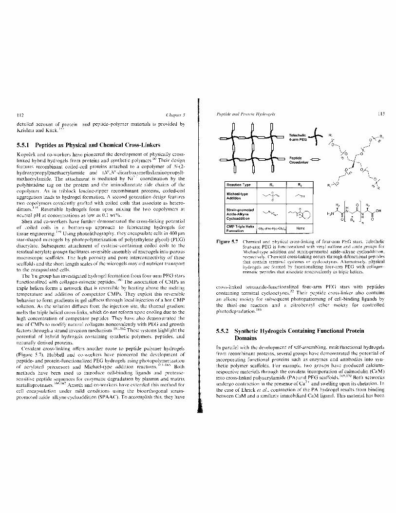
Covalent cross-linking offers another route to peptide polymer hydrogels (Figure 5.7). Hubbell and co-workers have pioneered the development of peptide- and protein-functionalized PEG hydro gels using photopolymerization of acrylated precursors and Michael-type addition reactions. 16 ·~- 165 Both methods have been used to introduce cell-binding ligands and proteasesensitive peptide sequences for enzymatic degradation by plasmin and matrix metalloproteases. 166
·167 Anseth and co-workers have extended this method for
cell encapsulation under mild conditions using the bioorthogonal strainpromoted azide alkyne cycloaddition (SPAAC). To accomplish this, they have
Peptide and Protein Hydroge/.1·
Reaction Type R,
Michael-type 0
Addition ~#~
Strain-promoted Azide-Aikyne
--........._..N.,.N~ 'W
Cycloaddition
CMP Triple Helix ·Giy3-(Pro-Hyp-Giy)9 Formation
Telechelic + 4-arm PEG
Peptide --c:::::J-Crosslinker
R,
/'oe~
0
~,;o ~ >
> -
None
113
Figure 5.7 Chemical and physical cross-linking of four-arm PEG stars. Telechelic four-arm PEG is functionalized with vinyl sulfone and azide groups for Michael-type addition and strain-promoted azide-alkyne cycloaddition, respectively. Chemical cross-linking occurs through difunctional peptides that contain terminal cysteines or cyclooctynes. Alternatively. physical hydrogels are formed by functionalizing four-arm PEG with collagenmimetic peptides that associate noncovalcntly as triple helices.
cross-linked tetraazide-functionalized four-arm PEG stars with peptides containing terminal cyclooctynes.n Their peptide cross-linker also contains an alkene moiety for subsequent photopatterning of cell-binding ligands by the thiol-ene reaction and a nitrobenzyl ether moiety for controlled photodegradation. 168
5.5.2 Synthetic Hydrogels Containing Functional Protein Domains
In parallel with the development of self-assembling, multifunctional hydrogels from recombinant proteins, several groups have demonstrated the potential of incorporating functional proteins such as enzymes and antibodies into synthetic polymer scaffolds. For example, two groups have produced calciumresponsive materials through the covalent incorporation of calmodulin (CaM) into cross-linked polyacrylamide (PA) and PEG scaffolds.''" 170 Both networks undergo contraction in the presence of Ca2
+ and swelling upon its chelation. In the case of Ehrick ct a/ .. contraction of the PA hydrogel results from binding between CaM and a similarly immobilized CaM ligand. This material has been
114 Chapter 5
used to create chemically tunable microlenses. 171 In the system developed by Murphy ct al .• conformational changes in CaM drive contraction of the PEG scaffold. This system has been used for the controlled release of encapsulated growth factors. 172
Other examples of functional protein polymer hybrids include hydrogels that swell and contract due to antigen-antibody binding 173 and catalytic activity of grafted enzymes. 174 In addition, there are several examples in which growth factors are covalently cross-linked to a polymer scaffold for tissue engineering applications. 175
-177 Although the protein components do not
necessarily contribute to hydrogel assembly or function, these systems still represent an important class of hybrid materials.
5.6 Future Directions and Challenges In the past 20 years. the field of peptide and protein hydrogels has grown from early observations of self-assembly of oligopeptides to the design of advanced materials with \\'ell-controlled biological activity and mechanical properties. Hydro gels arc now routinely produced from oligopeptides, recombinant proteins, and peptide polymer hybrids. Together with hydrogcls from naturally sourced biomolecules and synthetic polymers, these materials provide scientists, engineers, and clinicians with a multitude of options to address problems in medicine and basic biology. In this regard. an important challenge will be to match the most appropriate materials with each application, particularly when transitioning hydrogcls into clinical settings. Other challenges include assessing the immune response to peptide and protein hydrogcls and producing hydrogel precursors in sufficient quantity and purity for their intended applications. These and other challenges will guide the design of future generations of engineered hydrogcls.
5.6.1 Immune Response to Peptide and Protein Hydrogels
Peptide and protein hydrogels are frequently touted as biocompatible on the basis of the fact that their precursors resemble natural biopolymers and should be susceptible to enzymatic or hydrolytic degradation. However, implantation of hydrogels still has the potential to generate undesirable immune responses. While more thorough investigations will be required, initial in rivo and in vitro studies to analyze the immunogenicity of peptide hydrogels and their precursors have been encouraging. For example, using the RADI6 and EAKI6 pep tides. Holmes eta/. detected no inflammatory response after intramuscular injection in rats and no measureable antibody response when the peptides were conjugated to bovine serum albumin and injected in rabbits and goats. 178
Similarly, myocardial injection of RAD 16 hydro gels in mice did not generate significant inflammation. 179 while the MAX ~-hairpin hydrogels of the Schneider and Pochan groups did not elicit an inflammatory response from macrophages in an in vitro assay. 1xo Promising results have also been reported for elastin-like and silk-elastin-like materials. 1x1
•1x2
Peptide and Protein Hydrogel.\' II S
The Collier group has further investigated the immunogenicity of both coiled-coil and ~-sheet self-assembling oligopeptides183
-185 They have
demonstrated in a mouse model that ~-sheet oligopeptides stimulate antibody production only when displaying a strongly immunogenic epitope. 1s4 For example, undecorated ~-sheet fibrils and fibrils displaying an RGD motif do not elicit an immune response, whereas fibrils that display a 17-mer sequence from chicken egg albumin stimulate high titer antibody production. These findings suggest that self-assembling peptides may be safe for implantation or injection if strongly immunogenic epitopes are avoided. On the other hand. the ability of ~-sheet fibrils to enhance the immunogenicity of selected epitopes indicates that self-assembling peptidcs may be useful as well-defined adjuvants for vaccine delivery and immunotherapy.
In separate work. Collier and co-workers have demonstrated that, like ~
sheet fibrils, undecorated coiled coils do not elicit a measureable immune response. 185 However, triblock materials consisting of coiled-coil endblocks separated by a PEG spacer do stimulate a moderate level of antibody production. They suggest that higher molecular weight oligomers may present better targets to the immune system, a hypothesis that is consistent with the increased immunogcnicity of protein aggregates186 Although this study was conducted with peptide concentrations below the gel point, it still highlights a key challenge for peptide hydrogels. It may not be possible to predict the immunogenicity of hydrogel networks based on the immune response to precursor peptides and proteins. Further investigation with implanted or injected gels will be required.
5.6.2 New Methods for Peptide Synthesis
While solid-phase peptide synthesis has been used extensively for hydrogel applications. there are several limitations to this method that could hinder efforts to scale up peptide production to clinically and industrially useful quantities. The foremost limitation is the inverse relationship between peptide length and overall yield that precludes the synthesis of polypeptides that contain more than approximately 50 amino acids. Low overall yields and the poor atom economy of reactions involving large protecting groups and coupling reagents also result in significant amounts of wasted starting material. Pattabiraman and Bode have recently reviewed these and other challenges as well as promising new methods of amide bond formation and chemoselectivc ligation. 18 The development of new reagents and catalysts should be closely followed by the peptide hydro gels field.
Recombinant protein production offers an alternative to SPPS, even for small oligopeptides. Riley et a/. have recently described the bioproduction of ~-sheet peptides that self-assemble into hydrogels. 1
" To accomplish this, highly repetitive polypeptides are cleaved into oligopeptides at precise positions using cyanogen bromide. This method should be applicable to nearly any peptide sequence, assuming the appropriate selection of chemical or enzymatic cleavage agents. Like SPPS, there are also drawbacks to recombinant protein

116 Chapter 5
production. Each new recombinant protein requires cloning to produce the template DNA as well as optimization of expression and purification. Protein purification frequently involves an affinity chromatography step and the removal of lipopolysaccharide endotoxins for therapeutic applications. These factors must be considered when choosing between scaling up SPPS or binproduction of oligopeptides by fermentation.
5.6.3 Spatially Patterned Hydrogels and Epitopes Beyond RGD
Owing to their similar mechanical properties and ability to display bioactivc domains. peptide and protein hydro gels are excellent candidates to replace the extracellular matrix (ECM) in tissue engineering scaffolds and in vitro cell culture matrices. While the homogeneity and well-defined nature of peptide and protein hydrogels are typically advantageous properties, the natural ECM contains mixtures of physical and chemical signals that vary in each tissue. These signals are often arranged in gradients or spatial patterns that define the cellular microenvironment in such a way as to direct specific cell and tissue behaviors. Hydrogels that mimic these patterns will likely be key to recreating the morphogenic events observed during development and tissue repair.t\.JHS A number of research groups are developing strategies to accomplish this. 1
'9 The
West, Shoichet, and Anseth groups have created spatial patterns of cell-binding peptides and protein growth factors within hydrogels using three-dimensional photolithography16
'J90
·191 With these patterns it is possible to direct cell
behaviors such as spreading and migration. Straley ct a/. have patterned elastinlike protein hydrogels using spatially controlled enzymatic degradation. 192
Other potential strategies for hydrogel patterning include layer-by-layer assembly using 30 printers 19
J.194 and microfl.uidic approaches. 195
In addition to spatially patterned hydrogels. more realistic cellular microenvironments can be obtained by incorporating biologically active domains beyond the standard RGD cell-binding sequence. While this tripeptide motif is frequently used to demonstrate the cytocompatibility of hydrogels, it binds only a subset of integrin receptors and lacks the spatial context normally provided by neighboring domains in fibronectin, vitronectin, laminin, or collagen.196·197 More complex cell-binding domains can be engineered into hydrogels assembled from recombinant proteins. For example, Fong has demonstrated accelerated in l'itro wound healing on elastin-like protein films containing the full-length fibronectin type III domains 9 and 10. 1n Wound closure on these materials occurs more rapidly than on materials containing only the RGD sequence and approaches the rate observed on fibronectin. It is also possible to include more biological complexity in hydrogels from oligopeptides. Kokkoli and co-workers have developed hydrogels from peptide amphiphiles that contain both the RGD cell-binding domain and the PHRSN synergy sequence separated by the approximate distance observed in fibronectin (H =histidine, N = asparagine).~'"- 21111 Endothelial cells cultured on these materials exhibit high levels of spreading, extracellular matrix deposition, and cytoskeletal organization. Together, spatial patterning and more complex
Peptide and Protein Hydrogcls 117
epitopes will allow researchers and clinicians to engineer cellular microenvironments that more closely resemble real tissues without compromising the well-defined nature of peptide and protein hydrogels.
Acknowledgments Work on protein hydrogels at Caltech is supported by NIH grant UOI DK089533-0 I.
References I. S. Gomes, I. B. Leonor, J. F. Mano, R. L. Reis and D. L. Kaplan, Frog.
Polym. Sci., 2012, 37, I. 2. S. A. Maskarinec and D. A. Tirrell, Curr. Opin. Bioteclmol., 2005, 16, 422. 3. J. C. M. van Hestand D. A. Tirrell, Chern. Commun., 2001, 1897. 4. T. 0. Yeates and J. E. Padilla, Curr. Opin. Struct. Bioi., 2002, 12, 464. 5. A. M. Lomas and N. A. Peppas, in Encyclopedia ol Controlled Drug
Delivery, ed. E. Mathiowitz, Wiley, New York, 1999, p. 397. 6. B. V. Slaughter, S. S. Khurshid, 0. Z. Fisher, A. Khademhosseini and
N. A. Peppas, Adr. Mater., 2009, 21. 3307. 7. J. L. Drury and D. J. Mooney, Biomaterials, 2003, 24, 4337. 8. M.P. Lutolf and J. A. Hubbell, Nat. Biotechnol., 2005. 23, 47. 9. G. D. Nicodemus and S. J. Bryant, Tissue Eng., Part B, 2008, 14, 149.
10. J. K. Tessmar and A. M. Gi\pferich, Adr. Drug Delil'er\' Rn .. 2007, 59, 274.
II. M. W. Tibbitt and K. S. Anseth, Biotech. Bioeng., 2009, 103, 655. 12. M. K. Nguyen and D. S. Lee, Macrmnol. Biosci., 2010. 10, 563. 13. L. Yu and J. Ding, Ch"m. Soc. Rev., 2008, 37, 1473. 14. A. Mata, Y. Geng, K. J. Henrikson, C. Aparicio, S. R. Stock. R. L.
Satcher and S. I. Stupp, Biomat"rials, 2010, 31. 6004. 15. C. Y. Khripin, D. Pristinski, D. R. Dunphy, C. J. Brinker and B. Kaehr,
ACS Nano, 2011, 5, 1401. 16. D. W. P. M. Li\wik, E. H. P. Leunissen, M. van den Heuvel, M. B.
Hansen and J. C. M. van Hest, Chern. Soc. Rev., 2010, 39, 3394. 17. R. V. Ulijn, N. Bibi, V. Jayawarna, P. D. Thornton, S. J. Todd, R. J.
Mart, A.M. Smith and J. E. Gough, Mater. Todav, 2007, 10, 40. 18. V. R. Pattabiraman and J. W. Bode, Nature, 2011, 480, 471. 19. P. Dawson, T. Muir, I. Clark-Lewis and S. Kent, Science, 1994,266,776. 20. J. F. Atkins and R. Gesteland, Science, 2002, 296, 1409. 21. A. Bock, K. Forchhammer, J. Heider, W. Leinfelder, G. Sawers, B.
Veprek and F. Zinoni, Mal. Microhia/., 1991, 5, 515. 22. C. A. DeForest, B. D. Polizzotti and K. S. Anseth, Nat. Mater., 2009, 8,
659. 23. C. M. Nimmo and M. S. Shoichet Bioconiugate Chem., 2011, 22, 2199. 24. S. K. Holmgren, L. E. Bretscher, K. M. Taylor and R. T. Raines, Chem.
Bioi., 1999, 6, 63.

118 Chapter 5
25. Y. Tang, G. Ghirlanda, W. A. Pelka, T. Nakajima, W. F. DeGrade and D. A. Tirrell, Angew. Chern. Int. Ed., 2001, 40, 1494.
26. N. C. Yoder and K. Kumar, Chern. Soc. Rev., 2002, 31, 335. 27. I. S. Carrico, S. A. Maskarinec, S.C. Heilshorn. M. L. Mock, J. C. Liu,
P. 1. Nowatzki, C. Franck, G. Ravichandran and D. A. Tirrell, J. Am. Chern. Soc., 2007, 129, 4874.
28. D. K. Smith, Chern. Soc. Rev., 2009, 38, 684. 29. J. A. Johnson. Y. Y. Lu, J. A. VanDeventer and D. A. Tirrell, Curr. Opin.
Chern. Bioi., 2010, 14, 774. 30. C. C. Liu and P. G. Schultz, Ann. Rev. Biochem., 2010, 79, 413. 31. E. L. Bakota, L. Aulisa, K. M. Galler and J. D. Hartgerink, Biomacro
rno/ecules, 2011, 12, 82. 32. L. Hsu, G. L. Cvetanovich and S. I. Stupp, J. Am. Chern. Soc., 2008, 130,
3892. 33. T. Aida, E. W. Meijer and S. I. Stupp, Science, 2012, 335, 813. 34. S. Zhang, T. Holmes, C. Lockshin and A. Rich, Proc. Nat/. Acad. Sci.
U. S. A., 1993, 90. 3334. 35. S. Zhang, T. C. Holmes, C. M. DiPersio, R. 0. Hynes, X. Su and A. Rich,
Biomaterials. 1995, 16, 1385. 36. S. Zhang, R. Ellis-Behnke, X. Zhao and L. Spirio. in Scaffolding in Tissue
Engineering, ed. P. X. Ma and J. Elisseeff, CRC Press, Boca Raton, FL, 2005, p. 217.
37. A. Aggeli, M. Bell, N. Boden, J. N. Keen, P. F. Knowles, T. C. B. McLeish, M. Pitkeathly and S. E. Radford, Nature, 1997, 386, 259.
38. A. Aggeli, I. A. Nyrkova, M. Bell, R. Harding, L. Carrick, T. C. B. McLeish, A. N. Semenov and N. Boden, Proc. Nat/. Acad. Sci. U. S. A., 2001, 98. 11857.
39. C. W. G. Fishwick, A. J. Beevers, L. M. Carrick, C. D. Whitehouse, A. Aggeli and N. Boden, Nano Lett., 2003, 3, 1475.
40. J. Kopecek and J. Yang, Acta Biornater., 2009. 5. 805. 41. M. R. Caplan, P. N. Moore, S. Zhang, R. D. Kamm and D. A.
Lauffenburger, Biomacrornolecu/es, 2000, I, 627. 42. M. R. Caplan. E. M. Schwartzfarb, S. Zhang, R. D. Kamm and D. A.
Lauffenburger, Biomaterials. 2002, 23, 219. 43. J. H. Collier, B. H. Hu, J. W. Ruberti. J. Zhang, P. Shum, D. H.
Thompson and P. B. Messersmith, J. Am. Chern. Soc., 2001, 123, 9463. 44. S. Ramachandran, P. Flynn, Y. Tseng andY. B. Yu, Chern. Mater., 2005,
17. 6583. 45. J. P. Schneider, D. J. Pochan. B. Ozbas. K. Rajagopal, L. Pakstis and
1. Kretsinger, J. Am. Chern. Soc .. 2002. 124. 15030. 46. D. J. Pochan, J.P. Schneider. J. Kretsinger. B. Ozbas, K. Rajagopal and
L. Haines. J. Am. Chern. Soc., 2003, 125, 11802. 47. B. Ozbas. 1. Kretsinger, K. Rajagopal, 1. P. Schneider and D. J. Pochan,
Macromolecules, 2004, 37. 7331. 48. K. Rajagopal, M. S. Lamm, L. A. Haines-Butterick, D. J. Pochan and
J.P. Schneider. Biomacromo/ecules. 2009, 10, 2619.
Peptide and Protein Hydrogels 119
49. L. A. Haines, K. Rajagopal, B. Ozbas, D. A. Salick, D. J. Pochan and J. P. Schneider, J. Am. Chern. Soc., 2005, 127, 17025.
50. D. 1. Adams, Macromo/. Biosci., 2011, 11, 160. 51. V. Jayawarna, M. Ali, T. A. Jowitt, A. F. Miller, A. Saiani, 1. E. Gough
and R. V. Ulijn, Adv. Mater., 2006, 18, 611. 52. Y. Zhang, H. Gu, Z. Yang and B. Xu, J. Am. Chem. Soc., 2003, 125,
13680. 53. Z. Yang, H. Gu, Y. Zhang, L. Wang and B. Xu, Chern. Commun., 2004,
208. 54. A. Mahler, M. Reches, M. Rechter, S. Cohen and E. Gazit, Adv. Mater.,
2006, 18; 1365. 55. S. Toledano, R. J. Williams, V. Jayawarna and R. V. Ulijn, J. Am. Chern.
Soc., 2006, 128, I 070. 56. Z. Yang, H. Gu, D. Fu, P. Gao, J. K. Lam and B. Xu, Adv. Mater., 2004,
16, 1440. 57. A. M. Smith, R. J. Williams, C. Tang, P. Cappo, R. F. Collins. M. L.
Turner, A. Saiani and R. V. Ulijn, Adv. Mater., 2008, 20, 37. 58. M. Zhou, A. M. Smith, A. K. Das, N. W. Hodson, R. F. Collins, R. V.
Ulijn and J. E. Gough, Biomateria/s, 2009, 30, 2523. 59. W. A. Pelka, J. L. Harden, K. P. McGrath, D. Wirtz and D. A. Tirrell,
Science, 1998, 281, 389. 60. C. Wang, R. J. Stewart and J. Kopecek, Nature, 1999,397,417. 61. E. F. Banwell, E. S. Abelardo, D. J. Adams, M.A. Birchall, A. Corrigan,
A. M. Donald. M. Kirkland, L. C. Serpell, M. F. Butler and D. N. Woolfson, Nat. Mater .. 2009. 8, 596.
62. H. Dong, S. E. Paramonov and J. D. Hartgerink, J. Am. Chern. Soc., 2008, 130, 13691.
63. N. L. Fletcher, C. V. Lockett and A. F. Dexter, Soji Matter, 2011, 7, 10210.
64. 1. M. Mason and K. M. Arndt, ChemBioChem, 2004,5, 170. 65. W. Landschulz, P. Johnson and S. McKnight, Science, 1988, 240, 1759. 66. S. A. Potekhin, T. N. Melnik, V. Popov, N. F. Lanina, A. A. Vazina, P.
Rigler. A. S. Verdini, G. Corradin and A. V. Kajava, Chern. Bioi., 2001, 8, 1025.
67. Y. Zimenkov, V. P. Conticello, L. Guo and P. Thiyagarajan, Tetrahedron, 2004, 60, 7237.
68. M. J. Pandya, G. M. Spooner, M. Sunde, J. R. Thorpe, A. Rodger and D. N. Woolfson. Biochemistry, 2000, 39, 8728.
69. M. G. Ryadnov and D. N. Woolfson, Nat. Mater., 2003, 2, 329. 70. D. N. Woolfson, Biopolymers, 2010, 94, 118. 71. D. Papapostolou, A. M. Smith, E. D. T. Atkins. S. J. Oliver, M. G.
Ryadnov, L. C. Serpell and D. N. Woolfson, Proc. Nat/. Acad. Sci. U.S. A .. 2007. 104, 10853.
72. M. D. Shoulders and R. T. Raines, Annu. Rev. Biochem., 2009, 78, 929. 73. J. A. Fallas, L. E. R. O'Leary and J. D. Hartgerink, Chern. Soc. Rev.,
2010, 39, 3510.
120 Chapter 5
74. S. M. Yu, Y. Li and D. Kim, Soft Matter, 2011, 7, 7927. 75. K. Kar, P. Amin, M.A. Bryan, A. V. Persikov, A. Mohs, Y. H. Wang and
B. Brodsky. J. Bioi. Chem., 2006, 2S1, 33283. 76. F. W. Kotch and R. T. Raines, Proc. Nat/. Acad. Sci. U. S. A., 2006, 103,
3028.
77. T. Koide, D. L. Homma, S. Asada and K. Kitagawa. Bioorg. Med. Chem. Lett., 2005, 15, 5230.
78. C. M. Yamazaki, S. Asada, K. Kitagawa and T. Koide, Peptide Sci., 2008, 90, 816.
79. S. Rete, Y. Song, R. P. Apkarian, Z. Qu, V. P. Conticello and E. L. Chaikof, J. Am. Chem. Soc.. 2007, 129, 14780.
80. S. E. Paramonov, V. Gauba and J. D. Hartgerink, Macromolecules. 2005, 3S, 7555.
81. M. M. Pires and J. Chmielewski, J. Am. Chern. Soc., 2009, 131, 2706. 82. W. Hsu, Y.-L. Chen and J.-C. Horng. Langmuir, 2012. 2S. 3194. 83. M. A. Cejas, W. A. Kinney, C. Chen, G. C. Leo, B. A. Tounge, J. G.
Vinter, P. P. Joshi and B. E. Maryanoff, J. Am. Chem. Soc., 2007, 129, 2202.
84. C.-C. Chen, W. Hsu, T.-C. Kao and J.-C. Horng, Biochemistry, 2011, 50, 2381.
85. L. E. R. O'Leary, J. A. Fallas, E. L. Bakota. M. K. Kang and J. D. Hartgerink, Nat. Chem., 2011, 3, 821.
86. 0. D. Krishna and K. L. Kiick, Biomacromolecules, 2009, IO, 2626. 87. D. D. Buechler, D. N. Paolella, B. S. Leslie. M. S. Brown. K. A. Mehos
and E. A. Gruskin, J. Bioi. Chern .. 2003, 27S, 645. 88. D. M. Pinkas, S. Ding, R. T. Raines and A. E. Barron, ACS Chern. Bioi.,
2011,6,320.
89. P. Berndt, G. B. Fields and M. Tirrell, J. Am. Chern. Soc., 1995, ll7, 9515.
90. M.A. Biesalski. A. Knaebel, R. Tu and M. Tirrell, Biomaterials. 2006. 27, 1259.
91. Y.-C. Yu, P. Berndt, M. Tirrell and G. B. Fields. J. Am. Chern. Soc .. !996. liS, 12515.
92. S. Vauthey, S. Santoso. H. Gong, N. Watson and S. Zhang, Proc. Nat/. A cad. Sci. U. S. A., 2002, 99, 5355.
93. J. D. Hartgerink, E. Beniash and S. I. Stupp, Science. 200 I, 294, 1684. 94. J.D. Hartgerink, E. Beniash and S. I. Stupp. Proc. Nat/. A cad. Sci. U.S. A.,
2002,99, 5133.
95. J. B. Matson and S. I. Stupp. Chem. Commun., 2012, 4S, 26. 96. K. Rajangam. H. A. Behanna, M. J. Hui, X. Han, J. F. Hulvat. J. W.
Lomasney and S. I. Stupp, Nano Lett .. 2006, 6. 2086. 97. M. J. Webber. J. Tongers, C. J. Newcomb, K.-T. Marquardt, J. Bauersachs,
D. W. Losordo and S.!. Stupp. Proc. Nat/. Acad. Sci. U.S. A., 2011, lOS, 13438.
98. A. P. Nowak, V. Breedveld, L. Pakstis. B. Ozbas. D. J. Pine, D. Pochan and T. J. Deming, Nature, 2002, 417. 424.
Peptide and Protein Hydrogel.\' 121
99. A. P. Nowak, V. Breedveld. D. J. Pine and T. J. Deming. J. Am. Chem. Soc., 2003, 125, 15666.
!00. T. J. Deming. Sofi Matter, 2005, I, 28. 101. Z. Li and T. J. Deming, Soft Matter, 2010, 6, 2546. 102. S. Banta, I. R. Wheeldon and M. Blenner, Annu. RC\'. Biomed. Eng., 20!0,
12, 167. !03. C. Xu, V. Breedveld and J. Kopecek, Biomacromolccules, 2005. 6. 1739. 104. C. Xu and J. Kopecek, Pharm. Res .. 2008, 25, 674. 105. W. Sherr. J. A. Kornfield and D. A. Tirrell, Sofi Matter, 2007, 3, 99. 106. W. Shen, R. G. H. Lammertink, J. K. Sakata, J. A. Kornfield and D. A.
Tirrell, Macromolecules, 2005, 38, 3909. 107. W. Shen. K. Zhang, J. A. Kornfield and D. A. Tirrell, Nat. Mater., 2006,
5, 153. 108. B. D. Olsen, J. A. Kornfield and D. A. TirrelL Macromolecules, 20!0. 43,
9094. 109. L. Mi, S. Fischer, B. Chung, S. Sundelacruz and J. L. Harden, Bioma
cromolecules, 2006, 7, 38. 110. P. J. Skrzeszewska, F. A. de Wolf, M. W. T. Werten. A. P. H. A. Moers,
M.A. Cohen Stuart and J. van der Gucht. Sofi Matter, 2009, 5. 2057. Ill. P. J. Skrzeszewska, J. Sprakel, F. A. de Wolf. R. Fokkink. M.A. Cohen
Stuart and J. van der Gucht, Macromolecules, 20!0, 43. 3542. 112. M. W. T. Werten. H. Teles, A. P. H. A. Moers, E. J. H. Wolbert. J.
Sprakel, G. Eggink and F. A. de Wolf, Biomacromolecules, 2009, 10, 1106. 113. H. Teles, T. Vermonden, G. Eggink, W. E. Hennink and F. A. de Wolf,
J. Controlled Release, 2010. 147. 298. 114. P. J. Skrzeszewska, L. N. Jong, F. A. de Wolf, M. A. Cohen Stuart and
J. van der Gucht. Biomacromoiecules, 2011, 12, 2285. 115. C. T. S. Wong Po Foo, J. S. Lee, W. Mulyasasmita, A. Parisi-Amon and
S. C. Heilshorn. Proc. Nat/. Acad. Sci. U. S. A .. 2009, 106, 22067. 116. W. Mulyasasmita, J. S. Lee and S. C. Heilshorn, Biomacronwlecules,
20 II, 12, 3406. 117. D. E. Meyer and A. Chilkoti, Biomacromolecules, 2004, 5. 846. 118. D. W. Urry, J. Phys. Chem. B, 1997, 101, 11007. 119. R. A. McMillan. T. A. T. Lee and V. P. Conticello, Macromolecules. !999,
32, 3643. 120. A. Panitch. T. Yamaoka, M. J. Fournier. T. L. Mason and D. A. Tirrell,
Macromolecules, 1999. 32, 1701. 121. D. T. McPherson, J. Xu and D. W. Urry, Protein Expression Purif, !996,
7, 51. 122. E. R. Wright and V. P. Conticello. Adv. Drug Delivery Rer.. 2002. 54.
1057. 123. E. R. Wright, R. A. McMillan, A. Cooper, R. P. Apkarian and V. P.
Conticello, Adv. Funct. Mater .. 2002. 12, 149. 124. H. Betre, L.A. Setton. D. E. Meyer and A. Chilkoti, Biomacrmnolecules.
2002. 3. 910. 125. K. Di Zio and D. A. Tirrell, Macromolecules, 2003.36, 1553.
122 Chapter 5
126. P. J. Nowatzki and D. A. Tirrell, Biomaterials, 2004, 25, 1261. 127. E. R. Welsh and D. A. Tirrell, Biomacromolecules, 2000, 1, 23. 128. R. A. McMillan and V. P. Conticello, Macromolecules, 2000, 33, 4809. 129. R. E. Sallach, W. Cui, J. Wen, A. Martinez, V. P. Conticello and E. L.
Chaikof, Biomaterials, 2009, 30, 409. 130. D. W. Lim, D. L. Nettles, L.A. Setton and A. Chilkoti, Biomacromole
cu/es, 2007, 8, 1463. 131. K. Trabbic-Carlson, L. A. Setton and A. Chilkoti, Biomacromolecules,
2003, 4, 572. 132. M. K. McHale, L.A. Setton and A. Chilkoti, Tissue Eng., 2005, 11, 1768. 133. K. Nagapudi, W. T. Brinkman, J. E. Leisen, L. Huang, R. A. McMillan,
R. P. Apkarian, V. P. Conticello and E. L. Chaikof, Macromolecules, 2002, 35, 1730.
134. J. Lee, C. W. Macosko and D. W. Urry, Macromolecules, 2001, 34,4114. 135. C. Vepari and D. L. Kaplan, Prog. Polym. Sci., 2007, 32, 991. 136. H.-J. Jin and D. L. Kaplan, Nature, 2003. 424, 1057. 137. U.-J. Kim, J. Park, C. Li, H.-J. Jin, R. Valluzzi and D. L. Kaplan, Bio
macromolecu/es. 2004, 5, 786. 138. A. Matsumoto, J. Chen, A. L. Collette, U.-J. Kim, G. H. Altman, P. Cebe
and D. L. Kaplan, J. Phys. Chern. B, 2006, 110, 21630. 139. P.-H. G. Chao, S. Yodmuang, X. Wang, L. Sun, D. L. Kaplan and G.
Vunjak-Novakovic, J. Biomed. Mater. Res., B, 2010, 95, 84. 140. X. Wang, J. A. Kluge, G. G. Leisk and D. L. Kaplan, Biomaterials, 2008,
29, 1054. 141. X.-X. Xia, Z.-G. Qian, C. S. Ki, Y. H. Park, D. L. Kaplan and S. Y. Lee,
Proc. Nat/. A cad. Sci. U.S. A., 2010, 107, 14059. 142. J. Scheller, K.-H. Giihrs, F. Grosse and U. Conrad, Nat. Biotechnol.,
2001, 19, 573. 143. A. Lazaris, S. Arcidiacono, Y. Huang, J.-F. Zhou, F. Duguay, N.
Chretien, E. A. Welsh, J. W. Soares and C. N. Karatzas, Science, 2002, 295, 472.
144. F. Teule, Y.-G. Miao, B.-H. Sohn, Y.-S. Kim, J. J. Hull, M. J. Fraser, R. V. Lewis and D. L. Jarvis, Proc. Nat/. A cad. Sci. U.S. A., 2012, 109, 923.
145. S. Rammensee, D. Huemmerich, K. D. Hermanson, T. Scheibel and A. R. Bausch, Appl. Phys. A: Mater. Sci. Process., 2006, 82, 261.
146. K. Schacht and T. Scheibel, Biomacromolecules, 2011, 12, 2488. 147. E. Bini, C. W. P. Foo, J. Huang, V. Karageorgiou, B. Kitchel and D. L.
Kaplan, Biomacromolecules, 2006, 7, 3139. 148. S. Yanagisawa, Z. Zhu, I. Kobayashi, K. Uchino, Y. Tamada, T. Tamura
and T. Asakura, Biomacrorno/ecules, 2007, 8, 3487. 149. Z. Megeed, J. Cappello and H. Ghandehari, Adv. Drug Delivery Rev.,
2002, 54, 1075. 150. A. Nagarsekar, J. Crissman, M. Crissman, F. Ferrari, J. Cappello and
H. Ghandehari, Biomacromo/ecules, 2003, 4, 602. 151. K. Greish, K. Araki, D. Li, B. W. O'Malley, R. Dandu, J. Frandsen,
J. Cappello and H. Ghandehari, Biornacromolecules, 2009, 10, 2183.
Peptide and Protein H:vdrogels 123
152. I. R. Wheeldon. S. Calabrese Barton and S. Banta, Biomacromo/ecules, 2007, 8, 2990.
153. I. R. Wheeldon, J. W. Gallaway, S.C. Barton and S. Banta, Proc. Nat/. Acad. Sci. U.S. A., 2008, 105, 15275.
154. H. D. Lu, I. R. Wheeldon and S. Banta, Protein Eng., Des. Set., 2010, 23, 559.
155. I. R. Wheeldon, E. Campbell and S. Banta, J. Mol. Bioi., 2009, 392, 129.
156. S. Topp, V. Prasad, G. C. Cianci, E. R. Weeks and J.P. Gallivan, J. Am. Chem Soc., 2006, 128, 13994.
157. 0. D. Krishna and K. L. Kiick, Biopolymers, 2010, 94, 32. !58. J. Yang, C. Xu, C. Wang and J. Kopecek, Biomacromolecu/es, 2006, 7,
1187. 159. B. Liu, Y. Liu, A. K. Lewis and W. Sherr, Biomaterials, 2010, 31, 4918. 160. P. J. Stahl, N.H. Romano, D. Wirtz and S.M. Yu, Biomacromo/ecules,
2010, 11, 2336. 161. A. Y. Wang, S. Leong, Y.-C. Liang, R. C. C. Huang, C. S. Chen and
S. M. Yu, Biomacromolecules, 2008, 9, 2929. 162. A. Y. Wang, X. Mo, C. S. Chen and S. M. Yu, J. Am. Chern. Soc., 2005,
127, 4130. 163. S. Halstenberg, A. Panitch, S. Rizzi, H. Hall and J. A. Hubbell, Bioma-
cromolecules, 2002, 3, 710. 164. D. L. Hern and J. A. Hubbell, J. Biomed. Mater. Res., 1998, 39, 266. 165. M.P. Lutolf and J. A. Hubbell, Biomacromolecules, 2003, 4, 713. 166. M. P. Lutolf, J. L. Lauer-Fields, H. G. Schmoekel, A. T. Metters, F. E.
Weber, G. B. Fields and J. A. Hubbell, Proc. Nat!. Acad. Sci. U.S. A., 2003, 100, 5413.
167. J. L. West and J. A. Hubbell, Macromolecules, 1998, 32, 241. 168. C. A. DeForest and K. S. Anseth, Nat. Chern., 2011,3,925. 169. J.D. Ehrick, S. K. Deo, T. W. Browning, L. G. Bachas, M. J. Madou and
S. Daunert, Nat. Mater., 2005, 4, 298. 170. W. L. Murphy, W. S. Dillmore, J. Modica and M. Mrksich, Angew.
Chern. Int. Ed., 2007, 46, 3066. 171. J. D. Ehrick, S. Stokes, S. Bachas-Daunert, E. A. Moschou, S. K. Deo,
L. G. Bachas and S. Daunert, Adv. Mater., 2007, 19, 4024. 172. W. J. King, J. S. Mohammed and W. L. Murphy, Soft Matter, 2009, 5,
2399. 173. T. Miyata, N. Asami and T. Uragami, Nature, 1999, 399, 766. 174. W. Yuan, J. Yang, P. Kopeckova and J. Kopecek, J. Am. Chern. Soc.,
2008, 130, 15760. 175. A. H. Zisch, M.P. Lutolf, M. Ehrbar, G. P. Raeber, S. C. Rizzi, N.
Davies, H. Schmiikel, D. Bezuidenhout, V. Djonov, P. Zilla and J. A. Hubbell, FASEB J., 2003, 17, 2260.
176. J. J. Moon, S.-H. Lee and J. L. West, Biomacromolecules, 2007,8, 42. 177. J. E. Saik, D. J. Gould, E. M. Watkins, M. E. Dickinson and J. L. West,
Acta Biomater., 2011, 7, 133.

124
178.
179.
Chapter 5
T C. Holmes, S. de Lacalle, X. Su, G. Liu, A. Rich and S. Zhang. Proc. Nat/. Acad. Sci. U. S. A., 2000. 97. 6728. M. E. Davis. J. P. M. Motion, D. A. Narmoneva. T. Takahashi, D. Hakuno. R. D. Kamm, S. Zhang and R. T. Lee, Circulatian. 2005, Ill. 442.
180. LA. Haines-Butterick, D. A. Salick, D. J. Pochan and J.P. Schneider, Biomaterials, 2008. 29, 4164.
181. J. Cappello. J. W. Crissman. M. Crissman. F. A. Ferrari. G. Textor, 0. Wallis, J. R. Whitledge, X. Zhou, D. Burman. L Aukerman and E. R. Stedronsky, J. Controlled Release. 1998, 53. I 05.
182. D. W. Urry. T. M. Parker, M. C. Reid and D. C. Gowda, J. Bioact. Compo!. Po/ym., 1991, 6. 263.
183. J. S. Rudra. T Sun. K. C. Bird. M.D. Daniels, J. Z. Gasiorowski, A. S. Chong and J. H. Collier, ACS Nano. 2012, 6, 1557.
184. J. S. Rudra, Y. F. Tian. J.P. Jung and J. H. Collier, Proc. Nat/. A cod. Sci. U. S. A., 2010, 107. 622.
185. J. S. Rudra. P. K. Tripathi, D. A. Hildeman, J.P. Jung and J. H. Collier. Biomaterials, 2010. 31. 8475.
186. A. Rosenberg. AAPS .! .. 2006, 8. E501. 187. J. M. Riley. A. Aggeli. R. J. Koopmans and M. J. McPherson. Biotechno/.
Bioenf< .. 2009. 103, 241. 188. E. S. Place. N.D. Evans and M. M. Stevens. Nat. Mater .. 2009.8,457. 189. S. Khetan and J. A. Burdick. Soji Mauer, 2011. 7, 830. 190. M.S. Hahn. J. S. Miller and J. L West. Adv. Mater .. 2006. 18. 2679. 191. R. G. Wylie. S. Ahsan, Y. Aizawa. K. L Maxwell. C. M. Morshead and
M.S. Shoichet, Nat. Mater .. 2011, 10. 799. 192. K. S. Straley and S.C. Heilshorn. Ad1·. Mater .. 2009. 21. 4148. 193. S. Moon, S. K. Hasan, Y. S. Song, F. Xu. H. 0. Keles. F. Manzur.
S. Mikkilineni. J. W. Hong, J. Nagatomi, E. Haeggslrom. A. Khademhosseini and U. Demirci. Tissue Enf<., Parr C. 2010, 16, 157.
194. K. Pataky. T Braschler. A. Negro. P. Renaud. M. P. Lutolf and J. Brugger, Adv. Mater .. 2012, 24, 391.
195. S. Allazetta. S. Casson and M.P. Lutolf. Chun. Common .. 2011.47. 191. 196. E. F. Plow. T. A. Haas. L Zhang. J. Loftus and J. W. Smith. J. Bioi.
Chun .. 2000, 275. 21785. 197. E. Ruoslahti and M. D. Pierschbacher. Cell. 1986. 44. 517. 198. E. Fong and D. A. Tirrell. Adr. Mater .. 2010.22.5271. 199. E. L Rexeisen. W. Fan. T. 0. Pangburn. R. R. Taribagil, F. S. Bates.
T P. Lodge. M. Tsapatsis and E. Kokkoli. L1111f<111uir, 20~10, 26. 1953. 200. K. Shroff. E. L. Rexeisen. M. A. Arunagirinalhan and E. Kokkoli. Soft
Matler, 2010. 6. 5064.
CHAPTER 6
Chemomechanical Hydrogels: Selective Response towards External Effector Molecules
HANS-JORG SCHNEIDER
FR Organischc Chemie der Universit~i.t des Saarlandes, D-66041 Saarbriicken, Germany Email: chl2hs(wrz.uni-sb.de
6.1 Introduction Chemoresponsive materials exhibit size changes by interactions with substances in the surrounding medium, representing a special kind of artificial muscle. Hydrogels bearing basic or acidic groups in the polymer backbone undergo volume changes by pH variation. Such gels can be used to detect, for example, glucose after incorporation of glucose oxidase. 1 The application of such enzymes, however, suffers from their tendency· to degrade over time: in addition the in viro presence of physiologic buffers attenuates the pH response. Until recently, chemoresponsive hydrogels were mostly triggered by unspecific interactions, (!.f?. by salts or solvents.2 Glucose-sensitive hydrogels were already developed in 1992,1
on the basis of covalent interactions \vith boronic acids (see Section 6.4). In contrast the chemorcsponsive hydrogels mostly dealt with in the present chapter bear binding sites which enable selective non-covalent associations. A related principle is based on hydrogelators, which can exhibit a macroscopic gel-sol transition by external stimuli. 4 Furthermore, gelation itself can be stimulated or disrupted by competitive guests, including cavitands as additives. 5
Monographs in Supra molecular Chcmistr} 1\o. II Poly'mcric and Self Assemhled Hydrogcls: From Fundamental Understanding to Applicatiom Edited by Xian Jun Loh and Oren A. Scherman
(_ The Royal Society of Chemistry 2013 Published hy the Royal Society of Chemistry. www.rsc.org
125