Embed Size (px)
Citation preview

CARYOLOGIA Vol. 53, no. 1: 9-29, 2000
Penetration and early colonization in basidiospore-derived infection on needles of Pinus pinea L. byCronartium flaccidum (Alb. et Schw.) Wint.NICOLA LONGO*, SIMONETTA POGGIOLESI, BIANCAMARIA NALDINI and GABRIELE TANI
Dipartimento di Biologia Vegetale, Universita di Firenze, via la Pira 4, 50121 Firenze, Italia.
Abstract — The behaviour and the morphology of the infection structures of themonokaryotic phase of Cronartium flaccidum were studied on needles of Pinus pinea seedlingsinoculated with the rust basidiospore. It was found that the penetration and early colonizationstructures of C. flaccidum in the monokaryotic phase maintained the morphological andfunctional significance of the typical monokaryotic ones, even if some aspects of theirbehaviour seemed to recall those of the dikaryon. A possible hypothesis as to the reason for thedynamics of penetration carried out on the markedly cutinized needles of pine by C. flaccidum inthe monokaryotic phase is discussed. It can be concluded that in C. flaccidum in themonokaryotic phase it is the nuclear set which determines the morphology and function of thestructures involved in the infection process; this is true even if the histological characteristics ofthe host organ which this rust species has evolved to infect in nature, condition its way ofpenetration.Key words: Cronartium, monokaryotic infection structures, rust penetration.
INTRODUCTION
The germ tubes of rust fungi infect theirhosts by means of two ways of penetration: di-rectly through the intact wall of the epidermalcell and indirectly through the stomatal aper-tures. From the most of literature cited the firstway of penetration results peculiar to the basid-iospore germ tubes of the monokaryotic phase,whereas the second one is characteristic of theaeciospore and urediniospore germ tubes of thedikaryotic phase.
As a result of a direct penetration, an in-traepidermal infection structure (vesicle and in-fection hypha) develops, whereas an indirectpenetration produces a substomatal infectionstructure (LITTLEFIELD and HEATH 1979; BUSHNELLand ROELFS 1984; HOCH and STAPLES 1991; MENDGEN etal. 1996; EPSTEIN and NICHOLSON 1997; MENDGEN 1997).
Moreover, PATTON and JOHNSON (1970) in theirstudies on Cronartium ribicola J. C. Fisher
* Corresponding author: fax ++39 055 2757398; [email protected].
ex Rabenh. monokaryotic phase in Pinus stro-bus L. needles found that the penetration of thisrust fungus takes place indirectly through thestomatal apertures producing a substomatalvesicle, despite the fact that the latter was a ba-sidiospore-derived infection.
However, subsequent studies on the pen-etration of the basidiospore germ tubes of severalrust fungi on their hosts (MILLER et al. 1980; GRAY etal. 1983; GOLD and MENDGEN 1984, 1991; HOPKIN etal. 1988; LONGO et al. 1988, 1991, 1994, 1997;MORIN et al. 1992) confirmed direct penetrationfor these spores and considered the indirectpenetration of (i) C.ribicola on P.strobus (PATTONand JOHNSON 1970) and (ii) Cronartium comandraePk. on Pinus banksiana Lamb. (BERGDHAL andFRENCH 1985) peculiar events which needed to beclarified.
Infact, GOLD and MENDGEN (1984), referring totwo other cases of basidiospore-derived indirectpenetration (GRILL et al. 1978 for Crys-omyxaabietis on Picea abies; BAUER 1983 forColeosporium spp. on Pinus spp.), hypothesizedthat basidiospore-derived direct penetration wascharacteristic of rust fungi on angiosperms,whereas basidiospore-derived indirect penetra-

10 LONGO, POGGIOLESI, NALDINI and TANI
tion was characteristic for gymnosperms. Ac-cording to these authors, the difference be tweenthe two ways of penetration could be as cribed tothe heavily cutinized, even young leaves ofgymnosperms.
On the other hand, LONGO et al. (1988, 1991,1997), studying the basidiospore-derived pen-etration oiMelampsorapinitorqua (A Br.) Rostr.and Melampsora laricitremulae Kleb. respec-tively on Pinus sylvestris L. and Larix deciduaMill., observed that these two fungi penetrateddirectly whichever organs they would infect;such was the case also for Cronartium quercuum(Berk.) Miyabe ex Shirai f. sp. fusiforme(Cumm.) Burds. & Snow on Pinus elliotiiEngelm. var. elliottii (MILLER et al. 1980) andEndocronartium harknessii (J. P. Moore) Y. Hi-rat, on Pinus contorta Dougl. var. latifolia Englm.(HOPKIN et al. 1988). Furthermore, LONGO et al.(1991) emphasized the indirect penetration of C.ribicola on P. strobus (PATTON and JOHNSON 1970)and of C. comandrae on P. banksiana (BERGDHALand FRENCH 1985) whichever organs were infected.
From these reports it may be seen that thesame gymnosperm organs are penetrated di-rectly or indirectly by basidiospore germlingsdepending on the rust species. Therefore, it mayalso probably depend on the rust species modeof penetration into the organs usually infected innature: i.e., a direct penetration by M. pinitorqua,C. quercuum f.sp. fusiforme and E. harknessii ongrowing shoots (where too few or no stomata arepresent); and an indirect pen etration by C. ribicolaand C. comandrae on needles.
The aim of the observations of this work is toprovide further information on the early phasesof the monokaryotic stage infection of theUredinales in general and, more specifically, ofthose which involve the conifers. Indeed, sincesuch infection phases are still characterised bymany question marks, it would be interesting toclarify what links the "direct" and "in direct"penetration way with the type of spore(monokaryon and dikaryon) and/or with thehistological characteristics of the organ whichbecomes infected.
In the context of these questions, it seemeduseful to add to the observations on the basid-iospore-derived infection, already known forother species, the study of the behaviour and ofthe morphology of the infection structures of
the monokaryotic phase of Cronartium flacci-dum (Alb. et Schw.) Wint. in order to comparethese structures with those of the same phase inother rust species and with what is typical of the"indirect" penetration of the dikaryotic phase;the infection process of such a phase in the Lit-erature is considered to be, in all its aspects,more evolved when compared to that of themonokaryotic one (GOLD and MENDGEN 1991;MENDGEN and DEISING 1993; HEATH 1995; MENDGENet al. 1996; MENDGEN 1997).
C. flaccidum is a macrocyclic and heteroe-cious rust which forms spermogonia and aeciaon some species of the genus Pinus, the urediniaand telia on some angiosperms such as Vinc-etoxicum hirundinaria Medicus and Peonia spp.
The basidiospore-derived infection begins onthe primary and secondary needles of the pine(WILSON and HENDERSON 1966) and the haploidmycelium, proceeding in the colonization ofmesophyll, forms stromata in the transfusiontissue giving rise to yellowish spots, whichconstitute the first symptom, on the pine, of therust infection (RADDI 1976; RAGAZZI and MORIONDO1979, 1980). Following the forma tion of thestromata, the rust hyphae invade the tracheids,reaching the brachyblasts (NALDINI and LONGOunpublished data) to then form spermogonia andaecia in the young stem.
RAGAZZI et al. (1987) and RAGAZZI and DEL-LAVALLE FEDI (1992), after some observations ongermlings of C. flaccidum on needles of Pinuspinaster Kit. and Pinus nigra Arn. subsp. laricio(Poiret), concluded that such rust fungus seemedto behave like C. ribicola (PATTON and JOHNSON1970), given that the basidiospore germ tubespenetrated the stomata and not through the wallof the epidermal cells. The same authorshowever, underlined that nothing could beconcluded as to how the germ tube it selfproceeded after having invaded the stomatalaperture.
On the other hand, it should be noted that asregards the basidiospore-derived infection of E.harknessii on P. contorta (HOPKIN et al. 1988),and of C. quercuum f. sp. fusiforme on P. elliottiivat. elliottii (MILLER et al. 1980), the authorsreported that in some cases the germ tubeintroduced itself into the stomatal antechamberapparently starting an "indirect" type of pen-etration; then, however, it immediately pen-etrated the anticlinal wall of one of the subsidi arycells of the stoma, where it produced an in-

PENETRATION AND EARLY COLONIZATION IN BASIDIOSPORE-DERIVED INFECTION 11
tracellular structure typical of the "direct" typeof penetration through the wall of the epider malcell. Thus, these observations were not ex-haustive enough to clarify the C. flaccidum pen-etration dynamics.
MATERIALS AND METHODS
Artificial inoculations were' carried out on seed-lings of Pinus pinea L., pine species which is reportedto be highly susceptible to C. flaccidum (RADDI et al.1979), with basidiospores of this rust produced fromgerminating teliospores on leaves of Vincetoxicum hi-rundinaria Medicus.
Cotyledons, primary and secondary needles wereinoculated suspending the leaves of the Vincetoxicumon the whole potted seedlings.
The inoculated seedlings were kept in a green-house, at a nocturnal temperature of 13° — 16°C anda diurnal temperature of 24° — 26°C. Small glassholders covered with a film of distilled water wereplaced in the containers of the inoculated plants so asto check periodically the quantity of basidiosporesreleased from the germinating teliospores in order todecide the period and the frequency of the samplings.Checking took place approximately every 24 hours,starting with the moment of inoculation. To carry outthe samplings, observations with fluorescence opticsof the inoculated needles were made to check, not onlythe quantity of basidiospores present on the surface ofthe needle, but also the number of germ tubes whichappeared to introduce themselves into the stomatalantechamber. The material was sampled continuouslyfrom the third to the tenth day following inoculation.The repeated sampling of the material was necessarysince the length of time taken by the germ tubes topenetrate the internal part of the needle and form thefirst infection structures, was not known. About twomonths following inoculation, a further sampling ofthe primary needles was carried out: the material wastaken where the yellowish spots showing the firstsymptoms of the rust infection occurred.
Fluorescence light microsopy
This was used for (a) whole mounts of fresh ma-terial treated with Calcofluor and (b) cryosectionedfresh material treated with Nile-Red (BECCARI andMAZZI 1966; GREENSPAN et al. 1985).
(a) In order to highlight the basidiospores withtheir germ tubes (PATTON and JOHNSON 1970; DELL-AVALLEFEDI and RAGAZZI 1991), the inoculated needles weredivided into pieces measuring 30-40 mm in
length, treated with Calcofluor 0.05% in distilledwater for 10 minutes, and observed with epifluores-cence optics using a Leica microscope equipped with amercury arc lamp. With U.V. light exciter filter BP-340-380 nm, dichroic beam splitter RKP-400 nm andbarrier filter LP-430nm the basidiospores and thegerm tubes emitted bright blue fluorescence.
(b) In order to highlight the cutin in the internaltangential walls of the primary needle epidermalcells, sections obtained using a Cryocut microtomeAmerican Optical were treated with Nile-Red 0.01%in acetone for 5 minutes and observed using epi-fluorescence optics with Blue light exciter filter BP-450-490 nm, dichroic beam splitter RKP-510 nm andbarrier filter LP-520nm. The cutin in the cell wallswas detected on the basis of the fluorescence emitted.
Bright field light microscopy
Small fragments from (a) needles previously treatedwith Calcofluor and observed using epifluorescenceoptics and (b) needles with the first symp toms of theinfection were fixed in F.A.A. for 5 days; dehydratedin ethyl alcohol 95% for Ih; infiltrated and embeddedin "Historesin" (glycolmetacrylate).
The sections, 2.5 (µm thick, and obtained with aglass knife using an ultramicrotome Reichert Om U3,were stained with Toluidine Blue 0.1% + Nacarbonate 0.1% in distilled water for 5 minutes andobserved using Leitz Orthoplan and Leica optic mi-croscopes.
Trasmission electron microscopy
Small fragments of needle from the samplingstaken from the third to the tenth day, treated withCalcofluor and observed using epifluorescence optics,were prefixed in a solution of glutararaldehyde 2.5%+ paraformaldehyde 4% in a phosphate buffer0.015MpH6.9 (KARNOWSKY 1965) diluted 1:1 in the samebuffer. The dilution of the prefixative solution wasnecessary since the prefixation was varied from 16h to60h. The variable duration of sampling prefixationwas due to the need to collect, for each sam pling, asufficient number of specimens which might presentprobable penetrations. The prefixed speci mens werethen fixed in a solution of glutaraldehyde 2.5% +paraformaldehyde 4% in phosphate buffer 0.15MpH 6.9 for l-2h; postfixed in OsO4 2% in phosphatebuffer 0.15M pH 6.9 for 2h; dehydrated with theethanol series; infiltrated in Spurr (SPURR 1969) +propylene oxide and embedded in Spurr.
The needles with the first symptoms of infectionwere prefixed for about 12h, then fixed, post-fixed,

12 LONGO, POGGIOLESI, NALDINI and TANI
dehydrated and embedded as the preceding mate rial.Serial sections were cut using a Reichert Ultracut E
with a diamond knife. They were then stained withlead citrate (REYNOLDS 1963) and observed with aPhilips EM 3 00.
Scanning electron microscopy
Small fragments of needle from the samplingstaken from the third to the tenth day, treated withCalcofluor and observed using epifluorescence optics,were prefixed in a solution of glutaraldehyde 2.5% +paraformaldhyde 4% in phosphate buffer 0.15M pH6.9, diluted 1:1 in the same buffer, for a period ofbetween 6h and 7 days; fixed in a solution ofglutaraldehyde 2.5% + paraformaldhyde 4% inphosphate buffer 0.15M pH 6.9 for 4-5h; postfixed inOsO4 1% in phosphate buffer 0.15M pH 6.9 for 12-14h at 4°C; dehydrated in an acetone series; criti calpoint dried; fractured with epidermal stripping(HUGHES and RIJKEMBERG 1985); gold sputter coated, andobserved with a Philips S.E.M. 505.
RESULTS
Prepenetration
On the surface of the inoculated needles, thebasidiospores appear roundish to oval, 8-12 �min diameter (Figs. 1, 2), with a smooth wall.Their germ tube is generally single, unbranchedwith quite a uniform diameter. It grows and de-velops at random, without moving directly to-wards the stomata (Fig. 1,2).
The penetration of the germ tubes into a sto-matal antechamber is casual. Indeed, many havebeen observed, variable in length, and only someof them penetrate into the chamber itself. Insome cases, a germ tube appears initially tolodge itself in the stomatal antechamber andthen exit again continuing its growth without
penetrating the stomatal aperture (Fig. 1). Sev-eral germ tubes can enter the same chamber,but only one of them penetrates the stomatalaperture.
Once inside the stomatal antechamber, thegerm tube which will eventually penetrate thestomatal aperture moves towards it and remainsmore or less attached to the walls of the subsidiarycells which lie above the stomatal guard cells(Fig. 3).
Penetration
It is the uppermost tip of the germ tubewhich, without giving rise to particular struc-tures, effects the penetration of the stomatal ap-erture (Figs. 2, 3). At this point, the germ tubedecreases in diameter: indeed, when appearingin the substomatal chamber, its diameter is abouthalf the size it was at the stomatal antechamberlevel. At the stomatal aperture level, the germtube takes on a flattened appearance, ribbon-shaped (Figs. 3, 4) and occupies a lim ited area,at times peripheral, of the aperture it self. In thesame stoma, the germ tube appears even moreflattened if it is in the more periph eral area andless so if it involves the more cen tral portion ofthe aperture itself. Preferred stomatal aperturepenetration areas by the germ tube have not beenobserved.
The germ tube undergoes its maximum re-duction in diameter at the level of the "ledges"of the stomatal aperture, where the "lips" ap pearfolded towards the inner portion of the stomawhich is penetrated by the tube itself (Fig. 4).
The ultrastructure of the germ tube, at thelevel of the stomatal aperture, is that typical of ahypha of the vegetative mycelium, with a cyto-plasm which is rich in organelles and glycogengranules (Fig. 5).
Figs. 1-17 — All the Figures refer to Pinus pinea needles. Abbreviations — D: distal portion of germ tube; E: endoplasmic reticu-lum;FH: the first haustorium; FS: fungal stroma; GT: germ tube; H: haustorium; I: intercellular hypha; IH: infection hypha; IS: infectionstructure; M: extrahaustorial matrix; Mb: extrahaustorial membrane; N: nucleus; P: proximal portion of germ tube; PC: parenchimalcell; S: septum; SbC: subsidiary cell; SC: stomatal cell; V: vesicle.
Figs. 1,2 — Epifluorescence optics of Calcofluor stained basidiospores with germ tubes on the epidermis of a cotyledon (Fig. 1) and asecondary needle (Fig. 2). The germ tubes show a random behaviour; appressoria are not produced (x 256). Fig. 3 — L.M. of aToluidine Blue stained thin transverse section of a cotyledon. Stomatal penetration carried out by germ tube without appressorium;the germ tube is flattened at the stomatal aperture (x 1280).

PENETRATION AND EARLY COLONIZATION IN BASIDIOSPORE-DERIVED INFECTION 13

14 LONGO, POGGIOLESI, NALDINI and TANI
Once the sides of the stomatal aperture havebeen overcome, reaching the substomal cham-ber, the hypha which originates directly fromthe germ tube becomes the "infection struc ture";this term refers to the hyphal portion whichdevelops inside the substomatal chamber beforegiving rise to intracellular colonization (Figs. 6,7).
The infection structure, continuing its devel-opment in the substomatal chamber in order toreach the chlorenchyma, may move towards thecentral portion of the chamber itself or it mayremain more or less attached to one of the twostomatal guard cells, along the cuticular layerwhich extends from the epidermis as far as thebeginning of the lower tangential wall of thestomatal guard cells.
The infection structure, in its proximal por-tion, has an ultrastructure which is very like thatdescribed for the distal portion of the germ tubeacross the stomatal aperture (Fig. 5).
Following this initial tract, increasing in di-ameter, the hypha gradually gets bigger so as toform a fusiform structure, similar to a " substo-matal vesicle", which represents the median partof the infection structure (Fig. 6). This fusiformstructure appears in the proximal re gion of thesubstomatal chamber, usually at the point ofthickening of lignin presented by the lowertangential walls of the stomatal guard cells. The"vesicle" is always occupied by a large vacuolein central position and by a nucleus in proximalposition with respect to the vacuole (Fig. 5).
The infection structure, more or less distallyto its fusiform median portion, tends to reduceits diameter again (Fig. 6): in this area one findsthe formation of a septum beyond which the"infection hypha" originates (Figs. 7, 8). Thelatter, filled with vacuoles in its proximal por-tion, without the formation of further septa andwithout ever branching, moves towards the
nearest parenchyma cell until it comes into con-tact with the cell wall (Figs. 6, 7,8).
Initial phases of colonizationIn its distal portion the infection hypha,
given the lack of other septa above the firsthaustorium, takes on the role of a haustorial"mother cell" without however differentiatingitself as a completely separate compartment(Figs. 7, 8). A penetration peg originates fromthe haustorial "mother cell", in the wall of thehost cell inside which the first haustorium dif-ferentiates (Fig. 8).
The mother cell is surrounded by amor phousmaterial which noticeable increases in thicknessin the region in contact with the host cell walls(Fig. 8). The mother cell wall does not presentany specific thickenings, or neo-forma- tions oflayers in the penetration point and, crossing withall its thickness the host cell wall, appearscontinuous with the haustorial wall. The samehappens with the mother cell plasma membranewhich is continuous with that of the haustorium(Fig. 9).
In the host cell wall, the first haustoriumpresents a penetration hole (Fig. 9) which canmeasure up to 0,5 µm. In the portion closest tothe region of penetration, the haustorium takeson a more or less cylindrical form; developingfurther in the host cell, it very gradually in-creases in diameter so as to reach even 3 µm atquite a distance from the region of penetration: inthis way, it takes on the typical appearance of ahypha, growing at times noticeably in lengthwith a twisted pattern. A septum is present atthe base of the haustorium, but always at a dis-crete distance from the region of penetrationinto the host cell (Fig. 10).
Along the haustorium outline, in direct con-tact with the cytoplasm of the host cell, the ext-rahaustorial membrane continuous with the hostplasmamembrane and the extrahaustorial
Fig. 4 — T.E.M. of the penetration of Fig. 3. Note the germ tube flattened at the ledges of the stomatal aperture. Below the stomatalaperture, in the substomatal chamber, the hypha from the germ tube becomes the "infection structure" (x 8900). Fig. 5 — T.E.M. of astomatal penetration on a cotyledon. In sequence from left to right note: the ultrastructure of the distal portion of germ tube at thestomatal aperture; proximal and middle portions of the infection structure in the substomatal chamber. The middle portion ofinfection structure, the "substomatal vesicle", presents a characteristic large central vacuole (x 8000). Fig. 6 — S.E.M. of a stomatalpenetration observed on the underside of the stripped epidermis of a cotyledon. The germ tube exit from the stomatal aperture formingthe infection structure in the substomatal chamber. The middle portion of the infection struc ture, the substomatal vesicle, and the distalportion, the "infection hypha", are visible. The infection hypha comes into contact with the parenchyma cell wall (x 5800).

PENETRATION AND EARLY COLONIZATION IN BASIDIOSPORE-DERIVED INFECTION 15
16 LONGO, POGGIOLESI. NALDINI and TANI
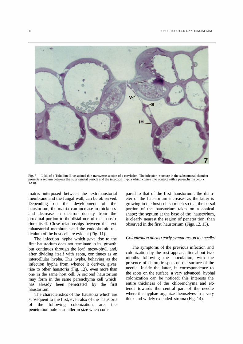
Fig. 7 — L.M. of a Toluidine Blue stained thin transverse section of a cotyledon. The infection stucture in the substomatal chamberpresents a septum between the substomatal vesicle and the infection hypha which comes into contact with a parenchyma cell (x1280).
matrix interposed between the extrahaustorialmembrane and the fungal wall, can be ob served.Depending on the development of thehaustorium, the matrix can increase in thicknessand decrease in electron density from theproximal portion to the distal one of the hausto-rium itself. Close relationships between the ext-rahaustorial membrane and the endoplasmic re-ticulum of the host cell are evident (Fig. 11).
The infection hypha which gave rise to thefirst haustorium does not terminate in its growth,but continues through the leaf meso-phyll and,after dividing itself with septa, con tinues as anintercellular hypha. This hypha, behaving as theinfection hypha from whence it derives, givesrise to other haustoria (Fig. 12), even more thanone in the same host cell. A sec ond haustoriummay form in the same parenchyma cell whichhas already been penetrated by the firsthaustorium.
The characteristics of the haustoria which aresubsequent to the first, even also of the haustoriaof the following colonization, are: thepenetration hole is smaller in size when com-
pared to that of the first haustorium; the diam-eter of the haustorium increases as the latter isgrowing in the host cell so much so that the ba salportion of the haustorium takes on a conicalshape; the septum at the base of the haustorium,is clearly nearest the region of penetra tion, thanobserved in the first haustorium (Figs. 12, 13).
Colonization during early symptoms on the needles
The symptoms of the previous infection andcolonization by the rust appear, after about twomonths following the inoculation, with thepresence of chlorotic spots on the surface of theneedle. Inside the latter, in correspondence tothe spots on the surface, a very advanced hyphalcolonization can be noticed; this interests theentire thickness of the chlorenchyma and ex-tends towards the central part of the needlewhere the hyphae organize themselves in a verythick and widely extended stroma (Fig. 14).

PENETRATION AND EARLY COLONIZATION IN BASIDIOSPORE-DERIVED INFECTION 17

18 LONGO, POGGIOLESI, NALDINI and TANI
Fig. 9 — T.E.M. of the penetration by the developing first haustorium through the parenchyma cell wall of a cotyledon. The "mothercell" wall, without ultrastructural modifications, is continuous with the haustorial wall. See the wide penetration hole in theparenchyma cell wall (x 33200).Fig. 10 — T.E.M. of two sectioned portions of the first haustorium in a parenchyma cell of a cotyledon. Note the septum in distalposition respect to haustorium penetration zone (x 6300).Fig. 11 — T.E.M. of a third portion of the first haustorium of Fig. 10. Note the haustorium ultrastructure, the extrahaustorial matrixand membrane, the relationships of haustorium with host endoplasmic reticulum and nucleus (x 23000).

PENETRATION AND EARLY COLONIZATION IN BASIDIOSPORE-DERIVED INFECTION 19
the formation of appressorium-like adhesionstructures. A "peg" of infection cannot be iden-tified, rather it is the apical part of the germtube which penetrates into the stomatal apertureand, inside the substomatal chamber, gives riseto a hypha which becomes the infectionstructure. The latter gradually gets bigger tak ingon a spindle-like shape, then forms a septum andgives rise to the infection hypha which, withoutbranchings or further septa, grows to wards thenearest parenchyma cell. The distal portion ofthe infection hypha, taking on the role of thehaustorial "mother cell", penetrates into the wallof the parenchyma cell thus giving rise to the firsthaustorium. This shows a mark edly hyphalappearance characterised by a wide penetrationhole in the host cell wall, a cylindri cal shape atleast in its basal part and a septum at a discretedistance from the point of penetra tion. Theextrahaustorial matrix is present from the pointof penetration.
The infection hypha then continues itsgrowth in the chlorenchyma, forming furthersepta and branching out into intercellular hy-phae which give rise to the subsequent hausto-ria. The latter, when compared to the first haus-torium, have a narrower penetration hole and amore basal septum.
In the advanced colonization stage of therust, in the primary needles, haustoria arepresent in the stomatal subsidiary cells, the onlyepidermal cells colonized by the fungus in themonokaryotic phase.
When one compares the characteristics de-scribed for C. flaccidum with the equivalents ofother rusts which have the same way of penetra-tion during the monokaryotic phase as well aswith those considered in the literature cited tobe typical of the monokaryotic and dikaryoticpenetration (Fig. 18 A, B, C), the following re-marks can be made.
The morphology of the basidiospores of C.flaccidum has shown itself to be similar to thatalready observed in the same rust by RAGAZZI et al.(1987); the only difference lies in their sizewhich is greater when compared to that re portedby those authors.
The growth of the germ tubes on the surfaceof the needle is at random and, therefore, nonoriented towards the stomatal apertures. TheLiterature considers such behaviour to be typicalof the monokaryotic infection phase, as re portedfor many rusts (GRAY et al. 1983; GOLD
and MENDGEN 1984; BERGDAHL and FRENCH 1985;HOPKIN et al. 1988; LONGO et al 1994, 1997), aswell as for C. flaccidum on P. pinaster and P.nigra subsp. laricio (RAGAZZI and DEL-LA VALLEFEDI 1992). Despite the fact that some of these authors(i.e. GOLD and MENDGEN 1984) have observed agrowth of germ tubes towards the anticlinalwalls of the epidermal cells, their wholebehaviour is always to be considered as beingcasual.
On the contrary, in the dikaryotic infectionphase, the germ tubes usually have an orientedpattern towards the stomata triggered by themorphological characteristics of the epidermissurface and, more specifically, by those of thestomata (Hocn and STAPLES 1991; TERHUNE et al.1991); the exceptions seem to be Cronartiumribicola J.C. Fisch. Ex Rabenh. on Kibes spp.(Woo and MARTIN 1981) and Melampsora larici-populina Klebahn and Melampsora medusaeThuem on Populus (SPIERS and HOPCROFT 1988)where a germ tube behaviour which is apparentlynon oriented has been ob served.
In C. flaccidum, as already observed by RA-GAZZI and BELLA VALLE FEDI (1992), also thepenetration of the germ tubes into the stomatalantechamber is casual; moreover, in accordancewith BERGDAHL and FRENCH (1985) for C. co-mandrae, several tubes can enter the samechamber, even if only one penetrates the sto-matal aperture.
Once inside the stomatal antechamber, thegerm tube of C. flaccidum grows towards thestomatal aperture without forming appressorium-like structures which are differentiated from thegerm tube; the lack of an appresso-rium hasalready been observed for the same rust byRAGAZZI and DELLAVALLE FEDI (1992) and alsoreported for C. ribicola (PATTON and JOHNSON1970) and C. comandrae (BERGDAHL and FRENCH1985); the same way of penetration as in C.flaccidum has been described for themonokaryotic phase of these two rust species.
The appressorium of the Uredinales (LIT-TLEFIELD and HEATH 1979; HARDER and CHONG 1984;HOCH and STAPLES 1991) is highly differentiated inthe dikaryotic infection phase. In this phase theappressorium is separated by a septum from thegerm tube from which it originates. In themonokaryotic phase, the appressorium, which isgenerally present, is instead a not welldifferentiated structure, and only appears

20 LONGO, POGGIOLESI, NALD INI and TANI
Fig. 12 — L.M. of a Toluidine Blue stained thin transverse section of a cotyledon. General view of epidermis with a stomatal pen-etration and mesophyll parenchyma showing intercellular colonization with one of the haustoria occurred after the first (x 1000).
as a swelling of the terminal portion of the germtube and is usually not separated by septa fromthe latter (MILLER et al. 1980; GRAY et al. 1983;GOLD and MENDGEN 1984; HOPKIN et al. 1988; LONGOet al. 1991, 1994; MORIN et al. 1992).
Given that in C. flaccidum the appressoriumis lacking, so too a differentiated infection peg, itis the apical portion of the germ tube itself whichpenetrates into the stomatal aperture.
In the dikaryotic phase the infection peg isdescribed as a thin outgrowth, which derivesfrom the innermost layer of the appressoriumwall, which grows apically introducing itself intothe stomatal aperture (LITTLEFIELD and HEATH1979).
Also the infection peg of the monokaryoticphase derives directly from the appressorium,but it is not differentiated like that of thedikaryotic one (GOLD and MENDGEN 1984;
MENDGEN 1997); it appears very thin at thepoint of penetration into the cuticle of the epi-dermal cell, then it expands in the wall and en-ters the host cell by invaginating its plasmamembrane.
In C. flaccidum the germ tube, growing in-side the stomatal aperture, decreases in diam eterand becomes clearly flattened, finally faces thesubstomatal chamber; a similar aspect is alsodescribed in C. ribicola (PATTON and JOHNSON 1970)and in C, comandrae (BERGDAHL and FRENCH 1985).
At this point, it is possible to conclude thatC. flaccidum, as far as the aspects related to thebasidiospore-derived prepenetration are con-cerned, differs from what has been observed inboth the monokaryotic and dikaryotic phases ofthe rusts in general and that the way of penetra tionof its monokaryotic phase, as observed in

PENETRATION AND EARLY COLONIZATION IN BASIDIOSPORE-DERIVED INFECTION 21
P. pinea, is always of the indirect type, as had al-ready been hypothesized by RAGAZZI and DEL-LAVALLE FEDI (1992) for other species of the ge nusPinus. This is analogous to what happens in Cribicola (PATTON and JOHNSON 1970) and in C.comandrae (BERGDAHL and FRENCH 1985), but notto what happens in E. harknessii (Hop-KIN et al.1988) and C. quercuum f. sp. fusiforme (MILLER etal. 1980) where, in some cases, the penetrationbegins in an indirect manner and involves thestomatal antechamber, but contin ues and ends ina direct manner through the anticlinal walls of thesubsidiary cells of the stoma. After havingreached the substomal chamber, the germ tube ofC. flaccidum gives rise to a hypha which growstowards the underlying chlorenchyma. Thefusiform portion of this hypha corresponds to themonokaryotic intraepi-dermal vesicle and to thedikaryotic substo-matal one. PATTON andJOHNSON (1970) and BERGDAHL and FRENCH (1985)describe, for C. ribicola and C. comandraerespectively (both of which penetrate indirectlyin the monokaryotic phase), a substomatal vesiclesimilar to that observed in C. flaccidum, at least asfar as the shape is concerned. Diversely, however,to what has
been reported for C. comandrae, in C. flaccidum aseptum has never been found in the proximalpart of the "vesicle" just as no multi- septatevesicles have ever been observed.
In the monokaryotic infection with directpenetration, the intraepidermal vesicle, de scribedin detail for Uromyces appendiculatus (Pers.)Unger var. appendiculatus by GOLD and MENDGEN(1984), for Puccinia xanthii Schw. by MORIN et al.(1992), for Melampsora pulcherrima (Bub) Maireby LONGO et al. (1994), for M. pimtorqua and M.larici tremulae by LONGO et al. (1997), is madeup of a thin neck which de rives from thepenetration peg and of a widened portion with alarge central vacuole. In accord ance with what hasbeen observed in the substomatal vesicle of C.flaccidum, also in the intraepidermal one there isno septum to separate the infection peg from thevesicle; only in C. quercuum f. sp. fusiforme aseptum as been observed between the infectionpeg and the neck of the vesicle (GRAY et al. 1983).
In the dikaryotic infection phase (LITTLE-FIELD and HEATH 1979; HARDER and CHONG 1984)with indirect penetration, a septum al waysseparates the substomatal vesicle from the
Fig. 13 — T.E.M. of another haustorium occurred after the first in a primary needle. Note the reduced penetration hole and theproximal position of the septum as in Fig. 12 (x 15300).

22 LONGO, POGGIOLESI, NALDINI and TANI
Fig. 14 — L.M. of a Toluidine Blue stained thin transverse section at level of a yellow spot on infected primary needle. Fungal stromawithin the transfusion tissue (x 128).
infection peg, as described in detail by LITTLE-FIELD and HEATH (1979), DAVIES and BUTLER (1986),SHAIN and JARLFORS (1987), MIMS et al. (1989), Huand RIJKEMBERG (1998). The size and shape ofsubstomatal vesicle may be, de pending on thedifferent rust species, roundish, ovoidal, spindle-shaped, lobed or even multi- septate andbranched (NiKS 1986).
A septum located immediately distal to thevesicle, as observed in C. flaccidum, is present inthe monokaryotic intraepidermal vesicle, andbehind it, the intracellular infection hypha, con-trary to what instead occurs in C. flaccidum, canfurther form other septa and branch out, thuscontinuing its growth with the production of in-tracellular hyphae and haustoria with hypha-likebehaviour in the neighbouring host cells, as wellas hyphae in the intercellular spaces. Theinfection hypha of the intraepidermal infectionstructure is surrounded by an extrahyphal matrixwhich is typical of all the intracellular struc tures;instead, its vesicle always appears matrix- lessdespite the fact that it too is an intracellularstructure (GOLD and MENDGEN 1984; MORIN et al.1992; LONGO et al 1994, 1997). The entirestructure of infection of C. flaccidum is matrix-less as are all the intercellular structures, be theymonokaryotic or dikaryotic.
The septum directly below the vesicle is notusually present for the dikaryotic substomatalvesicle; from the latter, as from the monokaryoticvesicle, one or more infection hyphae originatedepending on the different morphology ofvesicles, which in its turn depends on the spe ciesof rusts (SOTOMAYOR et al. 1983; NIKS 1986).These hyphae can also form septa and branchout before developing the haustorial mother celland the first haustorium in a paren chyma cell, asoccurs in P. porn (Sow.) Wint. (DAVIES andBUTLER 1986) and in Pucdnia recondita Rob.Ex Desm. f. sp. tritid (Hu and RIJKEMBERG 1998).
One of the characteristics of the dikaryoticphase, is the presence of a septum in the termi nalportion of the infection hypha so as to separatethis from the haustorial mother cell. Such aseptum, as well as the mother cell at the pen-etration point, shows a marked ultrastructuralspecialization (HEATH et al. 1975; LITTLEFIELD andHEATH 1979; HARDER and CHONG 1984).
In the infection hypha of C. flaccidum, theseptum outlining the haustorial "mother cell"has not been found: in this rust, the apical por-tion of the infection hypha penetrates into theparenchvma host cell without thickenings or

PENETRATION AND EARLY COLONIZATION IN BASIDIOSPORE-DERIVED INFECTION 23
Fig. 15 — L.M. of a Toluidine Blue stained transverse section of a primary needle. General view of epidermis with a stoma showing anhaustorium (arrow) in a subsidiary cell (x 1280).Figs. 16, 17 — L.M. of a Nile-Red stained cryosection of a fresh primary needle. General view of epidermis with a stoma which isobserved: in Fig. 16 with bright field and in Fig. 17 with epifluorescence optics. In Fig. 16: see the inner tangential wall of the twosubsidiary cells which appear thinner than that of the other epidermal cells. In Fig. 17: the red fluorescence, which showes the wallcutinization, is not detectable in the inner tangential wall of the two subsidiary cells (x 512).

24 LONGO, POGGIOLESI, NALDINI and TANI
new wall layers which are characteristic of thedikaryotic mother cell; thus, this apical portion ofthe infection hypha, at least morphologically,cannot be differentiated from the rest of the hy-pha, as occurs also with the haustorial mothercells of advanced colonization in rusts in themonokaryotic phase (LITTLEFIELD and HEATH 1979;HARDER and CHONG 1984) including C. flaccidum(LoNGO et al, 1982).
As regards this aspect, it is worth remember ingthat in the Literature detailed observationscarried out during the initial phases of coloniza-tion and thus pertaining to the first haustoriumand its mother cell, are not reported for the rustswhich penetrate indirectly during themonokaryotic phase, such as C. flaccidum.
The first haustorium of C. flaccidum showsdifferences when compared to typical hausto-ria, be they di- or monokaryotic.
According to the literature cited (LITTLE-FIELD and HEATH 1979; HARDER and CHONG 1984,1991; HARDER 1989), the dikaryotic haustorium ismade up of a thin neck with a neck band and arounded body; due to lack of septa, thehaustorium is continuous with its mother cell.
Differentiated neck and body were not foundin the monokaryotic haustorium; instead, this ischaracterised by a hypha-like behaviour and aseptum is always present at its base (LITTLEFIELDand HEATH 1979; HARDER and CHONG 1984, 1991;HARDER 1989). These morphological characteristicsdescribed for the two types of haustoria in theLiterature cited, were observed for thedikaryotic and monokaryotic haustoria of C.flaccidum during the advanced colonization,already studied by LONGO and BRUSCAGLIONI (1986)on leaves of Vincetoxicum hi-rundinaria Medicusand by LONGO et al. (1982) on primary needlesand young stems of some species of the genusPinus.
Even if in the first haustorium of C. flacci-dum in monokaryotic phase one finds the samecharacteristics as in the haustoria typical of thisphase, it has some particular aspects: a) the dis talposition of the septum with respect to the pointof penetration into the host cell; b) a pen etrationhole which is particularly wide; c) a more orless cylindrical shape in its basal por tion whichmakes it take on an even more hypha-likebehaviour.
In the first haustorium of C. flaccidum theextrahaustorial membrane and matrix and the
relationship between these structures and thecytoplasm of the host cell, appear to be typical.
The "mother cell" which gave rise to the firsthaustorium is not terminal and this behaviour istypically found in all rusts during themonokaryotic phase, while during the dikaryoticphase the haustorial mother cells are alwaysterminal (LITTLEFIELD and HEATH 1979; HARDER andCHONG 1984, 1991; HARDER 1989).
It was interesting to observe that, in C. flacci-dum, both the haustoria of the initial phases ofcolonization which develop after the first one, aswell as those in the subsidiary cells during theadvanced colonization, lack the specific aspectsfound in the first haustoria, and have the samemorphological characteristics of the typicalmonokaryotic haustoria, previously described byLONGO et al. (1982) for the same C. flaccidum, andby LITTLEFIELD and HEATH (1979), HARDER andCHONG (1984, 1991) and HARDER (1989) for manyother rusts in monokaryotic phase. Indeed, theseshow: a) the position of the septum clearly basalbehind the point of pen etration; b) a very narrowpenetration hole; c) a conical shape in their basalportion. Despite their different shape at the basewhen compared to that of the first haustorium, itcan be noted that also these haustoria maintain ahy-phal behaviour, even if not as marked as thatobserved in the first haustorium.
The presence of haustoria in subsidiary sto-matal cells was described in the Literature forthe dikaryotic phase of two species of Uredi-nales, Puccinia graminis Pers. f. sp. tritici Eriks.and Henn. (TIBURZY et al. 1990) and Hemileiavastatrix Berk, and Br. (COUTINHO et al. 1993).Indeed, these two rusts in the substomatalchamber produce an infection structure fromwhence the first haustoria in the subsidiary cellsoriginate directly, while the other epidermal cellsare not colonized.
The presence of haustoria only in the sub-sidiary cells of the stomata can be related to aspecific characteristic of these cells. Accordingto THERUNE et al. (1991), during the dikaryoticphase of the U. appendiculatus on P. vulgans, thechance that haustorial mother cells can dif-ferentiate at the level of the internal tangentialwall of the epidermical cells, can be related to thedegree of hydrophoby of those walls: the successof the development of the haustoria

PENETRATION AND EARLY COLONIZATION IN BASIDIOSPORE-DERIVED INFECTION 25
could, therefore, depend upon the lack or scar-sity of substances which when present modifythe walls of the epidermal cells, by making themimpermeable. As far as the subsidiary cells, the
same Authors then describe, always with refer-ence to P. vulgaris, "...a cuticular covering ex-tending slightly beyond the guard cell - subsidi arycell junction in the substomatal chamber", butwithout adding anything specific as regards thepresence of hydrophobic substances in theinternal tangential walls of the latter cells; on theother hand, in this last respect the Litera turedoes not offer exhaustive information.
In C. flaccidum, fluorescence followingstaining of sections of P. pinea primary needleswith Nile-Red has indicated that the internaltangential walls of the subsidiary cells do notseem to contain cutin, while the latter is presentin the walls of the other epidermal cells, includ ingthe stomatal ones.
Following on the hypothesis of TERHUNE et al.(1991) it can be concluded therefore that in C.flaccidum the presence of the haustoria in thesubsidiary cells of the stomata could be corre-lated to the scarsely hydrophobic nature of theirinternal tangential wall; instead, on the basis ofthe above, the formation of haustoria in the otherepidermal cells seems not to occur be cause thepresence of cutin.
CONCLUDING REMARKS
The literature cited covering the two typesof prepenetration, penetration and early coloni-zation respectively of the dikaryotic andmonokaryotic phases of the Uredinales, hashighlighted the fact that behaviour and struc-tures are substantially different for the twophases.
The characteristics of the indirectly pen-etrating dikaryotic phase on the host surfaceare: germ tubes which are oriented towards sto-mata; appressorium from which the tube itselfis separated by a septum; an infection pegwhich penetrates into the stomatal aperture.
The steps of this part of the infection proc essare mediated by a series of both physical andchemical stimuli, which are sent by the host sur face(HocH and STAPLES 1991; MENDGEN et al. 1996;EPSTEIN and NICHOLSON 1997) and more specificallyby the stomata (TERHUNE et al. 1991); thesestimuli, which guide the behaviour of the fungalstructures on the host surface are all the better,the stronger the adhesion of the fungus to thehost surface. Such adhesion
26 LONGO, POGGIOLESI, NALDINI and TANI
comes about as a result of mucilaginous exhu-dates produced by the fungus and containingnumerous lythic enzymes, especially the cuti-nase, which are considered to give a significantcontribution to the adhesion itself and, thus, tothe subsequent processes of rust structure dif-ferentiation (MENDGEN et al. 1996; HOWARD 1997;MENDGEN 1997). More specifically, ac cording toMENDGEN et al. (1996) and EPSTEIN and NICHOLSON(1997), the action of such en zymes modifies thecuticular surface of the host from hydrophobic tohydrophylic as that of the fungus is, and thusplays a positive role in the adhesion of the latter.The hydrophobic nature of the host surface thuswould seem to condition the entireprepenetration process of the fungus. In indirectpenetration, of marked im portance it is the roleof the adhesion of the ap-pressorium which mustmaintain a correct posi tion on the guard cells ofthe stoma (EPSTEIN and NICHOLSON 1997).
The characteristics of the dikaryotic phaseafter the penetration through the stoma are:substomatal vesicle divided from the infectionpeg by a septum; infection hypha with a distalseptum which divides it from the haustorialmother cell which is terminal originating thefirst haustorium.
In the substomatal chamber, the signalswhich regulate the interaction between the fun-gus and the host occur all along the entire ruststructure from the vesicle to the first haustoriumwhich, in the dikaryotic phase, is the firstintracellular structure (MENDGEN et al. 1988;HEATH 1989, 1995).
The characteristics of the directly penetrat ingmonokaryotic phase on the host surface are: germtubes with a non-oriented pattern on the leafsurface; slightly differentiated appresso-rium;penetration peg which penetrates the epi dermalcell wall.
The signals which occur in the monokaryoticphase between the host surface and the fun gusduring prepenetration are not as well known asthose of the dikaryon (MENDGEN 1997);nonetheless, the presence of some type of signalwhich guides the rust structures has beenhypothesized by some authors (DESPREZ-LOUSTEAU and EE MENN 1989; GOLD and MENDGEN1991; EONGO et al. 1994) also because, in somerusts, a preferential pattern of germ tubes alongthe anticlinal walls of the epi dermal cells hasbeen observed (GOLD and
MENDGEN 1984; HOPKIN et al. 1988; MORIN et al.1992). On the other hand, also in themonokaryon, a good adhesion of the fungus tothe host surface is considered important; espe-cially, according to EPSTEIN and NICHOLSON(1997), as regards the relationship between theappressorium and the epidermis of the host tobe penetrated. According to GOLD and MENDGEN(1991), the mucilaginous matrix commonlypresent around external rust structures is use ful,not only to favour the adhesion of the rust, but,above all, as an enzymatic reserve for thepenetration. It must be remembered that in thedirect penetration the pressure of the turgor in-side the appressorium plays a very importantrole, and thus the mechanical action togetherwith the enzymatic one enables the host wall tobe perforated (MENDGEN and DEISING 1993;HOWARD 1997).
The characteristics of the monokaryotic phaseafter the direct penetration are: intraepi-dermalvesicle continuous with the peg, but di vided by aseptum from the infection hypha; infection hyphafrom whose branches both intra cellular hyphaeand haustoria in contiguous cells, as well asintercellular hyphae which then give rise tofurther haustoria in the parenchyma cells, canoriginate.
Also in the monokaryotic phase signalswhich regulate the host-parasite interactiononce penetration has occurred can be found but,diversely to what happens in the dikaryon, all ofthem take place at the level of the epider mal cellin which the penetration occurs (HEATH 1989,1995).
As a result of this analysis, and in considera-tion of the fact that the infection process of thedikaryotic phase of the Uredinales presents a se-ries of well defined and differentiated struc turesboth from a morphological and a func tionalpoint of view (as the number and ar rangementof the septa which divide one from the otherclearly demonstrate) as well as the fact that eachof these structures expresses a pecu liarinteraction with the host, such a process is moreevolved when compared to that of themonokaryotic phase (GOLD and MENDGEN 1991;MENDGEN and DEISING 1993; HEATH 1995; MENDGENet al. 1996; MENDGEN 1997).
Taken as a whole, the monokaryotic infec-tion process of C. flaccidum presents a similar ity,even if only apparently, with what is typical ofthe rusts in dikaryotic phase. Indeed, also C.

PENETRATION AND EARLY COLONIZATION IN BASIDIOSPORE-DERIVED INFECTION 27
flaccidum shows an indirect penetration, theformation of a substomatal infection structureas well as of a first haustorium in a parenchymacell. However, this is a similarity of a behav-ioural type because the morpho-functionalcharacteristics which emerge in such an infec tiveprocess are essentially those of a monokaryoticphase. And in point of fact: the germ tubes onthe host surface do not have a specificorientation and only by chance do they penetrateinto a stomatal aperture without dif ferentiatingan appressorium and an infection peg; theinfection structures in the substomal chamberare characterised by a single septum betweenthe vesicle and the infection hypha; all thehaustoria, including those in the subsidiary cellsof the stoma, are typical monokaryoticstructures, especially the first one which, on ac-count of its particular morphology, presents aneven more marked hyphal behaviour. Moreo-ver, the peculiar aspects of the latter can be ob-served also for the monokaryotic intracellularstructures of M. pinitorqua (LoNGO et al. 1988,1991), which deriving directly from the infec tionhypha in the epidermal cell, colonise both theepidermal and the parenchymal contiguous cells.The first haustoria of C. flaccidum couldtherefore be considered the extension of thesubstomatal infection hypha of which it main-tains the behaviour even if, covering itself withextrahaustorial membrane and matrix, it takeson the function of exchange of substances be-tween host and parasite. This also takes place inthe intracellular infection hypha which derivesfrom the intraepidermal vesicle in monokaryoticdirect penetration; indeed, such a hypha, di-versely to what happens with the vesicle, is cov-ered by a matrix so as to indicate its haustorialfunction.
The penetration of the monokaryotic phaseof the C. flaccidum differs from both that of adikaryon and that of a typical monokaryonsince it is accomplished by the germ tube with outthe differentiation of an appressorium and,therefore, of a true penetration peg. In calling tomind that the function of the appressorium is thatof attaching the rust to the host, it's lack ap pearsconsequent if one considers the fact that C.flaccidum neither penetrates directly through theintact host epidermis ( monokaryotic charac-teristic), nor does its indirect penetrationthrough the stoma ( dikaryotic characteristic)
occur with an oriented behaviour of the germtube towards the stoma itself.
It is interesting to consider the similar be-haviour found between C. flaccidum and therusts in a dikaryon phase towards cutinized sur-faces. Indeed, with reference to the dikaryonphase, the hydrophobic nature of the cutinizedwalls of the host cells, which extends from theexternal surface of the epidermis as far as thesubstomal chamber given the continuity of thecuticle, seems to stimulate the penetration proc ess(MENDGEN et al. 1996; EPSTEIN and NICHOL-SON 1997)and the development of the intercel lular infectionstructures at the level of the substomatal chamber(TERHUNE et al. 1991). With reference to theintracellular colonization, the hydrophobicnature of the cutinized internal tangential wall ofthe epidermal cells prevents the differentiation ofthe haustorial mother cells and therefore theformation therein of the haustoria (TERHUNE et al.1991 for U. appendiculatus on P. vulgaris).
With regard to the monokaryotic phase of C.flaccidum we do not know what signals occurbetween the host cell walls and the structures ofprepenetration and of infection, nor do we knowthe role played by the enzymatic load in theexchange of such signals. We have seen,however, that this species in the monokaryoticphase, as the rusts in a dikaryotic phase, doesnot penetrate through the epidermal cell walls,but produces the first intracellular structures inthe parenchymal cells, in whose walls the cutin isscarse or absent, while it never does so in theepidermal cells which, instead, are impregnatedwith cutin in all their walls.
As a proof of what above considered, thefact of having found in C. flaccidum the haustoriaonly in the stomatal subsidiary cells, and the factof having revealed that their internal tangentialwall through which the haustoria penetrateseems to be lacking in cutin, are of the utmostimportance.
Following all considerations, a possible hy-pothesis as to the reason for the dynamics ofpenetration carried out by C. flaccidum in themonokaryotic phase could be as follows. Thebasidiospore germ tubes, having to infect "innature" organs such as the secondary needles ofPinus spp. which are markedly cutinized, butalso rich in stomata, and not being able to exer-cise a suitable mechanical action which inmonokaryotic penetration is always associated

28 LONGO, POGGIOLESI, NALDINI and TANI
with the enzymatic one, in order to fit them-selves on this substratum, modified their way ofpenetration from the direct one, which is typicalof the monokaryotic phase of the Uredinales, tothat of the indirect type. Nevertheless, the pen-etration structures of this rust in themonokaryotic phase have maintained the mor-phological and functional significance of thetypical monokaryotic ones, even if some aspectsof their behaviour seem to recall those of thedikaryon.
Hence, one can conclude that in C. flacci-dum in the monokaryotic phase it is the nuclearset which determines the morphology and func-tion of the structures involved in the infectionprocess; this is true even if the histological char-acteristics of the host organ which this rust spe-cies has evolved to infect in nature, condition itsway of penetration.
Acknowledgments — The authors wish to thank Dr. A.Paolillo, Mr. Pietro Di Falco and Mr. Fiorenzo Drovandi fortheir technical assistance and helpful cooperation. Researchgrants by the University of Florence are also acknowledged.
REFERENCES
BAUER R., 1983 — Experimentell - ontogenetische und karyolo-gische Untersuchungen an Uredinales. Doctoral disserta-tion, Universitat Tubingen, Tubingen, Federal Republic ofGermany.
BECCARI N. and MAZZI V., 1966 — Manuale di tecnica micro-scopica. Societa Editrice Libraria, Como.
BERGDAHL D.R and FRENCH D.W., 1985 — Penetration of theprimary tissues of Pinus banksiana by Cronartium coman-drae. In: J. BARROWS- BROADDUS and H.R. POWERS (Eds.), Proc.IUFRO Rusts of hard pines Conf., Oct. 1984, pp. 179-192,Athens, Univ. Georgia.
BUSHNELL W.R. and ROELFS A.P., 1984 — The cereal rusts. Vol.1. Academic Press, Orlando.
COUTINHO T.A., RIJKEMBERG F.H.J. and VAN ASCH M.A.J., 1993 —Development of infection structures by Hemileia vastatrix inresistant and susceptible selections ofCoffea and in Phaseolusvulgaris. Can. J. Bot, 71: 1001-1008.
DAVIES M.E. and BUTLER G.M., 1986 — Development of infectionstructures of the rust Puccinia porn, on leek leaves. Trans.Br. Mycol. Soc., 86: 475-515.
DELLAVALLE I. and RAGAZZI A., 1991 — Behaviour of Cronartiumflaccidum basidiospores on different organs of the same pinespecies. In: Y. HIRATSUKA et al. (Eds.), "Rusts of Pine".Proc. IUFRO Rusts of Pine Conf., Sept. 1989, Banff,Alberta. Inf. Rep. Nor - X - 317: 281-286.
DESPREZ-LOUSTEAU M. and LE MENN R., 1989 — Epicuticular waxesand Melampsora pinitorqua Rostr. preinfection behaviouron maritime pine shoots. Eur. J. For. Path., 19: 178-188.
EPSTEIN L. and NICHOLSON R., 1997 —Adhesion of spores andhyphae to plant surfaces. In: G.C. CARROL and P. TUDZYN-
SKI (Eds.), "The Mycota", Vol V, (part A): 11-25.Springer, Berlin.
GOLD R.E. and MENDGEN K., 1984 — Cytology of basidiosporegermination, penetration, and early colonization of Phaseolusvulgaris by Uromyces appendiculatus var. appendiculatus.Can. J. Bot., 62: 1989-2002.
—, 1991 — Rust basidiospore germlings and disease initiation.In: G.T. COLE and H.C. HOCH (Eds.), "The fungal spore anddisease initiation in plants and animals", pp. 67-99.Plenum Press, New York.
GRAY D.J., AMERSON H.V. and VAN DYKE C.G., 1983 — Ul-trastructure of the infection and early colonization of Pinustaeda by Cronartium quercuum f. sp. fusiforme. Mycologia,75: 117-130.
GREENSPAN P., MAYER E.P. and FOWLERS S.D., 1985 — Nile red: aselective fluorescent stain for intracellular lipid drop lets. J.Cell. Biol., 100: 965-973.
GRILL D., LACKNER E. und SCHARNER M., 1978 — Untersuchungen anmit Chrysomyxa abietis befallenen Fichten-nadeln.Phyton, 19: 71-82.
HARDER D.E., 1989 — Rust fungal haustoria - past, present, future.Can. J. Plant Path., 11: 91-99.
HARDER D.E. and CHONG J., 1984 — Structure and physiology ofhaustoria. In: W.R. BUSHNELL and A.P. ROELFS (Eds.), "Thecereal rusts", Vol I: 431-476. Academic Press, Or lando.
—, 1991 — Rust haustoria. In: K. MENDGEN and D.E. LESE-MANN (Eds.), "Electron microscopy of Plant Pathogens", pp.235-250. Springer, Berlin.
HEATH M.C., 1989 —A comparison of fungal growth and plantresponses in cowpea and bean cultivars inoculated with ure-diospores or basidiospores of the cowpea rust fungus.Physiol. Mol. Plant Path., 34: 415-426.
—, 1995 — Signal exchange between higher plants and rustfungi. Can. J. Bot., 73: 616-623.
HEATH M.C. and HEATH I.B., 1975 — Ultrastructural changesassociated with the haustorial mother cell septum duringhaustorium formation in Uromyces phaseoli var. vignae.Protoplasma, 84: 297-314.
HOCH H.C. and STAPLES R.C., 1991 — Signaling for infectionstructure formation in fungi. In: G.T. COLE and H.C. HOCH(Eds.), "The fungal spore and disease initiation in plantsand animals", pp. 25-46. Plenum Press, New York.
HOPKIN A.A., REID J., HIRATSUKA Y. and ALLEN E., 1988 — Initialinfection and early colonization of Pinus contorta byEndocronartium harknessii (western gall rust). Can. J.Plant Path., 10:221-227.
HOWARD R.J., 1997 — Breaching the outer barriers. Cuticle and cellwall penetration. In: G.C. CARROL and P. TUDZYNSKI (Eds.),"The Mycota", Vol V, (part A): 43-60. Springer, Berlin.
Hu G.G. and RIJKENBERG F.HJ., 1998 — Scanning electronmicroscopy of early infection structure formation by Puc-cinia reconditaf.sp. tritici on and in susceptible and resistantwheat lines. Mycol. Res., 102: 391-399.
HUGHES F.L. and RIJKEMBERG F.HJ., 1985 — Scanning electronmicroscopy of early infection in the medial stage of Pucciniasorghi in Zea mays. Plant path., 34: 61-68.
KARNOVSKYM.S., 1965 —A formaldehyde-glutaraldehyde fixative ofhigh osmolality for use in electron microscopy. J. Cell. Biol.,27: 137A.
LITTLEFIELD LJ. and HEATH M.C., 1979 — Ultrastructure of therust fungi. Academic Press, New York, S. Francisco.London.
LONGO N. and BRUSCAGLIONI L., 1986 — Ultrastructural observationson the dikaryotic haustorium of Cronartium flacci-

PENETRATION AND EARLY COLONIZATION IN BASIDIOSPORE-DERIVED INFECTION 29
dum (Alb. et Schw.) Wint. in Vincetoxicum himndinariaMedicus. Caryologia, 39: 51-64.
LONGO N., MORIONDO F. and NALDINI LONGO B., 1982 — Ul-trastructural observations on the hostpathogen interface ininfections of Cronartium flaccidum on fine. Caryologia, 35:307-326.
LONGO N., NALDINI B., DROVANDI F., GONNELLI T, and TANI G., 1994 —Penetration and early colonization in basid-iospore-derivedinfection of Melampsora pulcherrima (Bub) Maire onMercurialis annua L. Caryologia, 47: 207-222.
LONGO N., NALDINI B., PAOLILLO A., DROVANDI F., TANI G. andGONNELLI T., 1997 — Morphological aspects of early host-parasite interactions in infections ofMelampsora pinitorquaand Melampsora larici-tremulae on Pinus sylvestris.Implications in the taxonomical relationship of the two rustfungi. Caryologia, 50: 35-57.
LONGO N., NALDINI LONGO B., TANI G. and DROVANDI F., 1988 —Osservazioni sull'infezione basidiosporica di Melampsorapinitorqua Rostr. (Uredinales) in alcuni ospiti del gen. Pi-nus. Giorn. Bot. Ital., 122 (suppl. 1): 155.
—, 1991 — Penetration and early colonization in basidiospore-derived infection ofMelampsora pinitorqua Rostr. on Pinus.Structural and ultrastructural observations. In: Y. HIRAT-SUKA et al. (Eds.), "Rusts of Pine". Proc. IUFRO Rusts ofpine Conf., Sept. 1989, Banff, Alberta. Inf. Rep. Nor - X -317:120-127.
MENDGEN K., 1997 — The Uredinales. In: G.C. CARROL and P.TUDZYNSKI (Eds.), "The Mycota", Vol V (part B): 79-85.Springer, Berlin.
MENDGEN K. and DEISING H., 1993 — Infection structures offungal plant pathogens - a cytological and physiologicalevaluation. New Phytol., 124: 193-213.
MENDGEN K., HAHN M. and DEISING H., 1996 — Morphogenesis andmechanism of penetration by plant pathogenic fungi.Annu. Rev. Phytopathol., 34: 367-386.
MENDGEN K., SCHNEIDER A., STERK M. and FINK W., 1988 — Thedifferentiation of infection structures as a result of rec ognitionevents between some biotrophicparasites and their hosts.J.Phytopath., 123: 259-279.
MILLER T., PATTON R.F. and POWERS H.NJN., 1980 — Mode ofinfection and early colonization of slash pine seedlings byCronartium quercuumf. sp. fusiforme. Phytopathology, 70:1206-1208.
MIMS C.W., TAYLOR J. and RICHARDSON E.A., 1989 — Ul-trastructure of early stages of infection of peanut leaves bythe rust fungus Puccinia arachidis. Can. J. Bot., 67: 3570-3579.
MORIN L., BROWN J.F. and AULD B.A., 1992 — Teliospore germination,basidiospore formation and the infection process of Pucciniaxanthii on Xanthium occidentale. Mycol. Res., 96: 661-669.
NIKS E.R., 1986 — Variation of mycelial morphology betweenspecies and formae speciales of rust fungi of cereals andgrasses. Can. J. Bot., 64: 2976-2983.
PATTON R.F. and JOHNSON D.W., 1970 — Mode of penetration ofneedles of eastern white pine by Cronartium ribicola.Phytopathology, 60: 977-982.
RADDI P., 1976 — Indagine sui tipi di macchie fogliari e sul lorosignificato in discendenze di Pinus pinaster infette da Cron-artium flaccidum. Rivista Patologia vegetale, 12: 91-98.
RADDI P., MITTEMPERGHER L. and MORIONDO F., 1979 — Testing of Pinuspinea and P. pinaster progenies for resistance to Cronartiumflaccidum. Phytopathology, 69: 679-681.
RAGAZZI A. and DELLAVALLE FEDI L, 1992 — Penetration ofCronartium flaccidum into pine needles. Eur. J. For. Path.,22:278-283.
RAGAZZI A., FAGNANI A. and DELLAVALLE FEDI L, 1987 — Te-lial andbasidiospore stages of Cronartium flaccidum: light andscanning electron microscopy observations. Phytopath. medit,26: 81-84.
RAGAZZI A. and MORIONDO F., 1979 — Suscettibilitd del Pinussylvestris L. e del Pinus nigra Arn. alia ruggine vescicolosaCronartium flaccidum (Alb. et Schw.) Wint. LTtalia fore-stale e Montana, 34: 121-129.
—, 1980 — Results of Cronartium flaccidum (Alb. et Schw.)Wint. inoculations on eight-year-old plants of Pinus pinea L.In: H.R. POWERS et al. (Eds.), "Rusts of hard pines". Proc.IUFRO Meeting, Sept. 1979, Florence. Phytopath. Medit.,19:51-56.
REYNOLDS E.S., 1963 — The use of leadcytrate stain in electronmicroscopy.}. Cell. Biol., 17: 208-212.
SHAIN L. and JARLFORS U., 1987 — Ultrastructure of easterncottonwood clones susceptible or resistant to leaf rust. Can. J.Bot., 65: 1586-1598.
SOTOMAYOR LA., PURDY L.H. and TRESE A.T., 1983 — Infection ofSugarcane leaves by Puccinia melanocephala. Phy-topathology, 73: 695-699.
SPIERS A.G. and HOPCROFT D.H., 1988 — Penetration and infectionof poplar leaves by urediniospores of Melampsora larici-populina and Melampsora medusae. New Zeal. J. Bot, 26:101-111.
SPURR A.R., 1969 — A low-viscosity epoxy resin embedding me diumfor electron microscopy. J. Ultrastr. Res., 26: 31-43.
TERHUNE B.T., ALLEN E.A., HOCH H.C., WERGIN W.P. and ERBEE.F., 1991 — Stomatal ontogeny and morphology inPhaseolus vulgaris in relation to infection structure initia tionby Uromyces appendiculatus. Can. J. Bot., 69: 477-484.
TIBURZY R., NOLL V. and REISENER H.J., 1990 — Resistance of wheatto Puccinia graminis f.sp. tritici: histological investigation ofresistance caused by S2 5 gene. Physiol. Mol. Plant Path., 36:95-108.
WILSON M. and HENDERSON D.M., 1966 — British rust fungi.Cambridge University Press, Cambridge.
Woo J.Y. and MARTIN N.E., 1981 —Scanning electron microscopyof Cronartium ribicola infecting Ribes leaves. Eur. J. For.Path., 11:7-15.
Received 28 March 2000; accepted 25 May 2000