Embed Size (px)
Citation preview

“main” — 2011/2/10 — 13:35 — page 73 — #1
Anais da Academia Brasileira de Ciências (2011) 83(1): 73-98(Annals of the Brazilian Academy of Sciences)Printed version ISSN 0001-3765 / Online version ISSN 1678-2690www.scielo.br/aabc
Pelvic and hind limb musculature of Staurikosaurus pricei(Dinosauria: Saurischia)
ORLANDO N. GRILLO and SERGIO A.K. AZEVEDO
Departamento de Geologia e Paleontologia, Museu Nacional/UFRJQuinta da Boa Vista, s/n, São Cristóvão, 20940-040 Rio de Janeiro, RJ, Brasil
Manuscript received on January 15, 2010; accepted for publication on June 21, 2010
ABSTRACT
The study of pelvic and hind limb bones and muscles in basal dinosaurs is important for understanding the early
evolution of bipedal locomotion in the group. The use of data from both extant and extinct taxa placed into a phylo-
genetic context allowed to make well-supported inferences concerning most of the hind limb musculature of the basal
saurischian Staurikosaurus pricei Colbert, 1970 (Santa Maria Formation, Late Triassic of Rio Grande do Sul, Brazil).
Two large concavities in the lateral surface of the ilium represent the origin of the muscles iliotrochantericus caudalis
plus iliofemoralis externus (in the anterior concavity) and iliofibularis (in the posterior concavity). Muscle ambiens
has only one head and originates from the pubic tubercle. The origin of puboischiofemoralis internus 1 possibly cor-
responds to a fossa in the ventral margin of the preacetabular iliac process. This could represent an intermediate stage
prior to the origin of a true preacetabular fossa. Muscles caudofemorales longus et brevis were likely well developed,
and Staurikosaurus is unique in bearing a posteriorly projected surface for the origin of caudofemoralis brevis.
Key words: extant phylogenetic bracket, locomotion, muscular reconstruction, Saurischia, Staurikosaurus pricei.
INTRODUCTION
Bipedalism is a form of locomotion adopted by few
groups of animals (Alexander 2004, Gatesy and Bie-
wener 1991, Hutchinson and Gatesy 2006, McGowan
1999). Dinosaurs first evolved as bipedal animals and
all living representatives of this clade are bipeds. The
evolution of this type of locomotion is associated with
several modifications in posture, orientation of the hind
limbs, as well as correlated osteological and myologi-
cal modifications. Understanding bipedal locomotion in
dinosaurs requires multidisciplinary approach.
According to Lockley and Gillette (1989), studies
of trackways dating from the 19th century allowed the
estimate of velocity (Alexander 1976, Farlow 1981, Day
et al. 2002) and posture (Coombs 1980, Ishigaki 1989,
Proceedings of the Third Gondwanan Dinosaur SymposiumCorrespondence to: Orlando N. GrilloE-mail: [email protected]
Thulborn 1989, Wade 1989, Jones et al. 2000, Day et al.
2002) of dinosaurs. Comparisons with living animals
have often been used (e.g. Paul 1988, 1998, Carrano
1999, 2001, Jones et al. 2000, Hutchinson 2004a, b).
New studies using advanced graphic computing and
engineering principles (e.g., Gatesy et al. 1999, Stok-
stad 2001, Hutchinson and Garcia 2002, Wilhite 2003)
and computed tomography (e.g., Carrier et al. 2001,
Rayfield et al. 2001) also revealed important aspects of
posture and locomotion, such as mass and center of
mass position (e.g., Henderson 1999, Seebacher 2001).
In addition, muscle reconstructions have led to new pro-
positions about dinosaur locomotion (e.g., Hutchinson
et al. 2005).
The first reconstruction of dinosaur pelvic mus-
culature was made by Huene in 1908 (Romer 1923a),
followed by some authors that studied saurischian mus-
culature focusing on data obtained from living crocodiles
An Acad Bras Cienc (2011) 83 (1)

“main” — 2011/2/10 — 13:35 — page 74 — #2
74 ORLANDO N. GRILLO and SERGIO A.K. AZEVEDO
(Romer 1923a, b, Colbert 1964, Coombs 1979). More
recent works (e.g., Dilkes 2000, Hutchinson 2001a, b,
2002, Carrano and Hutchinson 2002, Langer 2003)
made more extensive use of avian data, resulting in re-
constructions that are consistent with the phylogenetic
positions of the studied taxa.
Witmer (1995, 1997) proposed a methodology
(Extant Phylogenetic Bracket, EPB) based on phylo-
genetic relationships and parsimony that allows the re-
construction of soft tissue features in extinct animals
using an accurate approach (see also Bryant and Rus-
sell 1992 for an independently-devised but similar ap-
proach). EPB is suitable for muscle reconstructions, re-
quiring a minimal level of speculation, and can be im-
proved if associated with data from extinct species with
close phylogenetic affinities. This association can re-
veal important osteological transformations that some-
times are not clear when the study relies only on data
from extant species (Hutchinson 2001a). Several works
on dinosaur limb muscle reconstruction have used the
EPB (Dilkes 2000, Gatesy 1990, Hutchinson and Gate-
sy 2000, Hutchinson 2001a, b, 2002, Carrano and Hut-
chinson 2002, Langer 2003, Jasinoski et al. 2006). Most
of these studies focused mainly on questions related
to the origin and evolution of avian locomotion (Gatesy
1999, Hutchinson 2001a, b, 2002). Some authors pre-
sented simplified propositions for musculature and lo-
comotion in basal dinosaurs (Carrano 2000, Hutchinson
and Gatesy 2000, Hutchinson 2001a, b, 2002), but no
detail on the locomotion in the earliest dinosaurs was
provided.
The evolutionary success of Dinosauria, includ-
ing birds, has often been attributed to their bipedal and
erect posture that freed their hands from a locomotor
function, allowing their use for capturing and manipu-
lating prey (Paul 1988) and later for flight. Accordingly,
the study of the locomotion on the early evolution of
Dinosauria is very important for understanding its suc-
cess of more than 225 million years. A detailed mus-
cular reconstruction of given taxa may help to resolve
specific points and may also contribute to understand-
ing major transformations that took place between basal
and avian dinosaurs.
Detailed EPB-based reconstructions of the pelvicand hind limb musculature of specific taxa have been
provided for only two species: Tyrannosaurus rex (seeCarrano and Hutchinson 2002) and Saturnalia tupini-quim (see Langer 2003). The work of Langer (2003)represents the most detailed muscular reconstruction fora basal dinosaur, and the results were presented as rep-resentative of a general condition shared by basal di-nosauriforms (e.g., Marasuchus and Pseudolagosuchus)and basal dinosaurs, such as Herrerasaurus, Stauriko-saurus, Guaibasaurus and basal species of the groupsTheropoda, Ornithischia and Sauropodomorpha (Langer2003).
Remains of basal dinosaurs are often very in-complete or poorly preserved, which may lead to uncer-tainties when muscular reconstructions are attempted.Therefore, it is important to evaluate muscle arrange-ment in other basal dinosaurs in order to complementprevious works. Also, the study of the pelvic and hindlimb musculature in other basal dinosaurs may confirmthe hypothesis of Langer (2003) of a shared generalconstruction in several basal members of the group.In this work we propose a detailed reconstruction ofthe pelvic and hind limb musculature of the basal Sau-rischian Staurikosaurus pricei Colbert, 1970. This tax-on represents one of the most complete basal dinosaursfound in south Brazil (Santa Maria Formation, Late Tri-assic, Rio Grande do Sul), and its remains may revealimportant features for understanding the early evolutionof locomotion in Dinosauria.
ABBREVIATIONS
ar adductor ridge (= linea aspera)bs brevis shelfC1-25 1st to 25th caudal vertebraD11-15 11th to 15th dorsal vertebradris dorsal ridge of ischiumEPB Extant Phylogenetic Bracketir ischial ridgeis ischiumit ischial tuberositylia linea intermuscularis cranialislip linea intermuscularis caudalisM. muscleMm. musclesmr1 first medial iliac ridgembbf medial blade of the brevis fossaop obturator processpa pubic apron
An Acad Bras Cienc (2011) 83 (1)

“main” — 2011/2/10 — 13:35 — page 75 — #3
PELVIC AND HIND LIMB MUSCLES OF STAURIKOSAURUS 75
pf preacetabular fossapib preacetabular iliac borderpst processus supratrochantericuspt pubic tuberclepu pubisrea rough expanded areaS1-2 1st and 2nd sacral vertebrastr striations
MATERIALS AND METHODS
In order to determine the areas of origin and insertion of
the pelvic and hind limb muscles of Staurikosaurus pri-
cei, the holotype MCZ 1669, deposited at the Museum
of Comparative Zoology (Harvard University), as well
as its cast (MN 6104-V), deposited at the Museu Na-
cional (Universidade Federal do Rio de Janeiro), were
examined.
Firstly, based on recent studies on the evolution of
the archosaur pelvic and hind limb osteology (Gatesy
1990, Hutchinson 2001a, b, 2002), the homologies be-
tween bone surfaces correlated with muscle attach-
ments, were traced between extant taxa (Crocodylia
and Aves) and Staurikosaurus. In this study we accept
the general conclusion that Staurikosaurus was a her-
rerasaurid, which is considered as a basal saurischian
(Fig. 1A) according to most recent works (Yates 2003,
Langer 2004, Leal et al. 2004, Bittencourt and Kell-
ner 2009). Additional osteological data were obtained
from the direct examination of specimens from the os-
teological collection of the Museu Nacional, namely:
Tupinambis sp. (Squamata, Teiidae; 04AC), Caiman
yacare (Crocodylia, Crocodylidae; 05AC, 06AC and
07AC) and Dendrocygna viduata (Aves, Anseriformes,
Anatidae; 14AC). Data was also gathered from the liter-
ature for the following taxa: fossils and living Crurotar-
si (Gregory and Camp 1918, Romer 1923c, Troxell
1925, Parrish 1987, Long and Murry 1995, Galton 2000,
Schwarz and Salisbury 2005), Dinosauromorpha and
basal dinosaurs, including Herrerasauridae (Novas 1992,
1993, 1996, Sereno and Arcucci 1993, 1994, Long and
Murry 1995, Bonaparte 1996, Hunt et al. 1998, Bona-
parte et al. 1999), non-avian Theropoda (Osborn 1905,
1916, Ostrom 1969, Brinkman and Sues 1987, Paul
1988, 2002, Colbert 1989, Barsbold and Osmólska
1990, Bonaparte et al. 1990, Molnar et al. 1990, Nor-
man 1990, Raath 1990, Rowe and Gauthier 1990, Mad-
sen 1993, Makovicky and Sues 1998, Sampson et al.
1998, Norell and Makovicky 1999, Currie 2000, Car-
rano and Hutchinson 2002, Carrano et al. 2002, Currie
and Chen 2001, Ji et al. 2003, Kobayashi and Lü 2003,
Calvo et al. 2004, Huang et al. 2004, Naish et al. 2004,
Coria and Currie 2006, Xu et al. 2006), Sauropodomor-
pha (Osborn 1904, Galton 1984, Ostrom and McIntosh
1999, Langer 2003, Yates 2003, Leal et al. 2004), and
other extinct and extant sauropsid taxa, including Aves
(Romer 1922, 1956, Goodrich 1958, Zaaf et al. 1999,
Russell and Bels 2001, Paul 2002, Sen 2003, Clarke
2004).
The phylogenetic framework adopted here (Fig.
1A) is congruent with the tree used by Hutchinson
(2001a, b, 2002) and those of Benton and Clark (1988),
Benton (1999), Sereno (1997, 1999), Holtz (1998), Pa-
dian et al. (1999), Norell et al. (2001), Huang et al.
(2004), Leal et al. (2004), Lloyd et al. (2008), and phy-
logenies presented in several of the works cited in the
previous paragraph.
In order to define the correlations between bone
surfaces and muscle origins and insertions we applied
the Extant Phylogenetic Bracket (EPB) methodology
(Witmer 1997). EPB allows the use of data from two
(or more) extant taxa, which represent the closest
groups to a given extinct taxon, in order to infer about
the latter with minimal speculation, i.e., with parsimony
(Fig. 1B). One of the extant taxa needs to be the liv-
ing sister group of the extinct taxon, and this branch
needs to have the other extant taxon as the living sister-
group. EPB was applied to verify the congruence of
the reconstruction for each muscle of Staurikosaurus.
As for any non-avian dinosaur, its closest extant taxa are
Crocodylia and Aves (Fig. 1B). EPB was applied with
the use of an extensive phylogenetic framework of fos-
sil taxa, which facilitates the identification of homolo-
gies when the extant taxa are highly divergent, as is the
case with Crocodylia and Aves.
We adopted the “levels of inference” of the EPB
as a metric of the level of speculation in the soft tissue
reconstruction, according to Witmer (1995, 1997).
We adopted the muscle homologies for Crocodylia
and Aves (Table I) presented by Hutchinson (2001a, b,
2002) and Carrano and Hutchinson (2002) that corre-
An Acad Bras Cienc (2011) 83 (1)

“main” — 2011/2/10 — 13:35 — page 76 — #4
76 ORLANDO N. GRILLO and SERGIO A.K. AZEVEDO
Fig. 1 – Phylogenetic framework adopted in this study, depicting the position of Herrerasauridae (A) and the application of the EPB to Staurikosaurus
muscle reconstruction (B): (1) Inference of the status of the osteological structure (s) and muscle (m) in the closest common ancestor of the extant
taxa from the observation of the extant taxa; (2) if the inference indicates that the muscle was present in the ancestor, the most parsimonious
condition indicates that it was also present in the extinct taxon (Staurikosaurus). Inferences are shown in gray circles (adapted from Witmer 1997).
spond to a revision of the work of Gadow (1880), Romer
(1923c) and Rowe (1986).
RESULTS
The reconstruction of the pelvic and hind limb mus-
culature of Staurikosaurus will be presented following
the order on Table I. For each muscle, the condition ob-
served in Crocodylia and Aves will be presented along
with the preserved osteological evidence that supports
the inferences for Staurikosaurus. The final reconstruc-
tion is presented in Table II and Figure 2.
TRICEPS FEMORIS
Mm. iliotibiales (IT1, IT2 and IT3) – Muscle (M.) ilio-
tibialis is a superficial, thin, large lamina in Crocodylia
and Aves, and is composed of three heads that originate
along the anterior and dorsal margins of the lateral ilium
(Romer 1923c, Carrano and Hutchinson 2002), superfi-
cially to other thigh muscles (Hutchinson 2002).
Langer (2003) noted a rough expanded area (rea)
in the anterodorsal surface of the cranial iliac process
in Saturnalia that he supposed to be homologous with
an expanded area in Herrerasaurus, Caseosaurus, and
other dinosaurs (Fig. 2 and 3F). This is continuous with
the dorsal border of the ilium and was reconstructed as
the origin of IT1 (Langer 2003).
This rough expanded area is also present, although
less expanded, in other Diapsida, including Lepidosau-
romorpha. It seems correlated with the preacetabular
iliac border (pib) because it is always adjacent to the
dorsal extremity of that structure (Fig. 3). In some Su-
chia (Poposauridae and Rauisuchidae), the rough ex-
panded area and the preacetabular iliac border are pos-
teriorly dislocated along the lateral surface of the ilium,
projecting over the supra-acetabular crest (Fig. 3D-F).
Apparently, this condition is also present in Crocodylo-
morpha, as can be observed in the material from extant
crocodiles, although an analysis of basal crocodiliforms
is necessary to confirm the series of transformations be-
tween these taxa. In living crocodiles this rough area is
less defined than in Poposauridae and Rauisuchidae and
is not correlated to the origin of IT1, but corresponds
to part of the area of IT2 (Fig. 3F). This rough area
An Acad Bras Cienc (2011) 83 (1)

“main” — 2011/2/10 — 13:35 — page 77 — #5
PELVIC AND HIND LIMB MUSCLES OF STAURIKOSAURUS 77
TABLE I
Homologies of the hind limb muscles in extant archosaurs (Modified from Hutchinson [2001a,
2002] and Carrano and Hutchinson [2002]). Although some variability exists within birds and
crocodilians regarding muscle size, shape, and even presence, the condition listed represents the
inferred condition for the common ancestor of each group (Carrano and Hutchinson 2002).
Crocodylia Aves
DORSAL GROUP
1. Triceps femoris
M. iliotibialis 1 (IT1) M. iliotibialis cranialis (IC)
Mm. iliotibiales 2, 3 (IT2, IT3) M. iliotibialis lateralis (IL)
M. ambiens (AMB) M. ambiens (AMB)
M. femorotibialis externus (FMTE) M. femorotibialis lateralis (FMTL)
M. femorotibialis internus (FMTI) M. femorotibialis intermedius (FMTIM)
and M. femorotibialis medialis (FMTM)
M. iliofibularis (ILFB) M. iliofibularis (ILFB)
2. Deep Dorsal
M. iliofemoralis (IF) M. iliofemoralis externus (IFE)
and M. iliotrochantericus caudalis (ITC)
M. puboischiofemoralis internus 1 (PIFI1) M. iliofemoralis internus (IFI)
M. puboischiofemoralis internus 2 (PIFI2) M. iliotrochantericus cranialis (ITCR)
and M. iliotrochantericus medius (ITM)
VENTRAL GROUP
3. Flexor cruris
M. puboischiotibialis (PIT) [absent]
M. flexor tibialis internus 1 (FTI1) [absent]
M. flexor tibialis internus 2 (FTI2) [absent]
M. flexor tibialis internus 3 (FTI3) M. flexor cruris medialis (FCM)
M. flexor tibialis internus 4 (FTI4) [absent]
M. flexor tibialis externus (FTE) M. flexor cruris lateralis pars pelvica (FCLP)
4. Mm. adductores femores
M. adductor femoris 1 (ADD1) M. puboischiofemoralis pars medialis (PIFM)
M. adductor femoris 2 (ADD2) M. puboischiofemoralis pars lateralis (PIFL)
5. Mm. puboischiofemorales externi
M. puboischiofemoralis externus 1 (PIFE1) M. obturatorius lateralis (OL)
M. puboischiofemoralis externus 2 (PIFE2) M. obturatorius medialis (OM)
M. puboischiofemoralis externus 3 (PIFE3) [absent]
6. M. ischiotrochantericus (ISTR) M. ischiofemoralis (ISF)
7. Mm. caudofemorales
M. caudofemoralis brevis (CFB) M. caudofemoralis pars pelvica (CFP)
M. caudofemoralis longus (CFL) M. caudofemoralis pars caudalis (CFC)
and the preacetabular iliac border are also adjacent to
the anterior limit of the M. iliofemoralis (Fig. 3F), as
seen in Lepidosauromorpha (Fig. 3A).
The hypothesis presented by Langer (2003) is in-
congruent with these observations, so we propose that
this rough expanded area is related to IT2 so that the an-
terior part of the origin of this muscle should be ventral
to IT1 in dinosaurs, as occurs in Alligator (Fig. 3F).
The rough area is preserved in both ilia of Stau-
rikosaurus and is located in the extremity of the preac-
etabular iliac border (Fig. 4C). It is triangular in shape,
similar to the rough area of Caseosaurus (Fig. 4G). In
An Acad Bras Cienc (2011) 83 (1)

“main” — 2011/2/10 — 13:35 — page 78 — #6
78 ORLANDO N. GRILLO and SERGIO A.K. AZEVEDO
TABLE II
Muscles inferred as present in Staurikosaurus pricei and levels of inference required.
IT1 anterodorsal border of the ilium (I), in a roughexpanded area ( )
tib ial cnemial crest (I)
IT2 dorsal border of the ilium (I); posterior limit undefined tib ial cnemial crest (I)IT3 dorsal border of the ilium (I); posterior limit between
ILFB and FTE (I’)tib ial cnemial crest (I)
AMB pubic tubercle (I) tib ial cnemial crest (I)FMTE lateral surface of femoral shaft, between and (I) tib ial cnemial crest (I)FMTI lateral surface of femoral shaft, between and (I) tib ial cnemial crest (I)ILFB concavity on the lateral postacetabular surface of the
ilium (I’)crest in the anterolateral margin of the fibula (I)
IFE subtriangular concavity on the lateral surface of theilium (I), posterior to ITC (II)
femoral trochanteric shelf (II)
ITC subtriangular concavity on the lateral surface of theilium (I), anterior to IFE (II)
anterior trochanter (II)
PIFI1 ? – medial surface of the ilium and in the sacral ribs(II) or in the iliac “preacetabular fossa” (II)
medial surface of the anteromedial proximalkeel of the femur (II)
PIFI2 last five (six?) dorsal vertebrae (II) lateral surface of the anteromedial proximal keelof the femur (II); posterior tendon absent?
PIT [probably absent] [probably absent]FTI1 if present, in the distal ischial tubercle (not preserved;
II’)if present, on a mark in the proximal
caudomedial surface of the tibia (II)FTI2 lateral postacetabular surface of the ilium, posterior to
FTE (II’)scar in the proximal caudomedial surface of the
tibia (II)FTI3 ischial tuberosity (II) and adjacent concavity (?) scar in the proximal medial surface of the tibia
(I)FTI4 ? ?FTE lateral postacetabular surface of the ilium, posterior to
ILFB (I’ )scar in the proximal medial surface of the tibia
(I)ADD1 ? – anterior margin of the ischial obturator process (I’) posterior surface of the femoral shaft, between
and (I)ADD2 scar on the lateral surface of the ischium, dorsal to the
ischiadic border (II)posterior surface of the femoral shaft, between
and (I)PIFE1 anterior surface of the pubic apron (II) femoral greater trochanter (I)PIFE2 posterior surface of the pubic apron (II) femoral greater trochanter (I)PIFE3 caudoventral to the ischiadic border, between ADD1
and ADD2, on the lateral surface of the obturatorprocess (II)
femoral greater trochanter (I)
ISTR medial and dorsal surfaces of the ischium, adjacent toADD2 (II)
proximal lateral surface of the femur (I), in agroove proximal to the trochanteric shelf
CFB expanded medial surface of the iliac brevis fossa (II) posterior lateral surface of the femur, betweenthe fourth trochanter and (I)
CFL caudal vertebral centra and transverse processes (atleast from 1 to 25 ; I)
medial surface of the fourth trochanter (I);secondary tendon absent (II)
Herrerasaurus, differently, this area is larger in the ven-
tral part, a condition also seen in Marasuchus. In Stau-
rikosaurus, the origin of IT1 is supposedly located in the
anterolateral margin of the cranial iliac process (Level I
inference), in the dorsal portion of the rough area. The
origin of IT2 extends along the ventral portion of this
surface and continues to the dorsal margin of the ilium.
The dorsal iliac border is not preserved in Staurikosau-
rus, so it is impossible to determine the exact limit be-
tween IT2 and IT3. Likewise, the posterior limit of IT3
is not observable, but, in Crocodylia, it is located dor-
sal to the origin of M. flexor tibialis externus (FTE) and
caudal to the origin of M. iliofibularis (ILFB; Fig. 2A).
In Aves, the posterior limit of M. iliotibialis lateralis
(IL = IT2+3) is located between the areas of origin of
M. flexor cruris lateralis pars pelvica (FCLP = FTE)
and ILFB (Fig. 2C). Accordingly, it is possible to in-
fer the posterior limit of IT3 in Staurikosaurus from the
position of ILFB and FTE (Level I’ inference).
In living archosaurs, the three heads of M. ilioti-
bialis converge together with M. ambiens and Mm. fe-
morotibiales, forming a common extensor tendon that
inserts onto the tibial cnemial crest (Romer 1923c,
Hutchinson 2002, Carrano and Hutchinson 2002). The
same condition is inferred for Staurikosaurus (Level I
inference).
An Acad Bras Cienc (2011) 83 (1)
“main” — 2011/2/10 — 13:35 — page 79 — #7
PELVIC AND HIND LIMB MUSCLES OF STAURIKOSAURUS 79
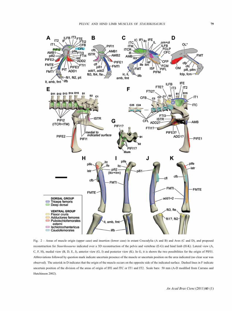
Fig. 2 – Areas of muscle origin (upper case) and insertion (lower case) in extant Crocodylia (A and B) and Aves (C and D), and proposed
reconstruction for Staurikosaurus indicated over a 3D reconstruction of the pelvis and vertebrae (E-G) and hind limb (H-K). Lateral view (A,
C, F, H), medial view (B, D, E, J), anterior view (G, I) and posterior view (K). In G, it is shown the two possibilities for the origin of PIFI1.
Abbreviations followed by question mark indicate uncertain presence of the muscle or uncertain position on the area indicated (no clear scar was
observed). The asterisk in D indicates that the origin of the muscle occurs on the opposite side of the indicated surface. Dashed lines in F indicate
uncertain position of the division of the areas of origin of IFE and ITC or IT1 and IT2. Scale bars: 50 mm (A-D modified from Carrano and
Hutchinson 2002).
An Acad Bras Cienc (2011) 83 (1)

“main” — 2011/2/10 — 13:35 — page 80 — #8
80 ORLANDO N. GRILLO and SERGIO A.K. AZEVEDO
M. ambiens (AMB) – In extant Reptilia (includingAves), the origin of the M. ambiens is anteroventralto the acetabulum, often from a pubic tubercle (pt; Hut-chinson 2001a). In Crocodylia, this structure is absentor reduced (Hutchinson 2001a), and M. ambiens is di-vided in two heads that originates on the cranial portionof the preacetabular cartilage and in the medial prox-imal region of the proximal pubis, but this conditionis derived in relation to other Reptilia (Romer 1923c,Hutchinson 2002). The pubic tubercle of Staurikosau-rus is preserved only on the left pubis (Fig. 5A) and issimilar in shape to that of Herrerasaurus, Saturnalia,and Lagerpeton. The right pubis of Staurikosaurus hasoften been used to illustrate this bone in the taxon, butit is damaged in the region of the pubic tubercle. Thisleaded several authors (e.g., Colbert 1970, Galton 1977,Novas 1993) to propose that this structure was absent inStaurikosaurus. AMB inserts in the tibial cnemial crest,together with the Triceps femoris group (Romer 1923c,Hutchinson 2002). In extant archosaurs, AMB also hasa secondary tendon that perforates the extensor tendon(Carrano and Hutchinson 2002, Hutchinson 2002). Thistendon was probably also present in Staurikosaurus.
Mm. femorotibiales (FMTE and FMTI) – M. femo-rotibialis has two divisions in Crocodylia (femorotibia-lis externus, FMTE; femorotibialis internus, FMTI) andthree in Aves (femorotibialis lateralis, FMTL; femoro-tibialis intermedius, FMTIM; femorotibialis medialis,FMTM), which originates from the main part of thefemoral shaft between the trochanteric region and thecondyles (Romer 1923c, Hutchinson 2002, Carrano andHutchinson 2002). Three ridges (linea intermusculariscranialis, lia; linea intermuscularis caudalis, lip ; lineaaspera = adductor ridge, ar) indicate the limits betweenthese muscles, defining three adjacent areas around thefemoral shaft: FMTE (= FMTL) is delimited by lia andlip, and FMTI (= FMTIM + FMTM) is limited by liaand ar (Hutchinson 2001b). In Staurikosaurus thesethree ridges are not complete, but the right femur andthe proximal part of the left femur have the major partof the lip and its distal part respectively preserved. Anirregular border is seen on the middle anterior portionof the left femur, exactly in the position where lia ofHerrerasaurus is located (Hutchinson 2001b). The dis-tal part of ar can be observed on the right femur of
Staurikosaurus, but most of its dorsal extension is oblit-erated due to distortions of the fossil. In the left femur,this portion of the shaft is concealed by the dorsal verte-brae. Accordingly, it is possible to determine the areasof origin of FMTE and FMTI with some precision, buttheir exact distal extension is uncertain.
In Aves, FMTI is divided in two parts (FMTIM andFMTM). Langer (2003) observed in Saturnalia a tenu-ous line that extends proximally from the medial condylealong the medial surface of the femur that could indicatea rudimentary division of FMTI. Due to poor preserva-tion, this structure is not observable in Staurikosaurus.
As in extant Archosauria, Mm. femorotibiales ofStaurikosaurus extended anterolaterally down to theproximal tibia, where they inserted onto the anterolat-eral cnemial crest, forming the knee extensor tendon(Romer 1923c, Carrano and Hutchinson 2002).
M. iliofibularis (ILFB) – M. iliofibularis originates onthe lateral surface of the ilium, between Mm. iliofemo-ralis and flexor tibialis externus (Hutchinson 2002,Carrano and Hutchinson 2002), slightly ventral to ilio-tibialis (Romer 1923c). Bittencourt and Kellner (2009)indicated that Staurikosaurus has one large concavityon the lateral surface of the ilium, but, this concavityappears to be divided in two by a smooth elevation(Fig. 4A-B), so that two concavities are present. Theanterior one is large and deep and is located just dor-sal to the acetabulum. The shallower posterior concav-ity probably corresponds to the ILFB origin because itis topographically equivalent to the surface where thismuscle originates in extant Archosauria. A smooth arc-shaped scar in the dorsoposterior limit of the posteriorconcavity may indicate the limits of ILFB origin (Fig.4C), whereas its ventral limit is indicated by the brevisshelf (Fig. 4C).
The anterolateral surface of the proximal part ofthe fibula of Staurikosaurus has an elongated crest thatcorresponds to the ILFB tubercle (Bittencourt and Kell-ner 2009), i.e., the insertion area of ILFB, as seen inextant Archosauria.
DEEP DORSAL
M. iliofemoralis externus (IFE) and M. iliotrochan-tericus caudalis (ITC) – In Crocodylia the M. iliofemo-ralis (IF) is not divided, but in Aves it has two parts:
An Acad Bras Cienc (2011) 83 (1)

“main” — 2011/2/10 — 13:35 — page 81 — #9
PELVIC AND HIND LIMB MUSCLES OF STAURIKOSAURUS 81
Fig. 3 – Iliac structures associated with muscle origin. A-J: Evolution of the preacetabular iliac border (pib) and the associated rough expanded
area (rea) in Diapsida and its relationship with the origin of the muscles IT, IC (blue areas in A, F and J) and IF, IFE and ITC (green areas in A, F,
and J). Number and letters correspond to the following taxa: (1) Diapsida (A – Iguana, Lepidosauromorpha), (2) Archosauria, (3) Crurotarsi (B –
Leptosuchus, Rutiodontidae), (4) Suchia (C – Stagonolepis, Aetosauria; D – Lythrosuchus, Poposauridae; E – Postosuchus [juvenile], Rauisuchidae;
F – Caiman, Crocodylomorpha), (5) Saurischia (G – Caseosaurus, Basal Saurischia [right ilium reversed]; H – Apatosaurus, Sauropodomorpha)
and (6) Avetheropoda (I – Allosaurus, Carnosauria; J – Meleagris, Aves). K-O: Relationship between the position of the areas of origin of
ITC, IFE and ILFB in Staurikosaurus (K, hypothesis adopted in this work; L, two hypothesis proposed by Langer 2003), Tyrannosaurus (M),
Sinornithomimus (N) and Crypturellus (O, indicating the relationship of IFE and the processus supratrochantericus, pst). Arrowheads in M and N
indicate the convex borders that may indicate anterior and posterior limits of IFE. Scale bars: 50 mm (A – after Romer 1922, 1923c, 1956; B-E
and G – from Long and Murry 1995; F – muscle disposition according to Romer 1923c; H – from Ostrom and McIntosh 1999; I – from Madsen
1993; J, O – from Hutchinson 2001a; M – from Osborn 1916; N – from Kobayashi and Lü 2003).
An Acad Bras Cienc (2011) 83 (1)
“main” — 2011/2/10 — 13:35 — page 82 — #10
82 ORLANDO N. GRILLO and SERGIO A.K. AZEVEDO
iliofemoralis externus (IFE) and iliotrochantericus cau-dalis (ITC) (Carrano and Hutchinson 2002). This sub-division is reflected on a differentiation in the area ofinsertion of IF in the femoral trochanteric shelf: in Di-nosauriformes, the trochanteric shelf has a cranial pro-tuberance (anterior or lesser trochanter) that is homol-ogous to the area of insertion of ITC in Aves, whichsuggests that IF was divided in this taxon (Hutchinson2001b). This structure is present in Staurikosaurus, butis reduced in size (Bittencourt and Kellner 2009), so wecan infer the presence of both IFE and ITC and indi-cate the area of insertion of ITC.
According to Hutchinson (2002), the insertion ofIFE occurs in a rough area of the trochanteric shelf,on the lateral surface of the femur. In the left femur ofStaurikosaurus there are some rough scars with unde-fined limits that may correspond to muscle insertion ar-eas (Fig. 5D). One of these is located on the trochantericshelf, exactly posterior to the anterior trochanter, and isinterpreted here as the insertion area of IFE.
IFE and ITC origins are located on the lateral sur-face of the ilium, but there is generally no scars that in-dicate the exact limits of their areas (Hutchinson 2001a,Carrano and Hutchinson 2002). As already mentioned,the ilium of Staurikosaurus has a large subtriangularconcavity on the anterior lateral surface of the ilium.This is dorsal to the acetabulum, bound anteriorly bythe preacetabular iliac border (Fig. 4C). This concavitycould hold a large muscle, similar to the condition ob-served in Tyrannosaurus by Carrano and Hutchinson(2002) and in Saturnalia by Langer (2003). A Level Iinference indicates that this area corresponds to the ori-gin of both parts of the iliofemoralis (IFE and ITC),contrary to the proposition of Langer (2003). Accord-ing to Langer (2003), ITC would occupy this entire con-cavity and IFE would originate from the dorsal borderof the acetabulum, immediately posterior to the supra-acetabular crest or from a small surface in the dorsallimit between this large anterior concavity and the con-cavity of origin of ILFB (Fig. 3L). The first hypothesisis not congruent with the position of the origin of IFEin Aves because it is located between ITC and ILFB,and is immediately ventral to the muscle iliotibialis.Also, Carrano and Hutchinson (2002) noted a verticalridge dividing the anterior cavity in two equally-sized
areas in Tyrannosaurus, and they interpreted this as thedivision of IF in IFE and ITC (Fig. 3M). The similarsize of these two muscles is corroborated by the sizeof their insertion areas in the femur. According to thepropositions of Langer (2003), ITC would be a verylarge muscle and IFE would be a very small one, andthis is not congruent with the size of their insertion areasin the femur of Staurikosaurus: the anterior trochanteris reduced and, although the limits of the insertion areaof IFE are not clear, the rough area appears to be equalin size to the anterior trochanter (Fig. 5D).
The anterior limit of ITC may be indicated by thepreacetabular iliac border that is adjacent to the anteriorlimit of the area of IF in lepidosaurs and Crocodylia,and of ITC in Aves (Fig. 3A, F, J). In Staurikosaurus,the preacetabular iliac border has striations (str) paral-lel to its long axis (Fig. 4C) that may be related to theorigin of ITC.
M. puboischiofemoralis internus 1 (PIFI1) – M. pubois-
chiofemoralis internus 1 of Crocodylia (= iliofemoralis
internus, IFI, in Aves) is homologous to the muscles
PIFI1 and PIFI2 of other Reptilia (Rowe 1986, Hut-
chinson 2002).
In Crocodylia, PIFI1 originates from the medial
surface of the ilium, in the medial proximal surface of
the ischium, and sacral ribs (Romer 1923c, Hutchinson
2001a, 2002, Carrano and Hutchinson 2002). In Aves,
IFI originates on the lateral surface of the ilium, from
a reduced preacetabular (“cuppedicus”) fossa (pf ; Hut-
chinson 2001a, 2002). The change in position of the
origin area of PIFI1 can be observed along the evo-
lution of Archosauria and is related to the expansion
of the cranial iliac process (Carrano 2000, Hutchinson
2001a). The appearance of the preacetabular fossa and
the reduction of the ventral portion of the pelvis also in-
dicate this transition (Hutchinson 2001a, 2002). These
changes probably produced the dorsolateral displace-
ment of PIFI1 origin in tetanuran theropods (as indi-
cated by the appearance of the preacetabular fossa). The
lateral displacement in Aves is indicated by the reduc-
tion of this fossa (Norell et al. 2001, Hutchinson 2002).
In basal dinosaurs, including Staurikosaurus, there
are few indications of these modifications. Compared to
Neotheropoda, the ventral portion of the pelvis is well
An Acad Bras Cienc (2011) 83 (1)

“main” — 2011/2/10 — 13:35 — page 83 — #11
PELVIC AND HIND LIMB MUSCLES OF STAURIKOSAURUS 83
Fig. 4 – Right (A-B) and left (C) ilium of Staurikosaurus in lateral (A, C) and dorsal (B) views indicating the existence of two concavities (1 and
2) on the lateral surface and the expansion of the posterior part of the medial blade of the brevis fossa (mbbf), indicated by the two directions arrow
(C). The dorsoposterior limit of ILFB origin (concavity 2) is indicated by a smooth border (dotted line in C). Right ilium of several taxa (D-I)
indicating the presence of a preacetabular fossa (pf) or a similar structure (pf?) on the ventral surface of the cranial iliac process: Staurikosaurus
(medial view [D]), Sellosaurus (lateral view [E]), Caseosaurus (medial [F] and lateral [G] views) and Tyrannosaurus (medial [H] and lateral [I]
views). The first medial iliac ridge (mr1) delimits the preacetabular fossa medially in Tyrannosaurus. In Staurikosaurus and Caseosaurus, this
fossa is delimited medially by a border (X) connected, but not equivalent to the mr1. Scale bars: 50 mm (E – from Galton 1984; F-G – from Long
and Murry 1995; H-I – from Osborn 1916).
developed and the cranial process of the ilium is not ex-
panded. Hutchinson (2001a) considers the preacetabular
fossa as an Avetheropoda character formed by the ex-
pansion of the first medial iliac ridge (articulation ridge
for the first sacral vertebra; mr1) that marks the medial
limit of this fossa (Fig. 4H-I). In Caseosaurus and Stau-
rikosaurus the first medial iliac ridge is in similar posi-
tion to this border in Crocodylia, i.e., horizontal and just
dorsal to the acetabulum (Fig. 4D, F, H). However, these
two forms bear another medial ridge in the ilium that ap-
pears to represent a dorsal extension of the first medial
iliac border and that also participates in the sacral ver-
An Acad Bras Cienc (2011) 83 (1)

“main” — 2011/2/10 — 13:35 — page 84 — #12
84 ORLANDO N. GRILLO and SERGIO A.K. AZEVEDO
tebra articulation (X in Fig. 4D, G). This ridge bounds
a shallow fossa, topographically equivalent to the preac-
etabular fossa, i.e., it is located in the ventromedial sur-
face of the cranial process of the ilium (Fig. 4). Despite
the topographical equivalence, the homology between
these structures is not clear because this fossa is medi-
ally limited by a ridge that cannot be certainly homolo-
gized with the first preacetabular medial ridge of the il-
ium. Accordingly, the origin of PIFI1 in Staurikosaurus
is uncertain (Fig. 2G): it could be equivalent to that of
Crocodylia (Level II inference), or may have shifted into
the aforementioned fossa (also Level II inference).
The PIFI1 of Crocodylia inserts at the proximal
part of the femur, anteromedially to the insertions of
PIFI2 (Romer 1923c, Hutchinson 2001b, 2002), on a
keel that separates the insertion of PIFI2 and FMTI
(Hutchinson 2001b). In Aves, IFI inserts on a rounded
mark at the medial proximal portion of the femur (Hut-
chinson 2001b, 2002). Herrerasaurus (Novas 1993,
Hutchinson 2001b) and Staurikosaurus possess a crest
on the anterior surface of the femur, distal and ante-
rior to the anterior trochanter, that is similar to that of
Crocodylia, indicating a similar insertion of PIFI1
(Level II inference).
M. puboischiofemoralis internus 2 (PIFI2) – There are
two homology hypothesis for the archosaur PIFI2 (Car-
rano and Hutchinson 2002, Hutchinson 2002): PIFI2 of
Crocodylia may be homologous to Mm. iliotrochanteri-
cus cranialis (ITCR) and medius (ITM) of Aves (Romer
1923b, Rowe 1986), with M. iliofemoralis (IF) of Cro-
codylia divided in two avian parts: iliofemoralis externus
(IFE) and iliotrochantericus caudalis (ITC); and PIFI2
may have been lost in Aves, and IF was divided in four
parts: IFE, ITC, ITCR and ITM (Gadow 1880). Because
the first hypothesis has more support from anatomical
and ontogenetic data and requires fewer transformations
in the number and position of muscles (Rowe 1986), we
will treat PIFI2 of Crocodylia as homologous to ITCR
and ITM of Aves. PIFI2 of Crocodylia should not be
confused with the homonymous muscle of other Rep-
tilia, but is homologous to their PIFI3 (Rowe 1986,
Romer 1923b).
In Crocodylia, PIFI2 originates from the centra and
transverse processes of the last six dorsal vertebrae (lum-
bar vertebrae; Romer 1923c). In Aves, the origins of the
homologous ITCR and ITM are located on the ventrolat-
eral surface of the preacetabular iliac process, anteriorly
to the origin of IFI. As previously presented, this tran-
sition is associated with the expansion of the preacetab-
ular iliac process and with the origin of the preacetabu-
lar fossa (Hutchinson 2001a, 2002). In Tyrannosaurus,
the centra of the dorsal vertebrae have large pleurocels
and little area for the attachment of muscles, and the
preacetabular fossa is present (Carrano and Hutchinson
2002). Staurikosaurus, on the other hand, has large ar-
eas for the attachment of PIFI2 on the dorsal vertebrae
that lack pleurocels. Also, the last five dorsal vertebrae
of Staurikosaurus have shallow depressions bellow the
infradiapophyseal fossae that could correspond to part of
PIFI2 origin. The eighth and ninth dorsal vertebrae are
partly covered by sediments and rib fragments, so it is
impossible to verify the presence of these depressions,
which are absent from the seventh to the more anterior
dorsal vertebrae. Accordingly, as for Crocodylia, PIFI2
of Staurikosaurus probably originated from the last five
(maybe six) dorsal vertebrae (Level II Inference).
In Crocodylia, PIFI2 inserts on the lateral surface
of a keel extending along the proximal femur, lateral
to the PIFI1 insertion, and its tendon is partly divided
by the proximal part of the origin of FMTI (Romer
1923c). In Tetanurae, PIFI2 inserts on a large process
(accessory trochanter), which is reduced to a small scar
in basal Aves (Hutchinson 2002). Despite this differ-
ence, the positions of these structures are the same. Bit-
tencourt and Kellner (2009) proposed that, in Stauri-
kosaurus, PIFI2 inserted on a proximodistally extended
and narrow crest located on the posterolateral surface
of the proximal femur, but it is not congruent with the
position observed in Crocodylia and Aves. In fact, this
crest corresponds to the medial limit of the insertion of
Mm. puboischiofemorales externi.
In Staurikosaurus, the surface of the anterior keel
of the femur is damaged and partly covered by sed-
iments, and it is impossible to identify muscle scars.
However, the same condition seen in Crocodylia, with
PIFI2 inserting on the lateral surface of this keel, is
likely to occur, since it is equivalent to the accessory
trochanter (Level I’ inference). It is not possible to con-
firm the presence of the posterior portion of the
An Acad Bras Cienc (2011) 83 (1)

“main” — 2011/2/10 — 13:35 — page 85 — #13
PELVIC AND HIND LIMB MUSCLES OF STAURIKOSAURUS 85
Fig. 5 – Right and left pubis in anterior view (A) indicating the pubic tubercle (pt) and the pubic apron (pa). Dashed line indicates the supposed
position of the unpreserved pt in the right pubis. Right (B) and left (C) ischium in lateral view. The dorsal ridge of the ischium (dris), ischial
ridge (ir), ischial tuberosity (it) and obturator process (op) are indicated, along with a scar that may indicate the origin of ADD2. Lateral view of
the proximal part of the left femur (D) indicating the approximate areas of insertion of the muscles ITC (on the anterior trochanter), IFE (on the
trochanteric shelf), ISTR (on a groove proximal to the trochanteric shelf) and PIFE (on the greater trochanter). The probable insertion of PIFI2 is
also indicated (on the lateral surface of the anterior keel of the femur). Proximal part of the right tibia of Caiman (E and F) and Staurikosaurus (G
and H) in medial (E and G) and posterior (F and H) views: the striations (str) in Staurikosaurus are topographically equivalent to the insertions of
FTI1-3, FTE, PIT and gastrocnemius internus (GI) in Caiman. Scale bars: 20 mm.
An Acad Bras Cienc (2011) 83 (1)

“main” — 2011/2/10 — 13:35 — page 86 — #14
86 ORLANDO N. GRILLO and SERGIO A.K. AZEVEDO
insertion tendon in the currently available material of
Staurikosaurus; the muscle scars on the trochanteric
region of the femur are not well defined.
FLEXOR CRURIS
Homologies of the Flexor cruris group are not well re-
solved (Romer 1923c, Hutchinson 2002). Here we fol-
low the hypothesis of Romer (1942). See Hutchinson
(2002) for a revision of different hypothesis and nomen-
clature.
The Flexor cruris muscles share two insertion
tendons in Crocodylia: FTI1 shares a tendon with FTI2
that connects to the tendon of PIT, and inserts on
the caudomedial surface of the proximal tibia (Romer
1923c, Hutchinson 2002), whereas FTI3, FTI4 and FTE
share a tendon that inserts on the posteromedial surface
of the proximal tibia, as occurs with the avian homo-
logues of these muscles (Hutchinson 2002, Carrano and
Hutchinson 2002). The proximal portion of the right
tibia of Staurikosaurus bears several striations that are
similar to the scars observed in extant Caiman tibiae
(Fig. 5E-H), which correspond to the insertion of FTI3
and FTE (posteromedially) , and of FTI1, FTI2 and PIT
(posterolaterally). Accordingly, the same condition is
inferred for Staurikosaurus. Considering the proposed
absence of PIT in Staurikosaurus (see below) , the pos-
terolateral striations seen on its tibia may correspond to
the insertion of FTI1 (if present) and FTI2.
On its medial side, the proximal tibia of Stauriko-
saurus also bears a scar (partly lost due to fragmentation
of the bone surface) distal to that of FTI3 and FTE (Fig.
5G), which can be attributed to the M. gastrocnemius
internus (that will not be treated here).
M. puboischiotibialis (PIT) – M. puboischiotibialis is
present in basal reptiles, reduced in Crocodylia and ab-
sent in Aves (Romer 1923c, Hutchinson 2002, Carrano
and Hutchinson 2002). In Crocodylia, there is only one
branch of PIT originating on a scar located on the prox-
imal tip of the obturator process (op) of the ischium
(Carrano and Hutchinson 2002), ventral to the acetab-
ulum (Romer 1923c). PIT inserts on the caudomedial
surface of the proximal tibia, as a tendon shared with
Mm. flexor tibiales interni 1 et 2 (Romer 1923c, Hut-
chinson 2002). The margin of the obturator process of
the ischium of Staurikosaurus in not preserved, and it is
impossible to determine the presence of PIT. Yet, Hut-
chinson (2002) points that the scar for PIT is absent in
all basal archosaurs and that there is no evidence of one
or more parts of PIT in Dinosauromorpha. Accordingly,
it was probably also absent in Staurikosaurus.
M. flexor tibialis internus 1 (FTI1) – M. flexor tibia-
lis internus 1 is absent in Aves and originates from the
caudolateral surface of the distal ischium of crocodiles
(Romer 1923c, Hutchinson 2002). Some theropods (e.g.,
Allosaurus, Piatnitzkysaurus, and Therizinosauroidea)
and Herrerasaurus possess a structure (distal ischial
tuberosity) on the caudolateral surface of the distal is-
chium that is topographically equivalent to FTI1 ori-
gin in Crocodylia (Hutchinson 2001a, 2002, Carrano
and Hutchinson 2002). The distal part of the ischium
of Staurikosaurus is not preserved, and the presence of
the distal ischial tuberosity cannot be confirmed. Yet,
it is present in Herrerasaurus and Saturnalia (Langer
2003), suggesting the presence of FTI1 in Staurikosau-
rus (Level II’ inference).
M. flexor tibialis internus 2 (FTI2) – M. flexor tibialis
internus 2, absent in Aves (Hutchinson 2002, Carrano
and Hutchinson 2002), originates from the lateral sur-
face of the postacetabular iliac process of crocodiles,
ventral to the origin of FTE (Romer 1923c, Hutchinson
2002, Carrano and Hutchinson 2002). Langer (2003)
indicated a division of muscle scars on the lateral sur-
face of the postacetabular iliac process in Saturnalia and
other dinosaurs (Herrerasaurus, Caseosaurus, basal or-
nithischias and ‘prosauropods’) that is topographically
equivalent to the origins of FTI2 and FTE in Crocodylia.
One of these marks is an extension of the dorsal iliac
margin (origin of IT3) that corresponds to the origin area
of FTE (Langer 2003). Posterior to this scar, on the cau-
dal most part of the ilium, there is another scar probably
associated with FTI2 (Langer 2003). These scars are not
visible in Staurikosaurus, but a Level II’ inference indi-
cates the presence of FTI2 and FTE originating from its
postacetabular iliac process, dorsal to the brevis shelf.
M. flexor tibialis internus 3 (FTI3) – M. flexor tibia-
lis internus 3 of Crocodylia is equivalent to the inner
part of FTI2 of basal Reptilia, in which the muscle is
not divided (Hutchinson 2002). Its origin is located
An Acad Bras Cienc (2011) 83 (1)

“main” — 2011/2/10 — 13:35 — page 87 — #15
PELVIC AND HIND LIMB MUSCLES OF STAURIKOSAURUS 87
on the ischial tuberosity (it; Hutchinson 2001a, 2002),
at the posterior margin of the ischium, proximal to the
origin of ADD2 (Romer 1923c). It is homologous to
the avian M. flexor cruris medialis (FCM), which origi-
nates from a similar (but distal) position, while the ischial
tuberosity is absent (Hutchinson 2001a, 2002, Carrano
and Hutchinson 2002). The ischium surface is not well
preserved in Staurikosaurus, with fractures hampering
the identification of muscle scars. However, both ischia
bear a crest (Fig. 5 B-C) near the articular surface of the
ilium that is slightly proximal in relation to the ischial
tuberosity of other dinosaurs, but may be a homologous
structure. Along with a depression lateral to the crest,
these structures could correspond to the origin area of
FTI3 as proposed by Langer (2003).
M. flexor tibialis internus 4 (FTI4) – This division of
the flexor tibialis internus is only present in Crocodylia,
and is equivalent to the superficial part of FTI2 of other
Reptilia (Romer 1942, Hutchinson 2001a, 2002). FTI4
originates on the fascia around the caudoventral ilium
and the caudodorsal ischium (Hutchinson 2002). Ac-
cordingly, its origin cannot be verified in Staurikosaurus
because it is not correlated to any bone scar. Its presence
is also equivocal, since it is absent in Aves.
M. flexor tibialis externus (FTE) – M. flexor tibialis ex-
ternus (= flexor cruris lateralis pars pelvica, FCLP, in
Aves) originates on the lateral surface of the ilium of
crocodiles, posterior to Mm. iliofibularis and iliofemo-
ralis externus (Romer 1923c, Carrano and Hutchinson
2002). As already mentioned, the ilium of Staurikosau-
rus has no preserved muscle scar posterior to the origin
of ILFB. The shape of the posterodorsal limit of ILFB
in Staurikosaurus suggests the posterior extension of
the dorsal border of the ilium (Fig. 4C), as seen in
other taxa (e.g., in Saturnalia and Herrerasaurus, Lan-
ger [2003]). Accordingly, it is assumed that the origin
of FTE in Staurikosaurus was posterior to ILFB and in
continuity to that of IT3. FTI2 origin may be posterior
to that of FTE, but their exact positions cannot be con-
firmed with current available material.
MM. ADDUCTORES FEMORES
The muscle adductor femoris is divided in two parts in
extant archosaurs: ADD1 and ADD2 in Crocodylia that
are homologous to, respectively, M. pubosichiofemora-
lis pars medialis (PIFM) and pars lateralis (PIFL) in
Aves (Romer 1923c, Hutchinson 2002). The two parts
originate from the lateral surface of the ischium (ADD1
near the cranial border of the bone) and are separated,
in Crocodylia, by the origin of PIFE3 (Romer 1923c).
In Aves, the position of PIFL origin is anteroventral in
relation to its crocodilian homologue, ADD2 (Hutchin-
son 2001a). This is probably related to the reduction of
the obturator process, and the change of the origin of M.
ischiotrochantericus to the lateral surface of the ischium
(Carrano and Hutchinson 2002).
According to Hutchinson (2001a), the ischial ridge
(ir) is located cranioventrally to the origin of FTI3 and
ventrally to ADD2. The bone surface of both ischia
of Staurikosaurus is damaged, and no muscle scar can
be safely identified. The ischial ridge is better seen in
the left bone (Fig. 5C). On the right ischium, dorsal to
the ischial ridge, in a well-preserved small area, a scar
(Fig. 5B) topographically equivalent to the origin of
ADD2 in Crocodylia may correspond to the origin of
this muscle. The origin of ADD1 is probably located
on the anterior margin of the obturator process, as in
extant archosaurs, but this structure is not preserved in
the holotype of Staurikosaurus.
The two ADD heads converge to a long and
narrow insertion area, on the caudal surface of the
distal femur (Romer 1923c), located between the linea
intermuscularis caudalis and the linea aspera (adduc-
tor ridge, Hutchinson 2001b). These structures, as al-
ready mentioned, are partly preserved in the femora of
Staurikosaurus and indicate the approximate position of
ADD insertion. Unfortunately there is no distinct scar
for either of the branches, as Carrano and Hutchinson
(2002) observed in Tyrannosaurus.
MM. PUBOISCHIOFEMORALES EXTERNI
Mm. Puboischiofemorales externi originate on the lat-
eral surface of the pubo-ischiadic plate in basal archo-
saurs, and is divided in two pubic parts, PIFE1 and
PIFE2. These are homologous to the avian Mm. obtu-
ratorius lateralis, OL, and obturatorius medialis, OM,
respectively. Its ischiadic part, PIFE3, is absent in Aves
(Hutchinson and Gatesy 2000). This plesiomorphic con-
dition is retained in Crocodylia (Carrano and Hutchinson
An Acad Bras Cienc (2011) 83 (1)
“main” — 2011/2/10 — 13:35 — page 88 — #16
88 ORLANDO N. GRILLO and SERGIO A.K. AZEVEDO
2002) and the modifications seen in Aves are attributed
to the division of the pubo-ischiadic plate and loss of the
obturator process (Hutchinson and Gatesy 2000).
The three heads of PIFE in Crocodylia shares an
insertion tendon that attaches to the greater trochanter of
the femur, as occurs with the avian homologues OL and
OM (Hutchinson and Gatesy 2000, Hutchinson 2001b,
Carrano and Hutchinson 2002). In Staurikosaurus, the
greater trochanter is a S-shape crest located on the cra-
niolateral region of the proximal region of the femur
(Fig. 5D; Bittencourt and Kellner 2009). Galton (1977,
2000) previously identified this structure as the anterior
trochanter (see Bittencourt and Kellner 2009, for a dis-
cussion).
M. puboischiofemoralis externus 1 (PIFE1) – In Stau-
rikosaurus, PIFE1 originates on the anterior surface of
the pubic apron (pa; Fig. 5A), as seen in Crocodylia
(Romer 1923c, Hutchinson 2002, Carrano and Hutchin-
son 2002). The apron corresponds to the dorsoventrally
expanded surface of the pubic symphysis (Hutchinson
2001a). In Aves, which lack a pubic symphysis, PIFE1
originates from the proximal lateral surface of the pu-
bis (Hutchinson 2002). Accordingly, the reconstrution
proposed for Staurikosaurus corresponds to a Level II
inference. Langer (2003) suggests the lateral surface of
the distal part of the pubis of Herrerasaurus (as proba-
bly in Staurikosaurus) as equivalent to the anterior sur-
face of the pubis of Saturnalia because it has a series
of striations that continues from the anterior surface of
the apron. These striations could indicate the origin of
PIFE1 (Langer 2003). Extant archosaurs do not have any
part of PIFE1 originating from the distal lateral surface
of the pubis. As this supposition requires a Level III
inference, it was not considered here.
Langer (2003) noted the laterally expanded lateral
border of the pubis of Saturnalia, Herrerasaurus, and
prosauropods, which gives the pubis a sinuous shape
in anterior view. Novas (1993) erroneously (see Fig.
5A) indicated that Staurikosaurus retains the primitively
straight dinosauromorph condition for the lateral border
of the pubis. Langer (2003) proposed that the proximal
margin of the lateral expansion of the pubis would in-
dicate the passage of PIFE1 from the pubis in direction
to the femur. In Staurikosaurus the expanded border is
more dorsal if compared to the other basal Saurischia, and
the origin of PIFE1 could be more dorsally expanded.
M. puboischiofemoralis externus 2 (PIFE2) – PIFE2
originates on the posterior surface of the pubic apron
(Romer 1923c, Hutchinson 2002, Carrano and Hutchin-
son 2002). In Aves, the homologue OM has moved
caudally to the pubo-ischiadic membrane (Hutchinson
2002). We consider (Level II inference) that Stauriko-
saurus has the same plesiomorphic condition of Croco-
dylia because the pubic apron is well developed.
M. puboischiofemoralis externus 3 (PIFE3) – PIFE3
originates from the lateral surface of the ischial obtu-
rator process, between the origin areas of ADD1 and
ADD2 (Romer 1923c, Hutchinson 2002, Carrano and
Hutchinson 2002). It is limited anterodorsally by the
ischial ridge (Hutchinson 2002). In Aves, this muscle
was lost together with the obturator process (Hutchin-
son 2001a, 2002). The obturator surface of the ischium
of Staurikosaurus is not well preserved and no muscle
scar is preserved. Accordingly, the origin of PIFE3 was
tentatively reconstructed based on the position of the is-
chial ridge, PIFE3, ADD1, and ADD2 in Crocodylia.
M. ISCHIOTROCHANTERICUS (ISTR)
M. ischiotrochantericus (ISTR) of crocodiles originates
on the medial surface of the caudal part of the ischium
(Romer 1923c, Hutchinson 2001a, 2002, Carrano and
Hutchinson 2002). In Aves, with the reduction of the
ischial symphysis, the origin of the homologue M. is-
chiofemoralis (ISF) was displaced to the lateral surface
of the ischium and to the ilio-ischiadic membrane (Hut-
chinson 2001a, 2002, Carrano and Hutchinson 2002).
In Aves, ISF is more cranial than in Crocodylia, in
which ISTR is located on the caudal extremity of the
ischium (Romer 1923c). But the distal part of the is-
chium in Aves corresponds to a caudoventral elongation
of the distal part of the ischial symphysis (Hutchinson
2001a). Besides, the avian ISF is located near the origin
of PIFM (= ADD1) and PIFL (= ADD2), as occurs in
Crocodylia, and ISTR origin is medial to that of ADD2
(Carrano and Hutchinson 2002).
Accordingly, it is possible to infer that the origin of
this muscle was displaced laterally in Aves, but not cra-
nially or caudally. Sereno and Arcucci (1993, 1994) and
An Acad Bras Cienc (2011) 83 (1)

“main” — 2011/2/10 — 13:35 — page 89 — #17
PELVIC AND HIND LIMB MUSCLES OF STAURIKOSAURUS 89
Langer (2003) suggest that the dorsal surface of the di-
nosaur ischium was laterally displaced, so that the dorsal
ridge of the ischium (dri), which separates the origins
of ISTR and ADD2, is placed on the lateral surface of
the ischium (Langer 2003). In Staurikosaurus, the ridge
that separates ISTR from ADD2 is visible and helps to
define the approximate position of ISTR origin, which
is near and dorsal to the scar that supposedly indicates
the origin of ADD2.
In Crocodylia, ISTR inserts on a scar on the cau-
dolateral surface of the proximal femur (Romer 1923c,
Hutchinson 2002), in a position almost equal to that
seen in Aves (Hutchinson 2002). Dinosauromorphs have
a sigmoid structure (trochanteric shelf) that corresponds
to the insertion of IFE. A groove proximal to the tro-
chanteric shelf corresponds to the insertion of ISTR
(Hutchinson 2001b, 2002, Carrano and Hutchinson
2002). The trochanteric shelf of Staurikosaurus is re-
duced (Bittencourt and Kellner 2009), but the groove
where ISTR is inserted is present and clearly seen on
the left femur (Fig. 5D).
MM. CAUDOFEMORALES
M. caudofemoralis brevis (CFB) – M. caudofemoralis
brevis of crocodiles (= caudofemoralis pars pelvica,
CFP, in Aves) originates on a shallow fossa on the
medioventral surface of the ilium, from the posterior
sacral ribs and the first caudal vertebra (Romer 1923c,
Carrano and Hutchinson 2002). In Aves, CFP origi-
nates only from the caudolateral surface of the ilium
(Hutchinson 2002). Changes in the origin of CFB/CFP
are apparently related to modifications of the medial
and lateral regions of the postacetabular ilium, which
include the transversal widening and deepening of both
the iliac blade and the medial shelf, forming the bre-
vis shelf (bs) and the brevis fossa of Dinosauria (Novas
1996, Hutchinson 2001a, 2002). Accordingly, CFB of
dinosaurs would have its origin from the brevis fossa,
which is reduced in taxa proximally related to Aves
(Hutchinson 2001a, 2002).
According to Novas (1992), Herrerasauridae re-
tains the plesiomorphic condition of basal Ornithodira
(e.g., Lagerpeton and Lagosuchus), in which the brevis
fossa is absent so that this structure would be novel for
Ornithischia + Saurischia. In following works, Novas
(1993, 1996) considered herrerasaurids as basal thero-
pods and suggested that a groove on the lateral surface
of the posterior part of the ilium of Herrerasaurus rep-
resents a reduced brevis fossa. Hutchinson (2001a) also
treats Herrerasauridae as basal Theropoda and suggests
that the most parsimonious condition would be the re-
duction of the brevis fossa in this group. Even if we con-
sider Herrerasauridae as basal Saurischia (Yates 2003,
Langer 2004, Leal et al. 2004), the reduction hypothe-
sis is more parsimonious since ornithischians also have
a well-developed fossa (Novas 1992, 1996).
The reduction of the brevis fossa appears to be more
pronounced in Staurikosaurus than in Herrerasaurus,
because just a shallow depression on the lateral post-
acetabular surface of the ilium is present. This depres-
sion is dorsally delimited by an elongated elevation that
starts at the dorsoposterior margin of the supra-acetabu-
lar crest, finishing on the posterior margin of the ilium.
This structure is interpreted as homologue to both the
ridge that bounds the groove of the posterior ilium of
Herrerasaurus, and the brevis shelf. Accordingly, the
surface ventral to this shelf (posteroventral margin of
the ilium) corresponds to the medial blade of the ilium,
which bounds the brevis fossa medially (medial blade
of the brevis fossa, mbbf; Fig. 4C) in other Dinosauria.
Therefore, the lateral surface of this blade would repre-
sent the origin of CFB in Staurikosaurus. Despite the
reduction of the brevis fossa, Staurikosaurus possesses
a marked dorsoventral expansion of the posterior part of
the posteroventral margin of the ilium (Fig. 4C), which
does not occur in Herrerasaurus. In both taxa, the medial
blade (posteroventral margin of the ilium) corresponds
to 29% of the length of the ilium, but the posterior part
in Herrerasaurus is not dorsoventrally expanded, sug-
gesting that the posterior part of CFB was enlarged in
Staurikosaurus.
CFB inserts on the posterolateral surface of the fe-
mur, between the fourth trochanter and the linea inter-
muscularis caudalis (Hutchinson 2001a, b, 2002, Car-
rano and Hutchinson 2002) in a position slightly prox-
imal and lateral to the insertion of M. caudofemoralis
longus (Romer 1923c). In Aves, the fourth trochanter
is reduced to a scar and the insertion of CFP is adja-
cent to this scar (Hutchinson 2002). The left femur of
Staurikosaurus has well-preserved fourth trochanter and
An Acad Bras Cienc (2011) 83 (1)

“main” — 2011/2/10 — 13:35 — page 90 — #18
90 ORLANDO N. GRILLO and SERGIO A.K. AZEVEDO
linea intermuscularis caudalis, but no muscle scars are
observed. Accordingly, the inferred position of CFB in-
sertion is approximate.
M. caudofemoralis longus (CFL) – M. caudofemoralis
longus (= caudofemoralis pars caudalis, CFC, in Aves)
originates on the centra and ventral surfaces of the cau-
dal vertebra transverse processes (Romer 1923c, Gatesy
1990, Hutchinson 2001b), approximately from the third
to the fifteenth element (Romer 1923c, Gatesy 1990).
In Aves, CFC was reduced along with the reduction of
the tail and the evolution of the pygostyle, and its origin
is restricted to this structure (Gatesy 1990, Hutchinson
2002). The presence of transverse processes would be an
indication of the minimal extension of the origin of CFL
in the tail of archosaurs, as are specializations of the ver-
tebrae and chevrons (Gatesy 1990). In Theropoda, the
tail has a transition point where the caudal vertebrae lose
the transverse processes (Russell 1972), so that the ori-
gin of the CFL is restricted to the area anterior to this
point. The transition is also marked by the elongation
of the prezygapophyses and dorsoventral compression
of the chevrons on the vertebrae posterior to the point
(Gauthier 1986, Gatesy 1990).
The holotype of Staurikosaurus does not have the
complete caudal series preserved: there are six proxi-
mal vertebrae; a block containing six articulated verte-
brae from the middle part of the tail; two blocks with
two vertebrae each, also from the middle of the tail;
and the last 19 caudal vertebrae (Bittencourt and Kell-
ner 2009). Vertebrae from one of the median blocks
with two vertebrae bear transverse processes. But the
19 terminal vertebrae lack these structures and have
elongated prezygapophyses (Gauthier 1986, Bittencourt
and Kellner 2009). Based on regression equations ob-
tained from data collected from the preserved caudal
vertebrae (height and length of the centrum), and consid-
ering that Staurikosaurus had the same number of caudal
vertebrae of Herrerasaurus (i.e., 47 vertebrae), it was
possible to estimate the position of the middle blocks
of vertebrae. This method also allowed estimating the
length of the non-preserved vertebrae and indicated that
the block containing the two vertebrae with transverse
processes corresponds to the 24th and 25th caudal ele-
ments (Grillo and Azevedo 2011). Accordingly, at least
25 caudal vertebrae of Staurikosaurus have transverse
processes, and this is the minimal extension of CFL ori-
gin (Grillo and Azevedo 2011).
CFL of crocodiles inserts on the medial surface
of the fourth trochanter, on an associated depression
(Romer 1923c, Gatesy 1990, Hutchinson 2001b, 2002).
In Aves, the reduction of CFC was accompanied by the
reduction of the fourth trochanter to a roughed area
(Gatesy 1990, Hutchinson 2001b, 2002). Staurikosaurus
possesses a well-developed crest-shaped fourth troch-
anter (Colbert 1970, Galton 1977, 2000, Bittencourt and
Kellner 2009) that indicates that the CFL was a large and
powerful muscle. This condition is congruent with the
expanded area of origin of this muscle in the tail.
In extant crocodiles the CFL has a secondary ten-
don that extends from the distal part of CFL to the caudal
region of the knee (Romer 1923c, Hutchinson 2001b,
2002), contributing to the origin of the M. gastrocne-
mius lateralis (Hutchinson 2002). In Theropoda, the
fourth trochanter is pendant and has a process ventrally
directed that could be an indication of the presence of
this secondary tendon (Hutchinson 2002, Carrano and
Hutchinson 2002). Staurikosaurus has a pendant fourth
trochanter, although its margin is damaged and the ven-
tral process cannot be observed, so that the secondary
tendon was probably present (Level II’ inference).
DISCUSSION
Reconstructions of the pelvic musculature of sauris-
chian dinosaurs have been attempted for more than a
century (Romer 1923b, c), but recent works (e.g. Hut-
chinson 2001a, b, 2002) clarified several aspects of the
evolution of the hind limb muscles in archosaurs. Some
studies (e.g. Coombs 1979, Dilkes 2000, Carrano and
Hutchinson 2002, Langer 2003) did not focus on higher
level taxa, but on particular species. This approach may
reveal exclusive adaptations, contributing to understand
the different locomotion adaptations of each species.
The muscle reconstruction of Staurikosaurus al-
lowed the identification of both modifications that dif-
ferentiate this taxon from other closely related species,
and of a series of osteological structures not observed
by other authors that studied this specimen. Differently
from other works, the reconstruction of the hind limb
musculature of Staurikosaurus was based on a single
An Acad Bras Cienc (2011) 83 (1)

“main” — 2011/2/10 — 13:35 — page 91 — #19
PELVIC AND HIND LIMB MUSCLES OF STAURIKOSAURUS 91
specimen, which has several problems of preservation
that hampered the observation of muscle scars. How-
ever, the comparison with other proximally related taxa
and the use of bones from both sides of the body, asso-
ciated to the EPB methodology, allowed to construct a
map of origin and insertion for the majority of the hind
limb muscles. The result is consistent to muscle arrange-
ment in extant archosaurs and other extinct dinosaurs that
have already been studied (see Carrano and Hutchinson
2002, Langer 2003).
Some muscle associated structures were described
for the first time for Staurikosaurus, complimenting
previous descriptions and studies about this taxon (Col-
bert 1970, Galton 1977, 2000, Bittencourt and Kellner
2009): the pubic tubercle (associated to AMB origin),
the lineae intermusculares on the femur (associated to
FMTE, FMTI, ADD1-2 and CFB), the two concavities
on the lateral surface of the ilium (associated to ITC, IFE
and ILFB origin), the concavities on the posterior dor-
sal vertebrae (associated to PIFI2 origin) and the fossa
on the ventral margin of the cranial iliac process (prob-
ably associated to PIFI1 origin). The pubic tubercle has
been figured as absent in Staurikosaurus (e.g., Colbert
1970, Galton 1977, Novas 1993), and a recent revision
(Bittencourt and Kellner 2009) did not recognize this tu-
bercle, perhaps as a result of taphonomic damage. All
these studies were based on the right pubis, but the pubic
tubercle is preserved only on the left pubis (Fig. 5A).
The concavities on the lateral surface of the ilium
described here are different from those mentioned by
Bittencourt and Kellner (2009). These authors indicate
that the ilium of Staurikosaurus has two concavities on
the lateral surface, the caudal one being interpreted as a
reduced brevis fossa. The cranial concavity indicated by
Bittencourt and Kellner (2009) was interpreted here as
comprising two concavities associated to the origins of
IFE, ITC, and ILFB (concavities 1 and 2, Fig. 4A-C).
The muscle reconstruction of Staurikosaurus may
be compared to that presented by Langer (2003) for an-
other basal Saurischian, Saturnalia tupiniquim. Langer
(2003) indicated that the hind limb and pelvic anatomy
of Saturnalia, and hence muscle arrangement and func-
tion, would be representative of a general construction
shared by basal dinosauriforms such as Marasuchus and
Pseudolagosuchus, and basal dinosaurs, including Her-
rerasaurus, Staurikosaurus, Guaibasaurus, and basal
sauropodomorphs. Most of the results presented are
very similar to those of Langer (2003). Differences are
mainly conceptual, rather than anatomical.
As proposed by Langer (2003), we consider that
the tenuous convexity that separates the two main con-
cavities on the lateral surface of the ilium indicates the
anterior limit of ILFB origin. However, Langer (2003)
proposed that the anterior concavity corresponds solely
to the origin of ITC, so that the origin of IFE would be
located on the surface immediately caudal to the supra-
acetabular crest or close to the dorsal margin of the
ilium, over the dorsal extremity of the convexity that
separates the two main lateral iliac concavities (Fig. 3L).
The first hypothesis is congruent with the arrangement
seen in Crocodylia, in which the origin of IF borders the
dorsoposterior margin of the acetabulum. In this con-
text, IFE would have originated as a posteroventral divi-
sion of IF. This is not congruent with the origin of IFE
in Aves, which is located on the dorsal margin of the il-
ium on the processus supra-trochantericus (pst; Fig. 3O;
Hutchinson 2001a). This indicates that IFE represents a
dorsoposterior separation of IF, which is congruent with
the second hypothesis of Langer (2003). If this last hy-
pothesis is accepted, it is also necessary to infer that IF
splited into a reduced IFE and a large ITC. Yet, osteo-
logical evidence suggests that ITC was initially of simi-
lar size to IFE, enlarging only along theropod evolution
until it was reduced again in Aves, and restricted to the
dorsal margin of the ilium. This is supported by the en-
largement of the anterior trochanter and cranial iliac pro-
cess in basal theropods, and by the later reduction of the
former in taxa closely related to Aves (Hutchinson and
Gatesy 2000, Hutchinson 2001b). Another indication
that IFE and ITC had similar sizes in non-maniraptoran
theropods is the presence of a dorsoventrally directed
convexity on the lateral surface of the anterior iliac pro-
cess, which divides the anterior concavity of the ilium,
separating the origins of ITC and IFE in Tyrannosaurus
(Carrano and Hutchinson 2002), as well as in the or-
nithomimosaur Sinornithomimus dongi. In this context,
the most parsimonious scenario for the evolution of these
muscles is the division of IF in two muscles of similar
size in basal dinosaurs whose origins shared the anterior
concavity of the ilium.
An Acad Bras Cienc (2011) 83 (1)

“main” — 2011/2/10 — 13:35 — page 92 — #20
92 ORLANDO N. GRILLO and SERGIO A.K. AZEVEDO
Another difference between the proposition of
Langer (2003) for Saturnalia and the one presented here
for Staurikosaurus is related to AMB. Langer (2003)
proposed that this muscle, as in Crocodylia, would have
two heads originating from the pubic tubercle and from
the dorsal margin of the pubis. On the contrary, we con-
sider that the AMB of dinosaurs, as in Aves, would have
just one head, originating from the pubic tubercle. Ac-
cording to Carrano and Hutchinson (2002), the presence
of two heads in Crocodylia represents an apomorphy of
this clade because both Aves and Lepidosauria have just
one AMB head (Romer 1922, 1923c).
Other differences relative to the reconstruction
presented by Langer (2003) are related to insufficient
information retrieved from the holotype of Staurikosau-
rus. The smooth division on the femur of Saturnalia
that could indicate the separation of FMTI in two parts
(FMTM and FMTIM) was not preserved or is absent
in Staurikosaurus. The phylogenetic positioning of Her-
rerasauridae is still uncertain. Some works (e.g. Yates
2003, Leal et al. 2004, Bittencourt and Kellner 2009)
place Herrerasauridae basal to Eusaurischia, but others
(e.g., Novas 1993, Sereno 1997, Sereno and Novas
1992, Benton 1999) suggest a closer relation to Thero-
poda. Accordingly, the presence of an initial division
of FMTI in Saturnalia does not bring additional evi-
dences for its division in Herrerasauridae until more sta-
ble phylogenetic hypotheses are obtained.
We restricted the origin of PIFE3 to the obturator
process, as in Crocodylia, because the obturator process
is not completely preserved and no muscle scar could be
observed. Yet, Langer (2003) observed striations on the
ventrolateral surface of the ischium of Saturnalia, which
were associated to PIFE3. This could also be the case for
Staurikosaurus, but new material is necessary to confirm
this hypothesis. Besides, Langer (2003) reconstructed
ISTR as originating from the entire mediodorsal surface
of the ischium, but the distal part of that bone forms an
elongated symphysis (Hutchinson 2001a). Moreover, in
Crocodylia, ISTR origin is restricted to the obturator pro-
cess and does not extend into the symphysis. In Aves, the
homologue ISF is also restricted to the proximal portion
of the ischium, so that there is no support from extant
taxa for a distal extension of this muscle in dinosaurs.
Finally, Langer (2003) attributed scars on the pu-
bic peduncle to the origin of PIFI1 or the dorsal part of
AMB. In the prosauropod Sellosaurus gracilis there is
a depression on the ventral margin of the cranial iliac
process that extends to the pubic peduncle (Fig. 4E).
This is equivalent to the fossa observed on the cranial
process of the ilium of Staurikosaurus (Fig. 4C-D) and
Caseosaurus (Fig. 4F-G), which are in the same position
of the preacetabular fossa of Avetheropoda (Fig. 4H-I).
The preacetabular fossa of Avetheropoda was attributed
to the origin of PIFI1, since it is the condition of the
avian homologue IFI (Hutchinson 2002). Accordingly,
it is possible to infer that early on dinosaur evolution
PIFI1 changed its origin from the medial surface of the
ilium, as is the case for Crocodylia, to the ventral margin
of the cranial iliac process and the dorsal part of the pu-
bic peduncle of the ilium. Later on Theropod evolution,
the incorporation of dorsal vertebrae to the sacrum led
to the rearrangement of the first medial iliac ridge and
to the development of the preacetabular fossa as defined
for Avetheropoda by Hutchinson (2001a).
In conclusion, disregarding the differences pointed
above, it is possible to confirm that basal dinosaurs shared
a similar muscle arrangement as proposed by Langer
(2003).
In the following paragraphs, this arrangement is
compared with that of extant Crocodylia and Aves and to
the reconstruction presented by Carrano and Hutchinson
(2002) for Tyrannosaurus, in the intent to discuss some
aspects of hind limb muscle evolution in Saurischia.
Relative to Crocodylia, Staurikosaurus differs in
various aspects: (1) the iliac surface for muscle origins is
proportionally larger, especially on the anterior portion
of the bone, due to the expansion of the cranial iliac pro-
cess; (2) the origin of ILFB is also enlarged, occupying
a large concavity on the lateral surface of the ilium; (3)
IF is divided; (4) AMB has just one head; and (5) PIFI1
apparently moved from the medial surface of the ilium to
a fossa on the ventral margin of the cranial iliac process
and dorsal part of the pubic peduncle. Other muscles in
Staurikosaurus, such as CFB, CFL, PIFE1-2, and PIFI2,
have origin and insertion areas in positions equivalent
to Crocodylia.
All the changes listed above for Staurikosaurus
were also observed in Tyrannosaurus (Carrano and
Hutchinson 2002). The most notable difference between
An Acad Bras Cienc (2011) 83 (1)

“main” — 2011/2/10 — 13:35 — page 93 — #21
PELVIC AND HIND LIMB MUSCLES OF STAURIKOSAURUS 93
these two taxa is the existence of a true preacetabular
fossa on the ventral margin of the cranial iliac process of
Tyrannosaurus, but Staurikosaurus also bears other dis-
similarities including: (1) reduced anterior trochanter;
(2) the femoral head in its plesiomorphic orientation, ro-
tated 45◦ posteriorly; (3) a more proximal insertion of
FTI3 and FTE and origin of ADD2 and FTI3; (4) more
posterior origin of IFE; (5) reduced brevis fossa; and
(6) a CFB origin extending to the posterior margin of
the ilium and with its posterior part dorsoventrally ex-
panded. The arrangement of other muscles are similar in
both taxa or is uncertain in Staurikosaurus (e.g., origins
of ADD1 and PIFE3) or in Tyrannosaurus (presence or
absence of FTI1 and FTI2). Other differences are re-
lated to the proportionately larger bone surfaces of Tyran-
nosaurus due to the expansion of the cranial and caudal
processes of the ilium.
Differences in muscle position observed between
Staurikosaurus and Tyrannosaurus are related to changes
in muscle function, as is the case of ADD2, FTI3, and
FTE, which have their origins in a more distal position.
This makes ADD2 to change its primary function from
adductor to extensor and increases the moment arm of
FTI3 and FTE (Carrano and Hutchinson 2002).
Based on the comparison between Staurikosaurus
and Tyrannosaurus, it is possible to infer that the main
features of the avian musculature present in Tyranno-
saurus were also present early on saurischian evolution,
e.g., a divided IF (Hutchinson 2001b) and the origin of
PIFI1 from the ventral surface of the cranial process
of the ilium. Modifications not present early on the
evolution of Saurischia, but present in Tyrannosaurus,
include: (1) the expanded anterior trochanter (Hutchin-
son and Gatesy 2000); (2) the expansion of the cra-
nial and caudal iliac processes (Carrano 2000), which
is less evident in basal dinosaurs; and (3) the rotation
of the femoral head, the lateral surface of which moved
to a more anterior position (Carrano 2000, Hutchinson
2001b).
The reconstruction of pelvic and hind limb muscu-
lature of Staurikosaurus also revealed a specific adap-
tation unknown to other taxa: the posterior part of the
origin of CFB (the medial blade of the brevis fossa)
is dorsoventrally expanded (Fig. 4C), which probably
allocated a larger posterior part of CFB. Compared to
Herrerasaurus, this expansion characterizes a volume
increase of CFB, since the length of the medial blade of
the brevis fossa corresponds to 29% of the total length
of the ilium in both taxa.
ACKNOWLEDGMENTS
The Museum of Comparative Zoology is thanked forthe loan of the S. pricei holotype to Alexander Kellner,Museu Nacional/UFRJ. Jonathas de Souza Bittencourtis thanked for providing the photographs of the S. priceiholotype and Riccardo Mugnai is thanked for preparingthe casts of the holotype. This research is part of theMSc thesis of one of the authors (ONG) at the MuseuNacional of Universidade Federal do Rio de Janeiro,funded by a fellowship of the Conselho Nacional de De-senvolvimento Científico e Tecnológico (CNPq) and ofthe Fundação Carlos Chagas Filho de Amparo à Pesquisado Estado do Rio de Janeiro (FAPERJ). The thesis wasexamined by the doctors Max Langer, Alexander Kell-ner and Oscar Rocha-Barbosa, who are thanked for pro-viding useful comments that greatly improved the finalversion of this paper.
RESUMO
O estudo da musculatura pélvica e do membro posterior em
dinossauros basais é importante para entender a evolução ini-
cial do bipedalismo em dinossauros Saurischia. Empregando
uma metodologia que tem como base dados obtidos a partir
de táxons viventes e extintos posicionados em um contexto
filogenético, foi possível fazer inferências bem suportadas re-
lativas à maior parte dos músculos do membro posterior do di-
nossauro Saurischia basal Staurikosaurus pricei Colbert, 1970
(Formação Santa Maria, Triássico Superior do Rio Grande do
Sul, Brasil). Duas grandes concavidades na superfície lateral
do ílio correspondem à origem dos músculos iliotrochantericus
caudalis e iliofemoralis externus (compartilhando a concavi-
dade anterior) e para o músculo iliofibularis (na concavidade
posterior). O músculo ambiens apresenta apenas um ramo que
se origina no tubérculo púbico. A origem provável do músculo
puboischiofemoralis internus 1 está localizada numa fossa na
margem ventral do processo pré-acetabular do ílio, represen-
tando um estágio anterior à formação da verdadeira fossa pré-
acetabular. Os músculos caudofemorales longus et brevis são
bem desenvolvidos e Staurikosaurus é único por apresentar a
superfície de origem do músculo caudofemoralis brevis proje-
tada e expandida posteriormente.
An Acad Bras Cienc (2011) 83 (1)

“main” — 2011/2/10 — 13:35 — page 94 — #22
94 ORLANDO N. GRILLO and SERGIO A.K. AZEVEDO
Palavras-chave: “extant phylogenetic bracket”, locomoção,
reconstrução muscular, Saurischia, Staurikosaurus pricei.
REFERENCES
ALEXANDER RMCN. 1976. Estimates of speeds of dino-
saurs. Nature 261: 129–130.
ALEXANDER RMCN. 2004. Bipedal animals, and their dif-
ferences from humans. J Anat 204: 321–330.
BARSBOLD R AND OSMÓLSKA H. 1990. Ornithomimosau-
ria. In: WEISHAMPEL DB ET AL. (Eds), The Dinosauria,
Berkeley: University of California Press, p. 225–244.
BENTON MJ. 1999. Scleromochlus taylori and the origin of
dinosaurs and pterosaurs. Philos T R Soc B 354: 1423–
1446.
BENTON MJ AND CLARK JM. 1988. Archosaur phylogeny
and the relationships of the Crocodylia. In: BENTON MJ
(Ed), The phylogeny and classification of the Tetrapods,
Vol. 1: Amphibians, Reptiles, Birds, Systematics Associ-
ation Special 35 A: 295–338.
BITTENCOURT JS AND KELLNER AWA. 2009. The anatomy
and phylogenetic position of the Triassic dinosaur Stau-
rikosaurus pricei Colbert, 1970. Zootaxa 2079: 1–56.
BONAPARTE JF. 1996. Dinosaurios de America del Sur.
Buenos Aires: Museo Argentino de Ciencias Naturales
“Bernardino Rivadavia”, 174 p.
BONAPARTE JF, FERIGOLO J AND RIBEIRO AM. 1999. A
new early Late Triassic saurischian dinosaur from Rio
Grande do Sul State, Brazil. In: TOMIDA Y ET AL. (Eds),
Proceedings of the Second Gondwanaland Dinosaur Sym-
posium, Tokyo: National Science Museum Monographs
15: 89–109.
BONAPARTE JF, NOVAS FE AND CORIA RA. 1990. Carno-
taurus sastrei Bonaparte, the horned, lightly built carno-
saur from the Middle Cretaceous of Patagonia. Contrib.
Sci Nat Hist Mus Los Angeles Co 416: 1–42.
BRINKMAN DB AND SUES H-D. 1987. A staurikosaurid di-
nosaur from the Upper Triassic Ischigualasto Formation
of Argentina and the relationships of the Staurikosauri-
dae. Palaeontology 30: 493–503.
BRYANT HN AND RUSSELL AP. 1992. The role of phyloge-
netic analysis in the inference of unpreserved attributes of
extinct taxa. Philos T R Soc B 337: 405–418.
CALVO JO, PORFIRI JD AND KELLNER AWA. 2004. On a
new maniraptoran dinosaur (Theropoda) from the Upper
Cretaceous of Neuquén, Patagonia, Argentina. Arq Mus
Nacional 62: 549–566.
CARRANO MT. 1999. What, if anything, is a cursor? Cat-
egories versus continua for determining locomotor habit
in mammals and dinosaurs. J Zool (London) 247: 29–42.
CARRANO MT. 2000. Homoplasy and the evolution of di-
nosaur locomotion. Paleobiology 26: 489–512.
CARRANO MT. 2001. Implications of limb bone scaling,
curvature and eccentricity in mammals and non-avian di-
nosaurs. J Zool (London) 254: 41–55.
CARRANO MT AND HUTCHINSON JR. 2002. Pelvic and
hindlimb musculature of Tyrannosaurus rex (Dinosauria:
Theropoda). J Morphol 253: 207–228.
CARRANO MT, SAMPSON SD AND FORSTER CA. 2002.
The osteology of Masiakasaurus knopfleri, a small abeli-
sauroid (Dinosauria: Theropoda) from the Late Cretaceous
of Madagascar. J Vert Paleontol 22: 510–534.
CARRIER DR, WALTER RM AND LEE DV. 2001. Influence
of rotational inertia on turning performance of theropod
dinosaurs: clues from humans with increased rotational
inertia. J Exp Biol 204: 3917–3926.
CLARKE JA. 2004. Morphology, phylogenetic taxonomy, and
systematics of Ichthyornis and Apatornis (Avialae: Orni-
thurae). Bull Am Mus Nat Hist 286: 1–179.
COLBERT EH. 1964. Relationships of the Saurischian Di-
nosaurs. Am Mus Novit 2181: 1–24.
COLBERT EH. 1970. A saurischian dinosaur from the Triassic
of Brazil. Am Mus Novit 2405: 1–60.
COLBERT EH. 1989. The Triassic dinosaur Coelophysis. Bull
Mus N Arizona 57: 1–160.
COOMBS JR WP. 1979. Osteology and myology of the hind-
limb in the Ankylosauria (Reptilia, Ornithischia). J Pale-
ontol 53: 666–684.
COOMBS JR WP. 1980. Swimming ability of carnivorous
dinosaurs. Science 207: 1198–1200.
CORIA RA AND CURRIE PJ. 2006. A new carcharodon-
tosaurid (Dinosauria, Theropoda) from the Upper Creta-
ceous of Argentina. Geodiversitas 28: 71–118.
CURRIE PJ. 2000. Theropods from the Cretaceous of Mon-
golia. In: BENTON MJ, SHISHKIN MA, UNWIN DM
AND KUROCHKIN EN (Eds), The age of dinosaurs in
Russia and Mongolia. Cambridge: Cambridge University
Press, p. 434–455.
CURRIE PJ AND CHEN P-J. 2001. Anatomy of Sinosauro-
pteryx prima from Liaoning, northeastern China. Can J
Earth Sci 38: 1705–1727.
DAY JS, NORMAN DB, UPCHURCH P AND POWELL HP.
2002. Dinosaur locomotion from a new trackway. Nature
415: 494–495.
An Acad Bras Cienc (2011) 83 (1)

“main” — 2011/2/10 — 13:35 — page 95 — #23
PELVIC AND HIND LIMB MUSCLES OF STAURIKOSAURUS 95
DILKES DW. 2000. Appendicular myology of the hadrosau-
rian dinosaur Maiasaura peeblesorum from the Late Cre-
taceous (Campanian) of Montana. Trans R Soc Edinb,
Earth Sci 90: 87–125.
FARLOW JO. 1981. Estimates of dinosaur speeds from a new
trackway site in Texas. Nature 294: 747–748.
GADOW H. 1880. Zur vergleichenden Anatomie der Musku-
latur des Beckens und der hinteren Gliedmasse der Ratiten.
Jena: G. Fischer.
GALTON PM. 1977. On Staurikosaurus pricei, an early sau-
rischian dinosaur from The Triassic of Brazil, with notes
on the Herrerasauridae and Poposauridae. Paläntol Z 51:
234–245.
GALTON PM. 1984. An early prosauropod dinosaur from
the upper Triassic of Nordwürttemberg, West Germany.
Stuttgarter Beitr Naturkd, ser B, 106: 1–25.
GALTON PM. 2000. Are Spondylosoma and Staurikosaurus
(Santa Maria Formation, Middle-Upper Triassic, Brazil)
the oldest saurischian dinosaurs? Paläntol Z 74: 393–423.
GATESY SM. 1990. Caudofemoral musculature and the
evolution of theropod locomotion. Paleobiology 16: 170–
186.
GATESY SM. 1999. Guinea fowl limb function. I: Cinera-
diographic analysis and speed effects. J Morphol 240:
115–125.
GATESY SM AND BIEWENER AA. 1991. bipedal locomo-
tion: effects of speed, size and limb posture in birds and
humans. j zool (london) 224: 127–147.
GATESY SM, MIDDLETON KM, JENKINS JR FA AND SHU-
BIN NH. 1999. Three-dimensional preservation of foot
movements in Triassic theropod dinosaurs. Nature 399:
141–144.
GAUTHIER J. 1986. Saurischian monophyly and the origin
of birds. In: PADIAN K (Ed), The origin of birds and the
evolution of flight, Mem California Acad Sci 8: 1–55.
GOODRICH ES. 1958. Studies on the structure and develop-
ment of vertebrates. New York: Dover Publications 1:
485.
GREGORY WK AND CAMP CL. 1918. Studies in compara-
tive myology and osteology. III. Bull Am Mus Nat Hist
38: 447–563.
GRILLO ON AND AZEVEDO SAK. 2011. Recovering miss-
ing data: estimating position and size of caudal vertebrae
in Staurikosaurus pricei Colbert, 1970. An Acad Bras
Cienc 83: 61–71.
HENDERSON DM. 1999. Estimating the masses and centers
of mass of extinct animals by 3-D mathematical slicing.
Paleobiology 25: 88–106.
HOLTZ JR TR. 1998. A new phylogeny of the carnivorous
dinosaurs. Gaia 15: 5–61.
HUANG SH, NORELL MA AND KEQIN G. 2004. A large
Compsognathid from the Early Cretaceous Yixian forma-
tion of China. J Syst Palaeont 2: 13–30.
HUENE F VON. 1908. Die Dinosaurier der Europaischen Tri-
asformation, mit Berucksichtigung der Aussereuropais-
chen vorkommnisse: Geologische und Palaeontologische
Abhandlungne, Supp band 1: 1–419.
HUNT AP, LUCAS SG, HECKERT AB, SULLIVAN RM AND
LOCKLEY MG. 1998. Late Triassic dinosaurs from the
western United States. Geobios 31: 511–531.
HUTCHINSON JR. 2001a. The evolution of pelvic osteology
and soft tissues on the line to extant birds (Neornithes).
Zool J Linn Soc 131: 123–168.
HUTCHINSON JR. 2001b. The evolution of femoral osteology
and soft tissues on the line to extant birds (Neornithes).
Zool J Linn Soc 131: 169–197.
HUTCHINSON JR. 2002. The evolution of hindlimb tendons
and muscles on the line to crown-group birds. Comp Bio-
chem Physiol A 133: 1051–1086.
HUTCHINSON JR. 2004a. Biomechanical modeling and sen-
sitivity analysis of bipedal running ability. II. Extinct taxa.
J Morphol 262: 441–461.
HUTCHINSON JR. 2004b. Biomechanical modeling and sen-
sitivity analysis of bipedal running ability. I. Extant taxa.
J Morphol 262: 421–440.
HUTCHINSON JR, ANDERSON FC, BLEMKER SS AND
DELP SL. 2005. Analysis of hindlimb muscle moment
arms in Tyrannosaurus rex using a three-dimensional
musculoskeletal computer model: implications for stance,
gait, and speed. Paleobiology 31: 676–701.
HUTCHINSON JR AND GARCIA M. 2002. Tyrannosaurus
was not a fast runner. Nature 415: 1018–1021.
HUTCHINSON JR AND GATESY SM. 2000. Adductors, ab-
ductors, and the evolution of archosaur locomotion. Pale-
obiology 26: 734–751.
HUTCHINSON JR AND GATESY SM. 2006. Beyond the
bones. Nature 440: 292–294.
ISHIGAKI S. 1989. Footprints of swimming sauropods from
Morocco. In: GILLETTE DD AND LOCKLEY MG (Eds),
Dinosaur Tracks and Traces, Cambridge: Cambridge Uni-
versity Press, p. 83–86.
JASINOSKI SC, RUSSELL AP AND CURRIE PJ. 2006. An
integrative phylogenetic and extrapolatory approach to
An Acad Bras Cienc (2011) 83 (1)

“main” — 2011/2/10 — 13:35 — page 96 — #24
96 ORLANDO N. GRILLO and SERGIO A.K. AZEVEDO
the reconstruction of dromaeosaur (Theropoda : Eumani-
raptora) shoulder musculature. Zool J Linn Soc 146: 301–
344.
JI Q, NORELL MA, MAKOVICKY PJ, GAO K-Q, JI SA AND
YUAN C. 2003. An early ostrich dinosaur and impli-
cations for ornithomimosaur phylogeny. Am Mus Novit
3420: 1–19.
JONES TD, FARLOW JO, RUBEN JA, HENDERSON DM
AND HILLENIUS WJ. 2000. Cursoriality in bipedal ar-
chosaurs. Nature 406: 716–718.
KOBAYASHI Y AND LÜ J-C. 2003. A new ornithomimid di-
nosaur with gregarious habits from the Late Cretaceous of
China. Acta Palaeont Polonica 48: 235–259.
LANGER MC. 2003. The pelvic and hind limb anatomy of the
stem-sauropodomorph Saturnalia tupiniquim (Late Trias-
sic, Brazil). PaleoBios 23: 1–40.
LANGER MC. 2004. Basal Saurischia. In: WEISHAMPEL
DB ET AL. (Eds), The Dinosauria, 2nd ed., Berkeley: Uni-
versity of California Press, p. 25–46.
LEAL LA, AZEVEDO SAK, KELLNER AWA AND DA ROSA
AAS. 2004. A new early dinosaur (Sauropodomorpha)
from the Caturrita Formation (Late Triassic), Paraná
Basin, Brazil. Zootaxa 690: 1–24.
LLOYD GT, DAVIS KE, PISANI D, TARVER JE, RUTA M,
SAKAMOTO M, HONE DWE, JENNINGS R AND BEN-
TON MJ. 2008. Dinosaurs and the cretaceous terrestrial
revolution. Proc R Soc B 275: 2483–2490.
LOCKLEY MG AND GILLETTE DD. 1989. Dinosaur tracks
and traces: an overview. In: GILLETTE DD AND LOCK-
LEY MG (Eds), Dinosaur Tracks and Traces, Cambridge:
Cambridge University Press, p. 3–10.
LONG RA AND MURRY PA. 1995. Late Triassic (Carnian and
Norian) Tetrapods from the Southwestern United States.
New Mexico Mus Nat Hist Sci Bull 4: 1–254.
MADSEN JR JH. 1993. Allosaurus fragilis: A revised osteol-
ogy, 2nd ed„ Utah Geol Survey Bull 109: 1–163.
MAKOVICKY PJ AND SUES H-D. 1998. Anatomy and phy-
logenetic relationships of the theropod dinosaur Microve-
nator celer from the Lower Cretaceous of Montana. Am
Mus Novit 3240: 1–27.
MCGOWAN C. 1999. A practical guide to vertebrate mechan-
ics. Cambridge: Cambridge University Press, 301 p.
MOLNAR RE, KURZANOV SM AND ZHIMING D. 1990.
Carnosauria. In: WEISHAMPEL DB ET AL. (Eds), The
Dinosauria, Berkeley: University of California Press,
p. 169–209.
NAISH D, MARTILL DM AND FREY E. 2004. Ecology, sys-
tematics and biogeographical relationships of dinosaurs,
including a new theropod, from the Santana Formation
(?Albian, Early Cretaceous) of Brazil. Hist Biol 16: 57–
70.
NORELL MA, CLARK JM AND MAKOVICKY PJ. 2001.
Phylogenetic relationships among coelurosaurian thero-
pods. In: GAUTHIER JA AND GALL LF (Eds), New Per-
spectives on the Origin and Early Evolution of Birds, New
Haven: Yale University Press, p. 49–67.
NORELL MA AND MAKOVICKY PJ. 1999. Important fea-
tures of the dromaeosaurid skeleton II: Information from
newly collected specimens of Velociraptor mongoliensis.
Am Mus Novit 3282: 1–45.
NORMAN DB. 1990. Problematic Theropoda: “Coelu-
rosaurs”. In: WEISHAMPEL DB ET AL. (Eds), The
Dinosauria, Berkeley: University of California Press,
p. 280–305.
NOVAS FE. 1992. Phylogenetic relationships of the basal
dinosaurs, the Herrerasauridae. Paleontology 35: 51–62.
NOVAS FE. 1993. New information on the systematics and
postcranial skeleton of Herrerasaurus ischigualastensis
(Theropoda: Herrerasauridae) from the Ischigualasto For-
mation (Upper Triassic) of Argentina. J Vert Paleontol 13:
400–423.
NOVAS FE. 1996. Dinosaur monophyly. J Vert Paleontol 16:
723–741.
OSBORN HF. 1904. Manus, sacrum, and caudals of sauro-
poda. Bull Am Mus Nat Hist 20: 181–190.
OSBORN HF. 1905. Tyrannosaurus and other Cretaceous car-
nivorous dinosaurs. Bull Am Mus Nat Hist 21: 259–265.
OSBORN HF. 1916. Skeletal adaptations of Ornitholestes,
Struthiomimus, Tyrannosaurus. Bull Am Mus Nat Hist
35: 733–771.
OSTROM JH. 1969. Osteology of Deinonychus antirrhopus,
an unusual theropod from the Lower Cretaceous of Mon-
tana. Peabody Museum of Natural History, Bull Peabody
Mus Nat Hist 30: 1–165.
OSTROM JH AND MCINTOSH JS. 1999. Marsh’s dinosaurs:
The Collections from Como Bluff. New Haven: Yale Uni-
versity Press, 388 p.
PADIAN K, HUTCHINSON JR AND HOLTZ JR TR. 1999.
Phylogenetic definitions and nomenclature of the major
taxonomic categories of the carnivorous Dinosauria
(Theropoda). J Vert Paleontol 19: 69–80.
PARRISH JM. 1987. The origin of crocodilian locomotion.
Paleobiology 13: 396–414.
An Acad Bras Cienc (2011) 83 (1)

“main” — 2011/2/10 — 13:35 — page 97 — #25
PELVIC AND HIND LIMB MUSCLES OF STAURIKOSAURUS 97
PAUL GS. 1988. Predatory dinosaurs of the world: A com-
plete illustrated guide. New York: Simon and Schuster,
464 p.
PAUL GS. 1998. Limb design, function and running per-
formance in ostrich-mimics and tyrannosaurs. Gaia 15:
257–270.
PAUL GS. 2002. Dinosaurs of the air: the evolution and loss
of flight in dinosaurs and birds. Baltimore: The Johns
Hopkins University Press, 472 p.
RAATH M. 1990. Morphological variation in small theropods
and its meaning in systematics: evidence from Syntar-
sus rhodesiensis. In: CARPENTER K AND CURRIE PJ
(Eds), Dinosaur systematics: Approaches and perspec-
tives, Cambridge: Cambridge University Press, p. 91–
105.
RAYFIELD EJ, NORMAN DB, HORNER CC, HORNER JR,
SMITH PM, THOMASON JJ AND UPCHURCH P. 2001.
Cranial design and function in a large theropod dinosaur.
Nature 409: 1033–1037.
ROMER AS. 1922. The locomotor apparatus of certain primi-
tive and mammal-like reptiles. Bull Am Mus Nat Hist 46:
517–606.
ROMER AS. 1923a. The pelvic musculature of saurischian
dinosaurs. Bull Am Mus Nat Hist 48: 605–617.
ROMER AS. 1923b. The ilium in dinosaurs and birds. Bull
Am Mus Nat Hist 48: 141–145.
ROMER AS. 1923c. Crocodilian pelvic muscles and their
avian and reptilian homologues. Bull Am Mus Nat Hist
48: 533–552.
ROMER AS. 1942. The development of tetrapod limb muscu-
lature – the thigh of Lacerta. J Morphol 71: 251–298.
ROMER AS. 1956. Osteology of the reptiles. Chicago: Uni-
versity of Chicago Press, 772 p.
ROWE T. 1986. Homology and evolution of the deep dorsal
thigh musculature in birds and other reptilian. J Morphol
189: 327–346.
ROWE T AND GAUTHIER J. 1990. Ceratosauria. In: WEI-
SHAMPEL DB ET AL. (Eds), The Dinosauria, Berkeley:
University of California Press, p. 151–168.
RUSSELL DA. 1972. Ostrich dinosaurs from the late Creta-
ceous of western Canada. Can J Earth Sci 9: 375–402.
RUSSELL AP AND BELS V. 2001. Biomechanics and kine-
matics of limb-based locomotion in lizards: review, syn-
thesis and prospectus. Comp Biochem Physiol A 131:
89–112.
SAMPSON SD, WITMER LM, FORSTER CA, KRAUSE DW,
OCONNOR PM, DODSON P AND RAVOAVY F. 1998.
Predatory dinosaur remains from Madagascar: implica-
tions for the Cretaceous biogeography of Gondwana.
Science 280: 1048–1051.
SCHWARZ D AND SALISBURY SW. 2005. A new species
of Theriosuchus (Atoposauridae, Crocodylomorpha) from
the Late Jurassic (Kimmeridgian) of Guimarota, Portugal.
Geobios 38: 779–802.
SEEBACHER F. 2001. A new method to calculate allometric
length-mass relationships of dinosaurs. J Vert Paleontol
21: 51–60.
SEN K. 2003. Pamelaria dolichotrachela, a new prolacertid
reptile from the Middle Triassic of India. J Asian Earth
Sci 21: 663–681.
SERENO PC. 1997. The origin and evolution of dinosaurs.
Annu Rev Earth Planet Sci 25: 435–489.
SERENO PC. 1999. The evolution of dinosaurs. Science 284:
2137–2147.
SERENO PC AND ARCUCCI AB. 1993. Dinosaurian precur-
sors from the Middle Triassic of Argentina: Lagerpeton
chanarensis. J Vert Paleontol 13: 385–399.
SERENO PC AND ARCUCCI AB. 1994. Dinosaurian precur-
sors from the Middle Triassic of Argentina: Marasuchus
lilloensis gen. nov. J Vert Paleontol 14: 53–73.
SERENO PC AND NOVAS FE. 1992. The complete skull and
skeleton of an early dinosaur. Science 258: 1137–1140.
STOKSTAD E. 2001. Did saurian predators fold up on turns?
Science 293: 1572.
THULBORN RA. 1989. The gaits of dinosaurs. In: GILLETTE
DD AND LOCKLEY MG (Eds), Dinosaur Tracks and
Traces, Cambridge: Cambridge University Press, p. 39–
50.
TROXELL EL. 1925. Hyposaurus, a marine crocodilian. Am
J Sci, 5th Series 9: 489–514.
WADE M. 1989. The stance of dinosaurs and the Cossack
Dancer Syndrome. In: GILLETTE DD AND LOCKLEY
MG (Eds), Dinosaur Tracks and Traces, Cambridge:
Cambridge University Press, p. 73–82.
WILHITE R. 2003. Digitizing large fossil skeletal elements for
three-dimensional applications. Palaeontologia Electron-
ica 5(2):10, 619KB; http://palaeo-electronica.org/2002_2/
scan/issue2_02.htm
WITMER LM. 1995. The extant phylogenetic bracket and
the importance of reconstructing soft tissues in fossils. In:
THOMASON JJ (Ed), Functional morphology in vertebrate
paleontology, Cambridge: Cambridge University Press,
p. 19–33.
An Acad Bras Cienc (2011) 83 (1)

“main” — 2011/2/10 — 13:35 — page 98 — #26
98 ORLANDO N. GRILLO and SERGIO A.K. AZEVEDO
WITMER LM. 1997. The evolution of the antorbital cavity of
archosaurs: a study in soft-tissue reconstruction in the fos-
sil record with an analysis of the function of pneumaticity.
J Vert Paleontol 17: 1–73.
XU X, CLARK JM, FORSTER CA, NORELL M, ERICKSON
GM, EBERTH DA, JIA C AND ZHAO Q. 2006. A basal
tyrannosauroid dinosaur from the Late Jurassic of China.
Nature 439: 715–718.
YATES AM. 2003. A new species of the primitive dinosaur
Thecodontosaurus (Saurischia: Sauropodomorpha) and
its implications for the systematics of early dinosaurs. J
Syst Palaeontol 1: 1–42.
ZAAF A, HERREL A, AERTS P AND DE VREE F. 1999. Mor-
phology and morphometrics of the appendicular muscu-
lature in geckoes with different locomotor habits (Lepi-
dosauria). Zoomorphology 119: 9–22.
An Acad Bras Cienc (2011) 83 (1)