Embed Size (px)
Citation preview

Pegylated Interferon andRibavirin Treatment for Hepatitis C Virus Infection
Hepatitis Research and Treatment
Guest Editors: Tatehiro Kagawa, Emmet B. Keeffe, and Yu Ming-Lung

Pegylated Interferon and Ribavirin Treatmentfor Hepatitis C Virus Infection

Hepatitis Research and Treatment
Pegylated Interferon and Ribavirin Treatmentfor Hepatitis C Virus Infection
Guest Editors: Tatehiro Kagawa, Emmet B. Keeffe,and Yu Ming-Lung

Copyright © 2010 Hindawi Publishing Corporation. All rights reserved.
This is a special issue published in volume 2010 of “Hepatitis Research and Treatment.” All articles are open access articles distributedunder the Creative Commons Attribution License, which permits unrestricted use, distribution, and reproduction in any medium, pro-vided the original work is properly cited.

Hepatitis Research and Treatment
Editorial Board
Piero Luigi Almasio, ItalyAlessandro Antonelli, ItalyVijayan Balan, USAMauro T. Bernardi, ItalyH. L. Bonkovsky, USASavino Bruno, ItalyB. R. Edlin, USAAnnarosa Floreani, ItalyAnnagiulia Gramenzi, ItalyDavid Gretch, USAMaria Guido, Italy
Steven-Huy Han, USAHie Won Hann, USAE. J. Heathcote, GermanyEva Herrmann, GermanyYoichi Hiasa, JapanWilliam Irving, UKTatehiro Kagawa, JapanNaoya Kato, JapanRaymond S. Koff, USAS. D. Lee, TaiwanAkihiro Matsumoto, Japan
Kohji Moriishi, JapanKeith Neal, UKAlfred M. Prince, USAP. Rosenthal, USAShiv Kumar Sarin, IndiaJorg Schlaak, GermanyGloria Taliani, ItalyDavid Hoffman Van Thiel, USAMan-Fung Yuen, Hong KongNizar Zein, USAMikio Zeniya, Japan

Contents
Pegylated Interferon and Ribavirin Treatment for Hepatitis C Virus Infection, Tatehiro Kagawa,Emmet B. Keeffe, and Yu Ming-LungVolume 2010, Article ID 275274, 2 pages
Treatment of Hepatitis C Infections with Interferon: A Historical Perspective, Robert M. Friedman andSara ContenteVolume 2010, Article ID 323926, 4 pages
Evolution of Interferon-Based Therapy for Chronic Hepatitis C, Chun-Hao Chen and Ming-Lung YuVolume 2010, Article ID 140953, 12 pages
Tatehiro Kagawa and Emmet B. Keeffe, Tatehiro Kagawa and Emmet B. KeeffeVolume 2010, Article ID 562578, 9 pages
Predictors of Virological Response to a Combination Therapy with Pegylated Interferon Plus RibavirinIncluding Virus and Host Factors, Namiki Izumi, Yasuhiro Asahina, and Masayuki KurosakiVolume 2010, Article ID 703602, 7 pages
Tumor Necrosis Factor Receptor 1 Expression Is Upregulated in Dendritic Cells in Patients with ChronicHCV Who Respond to Therapy, Raul Cubillas, Katherine Kintner, Frances Phillips, Nitin J. Karandikar,Dwain L. Thiele, and Geri R. BrownVolume 2010, Article ID 429243, 10 pages
Optimal Erythrocyte Ribavirin Level to Reduce the Risk of Anemia and Obtain an Early VirologicalResponse in Patients with Chronic Hepatitis C Caused by Genotype 1b Infection, Rie Kubota,Takako Komiyama, Naoki Kumagai, Miyuki Kimijima, Keiko Mitsuki, Junko Uetake, Fumihiko Kaneko,Satoshi Tsunematsu, and Kanji TsuchimotoVolume 2010, Article ID 495928, 5 pages
Differential Impact of Adherence to Pegylated Interferon and Ribavirin in the Treatment of Genotype 1High Viral Titer Chronic Hepatitis C, Makoto Numata, Tatehiro Kagawa, Sei-ichiro Kojima, Shunji Hirose,Naruhiko Nagata, Koichi Shiraishi, Norihito Watanabe, Hirokazu Shiozawa, Yasuhiro Nishizaki,Shigeyuki Motegi, Shinji Takashimizu, Jun-ichiro Kamochi, Mitsuru Wasada, Takashi Ohno, Yoshihiro Tei,Atsushi Nakano, Takuji Yamada, Kazuhiro Atsukawa, Tetsu Watanabe, and Tetsuya MineVolume 2010, Article ID 702748, 6 pages
Retreatment of Patients Nonresponsive to Pegylated Interferon and Ribavirin with Daily High-DoseConsensus Interferon, Douglas F. Meyer, Hillel Tobias, Albert D. Min, Arathi Rajendra, Ivanka Zic,Edward Brettholz, David J. Clain, Franklin Klion, David Bernstein, and Henry C. Bodenheimer Jr.Volume 2010, Article ID 537827, 5 pages
Hepatitis C in Haematological Patients, Y. Y. Hwang and R. H. S. LiangVolume 2010, Article ID 961359, 4 pages
Treatment of Chronic Hepatitis C Virus Infection in Dialysis Patients: An Update, Hugo Weclawiak,Nassim Kamar, Abdellatif Ould-Mohamed, Isabelle Cardeau-Desangles, Jacques Izopet, and Lionel RostaingVolume 2010, Article ID 267412, 6 pages
Antiviral Treatment for Hepatitis C Virus Infection after Liver Transplantation, Yasuhiko Sugawara,Sumihito Tamura, and Norihiro KokudoVolume 2010, Article ID 475746, 9 pages

Hindawi Publishing CorporationHepatitis Research and TreatmentVolume 2010, Article ID 275274, 2 pagesdoi:10.1155/2010/275274
Editorial
Pegylated Interferon and Ribavirin Treatment forHepatitis C Virus Infection
Tatehiro Kagawa,1 Emmet B. Keeffe,2 and Ming-Lung Yu3
1 Department of Gastroenterology, Tokai University School of Medicine, 143 Shimokasuya, Isehara 259-1193, Japan2 Division of Gastroenterology and Hepatology, Department of Medicine, Stanford University Medical Center,Palo Alto, CA 94304-1509, USA
3 Department of Internal Medicine, Kaohsiung Municipal Ta-Tung Hospital, Kaohsiung Medical University Hospital,Kaohsiung Medical University, Kaohsiung 801, Taiwan
Correspondence should be addressed to Tatehiro Kagawa, [email protected]
Received 25 November 2010; Accepted 25 November 2010
Copyright © 2010 Tatehiro Kagawa et al. This is an open access article distributed under the Creative Commons AttributionLicense, which permits unrestricted use, distribution, and reproduction in any medium, provided the original work is properlycited.
We are pleased to serve as editors of this special issue. Wewere gratified by the excellent response to the call for papersand the high quality of the eleven manuscripts that wereultimately accepted for this special issue.
This issue begins, as is appropriate, with a historicalperspective by R. M. Friedman and S. Contente from theUniformed Services University of the Health Sciences ontreatment of chronic hepatitis C with interferon, followedby a paper by C.-H. Chen and M.-L. Yu from KaohsiungMedical University tracing the evolution of interferon-basedtherapy, including the addition of ribavirin and pegylationof interferon. Both of these papers provide an interestingcapsule of where we began with the initial recognition ofthe antiviral activity of interferon in 1957, followed by theevolution interferon-based therapy of chronic hepatitis Cwith the addition of ribavirin in the mid-1990s and thedemonstration of the superiority of pegylated interferon(peginterferon) plus ribavirin in the early 2000s. The sus-tained virologic response (SVR) rate increased from 8%–9%with interferon monotherapy to 30% in genotype 1 patientswith the addition of ribavirin, and then to 40%–50% withthe transition to peginterferon plus ribavirin. As noted inseveral papers in this special issue, we are on the thresholdof increasing the SVR rate in 2011 up to 65%–75% withthe addition of a protease inhibitor to the standard of carewith peginterferon plus ribavirin. Thus, advances in thetreatment of chronic hepatitis C continue to march forward,with SVR rates increasing from less than 10% with interferonmonotherapy to close to 75% in the very near future with
triple therapy using a protease inhibitor. What is remarkableis that this progress has all taken place within the past 20years. In the next paper, T. Kagawa and E. Keeffe reviewthe recent literature evaluating the long-term outcomesof antiviral therapy in patients with chronic hepatitis Cthat convincingly demonstrate the impact of interferon-based treatment of chronic hepatitis C. The published datashows slowed disease progression in patients who achieve anSVR, including improved inflammation and fibrosis scoreson follow-up biopsy, reduced incidence of hepatocellularcarcinoma, and prolonged life expectancy with reduced liver-related deaths. Thus, the progress in treatment over the past20 years is paying dividends to our patients.
The prediction of response to interferon-based therapy isan important issue for patients considering embarking on acourse of therapy, and providers of care continue to improvetheir ability to predict success in individual patients by takinginto account baseline host (age, race, gender, histology (stageof fibrosis and presence or absence of steatosis), body weight,insulin resistance, and IL28B genotype) and viral factors(genotype and HCV RNA level). A number of papers inthis special issue address predictors of viral response. N.Izumi et al. provide a thorough and scholarly review ofthe various factors that predict an SVR and their relativeimportance. R. Cubillas et al. from Southwestern MedicalCenter in Dallas elegantly demonstrate that tumor necrosisfactor receptor 1 is upregulated in dendritic cells in patientswith chronic hepatitis C who respond to therapy. Althoughnot clinically available, R. Kubota et al. demonstrate that
2 Hepatitis Research and Treatment
erythrocyte ribavirin levels can predict which patients aremore likely to achieve an early virologic response after 12weeks of treatment.
A number of miscellaneous papers in this special issueaddress the treatment of chronic hepatitis C in a numberof special populations. M. Numata et al. from severalJapanese medical centers treated 122 patients with genotype1 infection and demonstrated by multivariate analysis thatadherence to peginterferon and ribavirin was the onlypredictor of SVR. The rate of SVR fell sharply when exposureto peginterferon was less than 80% and also decreased in astepwise fashion when ribavirin exposure was 60%–80% andless than 60% compared with greater than 80%. D. F. Meyeret al. from several New York medical centers report that dailyhigh-dose consensus interferon (24 µg) plus weight-basedribavirin was not successful in genotype 1 nonresponders toprior therapy and was associated with substantial side effects.Y. Y. Hwang and R. H. S. Liang from Queen Mary Hospital inHong Kong review antiviral therapy in hematologic patientsand point out that the published literature demonstratesthat HCV RNA levels increase during chemotherapy andimmunosuppression and that there is a risk of reboundimmunity against hepatitis C with liver injury after dis-continuation of immunosuppression. They recommend thatclose monitoring during chemotherapy is appropriate andthat antiviral therapy with peginterferon and ribavirinshould be deferred until complete of chemotherapy andrecovery of immunity. H. Weclawiak et al. focus on themanagement of chronic hepatitis C during hemodialysisand reinforce the recommendation to treat during dialysis,as there is a high rate of SVR that eliminates recurrenceof HCV infection after kidney transplantation. They alsoreinforce the standard recommendation not to treat chronichepatitis C after kidney transplantation because of the highrisk of acute allograft rejection. Finally, Y. Sugawara et al.from the University of Tokyo provide a thorough reviewof the controversies and challenges in the management ofrecurrent chronic hepatitis C after liver transplantation.Although there is no general consensus on who, when andhow to treat, the overall SVR rate is 25%–45% with standardpeginterferon plus ribavirin, in spite of a high prevalenceof intolerability. The newer antiviral therapies in 2011 maybring greater success in the management of patients withposttransplant recurrent HCV infection.
The editors are confident that the readers will enjoyreading this special issue and the diverse topics that areexpertly reviewed.
Tatehiro KagawaEmmet B. Keeffe
Ming-Lung Yu

Hindawi Publishing CorporationHepatitis Research and TreatmentVolume 2010, Article ID 323926, 4 pagesdoi:10.1155/2010/323926
Review Article
Treatment of Hepatitis C Infections with Interferon:A Historical Perspective
Robert M. Friedman and Sara Contente
Department of Pathology, Uniformed Services University of the Health Sciences, Bethesda, MD 20814, USA
Correspondence should be addressed to Sara Contente, [email protected]
Received 13 April 2010; Revised 2 July 2010; Accepted 30 July 2010
Academic Editor: Ming-Lung Yu
Copyright © 2010 R. M. Friedman and S. Contente. This is an open access article distributed under the Creative CommonsAttribution License, which permits unrestricted use, distribution, and reproduction in any medium, provided the original work isproperly cited.
Interferons were first described in 1957, but it was not until 34 years after their discovery that sufficient quantities of it were availablefor treatment of hepatitis C virus (HCV) infections, Clinicians now have an excellent understanding of the basis for the effectivenessof interferon alpha (IFN-α) in the therapy of this disease. Treatment with IFN-α is more efficient when it complemented by theantiviral ribavirin and the IFN-α is conjugated with polyethylene glycol to form peginterferon. In the near future treatment ofHCV with IFN-α may involve new anti-HCV agents that are currently under development.
The antiviral activity of interferon (IFN), first described in1957, was in a chick cell and inactivated influenza virussystem [1]. The inactivated virus induced a protein that hada broad spectrum of antiviral activity, which immediatelyattracted wide interest, so that there was expectation thatinterferons (IFNs) rapidly would develop clinically as agentsto treat a range of viral infections. In addition to theirantiviral activity, IFNs were later discovered to be importantregulators of both cellular growth and the immune response.A number of problems arose, however, that delayed theirclinical use for the treatment of virus infections for manyyears. The first of these was that the IFNs, with someexceptions, are species-specific in their biological activity[2], so that only human or primate interferons were foundto be active in humans. This meant that the single sourceof interferons for human use in the 1960s and 70s wasprimate cells, and the supply of such cells was quite limited.Another problem was that IFNs could only be assayed bymeans of their ability to inhibit virus replication in a tissueculture system [1]. In addition, IFNs were found to possessthen unprecedented biological activity, and it became evidentthat existing stocks of IFNs with very significant antiviralactivity actually were quite impure and so contained verylittle IFN. Because of the lack of even moderately clean IFN,it was impossible to accept any biological activity of an IFN
preparation, other than antiviral activity, as being due toits IFN content, although subsequently IFNs were shown tohave many biological functions. Despite such problems, andbecause of the promise IFNs held as a possible treatment forviral diseases, there were early clinical trials of the antiviralactivity of what IFN preparations were then available. Thesestudies tested the ability of an IFN produced by simiancells to inhibit the development of vaccinia virus lesionsin human skin or respiratory infections following exposureof volunteers to common cold viruses [3, 4]. The resultswere unimpressive, almost certainly because of the smallquantities of impure IFN used, so that for many years studieson IFNs were limited to experiments in tissue culture and toattempts to produce and purify sufficient quantities of IFNfrom human cells to carry out significant clinical studies. Tofurther complicate matters, it was discovered that there wereactually several forms of human IFN, IFNs-α, -β, and -γ.There are seven subtypes of human IFN-α, but only singlegenes coding for IFNs-β and -γ. Subsequently, additionalforms were discovered, but only IFNs -α, -β, and -γ arepresently used clinically.
Interest in IFNs was reignited in the mid-1970s whensufficient quantities of fairly clean human IFN-α, obtainedby Cantell’s group in Finland from the white blood cell buffycoats of donated blood, became available [5] for clinical

2 Hepatitis Research and Treatment
experiments. Many of these had promising, if not highlysignificant, results in studies on the prevention of commoncolds [6] and the treatment of several herpes virus infections,such as herpes keratoconjunctivitis and the varicella-zosterinfections, shingles and chickenpox [7, 8]. The discovery thatin tissue culture experiments mouse IFN-β inhibited chronicinfections with mouse leukemia viruses [9] prompted addi-tional studies employing interferon as therapy for humanchronic hepatitis B virus (HBV) infections. These had verypromising results [10].
A 1974 report that Cantell’s IFN-α was an effectivetreatment for cancer, although later shown to be flawed,nevertheless had profound effects on interferon research,both positive and negative [11]. That IFNs might be potentialanticancer drugs led to widespread, unwarranted, and laterdisappointed expectations of their being a general cure forcancer; on the positive side, however, interest in findingbetter sources for a potential wonder drug led directly tothe cloning of genes for human IFN-α [12], and later forIFNs-β and -γ [13, 14]. This in turn led to the productionof quantities of pure IFNs sufficient to carry out a largenumber of clinical trials with significant results. Such studieshave partially clarified what the role of IFNs might be in thetreatment of several diseases. Recombinant IFN-αs presentlyare widely employed with some success in the treatmentof chronic hepatitis B virus (HBV) and hepatitis C virus(HCV) infections and with limited effectiveness, in someforms of neoplasia such as melanomas [15]. IFN-β treatmentis regularly used to limit exacerbations of multiple sclerosis[16]. IFN-γ has been approved for clinical use only ina rare congenital disorder, chronic granulomatous disease,for which it is effective in preventing recurrent bacterialinfections. Current clinical trials are underway employing inthe treatment of chronic HBV and HCV infections IFN-λ,which is biologically similar to IFNs-α and -β, but employs adifferent membrane receptor [17, 18]. Phase 1 trials for IFN-λ were successfully completed in October, 2009, and Phase 2trials have been initiated.
By far the best understood clinical application of IFNsbiologically is against chronic HCV infections, for whichIFN-α has been an approved treatment since 1991, althoughIFN treatment for HCV was first employed in 1986 withsome promise, well before the viral cause of the infectionhad been identified [19, 20]. HCV is a widespread infec-tion spread by contaminated blood products or by druginjection. Although modern blood bank technology hasalmost eliminated the former, the latter remains a majorproblem. There are worldwide millions of HCV-infectedpatients. The progress of HCV infections is insidious, oftennot being clinically manifest for two or three decades afterinitial infection with the virus. Chronic HCV infectionmay cause serious hepatic malfunction eventually resultingin cirrhosis of the liver and in life-threatening esophagealvarices. In addition, a significant number of patients withchronic HCV infections eventually develop hepatocellularcancers (hepatomas) and have an increased risk for devel-oping renal cell carcinomas [21]. Chronic infections withHCV are a significant cause of death in patients withAIDS [22].
HCV is a small Flavivirus, the sole member of thehepacivirus ribovirus species, with seven genotypes, of whichgenotype 1, unfortunately the most common infection inNorth America, is relatively insensitive to IFN-α. It appearspossible to predict the response of a patient to infectionwith a genotype 1 HCV isolate by use of structural analysisof the infecting virus [23]. The core protein of genotype1 HCV induces cellular proliferation and transformationand so is associated with advanced hepatic cirrhosis andhepatocellular transformation [24]. The resistance of HCV toIFN resides in a nonstructural viral protein NS3/4A, a serineprotease that inactivates the signal leading to interferonproduction, thus apparently facilitating the developmentof chronic infections [25]. IFNs-α and -β production isinduced when a cellular protein receptor, RIG-1 (retinoicacid inducible gene), is activated by single-strand virusRNA. Activated RIG-1 in turn interacts with the adaptormitochondrial antiviral signaling (MAVS) protein that phos-phorylates IFN response factor 3 (IRF3), leading eventuallyto production of IFN [26]. The viral NS3/4 protease inhibitsinterferon production by hydrolyzing the attachment ofMAVS to its site on mitochondria. HCV growth is, how-ever, sensitive to the antiviral action of IFN although themechanism for this inhibition is presently uncertain. It mayinvolve two of the proteins induced by IFN treatment, aribonuclease that destroys HCV RNA or a protein kinase thatinactivates a factor required for virus protein synthesis [27].The expression of the gene for IL-28B, which codes for IFN-λ, is a predictor of the ability of patients to clear HCV or torespond to therapy for HCV infections [28]. Patients withsevere cases of HCV appear to respond to IFN therapy betterthan do patients with more moderate infections [29].
In order to augment the effectiveness of IFN-α employedin the treatment of HCV, two alterations in the protocolfor its treatment were initiated. Ribavirin, an oral purineanalogue that inhibits the growth of some RNA viruses, suchas flaviviruses, either by inhibiting the HCV polymerase orby inducing lethal virus mutations among other possiblemechanisms, was added to the regimen [30]; and IFN-αwas conjugated to polyethylene glycol to yield peginterferon.This conjugation decreases the renal clearance of the IFNand so significantly increases its half-life from about 5 hto almost 90 h, which in turn allows a reduction in therequired frequency of treatments [31, 32]. Of the two formsof peginterferon available, peginterferon alfa-2a appears tobe somewhat more effective than does peginterferon alfa-2b [33, 34]. With the combined ribavirin/peginterferontreatment, more than 75% of nongenotype 1 HCV patientsmaintain a sustained anti-HCV response, and up to 50% ofthe patients infected with the genotype 1 HCV respondedto this combined treatment; in those patients responding topeginterferon/ribavirin therapy, virus-induced liver damagefailed to progress, with some degree of healing taking place[31]. IFN-based treatment was associated with improvedsurvival and reduced the risk of hepatocellular cancer.Long-term followup indicated that once a particular HCV-infected patient attains a sustained response to peginter-feron/ribavirin therapy, defined as undetectable levels ofHCV RNA in the serum for six months, the risk for virologic

Hepatitis Research and Treatment 3
relapse is very low [35]. In one clinical study, low doses ofpeginterferon and ribavirin were as effective as higher doselevels [24, 36].
Current treatments for chronic HCV infections haveseveral limitations, as they result in rates of sustained virusresponses that were lower in black and Latino patients than innon-Latino whites [37, 38]. Long-term, IFN-based treatmentdid not halt the progression of chronic HCV infectionsin patients not responding to initial treatment [36]. Avariable percentage of patients treated with IFN develop anti-IFN antibodies, but surprisingly, there appears to be littlecorrelation between the presence of such antibodies and theresponse to IFN [39]. IFN-α is also useful in the treatment ofcryoglobulinemia and focal glomerulopathy, complicationsof chronic HCV infections [40].
Unfortunately, the prolonged peginterferon therapy nec-essary to control chronic HCV or HBV infections was oftenassociated with serious side effects such as fatigue, fever,and myalgias, symptoms of many acute virus infections,possibly because such effects are due to the inductionof IFNs by the infecting agents. Usually these symptomsrespond to treatment with nonsteroidal anti-inflammatoryagents [27]. In some patients, treatment with IFNs has alsoresulted in psychiatric problems such as depression, anxiety,and excessive irritability that may require treatment withpsychoactive pharmaceuticals. More severe toxicities, such ascytopenias and autoimmune disorders, also have rarely beenreported in patients treated with IFNs [41].
In patients who did not respond to standard pegin-terferon/ribavirin therapy, substitution of the consensusinterferon, alfacon-1, plus ribavirin proved effective in somecases [42]. Currently, new forms of therapy to augmenttreatment with ribavirin/peginterferon are under develop-ment, including inhibitors of the HCV protease, helicase,or polymerase and IFN-α conjugated to albumin [43].Telaprevir, an inhibitor of the HCV nonstructural proteaseNS3/4, has proven to be effective when employed withpeginterferon/ribavirin to treat patients with chronic HCVinfections that are unresponsive to conventional peginter-feron/ribavirin therapy [44]. Combinations of additionalnew agents with the currently employed therapies mayprovide effective treatment for a much larger percentage ofHCV patients than are currently responding to anti-HCVtreatment [31].
References
[1] A. Isaacs and J. Lindenmann, “Virus interference. I. Theinterferon,” Proceedings of the Royal Society B, vol. 147, no. 5,pp. 258–267, 1957.
[2] D. A. J. Tyrrell, “Interferon produced by cultures of calf kidneycells,” Nature, vol. 184, no. 4684, pp. 452–453, 1959.
[3] B. R. Jones, J. E. Galbraith, and M. K. Al-Hussaini, “Effectof interferon on vaccination in volunteers. A Report to theMedical Research Council from the Scientific Committee onInterferon,” The Lancet, vol. 279, no. 7235, pp. 873–875, 1962.
[4] J. W. Howie, “Experiments with interferon in man. A report tothe medical research council from the scientific committee oninterferon,” The Lancet, vol. 285, no. 7384, pp. 505–506, 1965.
[5] K. Cantell, S. Hirvonen, and V. Koistinen, “Partial purificationof human leukocyte interferon on a large scale,” in InterferonsPart A: Methods in Enzymology, S. Pestka, Ed., vol. 78, pp. 499–505, Academic Press, New York, NY, USA, 1981.
[6] T. C. Merigan, S. E. Reed, T. S. Hall, and D. A. Tyrrell,“Inhibition of respiratory virus infection by locally appliedinterferon,” The Lancet, vol. 1, no. 7803, pp. 563–567, 1973.
[7] R. Sundmacher, D. Neumann Haefelin, and K. Cantell,“Interferon treatment of dendritic keratitis,” The Lancet, vol.1, no. 7974, pp. 1406–1407, 1976.
[8] A. M. Arvin, S. Feldman, and T. C. Merigan, “Humanleukocyte interferon in the treatment of varicella in childrenwith cancer: a preliminary controlled trial,” AntimicrobialAgents and Chemotherapy, vol. 13, no. 4, pp. 605–607, 1978.
[9] R. M. Friedman and J. M. Ramseur, “Inhibition of murineleukemia virus production in chronically infected AKR cells: anovel effect of interferon,” Proceedings of the National Academyof Sciences of the United States of America, vol. 71, no. 9, pp.3542–3544, 1974.
[10] H. B. Greenberg, R. B. Pollard, and L. I. Lutwick, “Effectof human leukocyte interferon on hepatitis B virus infectionin patients with chronic active hepatitis,” The New EnglandJournal of Medicine, vol. 295, no. 10, pp. 517–522, 1976.
[11] H. Strander, K. Cantell, S. Ingimarsson, P. A. Jakobsson, U.Nilsonne, and G. Soderberg, “Exogenous interferon treatmentof osteogenic sarcoma,” Acta Orthopaedica Scandinavica, vol.45, part 6, pp. 958–959, 1974.
[12] S. Nagata, H. Taira, and A. Hall, “Synthesis in E. coli ofa polypeptide with human leukocyte interferon activity,”Nature, vol. 284, no. 5754, pp. 316–320, 1980.
[13] T. Taniguchi, L. Guarente, and T. M. Roberts, “Expressionof the human fibroblast interferon gene in Escherichia coli,”Proceedings of the National Academy of Sciences of the UnitedStates of America, vol. 77, no. 9 II, pp. 5230–5233, 1980.
[14] C. C. Simonsen, H. M. Shepard, P. W. Gray, et al., “Plasma-directed synthesis of human interferon-γ in E.coli and monkeycells,” in Interferons, T. C. Merigan and R. M. Friedman, Eds.,vol. 25, pp. 1–14, Academic Press, New York, NY, USA, 1982.
[15] H. Tsao, M. B. Atkins, and A. J. Sober, “Management ofcutaneous melanoma,” The New England Journal of Medicine,vol. 351, no. 10, pp. 998–1042, 2004.
[16] H. Panitch, D. S. Goodin, G. Francis et al., “Randomized,comparative study of interferon β-1a treatment regimens inMS: the evidence trial,” Neurology, vol. 59, no. 10, pp. 1496–1506, 2002.
[17] F. J. D. Mennechet and G. Uze, “Interferon-λ-treated dendriticcells specifically induce proliferation of FOXP3-expressingsuppressor T cells,” Blood, vol. 107, no. 11, pp. 4417–4423,2006.
[18] M. D. Robek, B. S. Boyd, and F. V. Chisari, “Lambda interferoninhibits hepatitis B and C virus replication,” Journal ofVirology, vol. 79, no. 6, pp. 3851–3854, 2005.
[19] J. H. Hoofnagle, K. D. Mullen, D. B. Jones, et al., “Treatmentof chronic non-A,non-B hepatitis with recombinant humanalpha interferon. A preliminary report,” The New EnglandJournal of Medicine, vol. 315, no. 25, pp. 1575–1578, 1986.
[20] J. H. Hoofnagle and L. B. Seeff, “Peginterferon and ribavirinfor chronic hepatitis C,” The New England Journal of Medicine,vol. 355, no. 23, pp. 2444–2451, 2006.
[21] S. C. Gordon, D. Moonka, K. A. Brown et al., “Risk forrenal cell carcinoma in chronic hepatitis C infection,” CancerEpidemiology Biomarkers and Prevention, vol. 19, no. 4, pp.1066–1073, 2010.

4 Hepatitis Research and Treatment
[22] J. L. Dienstag, “Drug therapy: hepatitis B virus infection,” TheNew England Journal of Medicine, vol. 359, no. 14, pp. 1486–1500, 2008.
[23] T. S. Oh and C. M. Rice, “Predicting response to hepatitis Ctherapy,” Journal of Clinical Investigation, vol. 119, no. 1, pp.5–7, 2009.
[24] S. L. Fishman, S. H. Factor, C. Balestrieri et al., “Mutations inthe hepatitis C virus core gene are associated with advancedliver disease and hepatocellular carcinoma,” Clinical CancerResearch, vol. 15, no. 9, pp. 3205–3213, 2009.
[25] E. Foy, K. Li, C. Wang et al., “Regulation of interferonregulatory factor-3 by the hepatitis C virus serine protease,”Science, vol. 300, no. 5622, pp. 1145–1148, 2003.
[26] J. Rehwinkel and C. Reis E Sousa, “RIGorous detection:exposing virus through RNA sensing,” Science, vol. 327, no.5963, pp. 284–286, 2010.
[27] S. D. Sharma, “Hepatitis c virus: molecular biology & currenttherapeutic options,” Indian Journal of Medical Research, vol.131, no. 1, pp. 17–34, 2010.
[28] A. Rauch, Z. Kutalik, P. Descombes et al., “Genetic variationin IL28B is associated with chronic hepatitis C and treatmentfailure: a genome-wide association study,” Gastroenterology,vol. 138, no. 4, pp. 1338–1345, 2010.
[29] K. Ikeda, Y. Arase, Y. Kawamura et al., “Necessities ofinterferon therapy in elderly patients with chronic hepatitisC,” American Journal of Medicine, vol. 122, no. 5, pp. 479–486,2009.
[30] J. G. McHutchison, E. J. Lawitz, M. L. Shiffman et al.,“Peginterferon alfa-2b or alfa-2a with ribavirin for treatmentof hepatitis C infection,” The New England Journal of Medicine,vol. 361, no. 6, pp. 580–593, 2009.
[31] J. H. Hoofnagle, “A step forward in therapy for hepatitis C,”The New England Journal of Medicine, vol. 360, no. 18, pp.1899–1901, 2009.
[32] M. W. Fried, M. L. Shiffman, K. R. Reddy et al., “Peginterferonalfa-2a plus ribavirin for chronic hepatitis C virus infection,”The New England Journal of Medicine, vol. 347, no. 13, pp. 975–982, 2002.
[33] M. G. Rumi, A. Aghemo, G. M. Prati et al., “Randomized studyof peginterferon-α2a plus ribavirin vs peginterferon-α2b plusribavirin in chronic hepatitis C,” Gastroenterology, vol. 138, no.1, pp. 108–115, 2010.
[34] L. Arcaini, M. Merli, F. Passamonti et al., “Impact oftreatment-related liver toxicity on the outcome of HCV-positive non-Hodgkin’s lymphomas,” American Journal ofHematology, vol. 85, no. 1, pp. 46–50, 2010.
[35] S. Maylin, M. Martinot-Peignoux, R. Moucari et al., “Eradi-cation of hepatitis C virus in patients successfully treated forchronic hepatitis C,” Gastroenterology, vol. 135, no. 3, pp. 821–829, 2008.
[36] A. M. Di Bisceglie, M. L. Shiffman, G. T. Everson et al.,“Prolonged therapy of advanced chronic hepatitis C with low-dose peginterferon,” The New England Journal of Medicine, vol.359, no. 23, pp. 2429–2441, 2008.
[37] A. W. Tai and R. T. Chung, “Racial differences in responseto interferon-based antiviral therapy for hepatitis C virusinfection: a hardwiring issue?” Journal of Infectious Diseases,vol. 199, no. 8, pp. 1101–1103, 2009.
[38] M. Rodriguez-Torres, L. J. Jeffers, M. Y. Sheikh et al.,“Peginterferon alfa-2a and ribavirin in latino and non-latinowhites with hepatitis C,” The New England Journal of Medicine,vol. 360, no. 3, pp. 257–267, 2009.
[39] A. A. Barone, R. A. Tosta, F. M. Tengan, J. H. Marins, N. P.Cavalheiro, and B. A. Cardi, “Are anti-interferon antibodiesthe cause of failure in: chronic HCV hepatitis treatment?” TheBrazilian Journal of Infectious Diseases, vol. 8, no. 1, pp. 10–17,2004.
[40] M. Casato, B. Lagana, G. Antonelli, F. Dianzani, and L.Bonomo, “Long-term results of therapy with interferon-α fortype II essential mixed cryoglobulinemia,” Blood, vol. 78, no.12, pp. 3142–3147, 1991.
[41] C.-L. Lai and M.-F. Yuen, “Chronic hepatitis B—new goals,new treatment,” The New England Journal of Medicine, vol.359, no. 23, pp. 2488–2491, 2008.
[42] B. R. Bacon, M. L. Shiffman, F. Mendes et al., “Retreatingchronic hepatitis C with daily interferon alfacon-1/ribavirinafter nonresponse to pegylated interferon/ribavirin: DIRECTresults,” Hepatology, vol. 49, no. 6, pp. 1838–1846, 2009.
[43] A. Traub, B. Payess, S. Reuveny, and A. Mizrahi, “Interferon-albumin conjugate with conserved biological activity,” Journalof General Virology, vol. 53, no. 2, pp. 389–392, 1981.
[44] J. G. McHutchison, M. P. Manns, A. J. Muir et al., “Telaprevirfor previously treated chronic HCV infection,” The NewEngland Journal of Medicine, vol. 362, no. 14, pp. 1292–1303,2010.

Hindawi Publishing CorporationHepatitis Research and TreatmentVolume 2010, Article ID 140953, 12 pagesdoi:10.1155/2010/140953
Review Article
Evolution of Interferon-Based Therapy for Chronic Hepatitis C
Chun-Hao Chen1 and Ming-Lung Yu2
1 Digestive Division, Department of Internal Medicine, Kaohsiung Municipal United Hospital, Kaohsiung 804, Taiwan2 Department of Internal Medicine, Kaohsiung Municipal Ta-Tung Hospital, Kaohsiung Medical University Hospital,Kaohsiung Medical University, Kaohsiung 807, Taiwan
Correspondence should be addressed to Ming-Lung Yu, [email protected]
Received 15 May 2010; Accepted 23 August 2010
Academic Editor: Tatehiro Kagawa
Copyright © 2010 C.-H. Chen and M.-L. Yu. This is an open access article distributed under the Creative Commons AttributionLicense, which permits unrestricted use, distribution, and reproduction in any medium, provided the original work is properlycited.
Since 1986, interferon-alfa (IFN-α) monotherapy has been administered for patients with chronic hepatitis C (CHC). However,sustained response rate is only about 8% to 9%. Subsequent introduction of ribavirin in combination with IFN-α was a majorbreakthrough in the treatment of CHC. Sustained virological responses (SVRs) rate is about 30% in hepatitis C virus genotype 1(HCV-1) patients, and is about 65% in HCV-2 or -3 patients. After 2000, pegylated interferon (PegIFN) much improved the ratesof SVR. Presently, PegIFN-α-ribavirin combination therapy has been current standard of care for patients infected with HCV. Inpatients with HCV-1, treatment for 48 weeks is optimal, but 24 weeks of treatment is sufficient in HCV-2 or -3 infected patients.Clinical factors have been identified as predictors for the efficacy of the IFN-based therapy. The baseline factor most stronglypredictive of an SVR is the presence of HCV-2 or -3 infections. Rapid virological response (RVR) is the single best predictor ofan SVR to PegIFN-ribavirin therapy. If patients can’t achieve a RVR but achieve a complete early virological response (cEVR),treatment with current standard of care can provide more than 90% SVR rate. HCV-1 patients who do not achieve an EVR shoulddiscontinue the therapy. Recent advances of protease inhibitor may contribute the development of a novel triple combinationtherapy.
1. Introduction
Interferon-alfa (IFN-α) monotherapy has been found withnormalization of alanine aminotransferase (ALT) levels ina few patients diagnosed as non-A, non-B hepatitis evenbefore hepatitis C virus (HCV) was identified as the chiefetiologic agent in this diagnosis [1]. In 1989, the first casesof successful treatment of documented chronic hepatitisC (CHC) with IFN-α monotherapy were reported, butrelapse after the cessation of treatment was common [2,3]. The introduction of combination therapy with IFN-αand ribavirin has markedly improved treatment response.Nevertheless, less than one-half of patients with CHC wereable to experience a favorable response to the combinationtherapy [4–6]. Since 2000, the attachment of inert polyethy-lene glycol to conventional IFN-α, pegylated IFN-α (PegIFN-α), reduced degradation and clearance, prolonging the half-life of IFN and permitting less frequent, weekly dosingwhile maintaining higher sustained IFN levels (comparedwith 3 times weekly for conventional IFN). Now, PegIFN-
α-ribavirin combination treatment has been recommendedfor all patients infected with HCV. For patients infected withHCV genotype 1 (HCV-1), the recommended treatmentduration is 48 weeks, whereas for patients infected withHCV-2 or HCV-3, the recommended treatment duration is24 weeks [7].
2. Approved Agents for Treatment of Hepatitis C
2.1. IFN-α. IFNs are natural cellular proteins with a varietyof actions. There are two distinct but complementarymechanisms for the antiviral effects of IFN-α: (a) inductionof a non-virus-specific antiviral state in infected cells,resulting in direct inhibition of viral replication, and (b)immunomodulatory effects that enhance the host’s specificantiviral immune responses and may accelerate the deathof infected cells [8]. A number of different IFNs have beenused [9]. The U.S. Food and Drug Administration (FDA)has approved 3 IFN preparations for treatment of HCV: (a)3 million units (MUs) IFN-α-2a 3 times weekly; (b) 3 MUs

2 Hepatitis Research and Treatment
of IFN-α-2b 3 times weekly; and (c) 9 μgs of IFN alfacon-1twice weekly, or 15 μg 3 times weekly in nonresponders [10].
2.2. Peginterferon (PegIFN). PegIFN is a product of pegyla-tion to conventional IFN (the attachment of inert polyethy-lene glycol (PEG) polymers to a therapeutic protein such asIFN). The larger molecular size of the compound results ina longer half-life due to reduced clearance, while retainingbiological activity, and allows more convenient once-weeklydosing. Two PegIFNs [11, 12] were studied: (a) PegIFN-α-2a, a 40 kDa branched molecule with a terminal half-life of80 hours (range: 50–140 hours) and a mean clearance of22 mL/hr·kg administered at a fixed 180 μg per week and(b) PegIFN-α-2b, a 12 kDa linear molecule with a meanterminal half-life of 40 hours (range: 22–60 hours) and amean clearance of 94 mL/hr · kg, administered on the basisof weight (1.5 μg/kg/week). Maximal serum concentrations(Cmax) occur between 15 and 44 hours post dose andare sustained for up to 48–72 hours. These two PegIFNsmuch improved the rates of SVR in comparison with theirnonpegylated counterparts [11, 12].
2.3. Ribavirin. Ribavirin (1-β-D-ribofuranosyl-1,2,4-triazo-le-3-carboxamide) is an oral purine nucleoside analoguewith broad activity against viral pathogens [13]. Clearanceof ribavirin is markedly reduced with renal insufficiency[14]. The mechanism of action of ribavirin in CHC remainscontroversial. Among the suggested, but not proven, rolesof ribavirin in the treatment of CHC are an immunologicmodulation through switching the T-cell phenotype fromtype 2 to type 1; inhibition of host inosine monophosphatedehydrogenase activity; depletion of intracellular guanosinetriphosphate pools; induction of mutational catastrophe;or a moderate, transient, early direct antiviral effect [15].Ribavirin may lead to rapid and lethal mutation of virionsor depletion of intracellular guanosine triphosphate, whichis necessary for viral RNA synthesis [16]. Additionally,ribavirin may act synergistically with IFN by upregulatingthe activity of double-stranded RNA-activated protein kinaseand enhances the action of interferon-alfa against hepatitis Cvirus [17].
The most interesting clinical observation is that ribavirinmonotherapy had a minimal effect on HCV viremia, despitethe fact that serum ALT levels were reduced significantlyin a considerable proportion of patients with chronic HCVinfection [18]. However, the combination of ribavirin andIFN provides a clinically synergistic anti-HCV effect. Henceit was proposed that ribavirin may exert its effect on thehost immune response. Several studies on virus-specific T-cell reactivity in association with IFN treatment have foundincreased numbers of patients with CHC with demonstrableHCV-specific Th responses either during treatment or after asustained therapeutic response. These findings raise the pos-sibility that enhancement of HCV-specific T-cell reactivitymay be one mechanism for successful antiviral treatment.HCV-specific T-cell reactivity was uncommon at baselinebut increased markedly during antiviral therapy, peakingaround treatment weeks 4–8 [19]. The main difference in
T-cell reactivity of patients treated with IFN-ribavirin wasa significant decrease of the expression of IFN-Υ, whereasINF-Υ expression was similar to that in patients receivingIFN monotherapy. The greater efficacy of ribavirin may exertan anti-inflammatory effect and may help reducing IFN-γ-driven T-cell activation and liver damage [20].
3. Assessment of Treatment Responsefor Hepatitis C
In earlier studies, the primary end point for HCV therapieswas a biochemical response, defined as the normalizationof ALT levels [2, 3]. The introduction of virologic assays todetect HCV RNA further allows the assessment of a virologicresponse, defined as polymerase chain reaction- (PCR-)seronegative (�50 IU/mL, or 100 copies/mL) for HCV RNA.Histological response has been assessed in some clinicalstudies, but there is little indication for posttreatment biopsyin clinical practice.
Four on-treatment and three patterns of off-treatmentvirological responses to antiviral therapy for hepatitis C haveemerged over the past decade [21–23]. They include thefollowing:
(1) rapid virologic response (RVR): PCR-seronegative ofHCV RNA at week 4;
(2) early virologic response (EVR): there are two kindsstratifications of EVR:
(a) complete EVR (cEVR): PCR-seronegative ofHCV RNA at week 12;
(b) partial EVR (pEVR): decrease of HCV RNA by>2 log from baseline values at week 12;
(3) end-of-treatment virologic response (ETVR): PCR-seronegative of HCV RNA at the end of therapy;
(4) virologic breakthrough: HCV RNA reappearance inserum while still on treatment;
(5) sustained virologic response (SVR): PCR-seronegativeof HCV RNA 6 months after completing therapy;
(6) virologic Relapse: PCR-seronegative of HCV RNA atthe end of therapy, with return of circulating virusafter completion of therapy;
(7) nonresponders: persistently seropositive for HCVRNA throughout treatment.
More than 97% of patients with SVR remain nonviremicby PCR for the subsequent 5–14 years [24, 25]. Thesepatients are regarded as having a high probability of a durablebiochemical, virologic, and histological response [26].
4. Evolution of IFN-Based Therapy forChronic Hepatitis C
4.1. IFN-α Monotherapy. Until the 1990s, the only therapy ofproven benefit for patients with CHC was IFN-α. Initially, a6-month course of 3 weekly injections of 3 MUs of IFN-α was

Hepatitis Research and Treatment 3
approved for treatment of CHC, and a biochemical response,defined as the normalization of ALT levels, was assigned asthe primary end point [2, 3]. IFN-α monotherapy suppressesserum HCV RNA to undetectable levels and normalizes theALT level in 25% to 40% of CHC patients, usually withinthe first 2-3 months of treatment. However, these initialresponses to IFN-α monotherapy are usually transient, andsustained response is documented in only about 8% to 9% ofpatients [27].
When virologic assays for detection of HCV RNA becameavailable, the virological response rates were observed tobe lower than those reported with biochemical end points.In the meta-analysis of IFN-α monotherapy [28], nor-malization of ALT levels at the end of treatment and 6months after stopping treatment was seen in 47% and 23%of treated patients, respectively. ETVR and SVR, however,were observed in only 29% and 8% of treated patients,respectively. Improvement of efficacy on CHC could beachieved with higher doses and/or a longer duration of IFN-α monotherapy. A doubling of the duration of therapy to 12months increased the frequency of SVRs to approximately20%. The best efficacy/risk ratio was in favor of 3 MUs ofIFN-α 3 times weekly for at least 12 months in treatment-naıve patients with CHC [27].
4.2. IFN-α and Ribavirin Combination Therapy. The intro-duction of ribavirin in combination with IFN-α was amajor breakthrough in the treatment of CHC. Even thoughribavirin monotherapy was shown to be ineffective [18],the rate of SVRs was 43% and 6% for the IFN-α-2a withand without ribavirin combination [4], respectively, and36% and 18% for the IFN-α-2b with and without ribavirincombination [29]. A meta-analysis in 1995 showed thatthe SVR rate was significantly higher for IFN-ribavirincombination therapy than for IFN or ribavirin monotherapy(odds ratio [OR]: IFN-ribavirin versus IFN = 9.8; 95%confidence interval [CI] = l.9–50) [30].
Several landmark studies then followed and consis-tently demonstrated the dramatically improved responsesto combination therapy, especially for HCV-2 or HCV-3patients. In 1998, two multicenter randomized controlledtrials (RCTs) (one U.S. study and one international study)totaling 1,744 previously untreated patients with compen-sated CHC compared 24- and 48-week drug regimens ofIFN-α-2b monotherapy (3 MUs 3 times weekly) with thoseof IFN-α-2b and ribavirin (1.000 mg/day or 1.200 mg/dayfor patients weighing <75 kg or >75 kg, resp.) combinationtherapy followed by 24 weeks of off-therapy followup [5, 6].The overall SVR rates for 24 and 48 weeks of therapy were33% and 41%, respectively, for patients receiving IFN-α-2b-ribavirin, compared with SVR rates of 6% at 24 weeksand 16% at 48 weeks IFN-α-2b monotherapy. In additionto definitively showing the benefit of combination therapyover IFN alone, these studies made several other impor-tant clinical points. First, a striking reduction in hepaticinflammation was seen in sustained virological responders.Second, the likelihood of response to treatment was relatedto pretreatment virus level and genotype. SVRs to 48 or24 weeks of combination therapy occurred in 29% and
17% of HCV-1 patients, respectively, and in 65% and 66%of HCV-2 or HCV-3 patients. The two studies reinforcedthe importance of longer duration therapy for 48 weeksin patients with HCV-1 infection. Similarly, SVRs to 48or 24 weeks of combination therapy occurred in 38% and27% of patients with pretreatment HCV RNA levels ofgreater than 2 × 10⁶ copies/mL, respectively, but the SVRrates were no different for those with lower levels (45%and 43%, resp.). A systematic review in 2001 included datafrom 15 trials in which patients received IFN-α monotherapyor IFN-α-ribavirin combination therapy. In comparisonwith IFN-α monotherapy, combination therapy reduced thenonresponse rate (absence of SVR) by 26% in treatment-naıve patients (relative risk = 0.74; 95% CI = 0.70–0.78).Morbidity and mortality showed a nonsignificant trendduring treatment in favor of combination therapy.
In 1998, the FDA approved the combination of IFN-α and ribavirin for patients with chronic HCV infection.In 1999, the EASL International Consensus Conference onHepatitis C recommended that, for patients with CHC whohave not been previously treated, (a) standard therapy shouldconsist of IFN-α and ribavirin in combination for 24 weeksand that (b) treatment should be extended to 48 weeks inpatients with both HCV-1 and HCV RNA levels greater than2 × 106 copies/mL [31].
4.3. PegIFN-α Monotherapy. Four RCTs compared the effi-cacy and safety of once-weekly PegIFN-α monotherapycompared with IFN-α monotherapy three times per weekfor the treatment of chronic HCV infection in treatment-naıve patients [11, 12, 32, 33]. The initial studies of PegIFN-α evaluated the dose-ranging efficacy of monotherapy. Therecommended dose of PegIFN-α-2a monotherapy, admin-istered fixed at 180 μg/week for 48 weeks, achieved higherSVR rates compared with IFN-α-2a monotherapy (30% to39% versus 8% to 19%) [12, 32, 33]; the PegIFN-α-2bmonotherapy, administered according to body weight at1.5 μg/kg/week for 48 weeks, achieved an SVR rate of 23%,compared to 12% with IFN-α-2b monotherapy [11].
Of note, Heathcote et al. [32] conducted the first substan-tive prospective study confined to patients with compensatedcirrhosis or advanced fibrosis. Cirrhosis has been a poorpredictor of responsiveness and is associated with a highrisk of leucopenia and thrombocytopenia [5, 6]. This study,however, showed that PegIFN monotherapy was both welltolerated and effective in cirrhotic CHC patients, with anSVR rate of 30%.
PegIFN monotherapy has been recommended forpatients with contraindications to ribavirin, such as thosewith renal insufficiency, hemoglobinopathies, and ischemiccardiovascular disease. Some clinical trials have beenreported to date in these populations [34, 35]. For patientswith contraindications to ribavirin but who have indicationsfor antiviral therapy, PegIFN represents the best option oftreatment.
4.4. PegIFN-α and Ribavirin Combination Therapy. Theresults of PegIFN-α monotherapy encouraged more clinical

4 Hepatitis Research and Treatment
trials to go on and anticipation that combination therapywith PegIFN-α and ribavirin would be even more effective.The earlier two large RCTs were applied with fixed durationsof 48 weeks [36, 37]. In these trials, PegIFN-α-2b was dosedby weight (1.5 μg/kg was FDA approved) and coupled with800 mg of ribavirin; PegIFN-α-2a was given at a fixed dose of180 μg along with a weight-adjusted, higher dose of ribavirin(1.000 mg/day or 1.200 mg/day for patients weighing <75 kgor >75 kg, resp.). The overall response rate in clinical trialswas 54% to 56%. These trials demonstrated that higher SVRrates could be achieved with the combination of PegIFN-α weekly plus oral ribavirin given twice daily than with thecombination of IFN-α given 3 times weekly plus ribavirin orthan with PegIFN-α monotherapy.
The issue of influence of ribavirin dose by body weighton the response rate was first addressed. In the PegIFN-α-2b study, a post hoc analysis demonstrated that an SVRof 61% was achieved in the subgroup whose daily dose ofribavirin exceeded 10.6 mg/kg. Logistic regression analysesobserved that the response rates generally increased asribavirin dose increased up to about 13 mg/kg/day. Actually,the optimal ribavirin dose has not been defined. Somestudies highlighted the potential importance of higher dosesof ribavirin [38, 39]. The first 4 weeks of weight-basedribavirin exposure (>13 mg/kg/day) have been associatedwith the achievement of an RVR [40]. In non-RVR patients,one post hoc analysis showed that providing and maintainingoptimal dose of ribavirin within 12 weeks of treatmentwas pivotal for the attainment of a cEVR [41]. Patientswith a cEVR in this study received a ribavirin dose of16.3 mg/kg/day. Moreover, a higher weight-based dose ofribavirin (15.2 mg/kilogram/day) was associated with a lowerrelapse rate and higher SVR rate [42].
Later, the optimal treatment duration and ribavirin dosewere investigated in a multicenter RCT in which all CHCpatients received PegIFN-α-2a at a dose of 180 μg, whilepatients in the four arms received either 24 or 48 weeks ofribavirin at a dose of 800 mg or at the higher, weight-baseddoses of 1.000 or 1.200 mg daily [43]. In the subsequentregistration trial, a high frequency of SVRs occurred inpatients with HCV-2 or HCV-3, regardless of the regimen(79% to 84%), but optimal frequencies of SVRs in HCV-1 (52%) required longer duration and full-dose ribavirin,independent of the level of HCV RNA. In patients withHCV-1 with a low viral load (<2 × 10⁶ copies/mL, or800.000 IU/mL), the SVR was highest in those who hadreceived the higher ribavirin dose and who were treated for48 weeks (61%). This regimen was also optimal for patientswith HCV-1 and a high viral load (SVR rate: 46%). Incontrast, in patients with HCV-2 or HCV-3, regardless ofthe pretreatment viral load, no differences were detectedwith the 4 treatment regimens. Another single-arm, open-label, historical-control study of 24 weeks of treatment withPegIFN-α-2b plus ribavirin limited to patients with HCV-2 or HCV-3 demonstrated that 24 weeks of treatment wassufficient in HCV-2- or HCV-3-infected patients, with anoverall SVR rate of 81% [44]. This study supports the currentrecommendations that patients with HCV-1 require 48 weeksof PegIFN-α therapy with higher doses of ribavirin, while
patients with HCV-2 or HCV-3 can be treated for only 24weeks and with only 800 mg daily of ribavirin [7, 45].
So far, there are 3 RCTs to compare the rates of SVRof these two PegIFNs. One RCT showed no significantdifference between the two available peginterferon-ribavirinregimens in patients infected with HCV genotype 1 [46].Two RCTs showed that SVR rates were significantly greater inHCV patients treated with PegIFN-α-2a than patients treatedwith PegIFN-α-2b [12, 47, 48]. One recent meta-analysisshowed that peginterferon alpfa-2a significantly increasedthe number of patients who achieved a sustained virologicalresponse (SVR) versus peginterferon alfa-2b (47% versus41%; risk ratio: 1.11; 95% confidence interval: 1.04–1.19;P = .004 (eight trials)) [49].
4.5. Contraindication and Adverse Events of IFN-Ribavirinand Management. Contraindications and adverse events ofIFN-ribavirin therapy are listed in Table 1. Physicians shouldlook specifically for contraindications to antiviral therapyand assess both therapeutic risk and benefit. Ribavirin iscontraindicated in pregnancy, necessitating strict precau-tions and contraception in women of childbearing age andtheir sexual partners and in HCV-infected men with femalepartners of childbearing age. Flu-like side effects of IFNcan be managed with acetaminophen or nonsteroidal anti-inflammatory drugs; antidepressants and hypnotics can beused for depression and insomnia, respectively. For manage-ment of neutropenia, dose reduction suffices; the additionof granulocyte colony-stimulating factor is generally notrecommended, although it may be considered in individualcases of severe neutropenia. Treatment with ribavirin shouldbe avoided in patients with ischemic cardiovascular and cere-brovascular disease and in patients with renal insufficiency.If anemia occurs, options include ribavirin dose reduction orthe addition of erythropoietin. Patients with decompensatedcirrhosis are at high risk of adverse events and relativelycontraindicated to IFN-ribavirin.
Patients receiving combination therapy had an increasedrisk for requiring medication dose reduction (RR = 2.44;95% CI = 1.58–3.75) or discontinuation (RR = 1.28;95% CI = 1.07–1.52) compared with those receiving IFNmonotherapy [50]. The rates of IFN dose reduction and dis-continuation were similar among subjects receiving PegIFNand conventional IFN [11, 12].
5. Factors Associated with Treatment Efficacy
With the great progress in the management of CHC, clinicalfactors have been identified as predictors for the efficacy ofthe IFN-based therapy. They could be divided into two majorcategories: baseline and on-treatment predictors (Table 2).
5.1. Baseline Predictors of Response to IFN-Based Therapy
5.1.1. Virologic Factors. The pretreatment variable moststrongly predictive of an SVR is the presence of HCV-2 orHCV-3 infection [51], whether with conventional IFNs orPegIFNs, alone or in combination with ribavirin [5, 6, 36,37]. On the basis of variations in the nucleotide sequence

Hepatitis Research and Treatment 5
Table 1: Contraindications and adverse effects of hepatitis C therapy.
Contraindications
Absolute contraindications
Major, uncontrolled depressive illness; autoimmune hepatitis or other condition known to beexacerbated by interferon and ribavirin; untreated hyperthyroidism; pregnant orunwilling/unable to comply with adequate contraception; severe concurrent disease such assevere hypertension, heart failure, significant coronary artery disease, poorly controlled diabetes,obstructive pulmonary disease; under 3 years of age; known hypersensitivity to drugs used totreat HCV
Relative contraindications
Decompensated liver disease; solid organ transplantation (except liver); coexisting medicalconditions: severe anemia (hemoglobin level < 100 g/L), neutropenia (neutrophil count < 0.75 ×10⁹/L), thrombocytopenia (platelet count < 40 × 109/L), hemoglobinopathy, uncontrolled heartdisease (angina, congestive heart failure, significant arrhythmias), cerebrovascular disease,advanced renal failure (creatinine clearance < 50 mL/min)
Adverse effects
Interferon or peginterferon
Flu-like symptoms (fever, fatigue, myalgia and headaches); mild bone marrow suppression(especially, leucopenia and thrombocytopenia); gastrointestinal manifestation (anorexia, nausea,vomiting and diarrhea); emotional effects (depression, irritability, difficulty concentrating,memory disturbance and insomnia); dermatological manifestation (skin irritation, rash andalopecia); autoimmune disorders (especially thyroid dysfunction); weight loss; tinnitus andhearing loss; retinopathy (usually not clinically significant); hyperglycemia; seizures; renalfunction impairment; pneumonitis.
Ribavirin Hemolytic anemia (dose dependent); cough and dyspnea; rash and pruritis; nausea; sinusdisorders; teratogenicity.
Table 2: Factors associated with response to interferon-basedtherapy for hepatitis C.
Baseline
Virological factors
Hepatitis C virus genotype
Hepatitis C viral loads
Quasispecies
Host factors
Bridging fibrosis/cirrhosis
Gender
Age
Ethnicity
Insulin resistance
Obesity
Hepatic steatosis
Host genetics: genetic variation in IL28B
Coinfection with HIV
Nonresponse to previous interferon-based therapy
On-treatment
Rapid virological response (RVR) at week 4
Early virological response (EVR) at week 12
Complete EVR (cEVR) versus Partial EVR (pEVR)
Medical adherence
of HCV, six genotypes (numbered 1–6) and more than 50subtypes (identified by lowercase letters, e.g., 1a and 1b)have been identified [52]. Why HCV-1 is harder to treatthan other HCV genotypes is not yet fully understood.
Several studies demonstrated that there exists a genotype-specific difference of viral kinetics [23, 53]. The turnoverof hepatocytes infected with HCV-1 is slower than thatof hepatocytes infected with other HCV genotypes afterinitiation of IFN-based therapy [53, 54], implying thatHCV-1 might be more resistant to antiviral therapy. Underthe current recommendation [7], SVR rates were 42% to60% for HCV-1 infection with a 48-week PegIFN-ribavirintreatment, compared with 76% to 95% for HCV-2 or HCV-3 infections with a 24-week regimen [23, 36, 37, 43, 44,55, 56]. Patients with HCV-4, which is common in Egypt,are intermediate in responsiveness to therapy between thoseinfected with HCV-1 and HCV-2 or HCV-3, and it issuggested that they should be treated for a full 48 weeks withfull-dose ribavirin, like patients with HCV-1 [4, 41]. Thereis insufficient experience to provide recommendations forthe treatment of persons with HCV-5 and HCV-6 so far.Experienced providers need to make treatment judgmentson a case-by-case basis. Since HCV genotype is the strongestpredictor of responses to IFN-based therapy for CHC, itshould be determined in all HCV-infected persons priorto treatment to determine the duration of therapy and thelikelihood of response [7].
Pretreatment HCV RNA level, even less important thanHCV genotype, is a predictor of sustained response in IFN-based therapy [5, 6, 11, 37, 57]. A higher HCV RNA levelpredicts a lower response rate. The impact of HCV RNAlevel on the response to combination therapy was differentbetween patients with different HCV genotype infections.High viral load (with a cutoff value of 200.000 copies/mL,or 40.000 IU/mL) influenced the response rate in patientswith HCV-1 (41% versus 56%) but not those in patientswith HCV-2 or HCV-3 (74% versus 81%) [36]. Under

6 Hepatitis Research and Treatment
the circumstances of a determined HCV genotype forCHC patients, testing HCV RNA levels is beneficial andrecommended for HCV-1 patients but seems variable forHCV-2 or HCV-3 patients [7].
HCV viral quasispecies evolution is considered anotherkey element determining treatment response [58]. Higherquasispecies complexity at baseline has been observed innonresponders than in sustained virological responders[59]. An increasing number of mutations within the car-boxyl terminal region of the HCV nonstructural 5A pro-tein, named the IFN-sensitivity-determining region (ISDR),were correlated with treatment response in HCV-1-infectedpatients [60]. Patients infected with the so-called mutanttype, defined by four or more amino acid substitutionsin the ISDR, showed a more favorable response towardIFN-based therapy in Japan and Taiwan [60, 61]. However,these findings were not observed in a European study [62].Additionally, a high degree (≥6) of sequence variation inthe variable region 3 (V3) plus its upstream flanking regionof NS5A (amino acid 2334–2379), referred to as IFN/RBVresistance-determining region (IRRDR), would be a usefulmarker for predicting SVR, whereas a less diverse (≤5)IRRDR sequence predicts non-SVR [63].
5.1.2. Host Factors. The presence of bridging fibrosis andcirrhosis has been reported as one of the most unfavorablepredictors for IFN-based therapy [5, 6, 12, 51, 64, 65].Patients with cirrhosis generally respond poorly to standardIFN monotherapy, with SVR rates of 5% to 20% [6, 32].Responses are improved when conventional IFNs or PegIFNsare combined with ribavirin, resulting in SVR rates of 33%to 44% [6, 36, 37].
A gender effect on response has been reported. Femalesex was a predictor of SVR in studies of conventional IFN-based therapy [51], but not in the studies of PegIFN-ribavirin[11, 36, 43]. Younger patients (<40 years) had higherSVR rates with PegIFN-ribavirin [36, 37, 43]. Sustainedresponders were younger than nonresponders by an averageof 5 years [66].
Several studies have demonstrated that SVR rates arelower in patients with coexistent insulin resistance and/orhepatic steatosis or steatohepatitis [67, 68]. In HCV-1patients treated with PegIFN-ribavirin, a lower SVR ratewas observed in patients with insulin resistance (homeostasismodel of assessment, HOMA-IR > 2) compared to thosewithout insulin resistance [69, 70].
CHC patients with body mass indexes >30 kg/m2 aremore likely to be insulin-resistant, to have more advancedhepatic steatosis or steatohepatitis and fibrosis, and toexperience a reduced response to combination therapy[71, 72]. Additionally, other possible mechanisms of theimpact of obesity on the therapeutic response include thelinear correlation of efficacy and body-weight-based dosesof ribavirin (10.6–15 mg/kg/day) [37]. To encourage weightloss and exercise before treatment, which has been associatedwith a reduction in steatosis fibrosis scores, is the most directapproach for formulating more effective treatment regimens[73].
Excessive alcohol use could reduce the likelihood of aresponse to therapy [74, 75]. To increase the efficacy ofantiviral therapy, it has been suggested that abstinence berecommended before and during treatment for CHC [45].
Racial differences in response to efficacy of IFN existand have been one of the host factors. A lower responserate to IFN monotherapy was observed among African-American patients compared with White patients [17, 76].A pool analysis of two clinical trials with IFN-ribavirincombination therapy demonstrated that SVRs were highestamong Asians (61%), followed by Whites (39%), Hispanics(23%), and African-Americans (14%) [77]. Hispanics andAfrican-Americans were less likely to respond to PegIFN-α-ribavirin compared to Whites [78]. In studies of TaiwaneseCHC patients, the SVR rate was 23.7%, 37.1%, and 63.6% fora 24-week treatment of 3 MUs of IFN-α 3 times weekly alone,6 MUs of 3 times weekly alone, and 3 MUs of 3 times weeklyplus ribavirin, respectively [65, 79]. The SVR rate of HCV-1bpatients to 24-week PegIFN-α-ribavirin was 48.9% to 65.8%and could be as high as 80% with a 48-week regimen inTaiwan [79, 80]. A relative lower body weight (67–70 kg) inAsian patients compared to U.S. patients (78–81 kg) may alsoplay an important role [71].
The different ethnic response rates may reflect theimportant role of genetics. Host genetic variations areprobably involved in the efficacy of IFN-based therapiesfor CHC [81]. Genetic polymorphisms of human leukocyteantigen, CC chemokine receptor 5, cytotoxic T lymphocyteantigen-4, interleukin-10, low molecular mass polypeptide 7,MxA, and transforming growth factor-β1 have been reportedto have significant associations with responsiveness [82–89]. TNF-α-308 polymorphism was associated with SVRsto IFN-ribavirin in patients with HCV-1b infection and ahigh viral load [90]. These results reflect the important roleof unique genetic predisposition, at least in part, in theresponse to IFN-based therapy for CHC. Recent advancesin pharmacogenomics have demonstrated the potentialapplications of genetic single nucleotide polymorphism andexpression patterns in determining treatment responsivenessin CHC [91, 92]. A recent candidate gene study showedthat genetic variation in the IL28B gene, which encodesIFN-λ3, is associated with spontaneous HCV clearance [93].Several genome-wide associated studies observed that IL28Bsingle nucleotide polymorphisms played an important rolein the treatment outcome of PegIFN-RBV for CHC [94–96].A genome-wide association study in 2010 confirmed thatIL28B genetic variation was the strongest genetic predictorin both natural and treatment-induced control of HCV. NoSNP outside the IL28B/A locus reached genome-wide signifi-cance [97]. The increasing evidence for the role of IFN-λ3 forboth spontaneous and treatment-induced control of HCVinfection opens new avenues for prognosis and treatment ofHCV infection. Individuals with HCV genotype 1 or 4 whocarry the risk allele, particularly in homozygosis, will have avery low probability of natural or treatment-induced clear-ance. These individuals would be prime candidates for noveltherapeutic strategies [97]. Half of the ethnic differences inresponse to interferon and ribavirin combination therapycan be explained by genetic polymorphism of IL28B [94].

Hepatitis Research and Treatment 7
Because of the presumably shared routes of transmission,approximately one-fourth to one-third of all persons infectedwith HIV are coinfected with HCV [98]. Patients with HIV-HCV coinfection have been shown to respond less favorablyto antiviral therapy than patients infected with HCV alone[98, 99]. Several RCTs recommended 48 weeks of PegIFN-ribavirin for HCV, regardless of HCV genotype, in HCV-HIVcoinfected patients [100, 101].
Dual infections of HCV and hepatitis B virus (HBV)are not uncommon and occur in up to 5% of the generalpopulation in HCV-endemic areas [102]. Combined chronichepatitis B and C leads to more severe liver disease andan increased risk of HCV [103]. Although HBV-HCV dualinfection was refractory to conventional IFN monotherapy[104], recent studies in Taiwan have demonstrated that con-ventional IFN-ribavirin combination therapy was effective inHCV clearance among HCV-dominant, HBV/HCV duallyinfected patients [105, 106]. Recently, a large, open-label,comparative, multicenter study confirmed the efficacy ofPegIFN-ribavirin for patients with chronic HCV-HBV dualinfection in Taiwan [107].
Nonresponders are more resistant to retreatment withsubsequent IFN-based therapy, compared to relapsers (OR= 3.912; 95% CI = 1.459–10.49) [108]. Retreatment withPegIFN-ribavirin could achieve an SVR rate of 47% to 60%for relapsers and 18% to 23% for nonresponders [109–112].
5.2. On-Treatment Predictors and Response-Guided Individ-ualized Therapy. During IFN-α-based therapy, HCV RNAlevels generally fall in a biphasic manner [74]. The firstrapid phase of viral suppression, from a few hours after thefirst IFN-α injection to the end of the first day, is relatedto an inhibition of viral replication by a direct, nonspecificaction of IFN-α. This early initial decline in HCV RNAlevels correlates poorly with the eventual response to IFN-based therapy [74, 113]. The second, slower phase of viralsuppression, beginning on day 2 and gradually leading toseroclearance of HCV RNA, is possibly related to the gradualclearance of infected cells by the patient’s immune system,while HCV replication is efficiently inhibited. This phase, lessinfluenced by the dosage of IFN and HCV genotype, exhibitsa good response to PegIFN and is an excellent marker of anSVR to the treatment [36, 54, 74].
An RVR at week 4 could predict an SVR to IFN-ribavirinwith a high degree of accuracy in both HCV-1 and HCV-2 patients, with positive predictive values of 78% and 92%,respectively [23]. Recent studies have demonstrated that anRVR is the single best predictor of an SVR to PegIFN-ribavirin for HCV-1 [114, 115] and HCV-2 or HCV-3patients [23, 55, 56, 116]. For HCV-1 or HCV-4 patients withlower baseline viral loads and an RVR, an abbreviated 24-week regimen could achieve a comparable SVR rate with astandard 48-week regimen [115, 117, 118]. Selected patientswith RVR might have their treatment courses abbreviated to16 weeks if they are infected with HCV-2 or HCV-3 [23, 56].But, the shortening of therapy duration for genotype 2/3with RVR is still controversial [119]. Abbreviated regimensmay be considered in patients with a low baseline viral loadwho achieve an RVR [120, 121].
Among patients with an EVR, the likelihood of an SVRis only 72% [22]. However, as a negative predictor, non-EVRis even an more robust predictor. In cases without an EVR,the likelihood of an SVR is approximately 0% to 2% [122].In Taiwan, the non-EVR is a significantly negative predictorin HCV-1 patients, but not in HCV-2 patients [23]. Thus itis recommended that HCV-1 patients who do not achievean EVR at week 12 should discontinue the therapy beyond12 weeks [22, 78]. Recently, stratification of early virologicalresponse (EVR) into complete EVR (cEVR) and partial EVR(pEVR) has been possible to further improve the predictionof an SVR and may allow for optimizing treatment durationin non-RVR patients [123]. Studies for HCV-1 non-RVRpatients have demonstrated that the current recommended48 weeks of treatment could achieve high SVR rates inpatients with a cEVR but could lower rates of SVR in patientswith a pEVR [124, 125]. The SVR rates would be more than90% if patients could reach a cEVR with a standard regimen(48 weeks for HCV-1 or 24 weeks for HCV-2) [41]. Fornon-RVR patients, HCV viral loads <104 IU/mL at week 4provided an early and accurate prediction of who would orwould not achieve a cEVR and following SVR [41]. In HCV-1-infected patients with a pEVR, the SVR rates were 10% and21% only and the relapse rates were up to 83% and 63% inthe 24-week and 48-week groups, respectively. The treatmentresponses were inadequate, even with a standard 48-weekregimen in these patients [124, 125].
Based on these predictors associated with treatmentefficacy, response-guided individualized therapy has becomea major consideration for clinicians. It is desirable to exposeCHC patients to the lowest doses and shortest durationsof treatment, to reduce the likelihood of adverse eventsand to minimize costs, without compromising treatmentefficacy. On the other hand, some difficult-to-treat patientshave to receive longer and/or higher dose therapy to achieveresponses. To date, HCV genotype, baseline viral loadas well as on-treatment virological responses will provideinformation for individualized therapy decisionmaking forCHC patients in the near future [115, 126]. People who havean RVR may have a chance to abbreviate their treatmentcourses to avoid unnecessary costs and preventable drug sideeffects. In patients without an RVR treated with standard ofcare, the SVR rate would be more than 90% if cEVR couldbe accomplished. In patients with only a pEVR, it has beensuggested to extend the treatment course to 72 weeks [124,125, 127] or adhere to high-dose peginterferon plus ribavirincombination therapy [128]. In the future, additional therapyother than interferon-based treatment, such as proteaseinhibitors, might be anticipated in those difficult-to-treatpatients. One would like to be able to evaluate whethera treatment response is likely as early as possible so thatindividualized strategies can be made or altered earlier beforeor during the treatment course. HCV viral loads <104 IU/mLat week 4 provided an accurate prediction of cEVR and SVRin non-RVR patients [41].
Medical adherence is an important factor associated withresponse to IFN-ribavirin, especially among patients withHCV-1 infection. In a retrospective analysis of data collectedin the large registration trials of IFN-ribavirin, SVRs have

8 Hepatitis Research and Treatment
been reported to be more likely in patients who had takenat least 80% of all projected IFN injections and at least 80%of all projected ribavirin for at least 80% of the anticipatedduration of treatment [39].
6. Protease Inhibitors and IFN-Based Therapy
Recent development of direct-acting antiviral agents, alsonamed “specifically targeted antiviral therapy for hepatitis C”(STAT-C) compounds, to treat HCV has focused predomi-nantly on inhibitors of the viral enzymes NS3/4A proteaseand the RNA-dependent RNA polymerase NS5B [129, 130].NS5B polymerase inhibitors in general have a lower antiviralefficacy than protease inhibitors [130]. The administrationof HCV NS3/4A protease inhibitors to patients with chronicHCV infections has demonstrated that they have dramaticantiviral effects and that compounds acting via this mech-anism are likely to form a key component of future anti-HCV therapy [131]. Newer data have demonstrated promisefor 2 protease inhibitors, SCH 503034 (boceprevir) and VX-950 (telaprevir), both of which appear to be able to improvesustained response while shortening duration of therapy[132]. Telaprevir (VX-950), the HCV protease inhibitor, isin the most advanced phase of clinical development [133].A first case of sustained virological response (SVR) achievedin a patient with chronic hepatitis C by monotherapy withtelaprevir without interferon therapy was reported [134].Owing to a low genetic barrier, resistant variants emergewithin a few days when used in monotherapy, therebydecreasing its efficacy. Consequently, telaprevir has beencombined with pegylated-interferon and ribavirin in clinicaltrials. This triple combination is more effective but has ahigher rate of adverse events (notably rash) than the standardof care, despite the shorter duration of therapy [133]. Resultsof the milestone studies PROVE 1 and 2 indicate that 12weeks of telaprevir-based triple therapy is too short becauseof the high rate of relapse after treatment completion.However, 24 to 48 weeks of total therapy including 12 weeksof triple therapy with telaprevir in addition to standardtreatment greatly improved SVR rates in treatment-naıvegenotype 1 patients compared with the standard of care.PROVE 3 has shown that telaprevir is also highly effectivein the treatment of prior nonresponders or relapsers infectedwith HCV genotype 1 [130, 135].
References
[1] J. H. Hoofnagle, K. D. Mullen, D. B. Jones et al., “Treatmentof chronic non-A,non-B hepatitis with recombinant humanalpha interferon. A preliminary report,” The New EnglandJournal of Medicine, vol. 315, no. 25, pp. 1575–1578, 1986.
[2] G. L. Davis, L. A. Balart, E. R. Schiff et al., “Treatmentof chronic hepatitis C with recombinant interferon alfa. amulticenter randomized, controlled trial,” The New EnglandJournal of Medicine, vol. 321, no. 22, pp. 1501–1506, 1989.
[3] A. M. Di Bisceglie, P. Martin, C. Kassianides et al., “Recom-binant interferon alfa therapy for chronic hepatitis C. Arandomized, double-blind, placebo-controlled trial,” TheNew England Journal of Medicine, vol. 321, no. 22, pp. 1506–1510, 1989.
[4] M.-Y. Lai, J.-H. Kao, P.-M. Yang et al., “Long-term efficacyof ribavirin plus interferon alfa in the treatment of chronichepatitis C,” Gastroenterology, vol. 111, no. 5, pp. 1307–1312,1996.
[5] J. G. McHutchison, S. C. Gordon, E. R. Schiff et al.,“Interferon alfa-2b alone or in combination with ribavirin asinitial treatment for chronic hepatitis C,” The New EnglandJournal of Medicine, vol. 339, no. 21, pp. 1485–1492, 1998.
[6] T. Poynard, P. Marcellin, S. S. Lee et al., “Randomised trialof interferon alpha2b plus ribavirin for 48 weeks or for 24weeks versus interferon alpha2b plus placebo for 48 weeksfor treatment of chronic infection with hepatitis C virus,” TheLancet, vol. 352, no. 9138, pp. 1426–1432, 1998.
[7] D. B. Strader, T. Wright, D. L. Thomas, and L. B. Seeff,“Diagnosis, management, and treatment of hepatitis C,”Hepatology, vol. 39, no. 4, pp. 1147–1171, 2004.
[8] G. C. Sen, “Viruses and interferons,” Annual Review ofMicrobiology, vol. 55, pp. 255–281, 2001.
[9] “National Institutes of Health Consensus DevelopmentConference panel statement: management of hepatitis C,”Hepatology, vol. 26, no. 3, supplement 1, pp. 2S–10S, 1997.
[10] B. Roehr, “Can science meet the challenges of the HCVpandemic: new treatment options for chronic hepatitis C,”Journal of the International Association of Physicians in AIDSCare, vol. 4, no. 7, pp. 24–29, 1998.
[11] K. L. Lindsay, C. Trepo, T. Heintges et al., “A randomized,double-blind trial comparing pegylated interferon alfa-2b tointerferon alfa-2b as initial treatment for chronic hepatitis C,”Hepatology, vol. 34, no. 2, pp. 395–403, 2001.
[12] S. Zeuzem, “Do differences in pegylation of interferon alfamatter?” Gastroenterology, vol. 138, no. 1, pp. 34–36, 2010.
[13] J. J. Feld and J. H. Hoofnagle, “Mechanism of action ofinterferon and ribavirin in treatment of hepatitis C,” Nature,vol. 436, no. 7053, pp. 967–972, 2005.
[14] A. Bruchfeld, K. Lindahl, R. Schvarcz, and L. Stahle, “Dosageof ribavirin in patients with hepatitis C should be basedon renal function: a population pharmacokinetic analysis,”Therapeutic Drug Monitoring, vol. 24, no. 6, pp. 701–708,2002.
[15] J. Y. N. Lau, R. C. Tam, T. J. Liang, and Z. Hong, “Mechanismof action of ribavirin in the combination treatment ofchronic HCV infection,” Hepatology, vol. 35, no. 5, pp. 1002–1009, 2002.
[16] S. Crotty, D. Maag, J. J. Arnold et al., “The broad-spectrumantiviral ribonucleoside ribavirin is an RNA virus mutagen,”Nature Medicine, vol. 6, no. 12, pp. 1375–1379, 2000.
[17] W.-L. Liu, W.-C. Su, C.-W. Cheng et al., “Ribavirin up-regulates the activity of double-stranded RNA-activatedprotein kinase and enhances the action of interferon-αagainst hepatitis C virus,” Journal of Infectious Diseases, vol.196, no. 3, pp. 425–434, 2007.
[18] H. C. Bodenheimer Jr., K. L. Lindsay, G. L. Davis, J. H.Lewis, S. N. Thung, and L. B. Seeff, “Tolerance and efficacy oforal ribavirin treatment of chronic hepatitis C: a multicentertrial,” Hepatology, vol. 26, no. 2, pp. 473–477, 1997.
[19] M. E. Cramp, S. Rossol, S. Chokshi, P. Carucci, R. Williams,and N. V. Naoumov, “Hepatitis C virus-specific T-cell reac-tivity during interferon and ribavirin treatment in chronichepatitis C,” Gastroenterology, vol. 118, no. 2, pp. 346–355,2000.
[20] A. Bergamini, F. Bolacchi, M. Cepparulo et al., “Treat-ment with ribavirin and interferon-α reduces interferon-γexpression in patients with chronic hepatitis C,” Clinical andExperimental Immunology, vol. 123, no. 3, pp. 459–464, 2001.

Hepatitis Research and Treatment 9
[21] G. L. Davis and J. Y. N. Lau, “Choice of appropriate endpoints of response to interferon therapy in chronic hepatitisC virus infection,” Journal of Hepatology, vol. 22, supplement1, pp. 110–114, 1995.
[22] G. L. Davis, J. B. Wong, J. G. McHutchison, M. P. Manns, J.Harvey, and J. Albrecht, “Early virologic response to treat-ment with peginterferon alfa-2b plus ribavirin in patientswith chronic hepatitis C,” Hepatology, vol. 38, no. 3, pp. 645–652, 2003.
[23] M.-L. Yu, C.-Y. Dai, J.-F. Huang et al., “A randomised study ofpeginterferon and ribavirin for 16 versus 24 weeks in patientswith genotype 2 chronic hepatitis C,” Gut, vol. 56, no. 4, pp.553–559, 2007.
[24] D. T.-Y. Lau, D. E. Kleiner, M. G. Ghany, Y. Park, P. Schmio,and J. H. Hoofnagle, “10-year follow-up after interferon-αtherapy for chronic hepatitis C,” Hepatology, vol. 28, no. 4,pp. 1121–1127, 1998.
[25] J. G. McHutchison, T. Poynard, R. Esteban-Mur et al., “Hep-atic HCV RNA before and after treatment with interferonalone or combined with ribavirin,” Hepatology, vol. 35, no.3, pp. 688–693, 2002.
[26] P. Marcellin, N. Boyer, A. Gervais et al., “Long-term his-tologic improvement and loss of detectable intrahepaticHCV RNA in patients with chronic hepatitis C and sus-tained response to interferon-α therapy,” Annals of InternalMedicine, vol. 127, no. 10, pp. 875–881, 1997.
[27] T. Poynard, V. Leroy, M. Cohard et al., “Meta-analysisof interferon randomized trials in the treatment of viralhepatitis C: effects of dose and duration,” Hepatology, vol. 24,no. 4, pp. 778–789, 1996.
[28] R. L. Carithers R.L. Jr. and S. S. Emerson, “Therapyof hepatitis C: meta-analysis of interferon alfa-2b trials,”Hepatology, vol. 26, no. 3, supplement 1, pp. 83S–88S, 1997.
[29] O. Reichard, G. Norkrans, A. Fryden et al., “Randomised,double-blind, placebo-controlled trial of interferon alpha-2b with and without ribavirin for chronic hepatitis C,” TheLancet, vol. 351, no. 9096, pp. 83–87, 1998.
[30] S. W. Schalm, B. E. Hansen, L. Chemello et al., “Ribavirinenhances the efficacy but not the adverse effects of interferonin chronic hepatitis C. Meta-analysis of individual patientdata from European centers,” Journal of Hepatology, vol. 26,no. 5, pp. 961–966, 1997.
[31] “EASL International Consensus Conference on hepatitis C.Paris, 26-27 February 1999. Consensus statement,” Journal ofHepatology, vol. 31, supplement 1, pp. 3–8, 1999.
[32] E. J. Heathcote, M. L. Shiffman, W. G. E. Cooksley et al.,“Peginterferon alfa-2a in patients with chronic hepatitis Cand cirrhosis,” The New England Journal of Medicine, vol. 343,no. 23, pp. 1673–1680, 2000.
[33] K. R. Reddy, T. L. Wright, P. J. Pockros et al., “Efficacyand safety of pegylated (40-kd) interferon α-2a comparedwith interferon α-2a in noncirrhotic patients with chronichepatitis C,” Hepatology, vol. 33, no. 2, pp. 433–438, 2001.
[34] S. S. Tan, M. R. Abu Hassan, A. Abdullah, B. P. Ooi, T.Korompis, and M. I. Merican, “Safety and efficacy of anescalating dose regimen of pegylated interferon alpha-2bin the treatment of haemodialysis patients with chronichepatitis C,” Journal of Viral Hepatitis, vol. 17, no. 6, pp. 410–418, 2010.
[35] T. Werner, B. Aqel, V. Balan et al., “Treatment of hepatitis C inrenal transplantation candidates: a single-center experience,”Transplantation, vol. 90, no. 4, pp. 407–411, 2010.
[36] M. W. Fried, M. L. Shiffman, K. Rajender Reddy et al.,“Peginterferon alfa-2a plus ribavirin for chronic hepatitis C
virus infection,” The New England Journal of Medicine, vol.347, no. 13, pp. 975–982, 2002.
[37] M. P. Manns, J. G. McHutchison, S. C. Gordon et al., “Pegin-terferon alfa-2b plus ribavirin compared with interferonalfa-2b plus ribavirin for initial treatment of chronic hepatitis C:a randomised trial,” The Lancet, vol. 358, no. 9286, pp. 958–965, 2001.
[38] K. Lindahl, L. Stahle, A. Bruchfeld, and R. Schvarcz, “High-dose ribavirin in combination with standard dose peginter-feron for treatment of patients with chronic hepatitis C,”Hepatology, vol. 41, no. 2, pp. 275–279, 2005.
[39] J. G. McHutchison, M. Manns, K. Patel et al., “Adher-ence to combination therapy enhances sustained responsein genotype-1-infected patients with chronic hepatitis C,”Gastroenterology, vol. 123, no. 4, pp. 1061–1069, 2002.
[40] V. Pattullo, N. C. Ravindran, and T. Mazzulli, “Pegylatedinterferon plus optimized weight-based ribavirin dosingnegate the influence of weight and body mass index on earlyviral kinetics and sustained virological response in chronichepatitis C,” Journal of Viral Hepatitis. In press.
[41] C. F. Huang, J. F. Yang, J. F. Huang et al., “Early identificationof achieving a sustained virological response in chronichepatitis C patients without a rapid virological response,”Journal of Gastroenterology and Hepatology, vol. 25, no. 4, pp.758–765, 2010.
[42] M. L. Shiffman, J. Salvatore, S. Hubbard et al., “Treatmentof chronic hepatitis C virus genotype 1 with peginterferon,ribavirin, and epoetin alpha,” Hepatology, vol. 46, no. 2, pp.371–379, 2007.
[43] S. J. Hadziyannis, H. Sette Jr., T. R. Morgan et al.,“Peginterferon-alpha2a and ribavirin combination therapyin chronic hepatitis C: a randomized study of treatmentduration and ribavirin dose,” Annals of Internal Medicine, vol.140, no. 5, pp. 346–I67, 2004.
[44] S. Zeuzem, R. Hultcrantz, M. Bourliere et al., “Peginterferonalfa-2b plus ribavirin for treatment of chronic hepatitis C inpreviously untreated patients infected with HCV genotypes 2or 3,” Journal of Hepatology, vol. 40, no. 6, pp. 993–999, 2004.
[45] “National Institutes of Health Consensus Development Con-ference statement: management of hepatitis C: 2002—June10–12, 2002,” Hepatology, vol. 36, no. 5, supplement 1, pp.S3–S20, 2002.
[46] J. G. McHutchison, E. J. Lawitz, M. L. Shiffman et al.,“Peginterferon alfa-2b or alfa-2a with ribavirin for treatmentof hepatitis C infection,” The New England Journal ofMedicine, vol. 361, no. 6, pp. 580–593, 2009.
[47] A. Ascione, M. De Luca, M. T. Tartaglione et al., “Peginter-feron alfa-2a plus ribavirin is more effective than peginter-feron alfa-2b plus ribavirin for treating chronic hepatitis Cvirus infection,” Gastroenterology, vol. 138, no. 1, pp. 116–122, 2010.
[48] M. G. Rumi, A. Aghemo, G. M. Prati et al., “Ran-domized study of peginterferon-alpha2a plus ribavirin vspeginterferon-alpha2b plus ribavirin in chronic hepatitis C,”Gastroenterology, vol. 138, no. 1, pp. 108–115, 2010.
[49] T. Awad, K. Thorlund, G. Hauser, D. Stimac, M. Mabrouk,and C. Gluud, “Peginterferon alpha-2a is associated withhigher sustained virological response than peginterferon alfa-2b in chronic hepatitis C: systematic review of randomizedtrials,” Hepatology, vol. 51, no. 4, pp. 1176–1184, 2010.
[50] L. L. Kjaergard, K. Krogsgaard, and C. Gluud, “Interferon alfawith or without ribavirin for chronic hepatitis C: systematicreview of randomised trials,” British Medical Journal, vol. 323,no. 7322, pp. 1151–1155, 2001.
10 Hepatitis Research and Treatment
[51] J. G. McHutchison and T. Poynard, “Combination therapywith interferon plus ribavirin for the initial treatment ofchronic hepatitis C,” Seminars in Liver Disease, vol. 19,supplement 1, pp. 57–66, 1999.
[52] J. Bukh, R. H. Miller, and R. H. Purcell, “Genetic heterogene-ity of hepatitis C virus: quasispecies and genotypes,” Seminarsin Liver Disease, vol. 15, no. 1, pp. 41–63, 1995.
[53] A. U. Neumann, N. P. Lam, H. Dahari et al., “Differencesin viral dynamics between genotypes 1 and 2 of hepatitis Cvirus,” Journal of Infectious Diseases, vol. 182, no. 1, pp. 28–35, 2000.
[54] S. Zeuzem, E. Herrmann, J.-H. Lee et al., “Viral kineticsin patients with chronic hepatitis C treated with standardor peginterferon α2a,” Gastroenterology, vol. 120, no. 6, pp.1438–1447, 2001.
[55] A. Mangia, R. Santoro, N. Minerva et al., “Peginterferon alfa-2b and ribavirin for 12 vs. 24 weeks in HCV genotype 2 or3,” The New England Journal of Medicine, vol. 352, no. 25, pp.2609–2617, 2005.
[56] M. Von Wagner, M. Huber, T. Berg et al., “Peginterferon-α-2a (40KD) and ribavirin for 16 or 24 weeks in patients withgenotype 2 or 3 chronic hepatitis C,” Gastroenterology, vol.129, no. 2, pp. 522–527, 2005.
[57] M.-L. Yu, C.-Y. Dai, S.-C. Chen et al., “A prospective study ontreatment of chronic hepatitis C with tailored and extendedinterferon-alpha regimens according to pretreatment viro-logical factors,” Antiviral Research, vol. 63, no. 1, pp. 25–32,2004.
[58] S.-I. Okada, Y. Akahane, H. Suzuki, H. Okamoto, and S.Mishiro, “The degree of variability in the amino terminalregion of the E2/NS1 protein of hepatitis C virus corre-lates with responsiveness to interferon therapy in viremicpatients,” Hepatology, vol. 16, no. 3, pp. 619–624, 1992.
[59] T. Moribe, N. Hayashi, Y. Kanazawa et al., “Hepatitis Cviral complexity detected by single-strand conformationpolymorphism and response to interferon therapy,” Gas-troenterology, vol. 108, no. 3, pp. 789–795, 1995.
[60] N. Enomoto, I. Sakuma, Y. Asahina et al., “Mutations in thenonstructural protein 5A gene and response to interferon inpatients with chronic hepatitis C virus 1b infection,” The NewEngland Journal of Medicine, vol. 334, no. 2, pp. 77–81, 1996.
[61] C.-H. Hung, C.-M. Lee, S.-N. Lu et al., “Mutations in theNS5A and E2-PePHD region of hepatitis C virus type 1b andcorrelation with the response to combination therapy withinterferon and ribavirin,” Journal of Viral Hepatitis, vol. 10,no. 2, pp. 87–94, 2003.
[62] M. Pascu, P. Martus, M. Hohne et al., “Sustained virologicalresponse in hepatitis C virus type 1b infected patients ispredicted by the number of mutations within the NS5A-ISDR: a meta-analysis focused on geographical differences,”Gut, vol. 53, no. 9, pp. 1345–1351, 2004.
[63] A. El-Shamy, M. Nagano-Fujii, N. Sasase, S. Imoto, S.-R.Kim, and H. Hotta, “Sequence variation in hepatitis C virusnonstructural protein 5A predicts clinical outcome of pegy-lated interferon/ribavirin combination therapy,” Hepatology,vol. 48, no. 1, pp. 38–47, 2008.
[64] S. W. Schalm, O. Weiland, B. E. Hansen et al., “Interferon-ribavirin for chronic hepatitis C with and without cirrhosis:analysis of individual patient data of six controlled trials,”Gastroenterology, vol. 117, no. 2, pp. 408–413, 1999.
[65] M.-L. Yu, C.-Y. Dai, S.-C. Chen et al., “High versus standarddoses interferon-alpha in the treatment of naıve chronichepatitis C patients in Taiwan: a 10-year cohort study,” BMCInfectious Diseases, vol. 5, no. 1, article 27, 2005.
[66] M. Martinot-Peignoux, P. Marcellin, M. Pouteau et al.,“Pretreatment serum hepatitis C virus RNA levels andhepatitis C virus genotype are the main and independentprognostic factors of sustained response to interferon alfatherapy in chronic hepatitis C,” Hepatology, vol. 22, no. 4,part 1, pp. 1050–1056, 1995.
[67] R. D’Souza, C. A. Sabin, and G. R. Foster, “Insulin resistanceplays a significant role in liver fibrosis in chronic hepatitis Cand in the response to antiviral therapy,” American Journal ofGastroenterology, vol. 100, no. 7, pp. 1509–1515, 2005.
[68] Y. J. Wu, L. S. Chen, and W. G. Qiang, “Effects of fattyliver and related factors on the efficacy of combinationantiviral therapy in patients with chronic hepatitis C,” LiverInternational, vol. 26, no. 2, pp. 166–172, 2006.
[69] C.-Y. Dai, J.-F. Huang, M.-Y. Hsieh et al., “Insulin resistancepredicts response to peginterferon-alpha/ribavirin combi-nation therapy in chronic hepatitis C patients,” Journal ofHepatology, vol. 50, no. 4, pp. 712–718, 2009.
[70] M. Romero-Gomez, M. Del Mar Viloria, R. J. Andrade etal., “Insulin resistance impairs sustained response rate topeginterferon plus ribavirin in chronic hepatitis C patients,”Gastroenterology, vol. 128, no. 3, pp. 636–641, 2005.
[71] B. L. Bressler, M. Guindi, G. Tomlinson, and J. Heathcote,“High body mass index is an independent risk factor fornonresponse to antiviral treatment in chronic hepatitis C,”Hepatology, vol. 38, no. 3, pp. 639–644, 2003.
[72] I. J. Hickman, E. E. Powell, J. B. Prins et al., “In overweightpatients with chronic hepatitis C, circulating insulin isassociated with hepatic fibrosis: implications for therapy,”Journal of Hepatology, vol. 39, no. 6, pp. 1042–1048, 2003.
[73] I. J. Hickman, A. D. Clouston, G. A. Macdonald et al., “Effectof weight reduction on liver histology and biochemistry inpatients with chronic hepatitis C,” Gut, vol. 51, no. 1, pp. 89–94, 2002.
[74] A. U. Neumann, N. P. Lam, H. Dahari et al., “Hepatitis Cviral dynamics in vivo and the antiviral efficacy of interferon-α therapy,” Science, vol. 282, no. 5386, pp. 103–107, 1998.
[75] M. Tabone, L. Sidoli, C. Laudi et al., “Alcohol abstinencedoes not offset the strong negative effect of lifetime alcoholconsumption on the outcome of interferon therapy,” Journalof Viral Hepatitis, vol. 9, no. 4, pp. 288–294, 2002.
[76] J. G. McHutchison, T. Poynard, S. Pianko et al., “The impactof interferon plus ribavirin on response to therapy in blackpatients with chronic hepatitis C,” Gastroenterology, vol. 119,no. 5, pp. 1317–1323, 2000.
[77] M. J. Hepburn, L. M. Hepburn, N. S. Cantu, M. G. Lapeer,and E. J. Lawitz, “Differences in treatment outcome forhepatitis C among ethnic groups,” American Journal ofMedicine, vol. 117, no. 3, pp. 163–168, 2004.
[78] A. J. Muir, J. D. Bornstein, and P. G. Killenberg, “Pegin-terferon alfa-2b and ribavirin for the treatment of chronichepatitis C in blacks and non-Hispanic whites,” The NewEngland Journal of Medicine, vol. 350, no. 22, pp. 2265–2271,2004.
[79] S.-D. Lee, M.-L. Yu, P.-N. Cheng et al., “Comparison ofa 6-month course peginterferon α-2b plus ribavirin andinterferon α-2b plus ribavirin in treating Chinese patientswith chronic hepatitis C in Taiwan,” Journal of Viral Hepatitis,vol. 12, no. 3, pp. 283–291, 2005.
[80] M.-L. Yu, C.-Y. Dai, Z.-Y. Z. Lin et al., “A randomized trial of24- vs. 48-week courses of PEG interferon α-2b plus ribavirinfor genotype-1b-infected chronic hepatitis C patients: a pilotstudy in Taiwan,” Liver International, vol. 26, no. 1, pp. 73–81,2006.

Hepatitis Research and Treatment 11
[81] J. Tang and R. A. Kaslow, “Pharmacogenomic perspectivesof chronic hepatitis C virus (HCV) infection,” Pharmacoge-nomics Journal, vol. 4, no. 3, pp. 171–174, 2004.
[82] I. Konishi, N. Horiike, Y. Hiasa, K. Michitaka, and M. Onji,“CCR5 promoter polymorphism influences the interferonresponse of patients with chronic hepatitis C in Japan,”Intervirology, vol. 47, no. 2, pp. 114–120, 2004.
[83] Y. Sugimoto, N. Kuzushita, T. Takehara et al., “A sin-gle nucleotide polymorphism of the low molecular masspolypeptide 7 gene influences the interferon response inpatients with chronic hepatitis C,” Journal of Viral Hepatitis,vol. 9, no. 5, pp. 377–384, 2002.
[84] F. Suzuki, Y. Arase, Y. Suzuki et al., “Single nucleotide poly-morphism of the MxA gene promoter influences the responseto interferon monotherapy in patients with hepatitis C viralinfection,” Journal of Viral Hepatitis, vol. 11, no. 3, pp. 271–276, 2004.
[85] M. Thursz, R. Yallop, R. Goldin, C. Trepo, and H. C. Thomas,“Influence of MHC class II genotype on outcome of infectionwith hepatitis C virus,” The Lancet, vol. 354, no. 9196, pp.2119–2124, 1999.
[86] P. G. Vidigal, J. J. Germer, and N. N. Zein, “Polymorphisms inthe interleukin-10, tumor necrosis factor-α, and transform-ing growth factor-β1 genes in chronic hepatitis C patientstreated with interferon and ribavirin,” Journal of Hepatology,vol. 36, no. 2, pp. 271–277, 2002.
[87] L. J. Yee, K. A. Perez, J. Tang, D. J. Van Leeuwen, andR. A. Kaslow, “Association of CTLA4 polymorphisms withsustained response to interferon and ribavirin therapy forchronic hepatitis C virus infection,” Journal of InfectiousDiseases, vol. 187, no. 8, pp. 1264–1271, 2003.
[88] L. J. Yee, J. Tang, A. W. Gibson, R. Kimberly, D. J. VanLeeuwen, and R. A. Kaslow, “Interleukin 10 polymorphismsas predictors of sustained response in antiviral therapy forchronic hepatitis C infection,” Hepatology, vol. 33, no. 3, pp.708–712, 2001.
[89] M.-L. Yu, C.-Y. Dai, S.-C. Chen et al., “Human leukocyteantigen class I and II alleles and response to interferon-αtreatment, in Taiwanese patients with chronic hepatitis Cvirus infection,” Journal of Infectious Diseases, vol. 188, no.1, pp. 62–65, 2003.
[90] C.-Y. Dai, W.-L. Chuang, W.-Y. Chang et al., “Tumornecrosis factor-α promoter polymorphism at position -308predicts response to combination therapy in hepatitis C virusinfection,” Journal of Infectious Diseases, vol. 193, no. 1, pp.98–101, 2006.
[91] L. Chen, I. Borozan, J. Feld et al., “Hepatic gene expressiondiscriminates responders and nonresponders in treatmentof chronic hepatitis C viral infection,” Gastroenterology, vol.128, no. 5, pp. 1437–1444, 2005.
[92] Y. Hwang, E. Y. Chen, Z. J. Gu et al., “Genetic predispositionof responsiveness to therapy for chronic hepatitis C,” Phar-macogenomics, vol. 7, no. 5, pp. 697–709, 2006.
[93] D. L. Thomas, C. L. Thio, M. P. Martin et al., “Geneticvariation in IL28B and spontaneous clearance of hepatitis Cvirus,” Nature, vol. 461, no. 7265, pp. 798–801, 2009.
[94] D. Ge, J. Fellay, A. J. Thompson et al., “Genetic variationin IL28B predicts hepatitis C treatment-induced viral clear-ance,” Expert Opinion on Investigational Drugs, vol. 461, no.7262, pp. 399–401, 2009.
[95] V. Suppiah, M. Moldovan, G. Ahlenstiel et al., “IL28B isassociated with response to chronic hepatitis C interferon-α and ribavirin therapy,” Nature Genetics, vol. 41, no. 10, pp.1100–1104, 2009.
[96] Y. Tanaka, N. Nishida, M. Sugiyama et al., “Genome-wideassociation of IL28B with response to pegylated interferon-α and ribavirin therapy for chronic hepatitis C,” NatureGenetics, vol. 41, no. 10, pp. 1105–1109, 2009.
[97] A. Rauch, Z. Kutalik, P. Descombes et al., “Genetic variationin IL28B is associated with chronic hepatitis C and treatmentfailure: a genome-wide association study,” Gastroenterology,vol. 138, no. 4, article e7, pp. 1338–1345, 2010.
[98] D. L. Thomas, “Hepatitis C and human immunodeficiencyvirus infection,” Hepatology, vol. 36, no. 5, supplement 1, pp.S201–S209, 2002.
[99] M. Perez-Olmeda, M. Nunez, M. Romero et al., “PegylatedIFN-α2b plus ribavirin as therapy for chronic hepatitis C inHIV-infected patients,” AIDS, vol. 17, no. 7, pp. 1023–1028,2003.
[100] F. Carrat, F. Bani-Sadr, S. Pol et al., “Pegylated interferonalfa-2b vs standard interferon alfa-2b, plus ribavirin, forchronic hepatitis C in HIV-infected patients: a randomizedcontrolled trial,” Journal of the American Medical Association,vol. 292, no. 23, pp. 2839–2848, 2004.
[101] R. T. Chung, J. Andersen, P. Volberding et al., “Peginterferonalfa-2a plus ribavirin versus interferon alfa-2a plus ribavirinfor chronic hepatitis C in HIV-coinfected persons,” The NewEngland Journal of Medicine, vol. 351, no. 5, pp. 451–459,2004.
[102] H. F. El-Sayed, S. M. Abaza, S. Mehanna, and P. J. Winch,“The prevalence of hepatitis B and C infections among immi-grants to a newly reclaimed area endemic for Schistosomamansoni in Sinai, Egypt,” Acta Tropica, vol. 68, no. 2, pp. 229–237, 1997.
[103] Y.-F. Liaw, Y.-C. Chen, I.-S. Sheen, R.-N. Chien, C.-T.Yeh, and C.-M. Chu, “Impact of acute hepatitis C virussuperinfection in patients with chronic hepatitis B virusinfection,” Gastroenterology, vol. 126, no. 4, pp. 1024–1029,2004.
[104] C.-J. Liu, J.-M. Liou, D.-S. Chen, and P.-J. Chen, “Naturalcourse and treatment of dual hepatitis B virus and hepatitis Cvirus infections,” Journal of the Formosan Medical Association,vol. 104, no. 11, pp. 783–791, 2005.
[105] W.-L. Chuang, C.-Y. Dai, W.-Y. Chang et al., “Viral interac-tion and responses in chronic hepatitis C and B coinfectedpatients with interferon-alpha plus ribavirin combinationtherapy,” Antiviral Therapy, vol. 10, no. 1, pp. 125–133, 2005.
[106] C.-J. Liu, P.-J. Chen, M.-Y. Lai, J.-H. Kao, Y.-M. Jeng, and D.-S. Chen, “Ribavirin and interferon is effective for hepatitis Cvirus clearance in hepatitis B and C dually infected patients,”Hepatology, vol. 37, no. 3, pp. 568–576, 2003.
[107] C. Liu, W. Chuang, C. Lee et al., “Peginterferon alfa-2a plusribavirin for the treatment of dual chronic infection withhepatitis B and C viruses,” Gastroenterology, vol. 136, no. 2,article e3, pp. 496–504, 2009.
[108] W.-L. Chuang, C.-Y. Dai, S.-C. Chen et al., “Randomizedtrial of three different regimens for 24 weeks for re-treatmentof chronic hepatitis C patients who failed to respond tointerferon-α monotherapy in Taiwan,” Liver International,vol. 24, no. 6, pp. 595–602, 2004.
[109] D. M. Jensen, P. Marcellin, B. Freilich et al., “Re-treatmentof patients with chronic hepatitis C who do not respondto peginterferon-α2b: a randomized trial,” Annals of InternalMedicine, vol. 150, no. 8, pp. 528–540, 2009.
[110] M. Sherman, E. M. Yoshida, M. Deschenes et al., “Peginter-feron alfa-2a (40KD) plus ribavirin in chronic hepatitis Cpatients who failed previous interferon therapy,” Gut, vol. 55,no. 11, pp. 1631–1638, 2006.

12 Hepatitis Research and Treatment
[111] M. L. Shiffman, “Retreatment of patients with chronichepatitis C,” Hepatology, vol. 36, no. 5, supplement 1, pp.S128–S134, 2002.
[112] M. L. Shiffman, A. M. Di Bisceglie, K. L. Lindsay et al.,“Peginterferon alfa-2a and ribavirin in patients with chronichepatitis C who have failed prior treatment,” Gastroenterol-ogy, vol. 126, no. 4, pp. 1015–1023, 2004.
[113] J. E. Layden and T. J. Layden, “How can mathematics helpus understand HCV?” Gastroenterology, vol. 120, no. 6, pp.1546–1549, 2001.
[114] D. M. Jensen, T. R. Morgan, P. Marcellin et al., “Earlyidentification of HCV genotype 1 patients responding to24 weeks peginterferon α-2a (40 kd)/ribavirin therapy,”Hepatology, vol. 43, no. 5, pp. 954–960, 2006.
[115] S. Zeuzem, M. Buti, P. Ferenci et al., “Efficacy of 24weeks treatment with peginterferon alfa-2b plus ribavirin inpatients with chronic hepatitis C infected with genotype 1and low pretreatment viremia,” Journal of Hepatology, vol. 44,no. 1, pp. 97–103, 2006.
[116] O. Dalgard, K. Bjøro, K. B. Hellum et al., “Treatment withpegylated interferon and ribavarin in HCV infection withgenotype 2 or 3 for 14 weeks: a pilot study,” Hepatology, vol.40, no. 6, pp. 1260–1265, 2004.
[117] A. Mangia, N. Minerva, D. Bacca et al., “Individualizedtreatment duration for hepatitis C genotype 1 patients: arandomized controlled trial,” Hepatology, vol. 47, no. 1, pp.43–50, 2008.
[118] M.-L. Yu, C.-Y. Dai, J.-F. Huang et al., “Rapid virologicalresponse and treatment duration for chronic hepatitis Cgenotype 1 patients: a randomized trial,” Hepatology, vol. 47,no. 6, pp. 1884–1893, 2008.
[119] M. Diago, M. L. Shiffman, J. P. Bronowicki et al., “Identifyinghepatitis C virus genotype 2/3 patients who can receive a16-week abbreviated course of peginterferon alfa-2a (40KD)plus ribavirin,” Hepatology, vol. 51, no. 6, pp. 1897–1903,2010.
[120] O. Dalgard, K. Bjøro, H. Ring-Larsen et al., “Pegylatedinterferon alfa and ribavirin for 14 versus 24 weeks in patientswith hepatitis C virus genotype 2 or 3 and rapid virologicalresponse,” Hepatology, vol. 47, no. 1, pp. 35–42, 2008.
[121] M. L. Shiffman, F. Suter, B. R. Bacon et al., “Peginterferonalfa-2a and ribavirin for 16 or 24 weeks in HCV genotype 2or 3,” The New England Journal of Medicine, vol. 357, no. 2,pp. 124–134, 2007.
[122] G. L. Davis, “Monitoring of viral levels during therapy ofhepatitis C,” Hepatology, vol. 36, no. 5 I, pp. S145–S151, 2002.
[123] S. S. Lee and P. Ferenci, “Optimizing outcomes in patientswith hepatitis C virus genotype 1 or 4,” Antiviral Therapy,vol. 13, supplement 1, pp. 9–16, 2008.
[124] T. Berg, M. von Wagner, S. Nasser et al., “Extended treatmentduration for hepatitis C virus type 1: comparing 48 versus 72weeks of peginterferon-alfa-2a plus ribavirin,” Gastroenterol-ogy, vol. 130, no. 4, pp. 1086–1097, 2006.
[125] J. M. Sanchez-Tapias, M. Diago, P. Escartın et al.,“Peginterferon-alfa2a plus ribavirin for 48 versus 72 weeksin patients with detectable hepatitis C virus RNA at week 4of treatment,” Gastroenterology, vol. 131, no. 2, pp. 451–460,2006.
[126] M.-L. Yu and W.-L. Chuang, “Treatment of chronic hepatitisC in Asia: when East meets West,” Journal of Gastroenterologyand Hepatology, vol. 24, no. 3, pp. 336–345, 2009.
[127] B. L. Pearlman, C. Ehleben, and S. Saifee, “Treatment exten-sion to 72 weeks of peginterferon and ribavirin in hepatitis
C genotype 1-infected slow responders,” Hepatology, vol. 46,no. 6, pp. 1688–1694, 2007.
[128] M. W. Fried, D. M. Jensen, M. Rodriguez-Torres et al.,“Improved outcomes in patients with hepatitis C withdifficult-to-treat characteristics: randomized study of higherdoses of peginterferon α-2a and ribavirin,” Hepatology, vol.48, no. 4, pp. 1033–1043, 2008.
[129] M. Gao, R. E. Nettles, M. Belema et al., “Chemical geneticsstrategy identifies an HCV NS5A inhibitor with a potentclinical effect,” Nature, vol. 465, no. 7294, pp. 96–100, 2010.
[130] C. M. Lange, C. Sarrazin, and S. Zeuzem, “Review article:specifically targeted anti-viral therapy for hepatitis C—a newera in therapy,” Alimentary Pharmacology & Therapeutics, vol.32, no. 1, pp. 14–28, 2010.
[131] N. J. Liverton, S. S. Carroll, J. Dimuzio et al., “MK-7009, apotent and selective inhibitor of hepatitis C virus NS3/4Aprotease,” Antimicrobial Agents and Chemotherapy, vol. 54,no. 1, pp. 305–311, 2010.
[132] K. Berman and P. Y. Kwo, “Boceprevir, an NS3 proteaseinhibitor of HCV,” Clinics in Liver Disease, vol. 13, no. 3, pp.429–439, 2009.
[133] I. Gentile, M. A. Carleo, F. Borgia, G. Castaldo, and G.Borgia, “The efficacy and safety of telaprevir—a new proteaseinhibitor against hepatitis C virus,” Expert Opinion onInvestigational Drugs, vol. 19, no. 1, pp. 151–159, 2010.
[134] F. Suzuki, Y. Suzuki, N. Akuta et al., “Sustained virologicalresponse in a patient with chronic hepatitis C treated bymonotherapy with the NS3-4A protease inhibitor telaprevir,”Journal of Clinical Virology, vol. 47, no. 1, pp. 76–78, 2010.
[135] J. G. McHutchison, M. P. Manns, A. J. Muir et al., “Telaprevirfor previously treated chronic HCV infection,” The NewEngland Journal of Medicine, vol. 362, no. 14, pp. 1292–1303,2010.

Hindawi Publishing CorporationHepatitis Research and TreatmentVolume 2010, Article ID 562578, 9 pagesdoi:10.1155/2010/562578
Review Article
Long-Term Effects of Antiviral Therapy in Patients withChronic Hepatitis C
Tatehiro Kagawa1 and Emmet B. Keeffe2
1 Department of Gastroenterology, Tokai University School of Medicine, Shimokasuya 143, Isehara 259-1193, Japan2 Division of Gastroenterology and Hepatology, Department of Medicine, Stanford University Medical Center, Palo Alto,CA 94304-1509, USA
Correspondence should be addressed to Tatehiro Kagawa, [email protected]
Received 22 June 2010; Accepted 25 July 2010
Academic Editor: Ming-Lung Yu
Copyright © 2010 T. Kagawa and E. B. Keeffe. This is an open access article distributed under the Creative Commons AttributionLicense, which permits unrestricted use, distribution, and reproduction in any medium, provided the original work is properlycited.
Chronic hepatitis C is a major cause of chronic liver disease globally, and the natural history of progression may lead to cirrhosiswith liver failure, hepatocellular carcinoma, and premature liver-related death. Emerging data demonstrates that interferon-basedtherapy, particularly among those achieving a sustained virologic response (SVR), is associated with long-term persistence of SVR,improved fibrosis and inflammation scores, reduced incidence of hepatocellular carcinoma, and prolonged life expectancy. Thisreduction in the rate of progression has also been demonstrated in patients with chronic hepatitis C and cirrhosis in some butnot all studies. The majority of these results are reported with standard interferon therapy, and long-term results of peginterferonplus ribavirin therapy with a higher likelihood of SVR should have a yet greater impact on the population of treated patients.The impact on slowing progression is greatest in patients with an SVR, less in relapsers, and equivocal in nonresponders. Thus,the natural history of chronic hepatitis C after completion of antiviral therapy is favorable with achievement of an SVR, althoughfurther data are needed to determine the likely incremental impact of peginterferon plus ribavirin, late long-term effects of therapy,and the benefit of treatment in patients with advanced hepatic fibrosis.
1. Introduction
Hepatitis C virus (HCV) is a leading cause of chronic liverdisease and a major public health problem, with 130 to170 million people infected worldwide [1]. Chronic HCVinfection may result in serious sequelae, such as end-stagecirrhosis, hepatocellular carcinoma (HCC), need for livertransplantation, and premature death [2]. Treatment of HCVinfection with interferon was first used successfully in 1986[3], and interferon in its pegylated formulation in combina-tion with ribavirin is the preferred therapy for the treatmentof patients with acute or chronic HCV infection [4]. Thecurrent treatment of chronic hepatitis C is peginterferonalfa-2a or alfa-2b plus ribavirin, with approximately 55% to65% of patients across all genotypes achieving a sustainedvirologic response (SVR) [5–7]. However, only 42% to 52%of patients with genotype 1 infection have an SVR after 48
weeks of combination therapy in pivotal trials [5–7]. SVR istraditionally defined as an undetectable serum HCV RNA 24weeks after the discontinuation of therapy and is regarded asa “cure” [4], although an undetectable HCV RNA 12 weeksafter completion of therapy appears to be equally as relevantfor prediction of an SVR in patients treated with peginter-feron plus ribavirin [8]. The primary goal of treatment ofchronic hepatitis C is to prevent late liver-related morbidityand mortality, and measurement of SVR is the short-termsurrogate used to predict the long-term efficacy of antiviraltherapy. The purpose of this concise paper is to summarizecurrent knowledge on the impact of antiviral therapy on per-sistence of SVR and long-term outcomes, including impacton hepatic fibrosis, incidence of HCC, and life expectancy.These data provide a rationale for the early use of antiviraltherapy not only to achieve an SVR but also to favorablyimpact the long-term prognosis of chronic hepatitis C.

2 Hepatitis Research and Treatment
Table 1: Durability of undetectable serum HCV RNA after SVR.
Author, year of publication (reference) Patients no. Followup period years Detectable HCV RNA no. (%)
Marcellin et al. [9] 80 4 (mean) 3 (4)
Reichard et al. [10] 26 5.4 (mean) 2 (8)
McHutchison et al. [11] 395; 151 5 (mean) 10 (2.5%); 2 (1.3)1
Veldt et al. [12] 286 Up to 4.9 12 (4)
Formann et al. [13] 187 2.4 (median) 0 (0)
Desmond et al. [14] 147 2.3 (mean) 1 (0.7)
Lindsay et al. [15] 366 4.8 (mean) 4 (1)
Maylin et al. [16] 344 3.3 (mean) 0 (0)
George et al. [17] 147 5.4 (median) 0 (0); 9 (6)2
Kim et al. [18] 73 Not reported 8 (11); 1 (1.4)3
Patients in the above studies who were followed after an SVR are heterogeneous and include those with all genotypes and various treatment regimens,including interferon monotherapy, interferon plus ribavirin and peginterferon plus ribavirin, and various treatment durations for 24 or 48 weeks; patientswere generally naıve to prior therapy, but relapsers were included in some studies.1395 patients with an SVR and participating in 4 studies were followed, including naıve and relapsed patients, and patients were treated with interferonmonotherapy or interferon plus ribavirin for 24 or 48 weeks; of these 395 patients, a subset of 151 patients were naıve and received interferon plus ribavirinfor 48 weeks.2No patient had detectable HCV RNA using PCR (sensitivity = 29 IU/mL); 9 patients had HCV RNA detectable by TMA (sensitivity = 5.3 IU/mL) on onesample (mean of 4 samples from the 9 patients), but all other samples of these 9 patients were negative by TMA.3HCV RNA was detectable by qualitative PCR in 8 patients, but only one patient had persistent viremia.
2. Sustained Virological Response PersistsLong Term
The earliest study demonstrating that a sustained response toantiviral therapy was associated with long-term biochemicaland virological responses as well as histologic improvementwas reported by Marcellin and colleagues from France [9].In this study of 80 patients who had a 6-month sustainedbiochemical and virologic response, mean followup of4 years showed that 93% of patients had a persistentlynormal alanine aminotransferase (ALT) level, and 96% hadundetectable serum HCV RNA. A comparison of hepatichistology before and 1 to 6.2 years after completion ofinterferon therapy showed improvement in 94% of patients,and HCV RNA was undetectable in the liver in 1 to 5years after treatment in all 27 patients tested. In an analysisof 4 large trials in which 395 patients were followed afterachieving an SVR with interferon alfa-2b with or withoutribavirin, the actuarial likelihood of maintaining responseafter a mean 5-year followup was 99% ± 1%, with overall10 patients (2.5%) developing detectable HCV RNA and allwithin 2 years of followup [11]. Thus, this analysis of a largestudy database confirmed that late relapse is rare in patientswho remain HCV RNA negative 24 weeks after completion ofinterferon-based therapy. Multiple other studies [10, 12–18],including one small study with a 10-year mean followup [19],showed that SVR predicts a high likelihood of long-termSVR (Table 1). One of these studies followed 344 patientsfor a median duration of 3.3 years (range, 0.5 to 18 years)after completion of interferon-based therapy with an SVR,and showed that serum HCV RNA remained undetectablein 1300 samples, indicating that none had a relapse with upto 18 years of followup [16]. It is unclear if late detectionof serum HCV RNA after an SVR in a small number of
patients represents true relapse, especially if the detectionoccurs only once or is intermittent and with use of a verysensitive assay. Although additional followup studies mayprovide further clarification of this distinction, an SVRappears for now to be durable and an accurate reflection of acure.
3. Natural History of Chronic Hepatitis C
The progression of HCV infection is relatively slow, andthe risk of developing cirrhosis ranges from 4%−25% ininfected persons over 20−30 years of followup [4, 26–28].Older age at the time of HCV infection, male gender, obesity,immunosuppression, and heavy alcohol intake are associatedwith a more rapid progression rate [28–32]. The annualrate of developing HCC in the presence of cirrhosis rangesfrom 1%−3% in Western countries to 5%−7% in Japan [33–38]. The higher proportion of elderly patients infected withHCV in Japan may explain the higher incidence rate in thiscountry, since older age accelerates the development of HCC[39]. Hepatic decompensation of cirrhosis occurs at a rate of3% to 4% per year [33, 34].
4. Antiviral Therapy Improves Hepatic Fibrosisand Inflammation
Changes in histologic findings following a course of inter-feron therapy have been studied extensively. Shiratori andcolleagues [40] analyzed paired liver biopsies of 593 patients;487 received interferon monotherapy for no longer than 6months and 106 were untreated. Fibrosis regressed at a rateof 0.28 Metavir units per year in those who achieved an SVR.A meta-analysis of data from 1,013 patients from 3 large

Hepatitis Research and Treatment 3
randomized trials [41–43] demonstrated that peginterferontreatment reduced inflammation and fibrosis in patientswith an SVR or who relapsed, but not in nonresponders[44]. This improvement was more prominent with use ofpeginterferon rather than interferon. Significant improve-ment has also been shown after use of peginterferon andribavirin combination therapy, with achievement of SVR[45]. Even in patients with advanced fibrosis or cirrhosis,inflammation and fibrosis were improved with interferonmonotherapy, peginterferon monotherapy, or peginterferonplus ribavirin combination therapy [44–46]. In a pooled setof data from 3,010 naıve hepatitis C patients with pretreat-ment and posttreatment biopsies form 4 randomized trialsof 10 different regimens using combinations of standardinterferon alfa-2b, peginterferon alfa-2b and ribavirin, itwas shown that there was a 39% to 73% improvement innecrosis and inflammation [45]. In addition, all regimenssignificantly reduced fibrosis progression rates in compar-ison to rates before treatment, with the best results notedwith peginterferon plus ribavirin. In particular, cirrhosiswas reversed in 49% of patients with baseline cirrhosis.Six factors were associated with the absence of significantfibrosis after treatment: baseline fibrosis stage, SVR, age <40years, body mass index <27 kg/m2, no or minimal baselineactivity, and HCV RNA level <3.5 million copies/mL [45].In contrast to the study of Camma et al. [44], the analysisof Poynard and colleagues showed slowing of the naturalprogression of fibrosis in nonresponders, as well as thosewith an SVR and relapsers [45]. A limitation of both of thesestudies is the relatively short time between paired biopsies,that is, before treatment and 24 week after completion oftherapy.
In summary, studies of antiviral therapy with inter-feron monotherapy, peginterferon monotherapy, or pegin-terferon plus ribavirin combination therapy demonstrate animprovement in inflammation and fibrosis in patients withan SVR, to a lesser extent in relapsers, and uncertain benefitin nonresponders.
5. Antiviral Therapy Is Associated witha Reduced Incidence of HepatocellularCarcinoma
The Inhibition of Hepatocarcinogenesis by Interferon Ther-apy (IHIT) Study Group in Japan has been studying thedevelopment of HCC in approximately 3,000 Japanesepatients with chronic hepatitis C and has demonstrated thatinterferon therapy reduces the risk of HCC by half includingdown to one-fifth in biochemical or virological responders,compared with untreated patients [37]. The preventive effectof interferon on development of HCC was also reportedby Imai et al. [39], who conducted a retrospective study of419 patients receiving interferon monotherapy for 6 monthsand compared the incidence rate of HCC with untreatedcontrols. The relative risk (RR) for HCC was 0.06 (P = .007)in patients with an SVR, and 0.51 (P = .15) in relapsers,and 0.95 (P > .2) in nonresponders, indicating that onlyan SVR was associated with a reduced risk of HCC. In
contrast, another study demonstrated that the incidence ofHCC was decreased not only in patients with an SVR, butalso in relapsers when compared with nonresponders [47].Compatible with the studies cited above, the IHIT StudyGroup has also demonstrated reduced inflammation andfibrosis in patients with an SVR [40]. However, it should benoted that a reduction in the risk of HCC does not necessarilyindicate improvement in overall survival, and interferon isless effective in patients with cirrhosis. In addition, cirrhoticpatients tend to be older, and liver-unrelated mortality maybe significant and obscure any potential benefit of interferontherapy.
6. Life Expectancy Is Prolonged withInterferon-Based Therapy
It would be expected that long-term durability of an SVR,improvement in fibrosis and inflammation, and a reducedincidence of HCC would translate into a prolonged lifeexpectancy. In fact, recent studies with sufficiently longfollowup are now reporting this ultimate end-point ofantiviral therapy [36, 48–50]. In a retrospective cohortstudy of 7 university hospitals and 1 regional core hospitalin Japan, 2,889 patients with biopsies, including 2,430patients receiving interferon and 459 untreated patientswere analyzed [49]. Compared with the general population,overall mortality was high among untreated patients butnot among the treated patients. In the interferon-treatedpatients, the risk of liver-related death was reduced comparedwith untreated patients, while the risk of liver-unrelateddeath remained unchanged. In another study of 459 patientsfollowed for a mean of 8.2 years, multivariate regressionanalysis revealed that interferon treatment decreased the riskratio for overall death and liver-related death, particularlyin patients with an SVR [48]. Once again, interferonshowed no association with liver-unrelated death. Anotherretrospective cohort study of 2,954 patients, including 2,698who were treated and 256 who were untreated, showedsimilar results [50]. Over a mean 6-year followup, interferontherapy improved survival in chronic hepatitis C patientsshowing a biochemical as well as a virological response bypreventing liver-related deaths. Finally, a prospective cohort6.8-year followup study of 345 patients with cirrhosis, ofwhom 271 were treated and 74 not treated, showed thatinterferon inhibited the development of HCC and alsoimproved survival [36]. In contrast to the above findings, anAustralian study demonstrated that IFN treatment reducedliver complications, but the beneficial effect of achieving anSVR was marginal (P < .058) in a multivariate analysis[51].
7. Impact of Antiviral Therapy in Patients withChronic Hepatitis C and Cirrhosis
What about the impact of antiviral therapy in patientswith chronic hepatitis C and cirrhosis? Shiratori et al. [36]conducted a prospective study on 345 cirrhotic patients, ofwhom 271 received interferon monotherapy for a median of

4 Hepatitis Research and Treatment
6.5 months, evaluating the cumulative incidence of HCC andsurvival relative to 74 untreated patients; SVR was attainedin 15% of these patients. In patients who achieved an SVR,the age-adjusted hazard ratio (HR) for developing HCCand death was significantly reduced to 0.31 (P < .001)and 0.05 (P = .003), respectively, whereas the outcomesfor those who did not attain an SVR were not significantlydifferent from the control group. Bruno et al. [52] performeda retrospective cohort study of 920 cirrhotic patients treatedwith interferon monotherapy for 1 year, which resulted in anSVR rate of 13.5%. Failure to achieve an SVR had a higherrisk of liver-related complications, HCC, and liver-relatedmortality. Thirty-three percent of patients achieved an SVRafter peginterferon plus ribavirin combination therapy andhad better outcomes than those who did not attain an SVR[53]. Multivariate analysis revealed that a failure to achievean SVR was associated with a higher risk of liver-relatedcomplications, HCC, and liver-related mortality comparedto those who achieved an SVR.
However, several studies have demonstrated no bene-ficial effect of IFN therapy on the prognosis of cirrhoticpatients [33, 54, 55]. A lower SVR rate (4%−9%) or shorterfollowup period (median, 2.1 years) in these studies mayhave underestimated the benefit of interferon therapy. Arecently published meta-analysis demonstrated that antiviraltreatment was associated with a reduced risk of HCCin patients who attained an SVR, compared with nonre-sponders; the best outcomes were seen in patients treatedwith ribavirin-based regimes, which confirms the results ofother meta-analyses [56–58]. The attainment of SVR alsodemonstrated prevention of the development of esophagealvarices [34].
There have been case reports [59] and long-term fol-lowup studies that have shown the development of HCC inpatients with advanced hepatic fibrosis after the achievementof an SVR [17, 36, 52, 55, 60]. In one followup study, thetwo patients who developed HCC were diagnosed 5.8 and7.3 years after having achieved an SVR. These observationsunderscore the continued risk of HCC and need for ongoingsurveillance with imaging and alpha-fetoprotein testing inpatients with chronic hepatitis C and advanced hepaticfibrosis, even after an SVR.
In summary, multiple studies have shown that theachievement of an SVR with antiviral therapy reduces, butdoes not eliminate, the incidence of HCC and decreasesliver-related complications and liver-related mortality inpatients with chronic hepatitis C, and in some studiesalso in cirrhotics. However, the effect of antiviral therapyis controversial in patients who were responders duringtreatment and subsequently relapsed after treatment andprobably has limited benefit in nonresponders.
8. Preventive Effect of Interferon on Recurrenceof Tumor after Treatment of HCC
To evaluate effects of interferon treatment on HCC recur-rence after resection of HCC, studies were performedin which patients were randomized to receive interferon
treatment or to remain untreated [61, 62]. Patients inthe interferon-treated group received interferon alpha forapproximately 2 years, but only 2 patients (13%) achieved anSVR. HCC recurrence was observed in 33% of the interferon-treated group and in 80% of the control group. Therecurrence rate was significantly lower and the cumulativesurvival rate significantly higher in the interferon groupthan in the control group [61, 62]. In another randomizedcontrolled study, 74 patients with cirrhosis and low HCVRNA loads were assigned to receive interferon for 1 year (n =49) or to an untreated control group (n = 25) after completeablation of HCC by percutaneous ethanol injection therapy[63]. SVR was achieved in 29% of patients. Interferontreatment seemed to suppress the rate of a second or thirdrecurrence of HCC, but not a first recurrence, and improvethe survival rate. Several studies, including one meta-analysiswith one exception, indicate that the achievement of SVRappears to prevent the recurrence of HCC [64–69]. However,as virtually all of these studies were conducted in Japan, adefinitive conclusion should await further reports from otherregions of the world.
9. Impact of Maintenance Therapy onOutcomes of Chronic Hepatitis C withAdvanced Fibrosis
An earlier randomized controlled trial revealed that long-term low-dose interferon treatment for patients who didnot obtain an SVR with previous standard interferon-basedtherapy improved liver histology [20] (Table 2). In addition,Nishiguchi et al. [70, 71] demonstrated that interferonmonotherapy for 12 to 24 weeks reduced the occurrenceof HCC and disease progression. Although these studiesincluded a small number of patients, the results encouragedclinicians to perform large studies to confirm the impact ofmaintenance therapy for chronic hepatitis C with advancedhepatic fibrosis.
The HALT-C study enrolled 1,050 patients with bridgingfibrosis or cirrhosis (defined as an Ishak fibrosis score of 3 ormore) who had not responded to a previous 6- or 12-monthcourse of therapy (lead-in phase) with peginterferon andribavirin [22, 72]. These patients were randomized to receiveeither no therapy or peginterferon alfa-2a at a dose of 90 μgper week for 3.5 years. Although serum aminotransferaselevels, serum HCV RNA levels, and necroinflammatoryscores decreased significantly with therapy, the rate ofprimary outcome (death, HCC, hepatic decompensation, oran increase in the Ishak score of 2 or more points) was similarin the treatment and control groups (34.1% and 33.8%,respectively), confirming the results of a previously reportedrandomized, controlled trial with a relatively small numberof patients [21].
In the HALT-C study, viral suppression of ≥4 log10
during the lead-in phase of therapy was associated with asignificant reduction in clinical outcomes, although a sig-nificant difference was not observed between the treatmentand control groups [73]. Unexpectedly, viral suppression of≥4 log10 during the phase of maintenance therapy did not

Hepatitis Research and Treatment 5
Ta
ble
2:R
ando
miz
ed,c
ontr
olle
dtr
ials
toev
alu
ate
the
effec
tof
mai
nte
nan
ceth
erap
yon
the
prog
ress
ion
ofH
CV
-rel
ated
chro
nic
liver
dise
ases
.
Au
thor
(stu
dyn
ame)
[ref
eren
cen
um
ber]
Year
Pati
ents
Pati
ent
no.
(tre
atm
ent/
con
trol
)IF
Nre
gim
enTr
eatm
ent
dura
tion
(yea
r)C
ontr
ol
Pre
ven
tin
geff
ect
Liv
erh
isto
logy
Dis
ease
prog
ress
ion
HC
Cin
cide
nce
Com
pen
sati
on-f
ree
surv
ival
Infl
amm
atio
nFi
bros
is
Shiff
man
etal
.[2
0]19
99C
hro
nic
hep
atit
is(a
)27
/26
IFN
alfa
-2b
3M
U3
tim
es/w
k2
obse
rvat
ion
Yes
Yes
N/A
(b)
N/A
N/A
Fart
oux
etal
.[21
]20
07C
irrh
osis
(c)
51/5
1IF
Nal
fa-2
a3
MU
3ti
mes
/wk
2ob
serv
atio
nN
/AN
/AN
oN
oN
o
DiB
isce
glie
etal
.[2
2](H
ALT
-C)
2008
Bri
dgin
gfi
bros
isor
cirr
hos
is(d
)
517/
533
PegI
FNal
fa-2
a90
μg/
wk
3.5
obse
rvat
ion
Yes
No
No
No
No
Car
den
aset
al.
[23]
and
Afd
hal
etal
.[24
](C
OP
ILO
T)
2009
(e)
Adv
ance
dfi
bros
isor
cirr
hos
is(f
)
282/
266
PegI
FNal
fa-2
b0.
5μ
g/kg
/wk
4C
olch
icin
es1.
2m
g/da
yN
/AN
/A
Yes
(red
uce
dva
rice
albl
eedi
ng)
No
No
(g)
Bru
ixet
al.
(EP
IC3)
[25]
2009
(e)
Cir
rhos
is(h
)63
1(t
otal
)Pe
gIFN
alfa
-2b
0.5μ
g/kg
/wk
3ob
serv
atio
nN
/AN
/AN
o(i
)N
/AN
o
(a)
Th
epa
tien
tsof
both
arm
sre
ceiv
ed6-
mon
thco
urs
eof
IFN
mon
oth
erap
ybe
fore
ran
dom
izat
ion
.(b
)N
/A=
not
avai
labl
e.(c
)36
%an
d44
%of
pati
ents
prev
iou
sly
faile
dto
resp
ond
toIF
Nm
onot
her
apy
and
IFN
/rib
avir
inco
mbi
nat
ion
ther
apy,
resp
ecti
vely
.(d
)Pa
tien
tsdi
dn
oth
ave
resp
onse
to6-
or12
-mon
thle
ad-i
nph
ase
peg
inte
rfer
onpl
us
riba
viri
nth
erap
y.(e
)O
nly
abst
ract
isav
aila
ble.
(f)
Pati
ents
did
not
hav
ere
spon
seto
the
prev
iou
sth
erap
y.(g
)Ev
ent-
free
surv
ival
was
bett
eron
lyin
pati
ents
wit
hp
orta
lhyp
erte
nsi
on.
(h)
Pati
ents
did
not
hav
ere
spon
seto
the
prev
iou
sin
terf
eron
plu
sri
bavi
rin
ther
apy.
(i)
Clin
ical
even
tsw
ere
obse
rved
less
freq
uen
tly
inth
etr
eate
dgr
oup
inpa
tien
tsw
ith
base
line
esop
hag
ealv
aric
es.

6 Hepatitis Research and Treatment
result in an improvement in clinical outcomes. A decrease inserum HCV RNA levels might improve the clinical outcome,even if short-term and transient; thus viral suppressionduring the lead-in phase may have obscured the beneficialeffect of maintenance therapy. The Nishiguchi study testedinterferon-naıve patients, which may have accentuated thebeneficial effects of interferon [70]. Sixteen percent of treatedpatients achieved an SVR in this study, which is unlikely instudies enrolling previous nonresponders.
The results from two other large randomized trials arecurrently unpublished. In the COPILOT study, patients withan Ishak fibrosis score of 3 or more who were nonrespondersto interferon therapy were randomized to receive eitherpeginterferon alfa-2b, at a dose of 0.5 μg/kg weekly, orcolchicine. This maintenance therapy significantly retardedthe development of varies and prevented variceal bleeding,but did not affect overall outcome [23, 24]. The EPIC3 studyalso revealed that maintenance therapy was not superiorto control except in reducing clinical events in patientswith esophageal varices [25]. Finally, a recent meta-analysisassessing the role of the maintenance interferon therapy innonresponders did not reduce the incidence of HCC (RR:0.58; 95% CI: 0.33−1.03) [56].
In summary, there are insufficient data to recommendmaintenance therapy for patients who did not respondto previous interferon treatment. In the HALT-C study,3.5 years of low-dose peginterferon improved histologicnecroinflammatory scores, but did not reduce the rate ofdisease progression. Liver histology, especially fibrosis, isclosely associated with prognosis [37, 48, 74, 75]. Therefore,further followup of patients in the HALT-C trial may confirmthe inhibitory effect of the maintenance therapy on theprogression of liver disease.
10. Conclusions
The long-term benefit of antiviral therapy, including reduc-tion in hepatic fibrosis, lower incidence of HCC, andprolonged life expectancy, appears to be limited primarilyto patients able to achieve an SVR. A recent systematicreview showed that health-related quality-of-life was alsoimproved and that antiviral treatment is reasonably costeffective in treatment-naıve patients as well as relapsers andnonresponders [76]. Therefore, clinicians should aim to treatwith antiviral therapy, with the goal of achieving an SVRearly in the natural history of chronic hepatitis C. Directacting antiviral (DAA) agents are expected to provide newtreatment options for management of chronic hepatitis C inthe near future. Telaprevir, an HCV NS3 protease inhibitor,in combination with peginterferon and ribavirin, inducedSVR in approximately 70% of treatment-naıve genotype1 patients and in 51% of patients who failed previouspeginterferon plus ribavirin therapy [77–79]. The increase inSVR rate using DAA agents may provide a long-term benefitto a wider variety of patients, including nonresponders topeginterferon and ribavirin therapy, and further improvethe long-term outcomes of therapy including slowed fibrosisprogression, reduced incidence of HCC, and prolonged lifeexpectancy.
References
[1] D. Lavanchy, “The global burden of hepatitis C,” LiverInternational, vol. 29, supplement 1, pp. 74–81, 2009.
[2] J. F. Perz, G. L. Armstrong, L. A. Farrington, Y. J. F. Hutin,and B. P. Bell, “The contributions of hepatitis B virus andhepatitis C virus infections to cirrhosis and primary livercancer worldwide,” Journal of Hepatology, vol. 45, no. 4, pp.529–538, 2006.
[3] J. H. Hoofnagle, K. D. Mullen, D. B. Jones, et al., “Treatmentof chronic non-A, non-B hepatitis with recombinant humanalpha interferon. A preliminary report,” New England Journalof Medicine, vol. 315, no. 25, pp. 1575–1578, 1986.
[4] M. G. Ghany, D. B. Strader, D. L. Thomas, and L. B. Seeff,“Diagnosis, management, and treatment of hepatitis C: anupdate,” Hepatology, vol. 49, no. 4, pp. 1335–1374, 2009.
[5] M. W. Fried, M. L. Shiffman, K. R. Reddy et al., “Peginterferonalfa-2a plus ribavirin for chronic hepatitis C virus infection,”New England Journal of Medicine, vol. 347, no. 13, pp. 975–982,2002.
[6] S. J. Hadziyannis, H. Sette Jr., T. R. Morgan et al.,“Peginterferon-α2a and ribavirin combination therapy inchronic hepatitis C: a randomized study of treatment durationand ribavirin dose,” Annals of Internal Medicine, vol. 140, no.5, pp. 346–355, 2004.
[7] M. P. Manns, J. G. McHutchison, S. C. Gordon et al., “Pegin-terferon alfa-2b plus ribavirin compared with interferonalfa-2b plus ribavirin for initial treatment of chronic hepatitis C:a randomised trial,” Lancet, vol. 358, no. 9286, pp. 958–965,2001.
[8] M. Martinot-Peignoux, C. Stern, S. Maylin et al., “Twelveweeks posttreatment follow-up is as relevant as 24 weeks todetermine the sustained virologic response in patients withhepatitis c virus receiving pegylated interferon and ribavirin,”Hepatology, vol. 51, no. 4, pp. 1122–1126, 2010.
[9] P. Marcellin, N. Boyer, A. Gervais et al., “Long-term histologicimprovement and loss of detectable intrahepatic HCV RNAin patients with chronic hepatitis C and sustained response tointerferon-α therapy,” Annals of Internal Medicine, vol. 127, no.10, pp. 875–881, 1997.
[10] O. Reichard, H. Glaumann, A. Fryden, G. Norkrans, R.Wejstal, and O. Weiland, “Long-term follow-up of chronichepatitis C patients with sustained virological response toalpha-interferon,” Journal of Hepatology, vol. 30, no. 5, pp.783–787, 1999.
[11] J. G. McHutchison, G. D. Davis, R. Esteban-Mur, T. Poynard,M. H. Ling, and J. Albrecht, “Durability of sustained virologicresponse in patients with chronic hepatitis C after treatmentwith interferon α-2b alone or in combination with ribavirin,”Hepatology, vol. 34, p. 244A, 2001.
[12] B. J. Veldt, G. Saracco, N. Boyer et al., “Long term clinicaloutcome of chronic hepatitis C patients with sustainedvirological response to interferon monotherapy,” Gut, vol. 53,no. 10, pp. 1504–1508, 2004.
[13] E. Formann, P. Steindl-Munda, H. Hofer et al., “Long-termfollow-up of chronic hepatitis C patients with sustainedvirological response to various forms of interferon-based anti-viral therapy,” Alimentary Pharmacology and Therapeutics, vol.23, no. 4, pp. 507–511, 2006.
[14] C. P. Desmond, S. K. Roberts, F. Dudley et al., “Sustainedvirological response rates and durability of the response tointerferon-based therapies in hepatitis C patients treated inthe clinical setting,” Journal of Viral Hepatitis, vol. 13, no. 5,pp. 311–315, 2006.

Hepatitis Research and Treatment 7
[15] K. Lindsay, M. Manns, S. Gordon, et al., “Clearance of HCVat 5 year follow-up for peginterferon alfa-2b± ribavirin ispredicted by sustained virologic response at 24 weeks posttreatment,” Gastroenterology, vol. 134, p. A-772, 2008.
[16] S. Maylin, M. Martinot-Peignoux, R. Moucari et al., “Eradi-cation of hepatitis C virus in patients successfully treated forchronic hepatitis C,” Gastroenterology, vol. 135, no. 3, pp. 821–829, 2008.
[17] S. L. George, B. R. Bacon, E. M. Brunt, K. L. Mihindukula-suriya, J. Hoffman, and A. M. Di Bisceglie, “Clinical, virologic,histologic, and biochemical outcomes after successful HCVtherapy: a 5-year follow-up of 150 patients,” Hepatology, vol.49, no. 3, pp. 729–738, 2009.
[18] C. H. Kim, B. D. Park, J. W. Lee et al., “Durability of asustained virologic response in combination therapy withinterferon/peginterferon and ribavirin for chronic hepatitisC,” The Korean Journal of Hepatology, vol. 15, no. 1, pp. 70–79, 2009.
[19] D. T.-Y. Lau, D. E. Kleiner, M. G. Ghany, Y. Park, P. Schmio,and J. H. Hoofnagle, “10-year follow-up after interferon-αtherapy for chronic hepatitis C,” Hepatology, vol. 28, pp. 1121–1127, 1998.
[20] M. L. Shiffman, C. M. Hofmann, M. J. Contos et al.,“A randomized, controlled trial of maintenance interferontherapy for patients with chronic hepatitis C virus andpersistent viremia,” Gastroenterology, vol. 117, no. 5, pp. 1164–1172, 1999.
[21] L. Fartoux, F. Degos, C. Trepo et al., “Effect of prolongedinterferon therapy on the outcome of hepatitis C virus-relatedcirrhosis: a randomized trial,” Clinical Gastroenterology andHepatology, vol. 5, no. 4, pp. 502–507, 2007.
[22] A. M. Di Bisceglie, M. L. Shiffman, G. T. Everson et al.,“Prolonged therapy of advanced chronic hepatitis C with low-dose peginterferon,” New England Journal of Medicine, vol.359, no. 23, pp. 2429–2441, 2008.
[23] A. Cardenas, S. Pritchett, R. S. Brown, R. A. Levin, M. P.Curry, and N. H. Afdhal, “The effects of long-term PEG-interferon therapy on the development of esophageal varicesand variceal bleeding in patients with chronic hepatitis C andadvanced fibrosis: final results from the CoPilot Trial,” Journalof Gastroenterology, vol. 136, 2009, abstract 259.
[24] N. H. Afdhal, R. Levine, R. Brown, B. Freilich, M. O’Brien,and C. Brass, “Colchicine versus peg-interferon alfa 2b longterm therapy: results of the 4 year copilot trial,” Journal ofHepatology, vol. 48, p. S4, 2008.
[25] J. Bruix, T. Poynard, M. Colombo, et al., “Pegintron mainte-nance therapy in cirrhotic (metavir F4) HCV patients, whofailed to respond to interferon/ribavirn (IR) therapy: finalresults of the EPIC3 cirrhosis maintenance trial,” Journal ofHepatology, vol. 50, p. S22, 2009.
[26] T. J. Liang, B. Rehermann, L. B. Seeff, and J. H. Hoofnagle,“Pathogenesis, natural history, treatment, and prevention ofhepatitis C,” Annals of Internal Medicine, vol. 132, no. 4, pp.296–305, 2000.
[27] L. B. Seeff, “Natural history of chronic hepatitis C,” Hepatol-ogy, vol. 36, no. 5, pp. S35–S46, 2002.
[28] A. J. Freeman, G. J. Dore, M. G. Law et al., “Estimatingprogression to cirrhosis in chronic hepatitis C virus infection,”Hepatology, vol. 34, pp. 809–816, 2001.
[29] Y. Benhamou, M. Bochet, V. Di Martino et al., “Liver fibrosisprogression in human immunodeficiency virus and hepatitisC virus coinfected patients. The Multivirc Group,” Hepatology,vol. 30, no. 4, pp. 1054–1058, 1999.
[30] D. R. Harris, R. Gonin, H. J. Alter et al., “The relationship ofacute transfusion-associated hepatitis to the development ofcirrhosis in the presence of alcohol abuse,” Annals of InternalMedicine, vol. 134, no. 2, pp. 120–124, 2001.
[31] E. E. Powell, J. R. Jonsson, and A. D. Clouston, “Steatosis: co-factor in other liver diseases,” Hepatology, vol. 42, no. 1, pp.5–13, 2005.
[32] T. Poynard, P. Bedossa, and P. Opolon, “Natural history of liverfibrosis progression in patients with chronic hepatitis C. TheOBSVIRC, METAVIR, CLINIVIR, and DOSVIRC groups,”The Lancet, vol. 349, no. 9055, pp. 825–832, 1997.
[33] G. Fattovich, G. Giustina, F. Degos et al., “Morbidity andmortality in compensated cirrhosis type C: a retrospectivefollow-up study of 384 patients,” Gastroenterology, vol. 112,no. 2, pp. 463–472, 1997.
[34] S. Bruno, A. Crosignani, C. Facciotto et al., “Sustainedvirologic response prevents the development of esophagealvarices in compensated, child-pugh class A hepatitis C virus-induced cirrhosis. A 12-year prospective follow-up study,”Hepatology, vol. 51, no. 6, pp. 2069–2076, 2010.
[35] H. Tsukuma, T. Hiyama, S. Tanaka et al., “Risk factors forhepatocellular carcinoma among patients with chronic liverdisease,” New England Journal of Medicine, vol. 328, no. 25, pp.1797–1801, 1993.
[36] Y. Shiratori, Y. Ito, O. Yokosuka et al., “Antiviral therapy forcirrhotic hepatitis C: association with reduced hepatocellularcarcinoma development and improved survival,” Annals ofInternal Medicine, vol. 142, no. 2, pp. 105–114, 2005.
[37] H. Yoshida, Y. Shiratori, M. Moriyama et al., “Interferontherapy reduces the risk for hepatocellular carcinoma: nationalsurveillance program of cirrhotic and noncirrhotic patientswith chronic hepatitis C in Japan. IHIT Study Group.Inhibition of Hepatocarcinogenesis by Interferon Therapy,”Annals of Internal Medicine, vol. 131, no. 3, pp. 174–181, 1999.
[38] T. Okanoue, Y. Itoh, M. Minami et al., “Interferon therapylowers the rate of progression to hepatocellular carcinoma inchronic hepatitis C but not significantly in an advanced stage:a retrospective study in 1148 patients. Viral Hepatitis TherapyStudy Group,” Journal of Hepatology, vol. 30, no. 4, pp. 653–659, 1999.
[39] Y. Imai, S. Kawata, S. Tamura et al., “Relation of interferontherapy and hepatocellular carcinoma in patients with chronichepatitis C. Osaka Hepatocellular Carcinoma PreventionStudy Group,” Annals of Internal Medicine, vol. 129, no. 2, pp.94–99, 1998.
[40] Y. Shiratori, F. Imazeki, M. Moriyama et al., “Histologicimprovement of fibrosis in patients with hepatitis C who havesustained response to interferon therapy,” Annals of InternalMedicine, vol. 132, no. 7, pp. 517–524, 2000.
[41] E. J. Heathcote, M. L. Shiffman, W. G. E. Cooksley et al.,“Peginterferon alfa-2a in patients with chronic hepatitis C andcirrhosis,” New England Journal of Medicine, vol. 343, no. 23,pp. 1673–1680, 2000.
[42] P. J. Pockros, R. Carithers, P. Desmond et al., “Efficacyand safety of two-dose regimens of peginterferon alpha-2acompared with interferon alpha-2a in chronic hepatitis C: amulticenter, randomized controlled trial,” American Journal ofGastroenterology, vol. 99, no. 7, pp. 1298–1305, 2004.
[43] S. Zeuzem, S. V. Feinman, J. Rasenack et al., “Peginterferonalfa-2a in patients with chronic hepatitis C,” New EnglandJournal of Medicine, vol. 343, no. 23, pp. 1666–1672, 2000.

8 Hepatitis Research and Treatment
[44] C. Camma, D. Di Bona, F. Schepis et al., “Effect of peginter-feron alfa-2a on liver histology in chronic hepatitis C: a meta-analysis of individual patient data,” Hepatology, vol. 39, no. 2,pp. 333–342, 2004.
[45] T. Poynard, J. McHutchison, M. Manns et al., “Impact ofpegylated interferon alfa-2b and ribavirin on liver fibrosis inpatients with chronic hepatitis C,” Gastroenterology, vol. 122,no. 5, pp. 1303–1313, 2002.
[46] G. T. Everson, L. Balart, S. S. Lee et al., “Histological benefitsof virological response to peginterferon alfa-2a monotherapyin patients with hepatitis C and advanced fibrosis or compen-sated cirrhosis,” Alimentary Pharmacology and Therapeutics,vol. 27, no. 7, pp. 542–551, 2008.
[47] T. Okanoue, Y. Itoh, T. Kirishima et al., “Transient biochemicalresponse in interferon therapy decreases the development ofhepatocellular carcinoma for five years and improves the long-term survival of chronic hepatitis C patients,” HepatologyResearch, vol. 23, no. 1, pp. 62–77, 2002.
[48] F. Imazeki, O. Yokosuka, K. Fukai, and H. Saisho, “Favorableprognosis of chronic hepatitis C after interferon therapy bylong-term cohort study,” Hepatology, vol. 38, no. 2, pp. 493–502, 2003.
[49] H. Yoshida, Y. Arakawa, M. Sata et al., “Interferon therapyprolonged life expectancy among chronic hepatitis C patients,”Gastroenterology, vol. 123, no. 2, pp. 483–491, 2002.
[50] A. Kasahara, H. Tanaka, T. Okanoue et al., “Interferontreatment improves survival in chronic hepatitis C patientsshowing biochemical as well as virological responses bypreventing liver-related death,” Journal of Viral Hepatitis, vol.11, no. 2, pp. 148–156, 2004.
[51] S. A. Coverdale, M. H. Khan, K. Byth et al., “Effectsof interferon treatment response on liver complications ofchronic hepatitis C: 9-year follow-up study,” American Journalof Gastroenterology, vol. 99, no. 4, pp. 636–644, 2004.
[52] S. Bruno, T. Stroffolini, M. Colombo et al., “Sustainedvirological response to interferon-α is with improved outcomein HCV-related cirrhosis: a retrospective study,” Hepatology,vol. 45, no. 3, pp. 579–587, 2007.
[53] A.-C. Cardoso, R. Moucari, C. Figueiredo-Mendes et al.,“Impact of peginterferon and ribavirin therapy on hepatocel-lular carcinoma: incidence and survival in hepatitis C patientswith advanced fibrosis,” Journal of Hepatology, vol. 52, no. 5,pp. 652–657, 2010.
[54] D.-C. Valla, M. Chevallier, P. Marcellin et al., “Treatment ofhepatitis C virus-related cirrhosis: a randomized, controlledtrial of interferon alfa-2b versus no treatment,” Hepatology,vol. 29, no. 6, pp. 1870–1875, 1999.
[55] B. J. Veldt, E. J. Heathcote, H. Wedemeyer et al., “Sustainedvirologic response and clinical outcomes in patients withchronic hepatitis C and advanced fibrosis,” Annals of InternalMedicine, vol. 147, no. 10, pp. 677–684, 2007.
[56] A. K. Singal, A. Singh, S. Jaganmohan et al., “Antiviral therapyreduces risk of hepatocellular carcinoma in patients withhepatitis C virus-related cirrhosis,” Clinical Gastroenterologyand Hepatology, vol. 8, no. 2, pp. 192–199, 2010.
[57] C. Camma, M. Giunta, P. Andreone, and A. Crax, “Interferonand prevention of hepatocellular carcinoma in viral cirrhosis:an evidence-based approach,” Journal of Hepatology, vol. 34,no. 4, pp. 593–602, 2001.
[58] G. V. Papatheodoridis, V. C. Papadimitropoulos, and S. J.Hadziyannis, “Effect of interferon therapy on the developmentof hepatocellular carcinoma in patients with hepatitis C virus-related cirrhosis: a meta-analysis,” Alimentary Pharmacologyand Therapeutics, vol. 15, no. 5, pp. 689–698, 2001.
[59] K. S. Sieja and G. T. Everson, “De novo hepatocellularcarcinoma in a patient with chronic hepatitis C 5 years aftersustained virologic response to interferon/ribavirin therapy,”Digestive Diseases and Sciences, vol. 51, no. 3, pp. 600–602,2006.
[60] T. R. Morgan, M. G. Ghany, H. Y. Kim, et al., “Outcome ofsustained virological responders with histologically advancedchronic hepatitis C,” Hepatology, vol. 52, no. 3, pp. 833–844,2010.
[61] S. Kubo, S. Nishiguchi, K. Hirohashi et al., “Effects oflong-term postoperative interferon-α therapy on intrahepaticrecurrence after resection of hepatitis C virus-related hepato-cellular carcinoma,” Annals of Internal Medicine, vol. 134, no.10, pp. 963–967, 2001.
[62] S. Kubo, S. Nishiguchi, K. Hirohashi, H. Tanaka, T. Shuto,and H. Kinoshita, “Randomized clinical trial of long-termoutcome after resection of hepatitis C virus-related hepatocel-lular carcinoma by postoperative interferon therapy,” BritishJournal of Surgery, vol. 89, no. 4, pp. 418–422, 2002.
[63] Y. Shiratori, S. Shiina, T. Teratani et al., “Interferon therapyafter tumor ablation improves prognosis in patients withhepatocellular carcinoma associated with hepatitis C virus,”Annals of Internal Medicine, vol. 138, no. 4, pp. 299–306, 2003.
[64] S. C. Jeong, H. Aikata, Y. Katamura et al., “Effects of a 24-week course of interferon-α therapy after curative treatment ofhepatitis C virus-associated hepatocellular carcinoma,” WorldJournal of Gastroenterology, vol. 13, no. 40, pp. 5343–5350,2007.
[65] M. Kudo, Y. Sakaguchi, H. Chung et al., “Long-term interferonmaintenance therapy improves survival in patients with HCV-related hepatocellular carcinoma after curative radiofrequencyablation: a matched case-control study,” Oncology, vol. 72,supplement 1, pp. 132–138, 2007.
[66] T. Uenishi, S. Nishiguchi, S. Tanaka, T. Yamamoto, S.Takemura, and S. Kubo, “Response to interferon therapyaffects risk factors for postoperative recurrence of hepatitisC virus-related hepatocellular carcinoma,” Journal of SurgicalOncology, vol. 98, no. 5, pp. 358–362, 2008.
[67] K. Ikeda, M. Kobayashi, S. Saitoh et al., “Recurrence rateand prognosis of patients with hepatocellular carcinoma thatdeveloped after elimination of hepatitis C virus RNA byinterferon therapy: a closed cohort study including matchedcontrol patients,” Oncology, vol. 65, no. 3, pp. 204–210, 2003.
[68] Y. Miyake, A. Takaki, Y. Iwasaki, and K. Yamamoto, “Meta-analysis: interferon-alpha prevents the recurrence after cura-tive treatment of hepatitis C virus-related hepatocellularcarcinoma,” Journal of Viral Hepatitis, vol. 17, no. 4, pp. 287–292, 2010.
[69] K. Sanefuji, H. Kayashima, T. Iguchi et al., “Characterizationof hepatocellular carcinoma developed after achieving sus-tained virological response to interferon therapy for hepatitisC,” Journal of Surgical Oncology, vol. 99, no. 1, pp. 32–37, 2009.
[70] S. Nishiguchi, T. Kuroki, S. Nakatani et al., “Randomisedtrial of effects of interferon-α on incidence of hepatocellularcarcinoma in chronic active hepatitis C with cirrhosis,” Lancet,vol. 346, no. 8982, pp. 1051–1055, 1995.
[71] S. Nishiguchi, S. Shiomi, S. Nakatani et al., “Preventionof hepatocellular carcinoma in patients with chronic activehepatitis C and cirrhosis,” The Lancet, vol. 357, no. 9251, pp.196–197, 2001.
[72] K. G. Ishak, “Chronic hepatitis: morphology and nomencla-ture,” Modern Pathology, vol. 7, no. 6, pp. 690–713, 1994.

Hepatitis Research and Treatment 9
[73] M. L. Shiffman, C. Morishima, J. L. Dienstag et al., “Effectof HCV RNA suppression during peginterferon alfa-2a main-tenance therapy on clinical outcomes in the HALT-C trial,”Gastroenterology, vol. 137, no. 6, pp. 1986–1994, 2009.
[74] J. E. Everhart, E. C. Wright, Z. D. Goodman et al., “Prognosticvalue of ishak fibrosis stage: findings from the hepatitis Cantiviral long-term treatment against cirrhosis trial,” Hepatol-ogy, vol. 51, no. 2, pp. 585–594, 2010.
[75] A. Lawson, S. Hagan, K. Rye et al., “The natural history ofhepatitis C with severe hepatic fibrosis,” Journal of Hepatology,vol. 47, no. 1, pp. 37–45, 2007.
[76] G. Sroczynski, E. Esteban, A. Conrads-Frank et al., “Long-term effectiveness and cost-effectiveness of antiviral treatmentin hepatitis C,” Journal of Viral Hepatitis, vol. 17, no. 1, pp.34–50, 2010.
[77] C. Hezode, N. Forestier, G. Dusheiko et al., “Telaprevir andpeginterferon with or without ribavirin for chronic HCVinfection,” New England Journal of Medicine, vol. 360, no. 18,pp. 1839–1850, 2009.
[78] J. G. McHutchison, G. T. Everson, S. C. Gordon et al.,“Telaprevir with peginterferon and ribavirin for chronic HCVgenotype 1 infection,” New England Journal of Medicine, vol.360, no. 18, pp. 1827–1838, 2009.
[79] J. G. McHutchison, M. P. Manns, A. J. Muir et al., “Telaprevirfor previously treated chronic HCV infection,” The NewEngland Journal of Medicine, vol. 362, no. 14, pp. 1292–1303,2010.

Hindawi Publishing CorporationHepatitis Research and TreatmentVolume 2010, Article ID 703602, 7 pagesdoi:10.1155/2010/703602
Review Article
Predictors of Virological Response to a Combination Therapywith Pegylated Interferon Plus Ribavirin IncludingVirus and Host Factors
Namiki Izumi, Yasuhiro Asahina, and Masayuki Kurosaki
Department of Gastroenterology and Hepatology, Musashino Red-Cross Hospital, Kyonancho 1-26-1,Musashinoshi, 180-8610 Tokyo, Japan
Correspondence should be addressed to Namiki Izumi, [email protected]
Received 25 April 2010; Revised 29 June 2010; Accepted 19 July 2010
Academic Editor: Ming-Lung Yu
Copyright © 2010 Namiki Izumi et al. This is an open access article distributed under the Creative Commons Attribution License,which permits unrestricted use, distribution, and reproduction in any medium, provided the original work is properly cited.
A combination therapy with pegylated interferon (PEG-IFN) plus ribavirin (RBV) has made it possible to achieve a sustainedvirological response (SVR) of 50% in refractory cases with genotype 1b and high levels of plasma HCVRNA. Several factorsincluding virus mutation and host factors such as age, gender, fibrosis of the liver, lipid metabolism, innate immunity, and singlenucleotide polymorphism (SNPs) are reported to be correlated to therapeutic effects. However, it is difficult to determine whichfactor is the most important predictor for an individual patient. Data mining analysis is useful for combining all these together topredict the therapeutic effects. It is important to analyze blood tests and to predict therapeutic effects prior to initiating treatment.Since new anti-HCV agents are under development, it will be necessary in the future to select the patients who have a highpossibility of achieving SVR if treatment is performed with standard regimen.
1. Progress in Virological Response in theDifficult-to-Treat Patients with Genotype 1Hepatitis C Virus (HCV) Infection andFactors Correlated to the Efficacy
Recently, the average age of the patients with chronichepatitis C has been increasing in Japan. Incidence ofhepatocellular carcinoma (HCC) in the elderly patients withchronic hepatitis C (65 years or older) has demonstrated tobe higher than younger ones when adjusted by the stage ofhepatic fibrosis [1]. In Japan, refractory cases with genotype1b and high HCVRNA levels are seen in as high as 70percent of chronic hepatitis C patients. The outcome ofconventional IFN monotherapy has been an SVR responseof 3%–5% after 6 months of treatment in genotype 1band high HCVRNA patients [2, 3], and virus mutationsuch as interferon sensitivity-determining region (ISDR) isshown to be correlated with the virological response [2].The association of ISDR mutations and virological responseto IFN monotherapy was denied in an Italian study [4];
however, it was confirmed by a Chinese study [5] and aninternational meta-analysis [6].
However, pegylated IFN (PEG-IFN) extends the dura-tion of therapy and reduces adverse effects, and for thisreason, PEG-IFN has become the cornerstone of therapy.Furthermore, by the combination therapy with PEG-IFNand ribavirin (RBV), the rate of SVR has dramaticallyimproved. Even in the patients with genotype 1b and highHCVRNA level, SVR rate reaches as high as 40%–50%,thereby improving the therapeutic effects both in Westerncountries [7, 8] and in Japan [9, 10].
It is important to predict the rate of achieving SVR in theindividual patient, before initiating treatment. Both virus-and host-related elements have been reported as factorscorrelated to therapeutic effects of combination therapy [11–13]. A particular focus has been placed on virus mutations,age, gender, fibrosis of the liver, lipid metabolism, and degreeof fatty metamorphosis of the liver.
Among these factors related to PEG-IFN and RBV, innateimmunity has been shown to be correlated in virological
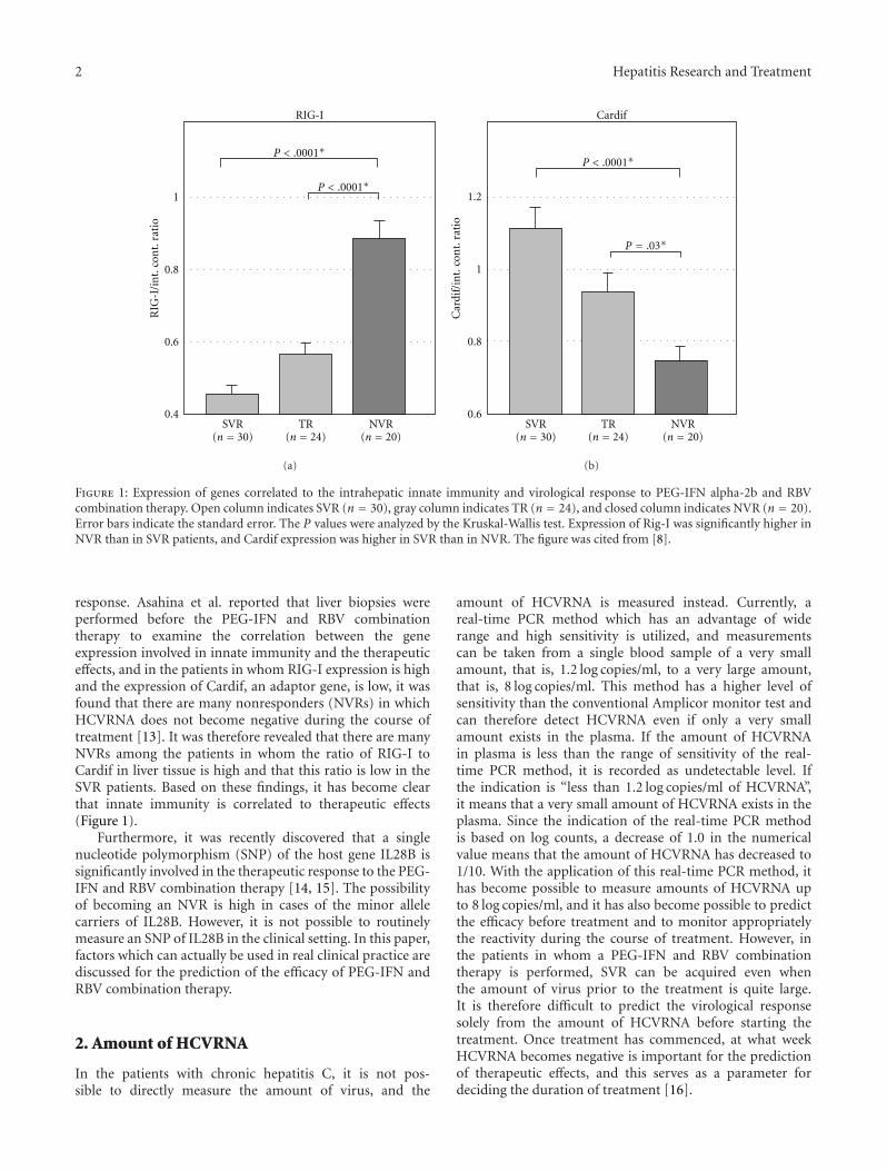
2 Hepatitis Research and Treatment
SVR(n = 30)
TR(n = 24)
NVR(n = 20)
RIG
-I/i
nt.
con
t.ra
tio
RIG-I
0.4
0.6
0.8
1
P < .0001∗
P < .0001∗
(a)
SVR(n = 30)
TR(n = 24)
NVR(n = 20)
Cardif
Car
dif/
int.
con
t.ra
tio
0.6
0.8
1
1.2
P < .0001∗
P = .03∗
(b)
Figure 1: Expression of genes correlated to the intrahepatic innate immunity and virological response to PEG-IFN alpha-2b and RBVcombination therapy. Open column indicates SVR (n = 30), gray column indicates TR (n = 24), and closed column indicates NVR (n = 20).Error bars indicate the standard error. The P values were analyzed by the Kruskal-Wallis test. Expression of Rig-I was significantly higher inNVR than in SVR patients, and Cardif expression was higher in SVR than in NVR. The figure was cited from [8].
response. Asahina et al. reported that liver biopsies wereperformed before the PEG-IFN and RBV combinationtherapy to examine the correlation between the geneexpression involved in innate immunity and the therapeuticeffects, and in the patients in whom RIG-I expression is highand the expression of Cardif, an adaptor gene, is low, it wasfound that there are many nonresponders (NVRs) in whichHCVRNA does not become negative during the course oftreatment [13]. It was therefore revealed that there are manyNVRs among the patients in whom the ratio of RIG-I toCardif in liver tissue is high and that this ratio is low in theSVR patients. Based on these findings, it has become clearthat innate immunity is correlated to therapeutic effects(Figure 1).
Furthermore, it was recently discovered that a singlenucleotide polymorphism (SNP) of the host gene IL28B issignificantly involved in the therapeutic response to the PEG-IFN and RBV combination therapy [14, 15]. The possibilityof becoming an NVR is high in cases of the minor allelecarriers of IL28B. However, it is not possible to routinelymeasure an SNP of IL28B in the clinical setting. In this paper,factors which can actually be used in real clinical practice arediscussed for the prediction of the efficacy of PEG-IFN andRBV combination therapy.
2. Amount of HCVRNA
In the patients with chronic hepatitis C, it is not pos-sible to directly measure the amount of virus, and the
amount of HCVRNA is measured instead. Currently, areal-time PCR method which has an advantage of widerange and high sensitivity is utilized, and measurementscan be taken from a single blood sample of a very smallamount, that is, 1.2 log copies/ml, to a very large amount,that is, 8 log copies/ml. This method has a higher level ofsensitivity than the conventional Amplicor monitor test andcan therefore detect HCVRNA even if only a very smallamount exists in the plasma. If the amount of HCVRNAin plasma is less than the range of sensitivity of the real-time PCR method, it is recorded as undetectable level. Ifthe indication is “less than 1.2 log copies/ml of HCVRNA”,it means that a very small amount of HCVRNA exists in theplasma. Since the indication of the real-time PCR methodis based on log counts, a decrease of 1.0 in the numericalvalue means that the amount of HCVRNA has decreased to1/10. With the application of this real-time PCR method, ithas become possible to measure amounts of HCVRNA upto 8 log copies/ml, and it has also become possible to predictthe efficacy before treatment and to monitor appropriatelythe reactivity during the course of treatment. However, inthe patients in whom a PEG-IFN and RBV combinationtherapy is performed, SVR can be acquired even whenthe amount of virus prior to the treatment is quite large.It is therefore difficult to predict the virological responsesolely from the amount of HCVRNA before starting thetreatment. Once treatment has commenced, at what weekHCVRNA becomes negative is important for the predictionof therapeutic effects, and this serves as a parameter fordeciding the duration of treatment [16].

Hepatitis Research and Treatment 3
0
10
20
30
40
50
60
70
80
90
100
Neg
ativ
era
teof
HC
V-R
NA
(%)
3 5
15
2424
5448
81
50
81
Core 70 mutant (n = 34)Core 70 wild (n = 84)
4 8 12 24 48
(w)
Figure 2: Comparison of aa70 mutations in the HCV core regionand the rate of HCVRNA becoming negative during the course oftreatment. Compared with the wild type, among the patients ofaa70 mutations, there were fewer patients in whom HCVRNA hadbecome negative during the course of treatment.
Measuring the rate of viral clearance from serum ishelpful for predicting the likelihood of a response to PGEIFNand RBV and useful for determining the optimal durationof therapy if the patients start to receive the treatment[17]. In the AASLD practice guideline, response-guidedtherapy is highly recommended [18]. In two nationwideregistration trials conducted in Japan [9, 10], the SVR ratewas high, from 76% to 100%, in patients whose HCVRNAwas cleared rapidly from serum by week 4 (rapid virologicalresponse; RVR), and 71% to 73% in patients who achievedundetectable HCVRNA from week 5 to week 12 (earlyvirological response; EVR). In contrast, the SVR rate inpatients with late clearance of HCVRNA from week 13to week 24 was low at 29% to 36%. No patients withoutclearance of HCVRNA by week 24 achieved SVR.
The strategy of extending therapy in patients withdelayed virological responses, defined as clearance of HCV-RNA between weeks 12 and 24, was evaluated in five studies[19–23]. These results cannot be compared directly witheach other because of the heterogeneous study populations,differences in the baseline characteristics, and the differentregimens utilized amongst them. Nevertheless, the resultsshowed a trend toward a higher SVR rate by extendingtherapy from 48 to 72 weeks in patients with delayedvirological response.
3. Viral Mutations in Core and NS5A Region
In the patients with genotype 1b HCV infection, the muta-tions in aa70 and aa91 in the core region have been shownto correlate with early virological response (EVR) duringPEG-IFN and RBV combination treatment [11]. If aa70 inthe core region is mutated to anything other than arginineand aa91 to anything other than leucine, it is difficult toachieve EVR, and it is thus highly possible that such cases
0
10
20
30
40
50
60
70
80
ETR SVR Relapse
ISDR 0–1 (n = 176)
Pati
ent
nu
mbe
r,n
ISDR ≧2 (n = 52)
76 72
44
55
17
P = .07
32
Figure 3: Number of ISDR substitutions and the comparison ofvirological response, SVR, and relapse at the end of the treatment.Compared with the patients with 2 or more sites of substitutions,the rate of SVR was lower and the rate of relapse was higher in thepatients in whom there were fewer substitutions in ISDR, that is, 0or 1 sites.
will become nonresponders. The examination results at ourinstitution including 292 patients with genotype 1b infectiondemonstrated that, in the cases with mutations in aa70 in thecore region, the rate of HCVRNA becoming negative duringthe course of combination treatment was low comparedto the wild type of aa70 (Figure 2). However, core aa70mutation is shown to have quasispecies detectable by cloning,and 70Q clone was positively selected during combinationtreatment with PEGIFN and RBV [24].
Furthermore, Enomoto et al. reported that the patientswith 4 or more amino acid mutations were observedin interferon sensitivity-determining region (ISDR) withinNS5A region [2]; SVR rate is higher than 90% by IFNmonotherapy, and SVR is less than 10% in the patients withno mutation in ISDR. It has also been reported that, in PEG-IFN and RBV combination therapy, the number of ISDRmutations is involved in the SVR [12].
We analyzed the relationship between virologicalresponse and ISDR mutations in the patients with genotype1b infection treated by PEG-IFN alpha-2b and BRVcombination therapy. In the patients with 0 or 1 ISDRmutation, even if the rate of HCVRNA becoming negativeat the end of treatment was the same, the rate of SVR wouldbe lower compared with the patients having 2 or moremutations (Figure 3). This demonstrates that there is ahigher incidence of relapse after the end of treatment in thepatients with 0 or 1 ISDR mutation.
Enomoto and Maekawa reported that mutations bothin NS5A-ISDR (interferon sensitivity-determining region)and core 70Q substitution are associated with no early viralresponse during PEGIFN and RBV combination therapy[25]. Association of core aa70 substitution and mutationsin NS5A region is confirmed to be associated with viralresponse by PEGIFN and RBV combination therapy in aJapanese multicenter cooperative study [26]. The number of

4 Hepatitis Research and Treatment
mutations in the interferon sensitivity-determining regionwas shown to be associated with the viral response toPEGIFN and RBV combination treatment not only in Japan[27], but also in Tunisia [28].
Recently, a consensus has been established that mutationsin aa70 in the core region are important for the predictionof HCVRNA becoming negative during the early course oftreatment, and the number of ISDR mutations is importantfor the prediction of relapse after the end of treatment.
4. Adherence
It has been confirmed that it is important to ensure 80%or more of the scheduled dose of both PEG-IFN and BBVin order to improve the rate of SVR, and together withthe duration of treatment, the 80 · 80 · 80 rule has beenestablished. However, Schiffman et al. recently reported thatthe dose of PEG-IFN in the initial stage of administrationis important and that, if a sufficient dose of PEG-IFN isadministered, then 60% or more of the RVB dose would beenough [29]. It is therefore of primary importance to ensurethe dose of PEG-IFN.
In Japan, the average age of patients with chronichepatitis C is increasing, and achieving good adherence isdifficult in many patients. Consequently, the rate of SVR islow in elderly patients. How to improve the rate of SVR inelderly patients is an important issue. With regard to thedose of RBV, reducing the RBV dose based on the calculationof the total body clearance (CL/F) has been proposed tobe useful for decreasing the discontinuation and improvingthe rate of SVR. Although there is no consensus on anappropriate dose of PEG-IFN in elderly patients, if the initialdose is set lower than the usual dose, discontinuation wouldbe reduced. Thus, it is necessary to investigate whether suchan initial dose would improve the rate of SVR.
Recently, the risk of hemolytic anemia was clearlydemonstrated to correlate with ITAP gene SNP [30]. Thepredictive implication should be analyzed prospectively inclinical practice.
5. Host Factors
Zeuzem et al. described the factors related to the lessresponse to interferon-based therapy, and he showed thatseveral host factors such as older age, race, and obesityare responsible factors for the poor response to IFN [31].Recently, insulin resistance which was examined by home-ostasis model assessment index (HOMA-IR) was shown tobe associated with a lower rate of SVR, and body mass index(BMI) was not identified as a significant factor for the poorresponse to PEGIFN and ribavirin combination therapy [32].Insulin resistance was confirmed as a related factor to thenonresponse to interferon-based treatment [33]. However,Charlton et al. reported that obesity itself is an associatedfactor for decreased efficacy of interferon-based therapies,and they discussed the possible mechanism [34], and obesitywas shown to be associated with the increased enhancement
of suppressor of cytokine signaling (SOCS) family in thehepatocytes [35].
6. Data Mining Analysis
Both virus- and host-related factors are correlated to thera-peutic effects of PEG-IFN and RBV. One important questionis which of these factors should be focused on in order topredict the therapeutic effects in an individual patient. Inaddition, in each individual patient, the host and virologicalfactors are different. It is therefore difficult to predictthe virological response in each case before treatment.Furthermore, although it is important to predict the relapserate when HCVRNA becomes within an undetectable levelin an individual patient, prediction of the rate of SVRincluding virological and host factors and adherence tothe treatment has never been carried out in an individualpatient.
A data mining analysis is the process of analyzing alarge amount of data by a computer in order to develop analgorithm. Conventional statics have been used to examinecertain hypothesis. Data mining is superior in that it can setan algorithm, using a computer, based on a large amount ofdata without a hypothesis.
We therefore conducted at our institute a data mininganalysis of the patients with genotype 1b infection havinghigh levels of HCVRNA to whom a PEG-IFN alpha-2b andRBV combination therapy was administered to investigatewhether by the 12th week after the commencement oftreatment HCVRNA became negative (EVR) (Figure 4) [36].The most important factor for the prediction of EVR wasthe steatosis of the liver: when steatosis was observed in 30%or more of hepatocytes, EVR was found to be difficult toachieve. In the patients in whom steatosis was not severe, thesecond most important factor was the serum LDL cholesterolvalue. While the rate of EVR was 57% in the patients inwhom this value was 100 mg/ml or above, the rate of EVRwas 32% in the patients in whom the LDL cholesterol wasless than 100 mg/dl.
The higher the LDL cholesterol value, the earlier theHCVRNA became negative. Among the patients with lowLDL cholesterol values, while the rate of EVR was 15% inpatients 60 years of age or older, the rate was high in thepatients under the age of 60 years old, that is, 49%. Amongpatients under the age of 60, the rate of EVR was low, that is,31%, in patients with a blood glucose level of 120 mg/dl orabove whereas EVR was achieved in 71% of the patients witha blood glucose level of less than 120 mg/dl (Figure 4).
On the other hand, in the patients with high LDLcholesterol values, the next most important factor was age.While the rate of EVR was 50% in patients 50 years of ageor older, EVR was achieved in 77% of the patients under theage of 50. Among patients of 50 years of age or older, thenext most important factor was the gamma GTP value. Whilethe rate of EVR was 35% in the patients in whom gammaGTP was 40 IU/L or above, EVR was achieved in 60% of thepatients where the value was less than 40 IU/L.
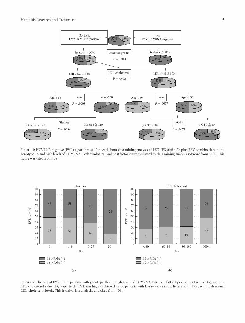
Hepatitis Research and Treatment 5
57% 43%
Steatosis < 30%
47%53% 82%18%
Steatosis grade
LDL-cholesterol
EVR12 w HCVRNA negative
No-EVR12 w HCVRNA positive
Age Age
γ-GTPGlucose
LDL-chol ≧100
68%32%
LDL-chol < 100
57%43%
Age < 60
49%51%
Age ≧60
85%15%
Glucose < 120
29%71%
Glucose ≧120
69%31%
Age < 50
77%23%
Age ≧50
50% 50%
60%40% 35%65%
P = .0014
P = .0002
P = .0008 P = .0057
P = .0084 P = .0171γ-GTP < 40 γ-GTP ≧40
Steatosis ≧30%
Figure 4: HCVRNA negative (EVR) algorithm at 12th week from data mining analysis of PEG-IFN alpha-2b plus RBV combination in thegenotype 1b and high levels of HCVRNA. Both virological and host factors were evaluated by data mining analysis software from SPSS. Thisfigure was cited from [36].
0
10
20
30
40
50
60
70
80
90
100
EV
Rra
te(%
)
0 1–9 10–29 30–
Steatosis
42
38
58
51
23
14
28
6
(%)
12 w RNA (+)12 w RNA (−)
(a)
0
10
20
30
40
50
60
70
80
90
100
EV
Rra
te(%
)
< 60 80–100 100 <
LDL-cholesterol
5
13 25
11 19
4139
35
(%)
12 w RNA (+)12 w RNA (−)
60–80
(b)
Figure 5: The rate of EVR in the patients with genotype 1b and high levels of HCVRNA, based on fatty deposition in the liver (a), and theLDL cholesterol value (b), respectively. EVR was highly achieved in the patients with less steatosis in the liver, and in those with high serumLDL-cholesterol levels. This is univariate analysis, and cited from [36].

6 Hepatitis Research and Treatment
We therefore compared these factors based on the orig-inal data. A univariate comparison of the fatty infiltrationof the liver and the rate of EVR demonstrated that therate of EVR was higher when the steatosis of the liver wasless severe (Figure 5(a)). In addition, a comparison of theLDL cholesterol value and the rate of EVR demonstrated asignificant correlation, confirming that the higher the LDLcholesterol value, the higher the rate of EVR (Figure 5(b)).Therefore, it was also proposed by the results of univariateanalysis of each factor extracted from the data mininganalysis that these factors were correlated to the rate of EVR.
From these observations, it is likely to improve the viralresponse to PEGIFN and ribavirin by reducing steatosis ofthe liver through daily walking or abstinating alcohol intakeor by refraining from high-fat diet.
By utilizing data mining, it is therefore possible to assessvirus- and host-related factors together and to predict thevirological response in each patient, and thereby clinicallyuseful information can be obtained. The algorithm shouldbe validated including a large number of the patients.
Acknowledgment
This paper was supported by a grant from the JapaneseMinistry of Health, Welfare and Labor.
References
[1] Y. Asahina, K. Tsuchiya, I. Hirayama, et al., “Effect of aging onrisk for hepatocellular carcinoma in chronic hepatitis C virusinfection,” Hepatology, vol. 52, no. 2, pp. 518–527, 2010.
[2] N. Enomoto, I. Sakuma, Y. Asahina et al., “Mutations in thenonstructural protein 5A gene and response to interferon inpatients with chronic hepatitis C virus 1b infection,” NewEngland Journal of Medicine, vol. 334, no. 2, pp. 77–81, 1996.
[3] S. Iino, F. Ichida, A. Sakuma et al., “A randomized clinical trialwith natural interferon-α monotherapy for 24 or 48 weeks onpatients with chronic hepatitis C having genotype 1b infectionin high viral titers,” Hepatology Research, vol. 24, no. 4, pp.338–345, 2002.
[4] G. Squadrito, M. E. Orlando, I. Cacciola et al., “Long-term response to interferon alpha is unrelated to “interferonsensitivity determining region” variability in patients withchronic hepatitis C virus-1b infection,” Journal of Hepatology,vol. 30, no. 6, pp. 1023–1027, 1999.
[5] C. Shen, T. Hu, L. Shen, L. Gao, W. Xie, and J. Zhang,“Mutations in ISDR of NS5A gene influence interferon efficacyin Chinese patients with chronic hepatitis C virus genotype 1binfection,” Journal of Gastroenterology and Hepatology, vol. 22,no. 11, pp. 1898–1903, 2007.
[6] M. Pascu, P. Martus, M. Hohne et al., “Sustained virologicalresponse in hepatitis C virus type 1b infected patients ispredicted by the number of mutations within the NS5A-ISDR:a meta-analysis focused on geographical differences,” Gut, vol.53, no. 9, pp. 1345–1351, 2004.
[7] S. J. Hadziyannis, H. Sette Jr., T. R. Morgan et al.,“Peginterferon-α2a and ribavirin combination therapy inchronic hepatitis C: a randomized study of treatment durationand ribavirin dose,” Annals of Internal Medicine, vol. 140, no.5, pp. 346–355, 2004.
[8] M. P. Manns, J. G. McHutchison, S. C. Gordon et al., “Pegin-terferon alfa-2b plus ribavirin compared with interferonalfa-2b plus ribavirin for initial treatment of chronic hepatitis C:a randomised trial,” Lancet, vol. 358, no. 9286, pp. 958–965,2001.
[9] S. Iino, E. Tomita, H. Kumada et al., “Prediction of treatmentoutcome with daily high-dose IFN α-2b plus ribavirin inpatients with chronic hepatitis C with genotype 1b and highHCV RNA levels: relationship of baseline viral levels and viraldynamics during and after therapy,” Hepatology Research, vol.30, no. 2, pp. 63–70, 2004.
[10] G. Yamada, S. Iino, T. Okuno et al., “Virological response inpatients with hepatitis C virus genotype 1b and a high viralload: impact of peginterferon-α-2a plus ribavirin dose reduc-tions and host-related factors,” Clinical Drug Investigation, vol.28, no. 1, pp. 9–16, 2008.
[11] N. Akuta, F. Suzuki, Y. Kawamura et al., “Predictive factors ofearly and sustained responses to peginterferon plus ribavirincombination therapy in Japanese patients infected with hep-atitis C virus genotype 1b: amino acid substitutions in the coreregion and low-density lipoprotein cholesterol levels,” Journalof Hepatology, vol. 46, no. 3, pp. 403–410, 2007.
[12] H. Shirakawa, A. Matsumoto, S. Joshita et al., “Pretreatmentprediction of virological response to peginterferon plus rib-avirin therapy in chronic hepatitis C patients using viral andhost factors,” Hepatology, vol. 48, no. 6, pp. 1753–1760, 2008.
[13] Y. Asahina, N. Izumi, I. Hirayama et al., “Potential relevanceof cytoplasmic viral sensors and related regulators involvinginnate immunity in antiviral response,” Gastroenterology, vol.134, no. 5, pp. 1396–1405, 2008.
[14] D. Ge, J. Fellay, A. J. Thompson et al., “Genetic variation inIL28B predicts hepatitis C treatment-induced viral clearance,”Nature, vol. 461, no. 7262, pp. 399–401, 2009.
[15] Y. Tanaka, N. Nishida, M. Sugiyama et al., “Genome-wideassociation of IL28B with response to pegylated interferon-αand ribavirin therapy for chronic hepatitis C,” Nature Genetics,vol. 41, no. 10, pp. 1105–1109, 2009.
[16] P. Ferenci, H. Laferl, T. Scherzer et al., “Peginterferon alfa-2aand ribavirin for 24 weeks in hepatitis C type 1 and 4 patientswith rapid virological response,” Gastroenterology, vol. 135, no.2, pp. 451–458, 2008.
[17] N. Izumi, S. Nishiguchi, K. Hino et al., “Management ofhepatitis C; Report of the Consensus Meeting at the 45thAnnual Meeting of the Japan Society of Hepatology (2009),”Hepatology Research, vol. 40, no. 4, pp. 347–368, 2010.
[18] M. G. Ghany, D. B. Strader, D. L. Thomas, and L. B. Seeff,“Diagnosis, management, and treatment of hepatitis C: anupdate,” Hepatology, vol. 49, no. 4, pp. 1335–1374, 2009.
[19] T. Berg, M. von Wagner, S. Nasser et al., “Extended treatmentduration for hepatitis C virus type 1: comparing 48 versus 72weeks of peginterferon-alfa-2a plus ribavirin,” Gastroenterol-ogy, vol. 130, no. 4, pp. 1086–1097, 2006.
[20] J. M. Sanchez-Tapias, M. Diago, P. Escartın et al.,“Peginterferon-alfa2a plus ribavirin for 48 versus 72 weeksin patients with detectable hepatitis C virus RNA at week 4of treatment,” Gastroenterology, vol. 131, no. 2, pp. 451–460,2006.
[21] P. Ferenci, H. Laferl, T. M. Scherzer, et al., “Customizingtreatment with peginterferon alfa-2a (40kD)(PEGASYS�)plus ribavirin (COPEGUS�) in patient with HCV genotype1 or 4 infection: interim results of a prospective randomizedtrial,” Hepatology, vol. 44, no. 336a, 2006.
[22] B. L. Pearlman, C. Ehleben, and S. Saifee, “Treatment exten-sion to 72 weeks of peginterferon and ribavirin in hepatitis C

Hepatitis Research and Treatment 7
genotype 1-infected slow responders,” Hepatology, vol. 46, no.6, pp. 1688–1694, 2007.
[23] M. Buti, Y. Lurie, N. G. Zakharova, et al., “Extended treatmentduration in chronic hepatitis C genotype 1-infected slowresponders: final results of the SUCCESS study,” Journal ofHepatology, vol. 50, supplement 1, p. S58, abstract 141, 2009.
[24] F. Kurbanov, Y. Tanaka, K. Matsuura et al., “Positive selectionof core 70Q variant genotype 1b hepatitis C virus strainsinduced by pegylated interferon and ribavirin,” Journal ofInfectious Diseases, vol. 201, no. 11, pp. 1663–1671, 2010.
[25] N. Enomoto and S. Maekawa, “HCV genetic elements deter-mining the early response to peginterferon and ribavirintherapy,” Intervirology, vol. 53, no. 1, pp. 66–69, 2010.
[26] T. Okanoue, Y. Itoh, H. Hashimoto et al., “Predictive valuesof amino acid sequences of the core and NS5A regions inantiviral therapy for hepatitis C: a Japanese multi-centerstudy,” Journal of Gastroenterology, vol. 44, no. 9, pp. 952–963,2009.
[27] M. Nakagawa, N. Sakamoto, M. Ueyama et al., “Mutations inthe interferon sensitivity determining region and virologicalresponse to combination therapy with pegylated-interferonalpha 2b plus ribavirin in patients with chronic hepatitis C-1b infection,” Journal of Gastroenterology, vol. 45, no. 6, pp.656–665, 2010.
[28] N. Bouzgarrou, E. Hassen, W. Mahfoudh et al., “NS5AISDR-V3 region genetic variability of Tunisian HCV-1b strains:correlation with the response to the combined inter-feron/ribavirin therapy,” Journal of Medical Virology, vol. 81,no. 12, pp. 2021–2028, 2009.
[29] M. L. Shiffman, M. G. Ghany, T. R. Morgan et al., “Impactof reducing peginterferon alfa-2a and ribavirin dose duringretreatment in patients with chronic hepatitis C,” Gastroen-terology, vol. 132, no. 1, pp. 103–112, 2007.
[30] J. Fellay, A. J. Thompson, D. Ge et al., “ITPA gene variants pro-tect against anaemia in patients treated for chronic hepatitisC,” Nature, vol. 464, no. 7287, pp. 405–408, 2010.
[31] S. Zeuzem, “Heterogeneous virologic response rates tointerferon-based therapy in patients with chronic hepatitis C:who responds less well?” Annals of Internal Medicine, vol. 140,no. 5, pp. 370–381, 2004.
[32] H. S. Conjeevaram, D. E. Kleiner, J. E. Everhart et al., “Race,insulin resistance and hepatic steatosis in chronic hepatitis C,”Hepatology, vol. 45, no. 1, pp. 80–87, 2007.
[33] G. Tarantino, P. Conca, P. Sorrentino, and M. Ariello,“Metabolic factors involved in the therapeutic response ofpatients with hepatitis C virus-related chronic hepatitis,”Journal of Gastroenterology and Hepatology, vol. 21, no. 8, pp.1266–1268, 2006.
[34] M. R. Charlton, P. J. Pockros, and S. A. Harrison, “Impact ofobesity on treatment of chronic hepatitis C,” Hepatology, vol.43, no. 6, pp. 1177–1186, 2006.
[35] M. J. Walsh, J. R. Jonsson, M. M. Richardson et al., “Non-response to antiviral therapy is associated with obesity andincreased hepatic expression of suppressor of cytokine sig-naling 3 (SOCS-3) in patients with chronic hepatitis C, viralgenotype 1,” Gut, vol. 55, no. 4, pp. 529–535, 2006.
[36] M. Kurosaki, K. Matsunaga, I. Hirayama, et al., “A predictivemodel of response to peginterferon ribavirin in chronichepatitis C using classification and regression tree analysis,”Hepatology Research, vol. 40, no. 3, pp. 251–260, 2010.

Hindawi Publishing CorporationHepatitis Research and TreatmentVolume 2010, Article ID 429243, 10 pagesdoi:10.1155/2010/429243
Clinical Study
Tumor Necrosis Factor Receptor 1 ExpressionIs Upregulated in Dendritic Cells in Patients withChronic HCV Who Respond to Therapy
Raul Cubillas,1 Katherine Kintner,1 Frances Phillips,2 Nitin J. Karandikar,1
Dwain L. Thiele,1 and Geri R. Brown1, 2
1 Division of Digestive and Liver Diseases, Department of Internal Medicine, Southwestern Medical Center at Dallas,University of Texas at Dallas, Dallas, TX 75390-9151, USA
2 Dallas Veterans Affairs Medical Center, 4500 S. Lancaster Road, Dallas, Texas 75216, USA
Correspondence should be addressed to Geri R. Brown, [email protected]
Received 25 March 2010; Revised 28 May 2010; Accepted 31 May 2010
Academic Editor: Tatehiro Kagawa
Copyright © 2010 Raul Cubillas et al. This is an open access article distributed under the Creative Commons Attribution License,which permits unrestricted use, distribution, and reproduction in any medium, provided the original work is properly cited.
The present studies assessed the level of tumor necrosis factor receptor (TNFR) expression in peripheral blood mononuclearcells (PBMCs) subsets from patients with chronic HCV undergoing interferon α/ribavirin-based therapy (Ifn/R). Methods. TNFRfamily member mRNA expression was determined using quantitative real-time PCR assays (RTPCRs) in PBMC from 39 HCV+patients and 21 control HCV− patients. Further subset analysis of HCV + patients (untreated (U), sustained virological responders(SVR), and nonresponders (NR)/relapsers (Rel)) PBMC was performed via staining with anti-CD123, anti-CD33, anti-TNFR1 orvia RTPCR for TNFR1 mRNA. Results. A similar level of TNFR1 mRNA in PBMC from untreated HCV+ genotype 1 patients andcontrols was noted. TNFR1 and TNFR2 mRNA levels in PBMC from HCV+ patients with SVR were statistically different thanlevels in HCV(−) patients. A significant difference was noted between the peak values of TNFR1 of the CD123+ PBMC isolatedfrom SVR and the NR/Rel. Conclusion. Upregulation of TNFR1 expression, occurring in a specific subset of CD123+ dendriticcells, appeared in HCV+ patients with SVR.
1. Introduction
Unlike infections by other human hepatitis viruses, hepatitisC virus (HCV) infection of healthy immunocompetentadults is persistent or prolonged in the majority of cases,unlike hepatitis B, where it persist in the minority ofcases. HCV appears to have evolved strategies to evadethe human immune responses. Investigators have suggestedthat viral interactions with tumor necrosis factor/tumornecrosis factor receptor (TNF/TNFR) family members maybe important in immune evasion. Investigators have reportedthat HCV core protein binds to the cytoplasmic domainof TNFR1, specifically the “death domain,” which mayhinder or promote cell death during HCV infection [1]. Inaddition, the HCV core protein binds to another TNFR fam-ily member, lymphotoxin β receptor’s (LTβR) intracellular
domain, which is involved in signal transduction [2]. Inves-tigators have also demonstrated that the HCV core proteinpotentiates NF6B activation initiated by TNFα/TNFR andLTα1β2/LTβR interactions, which, in turn may contribute tothe chronically activated, persistent state of HCV-infectedcells [3, 4]. Interference with signaling through membersof the TNF family of receptors may affect the chronicity ofHCV via affecting either cell death and/or activation of NFκB[1, 3, 4].
Of interest, other investigators have examined the rela-tionship between HCV and the soluble TNF receptors(sTNFR) in the blood. These sTNFR levels appear to peak9 hours after the first IFNα administration correlating withIFNα serum levels [5]. Other investigators have noted higherlevels of TNF and sTNFRs in HCV+ patients than in HCV(−) controls prior to treatment and postulated that high

2 Hepatitis Research and Treatment
levels of sTNFR might modify host responses but found nocorrelation between levels and response to therapy [6].
TNF receptors are present on the surface of a numberof cell subsets and play a role in a variety of functions.For example, TNF/TNFR interactions are imperative for theoptimal proliferation and effector functions of CD8 + T cells[7, 8], whose antiviral effects are essential for the clearanceof noncytopathic viruses, such as HCV. In addition, LTβR-LIGHT/LTα1β2 interactions have been implicated in bothoptimal growth and effector functions as well as costim-ulation of CD8+ T cells [9]. Similarly, TNFR is expressedon macrophages, peripheral blood monocytes, and antigenpresenting cells (APCs), cells known to be important in viralinfections. Recently, HCV negative strand RNA has beennoted in the macrophages of 67% of sustained virologicalresponders to interferon-based therapies [10]. Others havenoted the importance of monocytes and dendritic cells in theclearance of HCV [11–14]. The role of TNFR in HCV viralclearance may involve any of these cell subsets.
The largely chronic nature of HCV infection has beenattributed to an attenuated antiviral T-cell response. Ithas been postulated that APC’s may become dysfunctionalin some way during HCV infection contributing to thisattenuation. Specifically, large deficits in IFNα productionin plasmacytoid dendritic cells (PDCs) in HCV-infectedpatients have been reported in [15]. In the same studyHCV-infected PDC displayed distinct immunophenotypicfeatures including an increased ability to stimulate a mixedlymphocyte response (MLR) but lower HLA-DR and CD86expression. This profile suggested that HCV-infected PDCwere at an immature stage of differentiation [15].
The present studies were designed to quantify the mRNAand protein levels of the TNF receptor family members,including TNFR1 and TNFR2, in PBMCs during IFNα-basedtherapy of HCV+ patients. The results of these studies revealincreased peak levels of TNFR1 mRNA levels in respondersto IFN-based therapies. Furthermore, by flow cytometricstudies and western blot analysis, upregulation of TNFR1protein expression was noted in specific PBMC subsets. Theincrease in TNFR1 expression was specifically isolated inPDC (CD11− and CD123high) of HCV-infected patients whoresponded to IFNα based therapies compared to controlpatients and nonresponders.
2. Methods
2.1. Patients. Veteran patients >18 years of age with chronicHCV were recruited to IFNα based treatment trials con-ducted at the Dallas Veterans Affairs Medical Center fromFebruary 2002 through July 2005. Thirty-seven patients withgenotype 1 consented per institutional review board (IRB)guidelines for blood draws at one or more time points,including 0, 4, 8, 12, 24, and 48 weeks of therapy, wherethe standard therapy (48 weeks of weight-based dosing of1.5μg/kg of pegylated interferon α-2b and 13.5 mg/kg of(ribavirin) for genotype 1 patients was utilized. Thirteenpatients were sustained virological responders (SVR, HCVRNA negative 24 weeks after end of therapy), 20 patients
were nonresponders (NR, persistently HCV RNA positiveduring therapy), and 4 patients were relapsers (Rel, HCVRNA negative during therapy but HCV RNA positiveafter end of therapy). None of the patients analyzed hadreceived interferon prior to the initial blood draw. Similarly,10 HCV(−) patients and 11 patients with alcoholic liverdisease (ALD) or nonalcoholic fatty liver disease (NAFLD)consented per IRB guidelines for a blood draw.
In a second set of patients, samples from 13 genotype1 HCV+ patients (5 SVR, 6 NR/Rel and 2 HCV+ prior totherapy) and 2 HCV(−) control patients obtained duringtherapy at <12, 16–24, or 25–44 weeks were collected for aspecialized cell subset analysis.
2.2. RNA Isolation and Real-Time PCR Primers. RNA wasisolated from PBMC’s at 0, 4, 8, 12, 24, and 48 weeks ofinterferon/ribavirin (Ifn/R) treatment trials from 39 HCV+men, 10 age- and sex- matched HCV(−) controls, and 11age- and sex- matched controls with either alcoholic liverdisease (ALD) or nonalcoholic fatty liver disease (NAFLD).Initially, 40–60 cc of blood was obtained from HCV+patients and controls. PBMCs were then separated by densitygradient centrifugation and then stored in TRIzol reagentto stabilize the RNA. RNA was then extracted, precipitated,and subjected to DNase treatment, and then the treated RNAunderwent reverse transcription.
In order to assess whether there were differences inany specific cytokine receptors or TNF receptor familymembers, 101 cDNA samples were utilized in a quantitativereal time PCR-based assay assessing the relative quantitiesof cyclophilin, NGFR, CD30, TNFR1, TNFR2, LTβR, IL-2Rα, CD27, FAS, and CD3. RNA from PDC (CD11− andCD123high) and myeloid dendritic cells (MDC) (CD11+ andCD123dim) from a control, SVR, and NR were isolated in anidentical fashion and utilized in a quantitative PCR assay forTNFR1.
The primers utilized were obtained as follows. The NCBIdatabase was first interrogated to find the sequence of thecytokine receptor or other receptors of interest and thenentered into the Primer Express software program in orderto generate a primer which was then optimized according tothe parameters of the real time PCR (Table 1). The thresholdcycle or Ct value, the lowest cycle in which fluorescence wasdetected during the exponential phase, was calculated wherethe exponential phase of the reaction began.
2.3. Standardization and Comparison of Samples. The Ct
values were standardized using RNA isolated from cell linesknown to express high levels of the target genes. In order tonormalize across experiments, these values were comparedto the same standard for all experiments, that is RNA fromcell lines expressing high levels of TNFR or LTβR (PHAactivated lymphocytes and THP-1, resp.). The resulting cyclethreshold values were normalized to endogenous cyclophillinvalues and a calibrator value for each experiment. Raw cyclethreshold values for the no template control within eachexperiment were significantly different than the experimentalvalues for cyclophillin, CD3ε, NGFR, CD30, TNFR1, TNFR2,

Hepatitis Research and Treatment 3
Table 1: Real-Time PCR Primers.
Target Primer Sequence
Cyclophillin Fw 5′-TGCCATCGCCAAGGAGTAG-3′
Rv 5′-TGCACAGACGGTCACTCAAA- 3′
TNFR1 Fw 5′-CGCTACCAACGGTGGAAGTC- 3′
Rv 5′ -CAAGCTCCCCCTCTTTTTCAG-3′
LTβR Fw 5′-CGGGCCCCTCTAAAGGATT-3′
Rv 5′-GTGAAGTGTGGAACCCCAAAG-3′
TNFR2 Fw 5′-CAAGCCAGCTCCACAATGG-3′
Rv 5′-TGACCGAAAGGCACATTCCT-3′
NGFR Fw 5′-CCGTGGAGATGGGATGCTT-3′
Rv 5′-TTTCCACGAACCCCAAACC-3′
CD30R Fw 5′-GCTTTACTCTGGACCATAGGAAACA-3′
Rv 5′-CTCCTTAGCGTGAAATGTGAAAAA-3′
IL2Rα Fw 5′-CAGAAGTCATGAAGCCCAAGTG-3′
Rv 5′-GGCAAGCACAACGGATGTCT-3′
CD27 Fw 5′-TCGGCACTGTAACTCTGGTCTTC-3′
Rv 5′-CGACAGGCACACTCAGCATT-3′
FAS Fw 5′-CTTTTCGTGAGCTCGTCTCTGA-3′
Rv 5′-CTCCCCAGAAGCGTCTTTGA-3′
CD3 Fw 5′-CATCCCAAAGTATTCCATCTACTTTTC-3′
Rv 5′-CCCAGTCCATCCCCAGAGA-3′
LTβR, FAS, IL-2Rα, and CD27, verifying the presence ofcDNA for these receptors.
The comparison between control HCV(−) patients andSVR, NR, and Rel were expressed as fold elevation abovecontrol. The control population is defined as both HCV(−)patients with ALD or NAFLD (n = 9) and HCV(−) controlsubjects (n = 10).
2.4. Western Blot Analysis. Whole PBMC’s from one SVRwith a high level of TNFR1 mRNA by real time PCR wereanalyzed by Western blot in order to determine proteinlevels of TNFR1. Whole PBMC’s were freeze-thawed twice,suspended in PBS, and assayed for protein, and 20 μg fromone SVR and one control as well as 200 ng of recombinanthuman soluble TNF receptor 1 (R&D Systems, Minneapo-lis/St. Paul, MN) were prepared for sodium dodecyl sulfate-polyacrylamide gel electrophoresis (SDS-PAGE) in the pres-ence of 2-mercaptoethanol, resolved on a 10% acrylamidegel, and transferred to a nitrocellulose membrane. Afterblocking in 5% milk in TBS (0.1 mol/L NaCl and 0.4 mmol/LTris, pH 7.4), containing 0.05% sodium azide, the blotwas exposed to human soluble TNF receptor 1 polyclonaldetection antibody (R&D Systems). Bound antibody wasallowed to react with alkaline phosphatase-conjugated goatantimouse IgG (Sigma Chemical Co., St. Louis, MO) [15].
2.5. Flow Cytometry. At 44 weeks of therapy, whole PBMCsfrom a SVR and an NR were isolated from 40–60 ccwhole blood by density gradient separation. In anotherexperiment, PBMCs from an SVR at 48 weeks, NR at24 weeks, and control patient were isolated as previouslydescribed for subset analysis. The PBMC’s from all patients
were suspended in staining buffer (0.5 L HBSS, 0.5 L PBS,2% BSA, 0.02% sodium azide). Cells were labeled withthe FITC-labeled anti-TNFR1 Ab (FAB225F, R&D Systems)or control FITC IgG16 (555748l, BD Pharmingen). Thesamples designated for cell subset analysis were then stainedwith CD33 (555748l, BD Pharmingen), CD14 (555748l,BD Pharmingen), CD11 (555748l, BD Pharmingen), CD123(555748l, BD Pharmingen), or relative isotype controls. AftermAb staining and washing, all samples were fixed in PBScontaining 1% paraformaldehyde at room temperature for10 minutes and stored at 4◦C until flow cytometric analysisas previously described in [16, 17]. Flow cytometric analysiswas performed on an FACscan (Becton Dickinson) andused to determine the mean fluorescence intensity (MFI)of TNFR1 on all labeled cells. TNFR1 MFI ratios (TNFR1MFI/isotype control MFI) were determined.
RNA from FAC sorted plamacytoid dendritic cells(PDCs) (CD11− and CD123high) and myeloid dendritic cells(MDCs) (CD11+ and CD123dim from a control, SVRs andNR were utilized in a real time PCR assay for TNFR1.
2.6. Statistical Analysis. Descriptive data were reported as amean for all standard deviation (SD) and median and rangein Figures 1 and 2. All results were analyzed with an intentionto treat analysis. Characteristics of treatment groups werecompared using student’s t-test. All results were analyzedusing an exact Fisher test. A “P” value of less than .05 wasconsidered to be significant.
3. Results
3.1. PBMC Tumor Necrosis Factor Receptor (TNFR) mRNALevels from Responders to IFN Therapy Increased while OtherTNFR Family Members mRNA Levels Remained Stable. RNAfrom PBMC of 23 HCV+ patients was isolated prior tothe commencement of IFNα based therapy. Similarly, RNAfrom PBMC’s of 10 HCV(−) controls and 11 patients withALD or NAFLD were also isolated. During the course of48 weeks of IFNα based therapy, 80 RNA samples wereisolated from PBMC’s from 13 HCV+ responder [(SVR andend of treatment responders (ETR)] patients and from24 NR patients. Quantitative real time PCR levels of TNFR1,TNFR2, LTβR, FASR, CD30, CD27, and NGF, as well as CD3εand IL-2Rα mRNA were assessed in available samples at 8,12, 24, and/or 48 weeks of therapy (Table 2). In order tonormalize across experiments, these values were normalizedto the same standard for all experiments, that is, RNA fromcell lines expressing high levels of TNFR (PHA-activatedlymphocytes).
As noted, TNFR1 mRNA levels of PBMC from HCV+responder patients were significantly higher than levels fromcontrol age- and sex- matched HCV(−) control patientsduring the course of the IFNα based therapy (n = 26,P = .033), while TNFR1 mRNA levels of PBMC’s from HCVNR patients were not significantly different from controls(n = 38, P = .20). Similarly, during the course of IFNαbased therapy, TNFR2 mRNA levels of PBMC from HCV+responder patients were significantly higher than levels in

4 Hepatitis Research and Treatment
Table 2: TNFR1 Family Members’ mRNA Levels.
ControlHCV neg Responders∗ NR/Rel
Prior to Tx On Tx Prior to Tx On Tx
TNFR1
0.83± 0.9(n = 10)
1.2± 1.53(n = 11)∗∗
1.94 for all2.43 (n = 7)
7.09 for all8.82 (n = 26) (P = .033)∗∗
2.83 for all2.87 (n = 14)
2.31 for all3.53 (n = 38) (P = .20)∗∗∗
TNFR2
2.10± 2.93(n = 10)
2.03± 1.34(n = 5)∗∗
1.36 for all1.22 (n = 7)
5.93±7.61 (n = 22) (P = .045)∗∗2.30 for all
2.67 (n = 12)2.57 for all
4.60 (n = 33) (P = .41)∗∗
LTβR
1.67± 1.01(n = 10)
2.17± 2.66(n = 10)∗∗
1.21 for all1.24 (n = 7)
1.49± 1.64 (n = 22) (P = .75)∗∗1.35 for all
1.35 (n = 12)2.47 for all
5.41 (n = 34) (P = .66)∗∗
FASR 0.38 for all 0.54(n = 3)
0.001 (n = 1)0.14 for all
0.40 (n = 18) (P = .35)∗∗0.01 for all
0.01 (n = 6)0.03 for all
0.07 (n = 21) (P = .003)∗∗
CD30 0.37 for all0.41 (n = 6)
0.175 for all0.06 (n = 2)
0.61 for all1.22 (n = 21) (P = .65)∗∗
0.06 for all0.07 (n = 5)
0.61 for all1.51 (n = 20) (P = .71)∗∗
CD27 0.86 for all0.49 (n = 6)
0.77 for all0.37 (n = 2)
1.08 for all0.65 (n = 21) (P = .45)∗∗
1.10 for all0.62 (n = 5)
0.71 for all0.62 (n = 20) (P = .58)∗∗
NGF 0.75 for all1.22 (n = 6)
0.085 for all0.007 (n = 2)
1.85 for all4.80 (n = 22) (P = .59)∗∗
0.11 for all0.19 (n = 6)
1.37 for all2.86 (n = 18) (P = .62)∗∗
IL-2R for all 0.22 for all0.39 (n = 6)
0.175 for all0.18 (n = 2)
0.45 for all0.63 (n = 21) (P = .41)∗∗
0.10 for all0.22 (n = 6)
0.05 for all0.10 (n = 19) (P = .09)∗∗
CD3 1.43 for all0.71 (n = 3)
1.3 (n = 1)4.71 for all
12.62 (n = 17) (P = .67)∗∗1.67 for all
1.07 (n = 5)6.93 for all
21.06 (n = 18) (P = .66)∗∗
∗Include End of treatment responders (ETR) and SVR.∗∗Mean value for control liver disease patients, HCV negative.∗∗∗P-value compared to control (HCV negative, age and sexed matched).
the control age- and sex- matched HCV(−) patients (n =22, P = .045) while no difference was noted relative tolevels in HCV NR and controls (n = 33, P = .41).No other significant increases were observed in the otherTNFR family members or T-cell-associated mRNA (CD3εor IL-2Rα) levels of PBMC isolated from responders or NR(Table 2). In summary, PBMC’s mRNA levels of TNFR1and TNFR2 from responders to IFNα based therapy werestatistically different than levels in control HCV(−) age- andsex- matched patients.
3.2. TNFR1 and TNFR2 mRNA Levels from HCV+ PatientsPrior to Treatment and Control Patients Were Similar. Inlight of differences between levels of TNFR1 and TNFR2mRNA in PBMC of responders and nonresponders duringthe therapy, further analysis of the TNFR1 and TNFR2 levelswas performed. RNA was isolated from PBMCs of 23 HCV+untreated patients, 10 control HCV(−) patients, and 11patients with ALD or NAFLD. TNFR1 and TNFR2 mRNAlevels were similar in both HCV infected and control groups[1.02 ± 1.25 (n = 21) versus 2.53 for all 2.71 (n = 23) and1.95 for all 2.42 (n = 15) versus 2.07 for all 2.45 (n = 18)](Figure 1, panels A and B). Quantitative real time PCRrevealed no difference between initial levels of TNFR1 mRNAin the HCV+ genotype 1 patients and control non-HCV+patients (P = .08) nor between HCV+ genotype 1 patients
and patients with non-HCV liver disease (P = .17). Similarresults between initial TNFR1 RNA levels in control (HCVnegative and non-HCV liver disease) and initial levels inHCV+ patients were noted [median 0.59 (Range: 0.02–5.42)]versus [median 1.52 (Range: 0.06–11.65)] (Figure 1, panelA). TNFR2 mRNA levels were not significantly differentbetween HCV+ patients prior to therapy and either HCV(−)control patients or HCV(−) patients with liver disease (P =.88 and .95), (Figure 1, panel B).
3.3. Peak TNFR1 mRNA Levels Were Significantly Differentbetween SVR and Nonresponders. In order to determinewhether the elevation of TNFR1 or TNFR2 mRNA wasabove control levels at any point during IFNα based therapy(4, 8, 12, 24 and 48 weeks), 12 genotype 1 HCV+ SVRand 25 genotype 1 HCV+ patients NR/Rel were evaluated.TNFR1 and TNFR2 mRNA elevations were significantlymore frequent in SVR versus NR/Rel.
The peak TNFR1 mRNA levels in PBMC of genotype1 HCV+ patients at 4, 8, 12, 24, or 48 weeks were greater than2 SD (2.63 fold elevation) above mean TNFR1 mRNA levelsfrom HCV(−) controls in 7/12 (58%) sustained virologicalresponders (SVR), but in only 5/21 (28%) genotype 1 NRand 2/4 (50%) genotype 1 Rel. Furthermore, the peak TNFR1mRNA levels from responders were significantly higher thanthe peak TNFR1 mRNA levels in NR and Rel (P = .0023)

Hepatitis Research and Treatment 5
0
2
4
6
8
10
12
14
TN
FR1
mR
NA
Normal HCV−/alcohol/NAFL controls
HCV+prior to Txgenotype 1
Control and untreated HCV+
∗P = .08∗∗P = .17
HCV+Normal HCV−Liver disease HCV−
∗P value to normal HCV−∗∗P value to liver disease HCV−
n = 21 n = 23
(a)
0
2
4
6
8
10
12
TN
FR1
mR
NA
Normal HCV−/alcohol/NAFL controls
HCV+prior to Txgenotype 1
Control and untreated HCV+
∗P = .88∗∗P = .95
HCV+Normal HCV−Liver disease HCV−
∗P value to normal HCV−∗∗P value to liver disease HCV−
n = 15 n = 18
(b)
Figure 1: TNFR1 and TNFR2 mRNA levels were not different between HCV(−)/ALD/NAFLD control patients and genotype 1 HCV+patients prior to treatment. TNFR1 mRNA levels of PBMC’s isolated from 10 HCV(−) controls, 11 ALD/NAFLD controls, and 23 HCV+patients prior to therapy were ascertained by real time PCR (Panel A). TNFR2 mRNA levels of PBMC’s isolated from 10 HCV(−) controls,5 ALD/NAFLD controls, and 18 HCV+ patients prior to therapy were ascertained by real time PCR (Panel B). Statistical differences wereassessed by T- test.
[SVR: Median 7.37 (Range: 0.29–35.92) versus NR/Rel:Median 1.17 (Range: 0.08–15.91)] (Figure 2(a)).
The peak TNFR2 mRNA levels expressed in PBMC fromgenotype 1 HCV+ patients at 4, 8, 12, 24 or 48 weekswere greater than 2 SD (8.6 fold elevation) above meancontrol TNFR2 mRNA levels from HCV(−) patients in 3/12(25%) SVR versus 3/20 (15%) genotype 1 NR and 0/4 (0%)genotype 1 Rel. The peak TNFR2 mRNA levels from SVRwere not significantly higher than the peak TNFR2 mRNAlevels from NR and Rel (P = .085) (Figure 2(b)). Similarresults of peak TNFR levels in responders and nonresponderswere noted [median/range 6.1 (0.71–32.01) versus 2.98(0.01–18.57) in Figure 2(b)]. Therefore, unlike peak PBMCTNFR1 mRNA levels, peak PBMC TNFR2 mRNA levelsduring IFN for all therapy were not significantly differentbetween SVR and nonresponders.
3.4. TNFR1 Expression by HCV+ SVR Patient’s PBMCsWas Demonstrated by Western Blot Analysis. In order toassess whether TNFR1 mRNA levels directly correlated withdifferential expression of TNFR1 protein in a SVR or controlPBMCs, whole PBMC from a HCV+ SVR and a control
HCV(−) patient were lysed, assayed for protein content, and20μg of protein per sample was loaded on a SDS PAGEgel. Detection of the protein of −55 kDa molecular weightby anti-TNFR1 was observed in the PBMC of an HCV+responder patient but was not observed in the HCV(−)control PBMC lysate (Figure 3).
3.5. Increased TNFR1 Membrane Expression by SpecificPeripheral Blood Monocyte Subsets from SVR but Not NR WasObserved. The surface expression of TNFR1 by PBMC inHCV+ patients undergoing IFN/R has not been reported.However, soluble TNFR1 has been described to be elevatedin patients with chronic HCV [6]. Because of the differencesnoted in the TNFR1 mRNA levels between NR and the SVR,flow cytometric analysis of TNFR1 expression by PBMCfrom a SVR and a NR was assessed. Whole PBMCs fromNR and SVR were isolated and stained with an FITC-conjugated anti-TNFR1 Ab as described in the methods.The PBMC of the HCV+ SVR patient displayed 52% TNFR1positive PBMC with mean fluorescence intensity (MFI) of80. The non-responding patient displayed 0.0% TNFR1positive PBMC’s, with MFI of 0. As demonstrated by the flow

6 Hepatitis Research and Treatment
0
5
10
15
20
25
30
35
40T
NFR
1m
RN
A
Responders Non-responders/relapsers
Panel APeak values during therapy genotype 1
∗n = 25∗∗P = .0023
ResponderNon-responderRelapsers
n = 12
(a)
0
5
10
15
20
25
30
35
40
TN
FR2
mR
NA
Responders Non-responders/relapsers
Panel BPeak values during therapy genotype 1
∗n = 24∗∗P = .085
RespondersNon-responderRelapser
n = 12
(b)
Figure 2: Peak TNFR1 mRNA values were higher in responder patients than nonresponder patients. TNFR1 mRNA levels of genotype1 HCV+ patients PBMC’s were ascertained by real time PCR. Twelve responders, 25 NR/Rel and 12 responders, 24 NR/Rel were assessed forTNFR1 and TNFR2 mRNA levels, respectively (Panel A and B). Line indicates 2 SD above mean control levels. Levels did not vary betweensample triplicates. Statistical differences were assessed by T test.
cytometric figure, the increase of TNFR1 levels was notedin cells with light scatter characteristics of peripheral bloodmonocytes or dendritic cells (Figure 4).
In order to further delineate which PBMC subset hadelevated levels of TNFR1 expression, TNFR1 MFI ratios(TNFR1 MFI/isotype control MFI) were compared betweenSVR, NR/Rel, and controls. HCV+ patients prior to therapyand uninfected controls were noted to have similar TNFR1MFI ratios in both the CD33+ (1.2 ± 0.01 (n = 2) versus1.56 ± 0.37 (n = 2), resp. P = .19) and CD123+ PBMC(1.14±0.56 (n = 2) versus 1.40±0.09, (n = 2) resp., P = .57).No difference was observed between the TNFR1 MFI ratiosfrom both CD33+ and CD123+ PBMC of the uninfectedcontrols and the NR (P = .4 and P = 1.0, resp.).
In order to further assess the specific cell subset affectedby IFNα, 24 PBMC samples were stained with CD123, CD33,and TNFR-1 (16 SVR and 8 NR/Rel). Sixteen samples wereobtained from 5 SVR from weeks 62–48, and 8 sampleswere taken from NR/Rel from weeks 5–81. A trend towarda lower TNFR1 expression in CD123+ dendritic cells subsetwas noted in the NR/Rel group (1.25 ± 0.45 versus 1.59 ±0.42, P = .052). Of interest, a significant difference wasnoted between the peak value of TNFR MFI ratio (highest
TNFR1 MFI ratio observed during therapy) in CD123+PBMC isolated from SVR and NR/Rel (Table 3).
Further cell subset identification was performed utilizingan analysis by flow sort using three cell markers to identifythe two cell subsets of dendritic cells: myeloid (MDC) andplasmacytoid dendritic cells (PDCs). In the first panel, thecontrol patient without chronic liver disease had virtually noTNFR1 expression, in all 3 subsets, CD14 + CD123+, MDC(CD11+ and CD123dim), and PDC (CD11− and CD123high)(Figure 5(a)). However, in the second panel, after 48 weeksof Ifn/R therapy, expression of TNFR1 in MDC and PDC inthe SVR was higher compared to the control (Figure 5(b))In the non responder group the expression of TNFR1 wassimilar to controls, low in all three subsets (Figure 5(c)).Hence, upregulation of TNFR1 membrane expression in thedendritic cell subset isolated from the SVR was noted.
3.6. TNFR1 mRNA Levels Increased during IFNα BasedTherapy and Was Isolated to the Plasmacytoid Dendritic Cells.In order to determine when TNFR1 mRNA levels increasedduring IFN for all based therapy, 13 genotype 1 SVR and12 genotype 1 NR or Rel were evaluated. In twenty-fivegenotype 1 HCV+ patients enrolled in IFN/R therapy with

Hepatitis Research and Treatment 7
Res
pon
der
PB
MC
Con
trol
PB
MC
TN
FR1
pos
itiv
e
45
55
(kD)
Figure 3: TNFR1 protein expression was observed in PBMC’s fromHCV+ responder patient. Whole PBMC’s from a responding HCV+patient and a control HCV(−) patient were lysed and assayed forprotein content. Protein (20 μg) was loaded for responder (lane1) and HCV(−) patient (lane 2) and 0.2 μg TNFR1 chimericprotein positive control (lane 3) was loaded and subjected to SDS-PAGE.
PBMC mRNA samples collected during the 48 weeks oftherapy, patients with SVR (n = 10) were more likely to haveincreases in TNFR1 mRNA levels above mean control levelsthan patients with NR/Rel (n = 3) versus 3/12 (25%) NR/Rel.Importantly, the peak levels in the NR/Rel were lower thanin the SVR (Tables 4(a) and 4(b)). Of interest, 4/7 (56%)patients with elevated TNFR1 mRNA values during therapyremained above mean control levels weeks after interferontherapy discontinuation.
RNA from FAC sorted plasmacytoid dendritic cells(PDCs) (CD11− and CD123high) and myeloid dendritic cells(MDCs) (CD11+ and CD123dim) from a control, SVR, andNR at 28 weeks of therapy were utilized in a real time PCRassay for TNFR1. TNFR1 mRNA levels in PDC isolated froma responder were 6.7 fold higher than the level of the controlPDC. No difference in TNFR1 mRNA levels was noted inthe control compared to responders MDC, non respondersMDC, or non responders PDC. TNFR 1 RNA level is higherin the PDC isolated from patients with SVR than patientswith NR at 28 weeks (Figure 6).
4. Discussion
This study is the first paper detailing the levels of TNFRfamily member expression in PBMC samples from respon-ders and nonresponders to IFN therapy for chronic HCVover a 12-month period. Both TNFR1 and TNFR2 mRNAlevels in PBMC isolated during therapy were higher inresponders than control HCV(−) patients. Notably, the peakvalues of PBMC TNFR1 mRNA during therapy were higherin sustained virological responders than nonresponders orrelapsers. Furthermore, PBMC expression of other membersof the TNFR family remained stable during the course ofthe therapy. Importantly, the TNFR1 mRNA upregulation
Table 3
Peak Values SVR NR/Rel P value
TNFR1 MFI Ratio N = 5 N = 6
CD33+ 2.2± 0.56 1.54± 0.57 .06
CD123+ 2.06± 0.59 1.21± 0.43 .01
Table 4: (a) TNFR1 mRNA Levels in genotype 1 SVR dur-ing IFN based therapy. (b) TNFR1 levels in genotype 1 non-responders/relapsers during IFN-based therapy.
(a)
week 0 week 4–12 14–24 wks 25–48 wks
Patient 1 0.96 7.83
Patient 2 1.39 7.13 1.97 0.71
Patient 3 4.23 7.61 3.86
Patient 4 8.44 23.48
Patient 5 0.15 0.63
Patient 6 6.42 19.77 11.73
Patient 7 0.87 0.82
Patient 8 0.29 1.45
Patient 9 0.15 11.46
Patient 10 1.24 1.52
Patient 11 0.43 35.93 8.04
Patient 12 0.06 18.20
Patient 13 0.29 0.22
(b)
week 0 week 4–12 14–24 25–48
Patient 1 1.53 4.73
Patient 2 0.67 1.14
Patient 3 2.73 0.17
Patient 4 2.61 0.98
Patient 5 2.97 6.54
Patient 6 0.64 4.07 0.16
Patient 7 1.25 2.71
Patient 8 2.83 1.20 0.34
Patient 9 0.26 0.10 0.11
Patient 10 0.73 0.15
Patient 11 0.08 0.05
Patient 12 0.46 0.14
occurred in a subset of PBMC, with cell surface markersconsistent with the dendritic cells.
Though other investigators have noted an increase insoluble TNFR1 (sTNFR1) and soluble TNFR2 (sTNFR2)in the serum of chronic HCV patients (18–21), this studyis the first to note that the level of PBMC TNFR mRNArises over the course of IFN based therapy in responders.Furthermore, others have noted that sTNFR2 appears to besignificantly correlated with the severity of the disease and

8 Hepatitis Research and Treatment
100
101
102
103
104Non-responder 44 weeks
0 50 100 150 200 250
FSC-
SSC
-hei
ght
(a)
100
101
102
103
104
R4
Responder 44 weeks
0 50 100 150 200 250
FSC-
SSC
-hei
ght
(b)
0
30
60
90
120
150
100 101 102 103 104
FL1-
Cou
nts
(c)
0
150
300
450
600
750
100 101 102 103 104
FL1
Cou
nts
(d)
Figure 4: Flow cytometry revealed an increased frequency of PBMC’s that expressed TNFR1 among responders. Whole PBMCs were stainedwith FITC TNFR1 as described in the methods. Dot plots are noted on (a) and (b) with regions analyzed on each dot plot. (c) and (d)demonstrate the responder and the nonresponder histograms of TNFR1 in the specific region outlined in (a) and (b).
fibrosis [5]. As noted in our previous publications, 50%–60%of patients treated at the Dallas VA have stage 3 fibrosis [18],and in part enhanced TNFR1 expression at baseline maybe associated with fibrosis scores. However, there have beenno significant differences in fibrosis between responders andnonresponders to therapy in our patient population [18].
Though the upregulation of TNFR1 may be associatedwith prolonged exposure to IFN, patients with multiplesclerosis (MS) treated with long term type 1 IFN (interferonbeta) had stable PBMC TNFR 1 mRNA levels (data notshown). Other mechanisms besides prolonged exposure totype 1 IFN, involved in TNFR1 upregulation may includeclearance of HCV virus from specific cell types, allowingfurther signaling from TNFR1 to NF6B, inducing TNFR1upregulation [4]. Other mechanisms may relate to improvedantigen presentation by APC’s in specific patient populationsafter IFN exposure. Finally, specific patient populations mayhave different genetic polymorphisms of TNFR1, which
allow for enhanced viral clearance and upregulation of thereceptor’s mRNA, accounting for differences in response toIFN based therapy.
Though there are limitations in the study in that lessthan half of the patients with SVR had both initial andlong term followup, there is a suggestion that TNFR1 RNAlevels may increase in patients that respond and that thisincrease appears be related to an upregulation of TNFR1 ona dendritic cell subset. Though this represents only a portionof the patients, the authors are suggesting that the findings inthis study bear reporting.
The finding that dendritic cell subset appears to be thesite of TNFR1 upregulation may provide insight into thecause, these cells have been implicated as the cell amongPBMC that contains HCV negative RNA strand. Thoughonly a portion of the patients had extensive flow cytometricanalysis, the results suggest that the dendritic cell is at leastone of the cells, where upregulation of TNFR1 occurs. While

Hepatitis Research and Treatment 9
0
5
10
15
20
99.90.14
25
ControlN
um
ber
ofce
lls
100 101 102 103 104
TNFR1 FITC
(a)
0
30
60
90
98.2 1.8
120
Responder
Nu
mbe
rof
cells
100 101 102 103 104
TNFR1 FITC
(b)
0
200
400
600
100 0.035
Relapser
Nu
mbe
rof
cells
CD
14+
CD
123 −
100 101 102 103 104
TNFR1 FITC
(c)
0
2
4
1000
6
Nu
mbe
rof
cells
100 101 102 103 104
TNFR1 FITC
(d)
0
2
4
6
8
95.24.84
10
Nu
mbe
rof
cells
100 101 102 103 104
TNFR1 FITC
(e)
0
5
10
15
20
25
99.6 0.37
MD
C
Nu
mbe
rof
cells
100 101 102 103 104
TNFR1 FITC
(f)
0
1
2
3
4
5
1000
Nu
mbe
rof
cells
100 101 102 103 104
TNFR1 FITC
(g)
0
1
2
3
4
5
99.30.73
Nu
mbe
rof
cells
100 101 102 103 104
TNFR1 FITC
(h)
0
5
10
15
20
100 0
PD
C
Nu
mbe
rof
cells
100 101 102 103 104
TNFR1 FITC
(i)
Figure 5: TNFR1 membrane expression is higher in dendritic cell subset in SVR compared to NR. Whole PBMCs from control HCV−, SVRand NR were stained with PE-labeled CD123, FITC-labeled CD11, Texas Red-labeled CD14, and/or relative isotype controls as previouslydescribed in the methods. CD11+, CD123dim , CD11−, CD123high, and CD14+, CD11+ cell subsets were identified by 2-color analysis.Significant differences were noted in the expression of TNFR1 in the histogram from selected cell subset from a control HCV− (column a),SVR (column b), and NR (column c).
recent reports suggest that PBMC’s from up to 67% of SVRpatients still express residual HCV RNA, these levels are likelygreatly reduced from pretherapy levels and relief of HCV-mediated expression of TNFR family signaling may explainrebound enhanced expression of TNFR1. Furthermore, theseAPC’s have also been implicated as being critical for viralclearance. Our study supports that within these cells, acell surface receptor, TNFR1, is disparately upregulated inpatients responding to IFN based therapy.
Initially, TNFR1 expression was noted to be upregulatedin CD123+ dendritic cells in patients with sustained virologi-cal response. The data supports that at least in some patientsthe increase in TNFR1 mRNA is predominantly found in theplasmacytoid dendritic cells, an avid antigen presenting cell.Importantly, upregulation of TNFR1 surface expression wasnoted in one SVR patient’s myeloid dendritic cell, suggestingthat both types of dendritic cells may be important in HCVviral clearance.
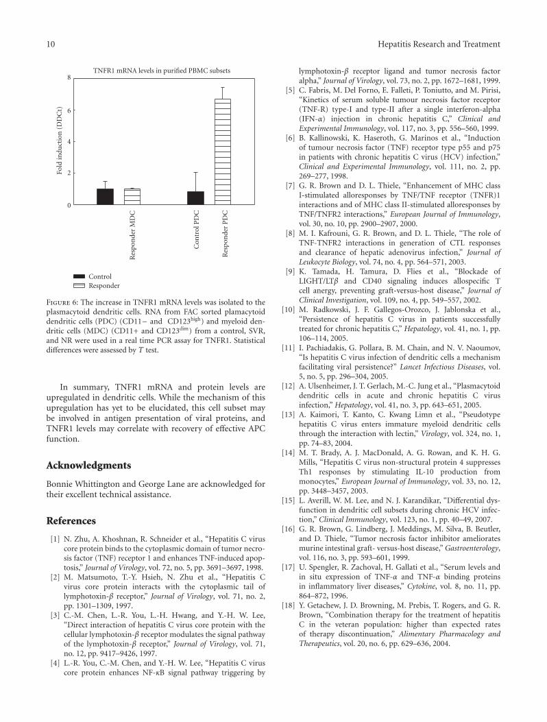
10 Hepatitis Research and Treatment
0
2
4
6
8TNFR1 mRNA levels in purified PBMC subsets
Res
pon
der
MD
C
Con
trol
PD
C
Res
pon
der
PD
C
Fold
indu
ctio
n(D
DC
t)
ControlResponder
Figure 6: The increase in TNFR1 mRNA levels was isolated to theplasmacytoid dendritic cells. RNA from FAC sorted plamacytoiddendritic cells (PDC) (CD11− and CD123high) and myeloid den-dritic cells (MDC) (CD11+ and CD123dim) from a control, SVR,and NR were used in a real time PCR assay for TNFR1. Statisticaldifferences were assessed by T test.
In summary, TNFR1 mRNA and protein levels areupregulated in dendritic cells. While the mechanism of thisupregulation has yet to be elucidated, this cell subset maybe involved in antigen presentation of viral proteins, andTNFR1 levels may correlate with recovery of effective APCfunction.
Acknowledgments
Bonnie Whittington and George Lane are acknowledged fortheir excellent technical assistance.
References
[1] N. Zhu, A. Khoshnan, R. Schneider et al., “Hepatitis C viruscore protein binds to the cytoplasmic domain of tumor necro-sis factor (TNF) receptor 1 and enhances TNF-induced apop-tosis,” Journal of Virology, vol. 72, no. 5, pp. 3691–3697, 1998.
[2] M. Matsumoto, T.-Y. Hsieh, N. Zhu et al., “Hepatitis Cvirus core protein interacts with the cytoplasmic tail oflymphotoxin-β receptor,” Journal of Virology, vol. 71, no. 2,pp. 1301–1309, 1997.
[3] C.-M. Chen, L.-R. You, L.-H. Hwang, and Y.-H. W. Lee,“Direct interaction of hepatitis C virus core protein with thecellular lymphotoxin-β receptor modulates the signal pathwayof the lymphotoxin-β receptor,” Journal of Virology, vol. 71,no. 12, pp. 9417–9426, 1997.
[4] L.-R. You, C.-M. Chen, and Y.-H. W. Lee, “Hepatitis C viruscore protein enhances NF-κB signal pathway triggering by
lymphotoxin-β receptor ligand and tumor necrosis factoralpha,” Journal of Virology, vol. 73, no. 2, pp. 1672–1681, 1999.
[5] C. Fabris, M. Del Forno, E. Falleti, P. Toniutto, and M. Pirisi,“Kinetics of serum soluble tumour necrosis factor receptor(TNF-R) type-I and type-II after a single interferon-alpha(IFN-α) injection in chronic hepatitis C,” Clinical andExperimental Immunology, vol. 117, no. 3, pp. 556–560, 1999.
[6] B. Kallinowski, K. Haseroth, G. Marinos et al., “Inductionof tumour necrosis factor (TNF) receptor type p55 and p75in patients with chronic hepatitis C virus (HCV) infection,”Clinical and Experimental Immunology, vol. 111, no. 2, pp.269–277, 1998.
[7] G. R. Brown and D. L. Thiele, “Enhancement of MHC classI-stimulated alloresponses by TNF/TNF receptor (TNFR)1interactions and of MHC class II-stimulated alloresponses byTNF/TNFR2 interactions,” European Journal of Immunology,vol. 30, no. 10, pp. 2900–2907, 2000.
[8] M. I. Kafrouni, G. R. Brown, and D. L. Thiele, “The role ofTNF-TNFR2 interactions in generation of CTL responsesand clearance of hepatic adenovirus infection,” Journal ofLeukocyte Biology, vol. 74, no. 4, pp. 564–571, 2003.
[9] K. Tamada, H. Tamura, D. Flies et al., “Blockade ofLIGHT/LTβ and CD40 signaling induces allospecific Tcell anergy, preventing graft-versus-host disease,” Journal ofClinical Investigation, vol. 109, no. 4, pp. 549–557, 2002.
[10] M. Radkowski, J. F. Gallegos-Orozco, J. Jablonska et al.,“Persistence of hepatitis C virus in patients successfullytreated for chronic hepatitis C,” Hepatology, vol. 41, no. 1, pp.106–114, 2005.
[11] I. Pachiadakis, G. Pollara, B. M. Chain, and N. V. Naoumov,“Is hepatitis C virus infection of dendritic cells a mechanismfacilitating viral persistence?” Lancet Infectious Diseases, vol.5, no. 5, pp. 296–304, 2005.
[12] A. Ulsenheimer, J. T. Gerlach, M.-C. Jung et al., “Plasmacytoiddendritic cells in acute and chronic hepatitis C virusinfection,” Hepatology, vol. 41, no. 3, pp. 643–651, 2005.
[13] A. Kaimori, T. Kanto, C. Kwang Limn et al., “Pseudotypehepatitis C virus enters immature myeloid dendritic cellsthrough the interaction with lectin,” Virology, vol. 324, no. 1,pp. 74–83, 2004.
[14] M. T. Brady, A. J. MacDonald, A. G. Rowan, and K. H. G.Mills, “Hepatitis C virus non-structural protein 4 suppressesTh1 responses by stimulating IL-10 production frommonocytes,” European Journal of Immunology, vol. 33, no. 12,pp. 3448–3457, 2003.
[15] L. Averill, W. M. Lee, and N. J. Karandikar, “Differential dys-function in dendritic cell subsets during chronic HCV infec-tion,” Clinical Immunology, vol. 123, no. 1, pp. 40–49, 2007.
[16] G. R. Brown, G. Lindberg, J. Meddings, M. Silva, B. Beutler,and D. Thiele, “Tumor necrosis factor inhibitor amelioratesmurine intestinal graft- versus-host disease,” Gastroenterology,vol. 116, no. 3, pp. 593–601, 1999.
[17] U. Spengler, R. Zachoval, H. Gallati et al., “Serum levels andin situ expression of TNF-α and TNF-α binding proteinsin inflammatory liver diseases,” Cytokine, vol. 8, no. 11, pp.864–872, 1996.
[18] Y. Getachew, J. D. Browning, M. Prebis, T. Rogers, and G. R.Brown, “Combination therapy for the treatment of hepatitisC in the veteran population: higher than expected ratesof therapy discontinuation,” Alimentary Pharmacology andTherapeutics, vol. 20, no. 6, pp. 629–636, 2004.

Hindawi Publishing CorporationHepatitis Research and TreatmentVolume 2010, Article ID 495928, 5 pagesdoi:10.1155/2010/495928
Clinical Study
Optimal Erythrocyte Ribavirin Level to Reduce the Risk ofAnemia and Obtain an Early Virological Response in Patientswith Chronic Hepatitis C Caused by Genotype 1b Infection
Rie Kubota,1 Takako Komiyama,1 Naoki Kumagai,2 Miyuki Kimijima,1 Keiko Mitsuki,1
Junko Uetake,1 Fumihiko Kaneko,2, 3 Satoshi Tsunematsu,2 and Kanji Tsuchimoto2
1 Pharmacy Practice and Sciences, Department of Clinical Pharmacy, Center for Clinical Pharmacy and Sciences, School of Pharmacy,Kitasato University, 5-9-1 Shirokane, Minato-ku, Tokyo 108-8641, Japan
2 Research Center for Liver Diseases, The Kitasato Institute Hospital, Kitasato University, 5-9-1 Shirokane,Minato-ku, Tokyo 108-8642, Japan
3 Department of Internal Medicine, The Kitasato Institute Medical Center Hospital, Kitasato University, 6-100 Arai,Kitamoto-shi, Saitama 364-8501, Japan
Correspondence should be addressed to Rie Kubota, [email protected]
Received 13 May 2010; Revised 28 July 2010; Accepted 4 August 2010
Academic Editor: Tatehiro Kagawa
Copyright © 2010 Rie Kubota et al. This is an open access article distributed under the Creative Commons Attribution License,which permits unrestricted use, distribution, and reproduction in any medium, provided the original work is properly cited.
Aims. To determine whether the erythrocyte phosphorylated ribavirin (RBV) level might be a useful index of EVR and risk ofanemia and to determine the optimal dose of RBV in 24 patients with hepatitis C with pegylated interferon and RBV. Methodology.The RBV level was measured by a high-performance liquid chromatography. Results and Conclusion. In patients aged 50 years orover, a negative correlation (r = −0.548, P < .05) was observed between the RBV level at week 2 and rate of Hb reduction (ΔHb)at week 4. The ΔHb at week 4 was significantly greater in patients with RBV levels of ≥800 μM (−25.5 ± 10.1%) than in patientswith RBV levels <800 μM (−15.6± 7.7%). None of the patients with RBV levels <600 μM at week 2 achieved EVR and SVR. Thusthe optimal levels of erythrocyte phosphorylated RBV at week 2 of therapy in order to achieve EVR without anemia seemed to be600–800 μM.
1. Introduction
The combination therapy with pegylated interferon (PEG-IFN) and ribavirin (RBV) has come to be established asthe standard treatment for chronic hepatitis C. A sustainedvirological response (SVR) has been reported with thistreatment in 30%–50% of patients with HCV genotype 1binfection, which accounts for 70% of all Japanese patientswith chronic hepatitis C [1]. However, the treatment oftenneeds to be discontinued, or the dose of RBV changed, inthese patients due to the development of hemolytic anemia.On the other hand, continuous treatment is important toobtain SVR with the treatment [2].
RBV is incorporated into the cells via the equilibrativenucleoside transporter (ENT) and converted to phosphateswithin the cells. RBV monophosphate (RMP) and RBV
triphosphate (RTP) are considered to have antiviral activ-ity [3, 4]. In nucleated cells, the phosphorylated RBVis subsequently dephosphorylated by the dephosphorylat-ing enzyme, and RBV is eliminated from the cells viathe ENT. However, in akaryocytes such as erythrocytes,which lack the dephosphorylating enzyme, accumulation ofphosphorylated RBV occurs, which diminishes the cellularATP and alters the cellular characteristics; these changesin the characteristics of the erythrocytes activate the cell-elimination activity of the reticuloendothelial system, result-ing in hemolysis [5, 6].
In this study, with the objective of reducing the adverseeffects and improving the treatment completion rate inpatients receiving combined PEG-IFN and RBV therapy, weattempted to evaluate whether the erythrocyte phosphory-lated RBV level might be useful as an index for the rate

2 Hepatitis Research and Treatment
of Early Virological Response (EVR) and SVR, the risk ofanemia.
2. Materials and Methods
2.1. Subjects. Among the patients with chronic hepatitis Ccaused by genotype 1b infection in whom combined PEG-IFNα2b and RBV therapy was started, 24 patients whoprovided written informed consent for participation in thisstudy were enrolled. The dosage regimen for the combinedPEG-IFN and RBV therapy was determined in accordancewith the standard dosing recommended for Japanese HCVpatients. RBV was started at the initial dose of 600 mg/day inpatients with a body weight of ≤60 kg, 800 mg/day in thosewith a body weight of >60 kg and ≤80 kg, and 1,000 mg/dayin those with a body weight of >80 kg. In patients with nocardiovascular disease, the RBV dosage of 600 mg/day wasdecreased to 400 mg/day, and 800 mg or 1000 mg/day wasreduced to 600 mg/day, if hemoglobin (Hb) level decreasedto less than 10 g/dL; permanently discontinued the drug ifHb decreased to less than 8.5 g/dL. In those with historyof stable cardiovascular disease, the dosage of RBV wasdecreased to 400 mg or 600 mg if Hb decreased by 2 g/dL ormore during any 4-week period; permanently discontinuedthe drug if Hb was less than 12 g/dL after 4weeks of a reducedRBV dosage. The patients were followed up until 48 weeksafter the start of the combined IFN and RBV therapy, andHb level and HCV RNA level were examined at week 12 afterthe start of treatment.
Serum HCV RNA negativity until 12 week of thetherapy was defined as EVR. Additionally, serum HCV RNAnegativity until 24 weeks after the therapy was completedand was defined as SVR. This study was performed with theapproval of the hospital research committee, in compliancewith the ethical principles laid out in the Declaration ofHelsinki.
2.2. Measurement of the Erythrocyte Level of PhosphorylatedRBV. Ten-mL samples of venous blood were obtained at2, 4, and 8 weeks after the start of the therapy, and theerythrocyte level of phosphorylated RBV was measured bythe HPLC method described by Homma et al. [7]. In thismethod, all phosphorylated RBV (RMP, RDP, and RTP) isconverted back to RBV by treatment with an erythrocytedephosphorylating enzyme, and the erythrocyte level ofphosphorylated RBV is calculated as the difference in theRBV levels measured before and after the enzyme treatment.
2.3. Statistical Analysis. The changes in the Hb level wereanalyzed by repeated-measures ANOVA and Dunnett’s test.The relationships of the RBV level to the risk of anemia andthe drug efficacy were examined by Student’s t-test, Pearson’scorrelation coefficient, χ2-test, and Fisher’s exact probabilitytest. P < .05 was regarded as denoting clinical significance.
3. Results
3.1. Patient Characteristics and Therapeutic Course. Thesubjects comprised 24 patients, and their demographic
Table 1: Baseline characteristics of patients.
Characteristics (n = 24)
Age (years) 59.9± 10.3
Sex (M/F) 12/12
Past treatment (Yes/No) 15/9
Body weight (kg) 57.7± 9.1
Ribavirin (mg/kg/day) 11.7± 1.5
Hemoglobin (g/dL) 13.6± 1.3
HCV RNA (KIU/mL)
<100 1
100∼500 3
500∼850 4
850 � 16
Data are expressed as mean ± S.D. or number of patientsHCV: Hepatitis C virus.
characteristics are indicated in Table 1. The combined PEG-IFN and RBV therapy needed to be discontinued in 3 ofthe 24 patients (12.5%), and the RBV dose needed to bereduced in 7 of the patients (29.2%) due to the developmentof anemia (Hb ≤ 10 g/dL). One patient was discontinuedthe therapy due to adverse reaction except anemia. However,the conditions of RBV administration for the initial 4 weekswere not changed. None of those in whom this therapywas discontinued achieved EVR. There were no significantdifference in the EVR rates between the subjects in whomthe RBV dose was reduced (72.7%) and those in whom thetherapy continued at the initial dose (64.0%). The dosage ofPEG-IFN was 1.5 ± 0.2μg/kg in accordance with a standardregimen, and the conditions of IFN administration have notchanged for 48 weeks after the combination therapy wasstarted in the subjects except for 4 patients who discontinuedthe therapy.
3.2. Changes in the Hb Level. The Hb levels (11.0± 1.3 g/dL)were significantly lower at week 4 of therapy as comparedwith 13.6 ± 1.3 g/dL at the start. In addition, the rate ofHb reduction [(Hb level-Hb level before administration)/Hblevel before administration] at week 4 of therapy was−12.4%in those aged less than 50 years, whereas it was −21.0% inthose aged 50 years or over (P < .05).
3.3. Changes in the Erythrocyte Phosphorylated RBV Level.The phosphorylated RBV and nonphosphorylated RBVlevels in the erythrocytes were 749.3±244.3 and 6.9±3.6μMat week 2, 1039.9 ± 239.6 and 8.4 ± 7.0μM at week 4,and 907.9 ± 292.1 and 8.9 ± 8.0μM at week 8 of therapy,respectively; thus, about 99% of the RBV in the erythrocyteswas phosphorylated (Figure 1).
3.4. Relationship between the Erythrocyte PhosphorylatedRBV Level at Week 2 and the Frequency of Anemia. Therelationship between the RBV level at week 2 and the rateof reduction of the Hb level was examined in the 19 patientsaged 50 years or over. There was a negative correlation (r =−0.548, P < .05) between the RBV level at week 2 and the rate

Hepatitis Research and Treatment 3
0
200
400
600
800
1000
1200
1400
0 2 4 8Ery
thro
cyte
phos
phor
ylat
edri
bavi
rin
con
cen
trat
ion
(µM
)
Weeks after starting combination therapy
Figure 1: Time course of erythrocyte phosphorylated ribavirin con-centration after starting PEG-IFNα-2b and ribavirin combinationtherapy.
of Hb reduction (ΔHb) at week 4 in the subjects in whomthe dose of RBV did not reduce or the combination therapydid not discontinue until week 4 (Figure 2). As shown inFigure 3, the ΔHg at week 4 was significantly higher (P < .05)in those with RBV level of≥800 μM (−25.5±10.1%) than inthose with the level of <800 μM (−15.6± 7.7%).
3.5. Relationship between the Erythrocyte Phosphorylated RBVLevel at Week 2 and the EVR. The relationship betweenthe phosphorylated RBV level at week 2 and the EVR wasevaluated in 20 of the 24 patients (Four cases were excludedbecause of the discontinuation of the therapy). The meanRBV level at week 2 was significantly lower (P < .05) in thenon-EVR patients (634.6± 236.6μM) than the EVR patients(889.7± 210.6μM).
As shown in Table 2, 3 cases with the phosphorylatedRBV level in erythrocytes ≥800 μM discontinued the com-bination therapy prematurely due to anemia, whereas noneof 14 cases with a levels <800 μM discontinued prematurely.
There were no EVR or SVR cases (0 of 7 cases) in patientswith erythrocyte phosphorylated RBV levels <600 μM atweek 2, whereas, in those with levels ≥600 μM, 11 of 17cases (64.7%) had EVR and 6 of 17 cases (35.3%) had SVR(P < .05).
Five of 7 cases (71.4%) with erythrocyte phosphorylatedRBV level at week 2 of 600–800 μM achieved EVR and 3cases (42.9%) achieved SVR without development of markedanemia. None of those patients discontinued RBV due todevelopment of anemia.
4. Discussion
In this study, combined PEG-IFN and RBV therapy neededto be discontinued, or the RBV dose needed to be reduced,in about 40% of the study subjects due to the developmentof hemolytic anemia (Hb ≤ 10 g/dL). None of the patientsin whom the combined PEG-IFN and RBV therapy wasdiscontinued by Week 12 showed SVR. However, no dif-ference in the rate of SVR was noted between the subjectsin whom the RBV dose was reduced and those in whomthe treatment could be continued at the initial dose. This
r = −0.548
−45
−40
−35
−30
−25
−20
−15
−10
−5
0
5
0 200 400 600 800 1000 1200 1400
2 week erythrocyte phosphorylated ribavirin concentration (µM)
Perc
ent
chan
gein
bloo
dh
emog
lobi
nbe
twee
nba
selin
ean
d4
wee
ks
Figure 2: Correlation between 4-week hemoglobin reduction ratefrom the baseline and 2-week erythrocyte phosphorylated ribavirinconcentration in patients aged 50 and over. r = −0.548 (P < .05).
−45
−40
−35
−30
−25
−20
−15
−10
−5
0
5
Concentration of phosphorylated RBV of erythrocyte at 2 weeks
P < .05
< 800µM
(n = 10)
800µM ≦(n = 9)
−15.6± 7.7
−25.5± 10.1
Perc
ent
chan
gein
bloo
dh
emog
lobi
nbe
twee
nba
selin
ean
d4
wee
ks
Figure 3: Comparison of 4-week hemoglobin reduction rate fromthe baseline of the patients with 2-week erythrocyte phosphorylatedribavirin concentration(<800 μM) to 4-week hemoglobin reductionrate from the baseline of the patients with 2-week erythrocytephosphorylated ribavirin concentration(800 μM �).
suggests that continuation of the combination therapy wasthe most important factor for achieving the desired clinicaloutcome. Clinically, the RBV dose reduction is performedbased on the present Hb level. However, it has been notedthat such dose adjustment does not effectively prevent theprogression of anemia; that is, once a decrease of the Hblevel has occurred, it is too late to stop the decline throughRBV dose reduction, presumably because of erythropoieticdelay.
In our subjects, the phosphorylated RBV level reacheda steady-state by 4 weeks of RBV therapy (1040 ± 240μM),implying gradual accumulation of phosphorylated RBV.Inoue et al. [8] reported that the erythrocyte phosphorylatedRBV level at the steady-state at week 4 was 1218 ± 234μMand was well correlated with Hb reduction.

4 Hepatitis Research and Treatment
Table 2: Comparisons of the rate in which RBV was discontinued, the rate in which RBV dosage was reduced due to development of anemia,4-week hemoglobin reduction rate from the baseline, the EVR rate and the SVR rate of the patients with 2-week erythrocyte phosphorylatedRBV concentration <600 μM to 600–800 μM, and 800 μM �.
<600 μM (n = 7) 600–800 μM (n = 7) 800μM � (n = 10)
Rate of RBV discontinuation (%) 0/7 (0) 0/7 (0) 3/10 (30.0)
Rate of RBV reduction (%) 1/7 (14.3) 3/7 (42.9) 3/10 (30.0)
ΔHb by 4 weeks (%) −13.5± 8.8 −15.7± 6.5 −23.4± 11.6
Rate of EVR(%) 0/7 (0) 5/7 (71.4) 6/10 (60.0)
Rate of SVR (%) 0/7 (0) 3/7 (42.6) 3/10 (30.0)
RBV: ribavirin.ΔHb: 4-week hemoglobin reduction rate from the baseline.EVR: Early Virological Response; Serum HCV RNA negativity until 12 weeks of therapy.SVR: Sustained Virological Response; Serum HCV RNA negativity at 24 weeks after completed therapy.ΔHb is expressed as mean ± S.D.
However, as the Hb level had already decreased sig-nificantly by week 4, the RBV level at week 4 does notpredict anemia. Therefore, we decided to evaluate whetherthe phosphorylated RBV level at week 2 might be useful forprediction of the subsequent development of anemia.
A close negative correlation was observed between theerythrocyte phosphorylated RBV level at week 2 and the ΔHbat week 4 in patients aged 50 years or over. In general, inelderly people, the percentage of fat cells in the bone marrowincreases, and the reserve of marrow stem cells decreaseswith reduction of the hematopoietic mass and reductionin the ability for formation of erythroid colony-formingunits [9, 10]. Additionally, RBV is known to be substantiallyexcreted by kidney, and renal function decreases in elderlypatients. Therefore, RBV accumulation is considered to bemore likely to cause anemia in the elderly, especially dueto the erythropoietic delay and the delay of RBV excretion.Nomura et al. reported that one of the higher risk of severeanemia was age higher than about 60 years [11].
In patients with RBV level of≥800 μM at week 2, theΔHbat week 4 was significantly higher, and a higher percentageof patients needed discontinuation of the RBV due to thedevelopment of anemia. Thus, we recommend that theerythrocyte phosphorylated RBV level be maintained at alevel of less than 800 μM at week 2 for treatment safety.
The plasma RBV level has been reported not to becorrelated with the SVR [12], but there have been few reportson the relationship between the erythrocyte phosphorylatedRBV level and the treatment efficacy. The erythrocyte RBVlevel is about 150 times higher than the plasma RBV concen-tration, and most of the administered RBV is considered tobe secreted into the urine. Of the proportion that remainsin the body, most of it does accumulate as phosphorylatedproduct within erythrocytes [13]. Furthermore, Homma etal. reported that little phosphorylated RBV existed in theplasma [13]. Since RBV taken up by cells is considered to bephosphorylated, and RMP and RTP are considered to haveantiviral activities, the erythrocyte phosphorylated RBV levelis a useful index for the antiviral effect of the drug. Since EVR,defined as a decrease of the HCV RNA level to 1/100 or zeroat week 12 after the start of therapy, has been reported to beuseful as a prognostic factor [14], we focused on not onlySVR but also EVR.
In this study, the erythrocyte phosphorylated RBV levelat week 2 was predictive of the EVR. Moreover, none of thepatients in whom the phosphorylated RBV level at week 2was <600 μM showed both EVR and SVR. Adjustment ofthe RBV dose to obtain an erythrocyte phosphorylated RBVlevel of ≥600 μM at week 2 is considered to be requiredto obtain an EVR. It would be necessary to have a largesample size, study of quality of life, and demonstrationof better EVR and SVR in a prospective randomized trial.Recently, Fellay et al. reported that genetic variants leadingto inosine triphosphatase (ITPA) deficiency protects againstclinically significant decline in Hb level induced by HCVantiviral treatment [15]. We should examine the relation-ships between ITPA gene variants and RBV-induced anemiain Japanese populations, and evaluate the usefulness as anindex to reduce the risk of anemia with erythrocyte RBVlevel.
5. Conclusion
In this study, the erythrocyte phosphorylated RBV level atweek 2 is proposed as a useful indicator to determine anoptimal dosage of ribavirin in patients with chronic hepatitisC under treatment with combination therapy with pegylatedinterferon and RBV.
References
[1] M. W. Fried, M. L. Shiffman, K. R. Reddy et al., “Peginterferonalfa-2a plus ribavirin for chronic hepatitis C virus infection,”The New England Journal of Medicine, vol. 347, no. 13, pp. 975–982, 2002.
[2] S. Iino, K. Okita, M. Omata et al., “Clinical efficacy of peg-interferon α-2b and ribabirin combination therapy for 48weeks in chronic hepatitis C patients with genotype 1 and highviral load.-retrospective comparison with interferon α-2b andribabirin combination therapy for 24 weeks,” Kan Tan Sui, vol.49, no. 6, pp. 1099–1121, 2004.
[3] P. Glue, “The clinical pharmacology of ribavirin,” Seminars inLiver Disease, vol. 19, no. 1, pp. 17–24, 1999.
[4] J. Y. N. Lau, R. C. Tam, T. J. Liang, and Z. Hong, “Mechanismof action of ribavirin in the combination treatment of chronicHCV infection,” Hepatology, vol. 35, no. 5, pp. 1002–1009,2002.

Hepatitis Research and Treatment 5
[5] L. De Franceschi, G. Fattovich, F. Turrini et al., “Hemolyticanemia induced by ribavirin therapy in patients with chronichepatitis C virus infection: role of membrane oxidativedamage,” Hepatology, vol. 31, no. 4, pp. 997–1004, 2000.
[6] H. Hosono, M. Homma, and Y. Inoue, “Effects of ribavirinuptake by red cells via a nucleoside transporter on redcell morphological changes and membrane phospholipids-their relationships with ribavirin induced hemolytic anemia,”Organ Biology, vol. 12, no. 3, p. 256, 2005.
[7] M. Homma, A. L. Jayewardene, J. Gambertoglio, and F.Aweeka, “High-performance liquid chromatographic deter-mination of ribavirin in whole blood to assess disposition inerythrocytes,” Antimicrobial Agents and Chemotherapy, vol. 43,no. 11, pp. 2716–2719, 1999.
[8] Y. Inoue, M. Homma, Y. Matsuzaki et al., “Erythrocyteribavirin concentration for assessing hemoglobin reductionin interferon and ribavirin combination therapy,” HepatologyResearch, vol. 34, no. 1, pp. 23–27, 2006.
[9] S. Okuma, “Aging of erythrocytes,” Journal of Practical Phar-macy, vol. 45, no. 10, pp. 59–64, 1994.
[10] Y. Takasaki, “Anemia in the elderly,” Internal Medicine, vol. 87,no. 2, pp. 302–306, 2001.
[11] H. Nomura, H. Tanimoto, E. Kajiwara et al., “Factors con-tributing to ribavirin-induced anemia,” Journal of Gastroen-terology and Hepatology, vol. 19, no. 11, pp. 1312–1317, 2004.
[12] N. Nakamoto, “Multi-facility joint study of the results ofcombination therapies for chronic hepatitis C: significance ofthe measurement of the plasma ribavirin concentration,” ActaHepatologica Japonica, vol. 45, supplement 1, p. A101, 2004.
[13] M. Homma, Y. Matsuzaki, Y. Inoue et al., “Marked elevationof erythrocyte ribavirin levels in interferon and ribavirin-induced anemia,” Clinical Gastroenterology and Hepatology,vol. 2, no. 4, pp. 337–339, 2004.
[14] N. Hiramatsu and N. Hayashi, “PEGIFN-ribavirin combina-tion therapy,” Biomedicine & Therapeutics, vol. 38, no. 9, pp.991–994, 2004.
[15] J. Fellay, A. J. Thompson, D. Ge et al., “ITPA gene variants pro-tect against anaemia in patients treated for chronic hepatitisC,” Nature, vol. 464, no. 7287, pp. 405–408, 2010.

Hindawi Publishing CorporationHepatitis Research and TreatmentVolume 2010, Article ID 702748, 6 pagesdoi:10.1155/2010/702748
Clinical Study
Differential Impact of Adherence to Pegylated Interferonand Ribavirin in the Treatment of Genotype 1 High Viral TiterChronic Hepatitis C
Makoto Numata,1 Tatehiro Kagawa,1 Sei-ichiro Kojima,1 Shunji Hirose,2 Naruhiko Nagata,1
Koichi Shiraishi,2 Norihito Watanabe,1 Hirokazu Shiozawa,3 Yasuhiro Nishizaki,3
Shigeyuki Motegi,3 Shinji Takashimizu,4 Jun-ichiro Kamochi,5 Mitsuru Wasada,5
Takashi Ohno,6 Yoshihiro Tei,7 Atsushi Nakano,8 Takuji Yamada,8 Kazuhiro Atsukawa,9
Tetsu Watanabe,10 and Tetsuya Mine1
1Department of Gastroenterology, Tokai University School of Medicine, Isehara 259-1193, Japan2Department of Gastroenterology, Tokai University Hachioji Hospital, Tokyo 192-0032, Japan3Department of Gastroenterology, Tokai University Tokyo Hospital, Tokyo 151-0053, Japan4Department of Gastroenterology, Tokai University Oiso Hospital, Oiso 259-0198, Japan5Ikegami General Hospital, Tokyo 146-8531, Japan6Isehara Kyodo Hospital, Isehara 259-1132, Japan7Japan Medical Alliance Ebina General Hospital, Ebina 243-0433, Japan8Tomei-Atsugi Hospital, Atsugi 243-8571, Japan9Hiratsuka City Hospital, Hiratsuka 254-0065, Japan
10Department of Community Health, Tokai University School of Medicine, Isehara 259-1193, Japan
Correspondence should be addressed to Tatehiro Kagawa, [email protected]
Received 3 May 2010; Revised 26 June 2010; Accepted 29 June 2010
Academic Editor: Emmet B. Keeffe
Copyright © 2010 Makoto Numata et al. This is an open access article distributed under the Creative Commons AttributionLicense, which permits unrestricted use, distribution, and reproduction in any medium, provided the original work is properlycited.
To clarify the impact of adherence, we treated 122 genotype 1 high viral titer chronic hepatitis C patients with pegylated interferon(peg-IFN) and ribavirin for 48 weeks at nine referral hospitals, and evaluated the prognostic factors with a focus on the adherenceto the treatment. This study included 68 (55.7%) treatment-naıve patients and 54 (44.3%) patients who did not respond to theprevious treatment. Multivariate analysis revealed adherence to peg-IFN and ribavirin as the only significant predictor. Sustainedvirological response (SVR) rate was 72.2%, 19.0%, and 27.3% in patients given ≥80%, 60%–80%, and <60% dose peg-IFN,respectively, and was 68.6%, 41.2%, and 5.3% in those given ≥80%, 60%–80%, and <60% dose ribavirin, respectively. SVR ratesharply fell when exposure to peg-IFN was below 80% whereas it decreased in a stepwise manner as for ribavirin. Therefore,≥80%of peg-IFN and as much as possible dose of ribavirin are desired to achieve SVR in the treatment of genotype 1 high viral titerchronic hepatitis C.
1. Introduction
Although the combination of pegylated interferon (peg-IFN) and ribavirin (RBV) is the standard-of-care therapy forchronic hepatitis C, the sustained virological response (SVR)rate is still 40%–50% [1–3] for patients who are infectedwith genotype 1 and have high viral load in their sera.
Adherence to the therapy is an important factor associatedwith a favorable outcome. McHutchison et al. reported thatthe patients who received ≥80% of the scheduled doses ofpeg-IFN and RBV for ≥80% of the planned duration oftherapy had an SVR rate of 51% compared with 34% in lessadherent patients [4]. In contrast, a study on patients withadvanced fibrosis revealed that reducing RBV dose did not

2 Hepatitis Research and Treatment
affect SVR rate as long as peg-IFN dose was maintained [5].Reddy et al. also reported that SVR rate was affected adverselyby RBV dose reduction when cumulative exposure was lessthan 60%, and that RBV dose reduction raised the relapserate [6]. The significant impact of adherence to both peg-IFNand RBV on SVR is well understood, however, there may bedifference between these two drugs in the way they effect theresponse.
Until now, many host factors including younger age (40years or less) [2], female gender [7], lighter body weight[1, 2], the absence of insulin resistance [8], elevated ALTlevels [2], less advanced liver histology [2, 7], and non-African American race [7, 9] are reported to be associatedwith favorable response. Recently the association of geneticvariation of IL28B with response has been reported [10–12].
Japanese elderly women were reported to be resistant tothis therapy [13, 14]. Japanese patients are approximately 10years older than those in other countries and our reportswould provide useful information when considering therapyfor elderly patients in other countries. The lower SVR rate inelderly women might be attributable to lower adherence topeg-IFN or RBV. However, few studies analyzed relationshipbetween SVR rate and the adherence in elderly patients.
In this study, we treated genotype 1 high viral titerchronic hepatitis C patients with peg-IFN and RBV combi-nation therapy, and evaluated the prognostic factors with afocus on the adherence to the treatment.
2. Materials and Methods
2.1. Patients. This study was performed at nine referralhospitals. Patients with hepatitis C virus (HCV) geno-type 1 and high viral load (≥100,000 IU/mL) who receivedpeg-IFN alfa-2b (Pegintron, Schering-Plough Corporation,Kenilworth, NJ) and RBV (Rebetol, Schering-Plough Cor-poration) combination therapy for 48 weeks from January2004 to December 2006 were consecutively enrolled into thestudy. Exclusion criteria were as follows: (1) patients withleukopenia (<3,000/μL), neutropenia (<1,500/μL), throm-bocytopenia (<90,000/μL), or anemia (hemoglobin con-centration <12 g/dL), (2) patients with creatinine clearance<50 mL/min, and (3) existence of cirrhosis, autoimmunediseases, uncontrolled mental disorders, uncontrolled malig-nancy, or severe heart or lung diseases. Written informedconsent was obtained from all patients.
2.2. Treatment. The patients were given peg-IFN alfa-2b ata dosage of 1.5 mg/kg every week subcutaneously for 48weeks. Daily RBV was administered orally for 48 weeksaccording to the labeling approved by the Japanese Ministryof Health, Labour and Welfare; 600 mg for patients ≤60 kg,800 mg for patients weighing 60 to 80 kg, and 1000 mg forpatients >80 kg. The use of hematopoietic growth factorssuch as G-CSF and erythropoietin was not permitted inthis study. Blood samples were collected every four weeksand parameters including complete blood cell counts, bio-chemistries, and the amount of HCV-RNA were determined.HCV serotype was tested with a serological genotyping assaykit (Immunocheck F-HCV Grouping; International Reagents
Co., Tokyo, Japan) [15]. If HCV serotype was not definitive,HCV genotyping was performed (HCV Core Genotype; SRL,Tokyo, Japan). The response to the treatment was evaluatedby an intention-to-treat analysis.
2.3. Statistical Analysis. The factors associated with SVR wereanalyzed by logistic regression using SPSS version 16 (SPSSJapan, Tokyo, Japan). Univariate or multivariate logisticregression analyses were performed to establish the factorscontributing to SVR. All reported P-values are 2-sided, withP < .05 considered statistically significant. The difference inthe rates of relapse or SVR was evaluated by chi-square test.
3. Results
3.1. SVR. A total of 122 patients were enrolled into thestudy. Forty-five patients (36.9%) were female and mean ±standard deviation (S.D.) of age was 54.0 ± 10.6 (min19–max 70) years. Sixty-eight patients (55.7%) were naıvepatients. The mean ± S.D. of weight and body mass index(BMI) was 63.5 ± 11.2 kg and 23.7 ± 3.3, respectively. High(100,000–800,000 IU/mL) and very high (≥800,000 IU/mL)HCV-RNA levels were observed in 36 (29.5%) and 86(70.5%) patients, respectively. This study included 68(55.7%) treatment-naıve patients and 54 (44.3%) patientswho did not respond to the previous treatment. The previoustreatment included a 24-week course of IFN alfa-2b and RBVcombination therapy for 36 patients and a 24-week courseof IFN alfa-2b or natural IFN alfa (human lymphoblastoidIFN) monotherapy for 18 patients. Forty-seven patientsrelapsed after the discontinuation of treatment, and theother 7 patients were nonresponders, in whom serum HCV-RNA were positive throughout the treatment. The SVRrate was 60.3%, 51.1%, and 28.6% in naıve patients, thosewith relapse, and nonresponders, respectively. In this study,the SVR rate was not significantly different between naıvepatients and those treated previously. Liver biopsy wasperformed before treatment in 87 (71.3%) patients; 75(86.2%) and 12 (13.8%) patients revealed METAVIR fibrosisscore of 0–2 and 3-4, respectively. The SVR rate was notsignificantly different between these two groups; 57.3% inpatients with F0–2 and 41.7% in those with F3-4. Finally 67patients (54.9%) achieved SVR in the entire cohort.
3.2. Factors Associated with SVR (Table 1). Analyzed factorsincluded gender, age, body weight, BMI, viral load, historyof IFN treatment, and adherence to the treatment. Youngerage, heavier weight, lower viral load, peg-IFN adherence,and RBV adherence were significant factors associated withSVR by univariate analysis. Multivariate analysis revealedadherence to peg-IFN and adherence to RBV as a significantpredictor. We performed the same analysis after stratifyingtreatment-naıve and previously treated patients, and foundadherence to peg-IFN and RBV as only factors significantlyassociated with SVR (data not shown) as shown in the entirecohort.
Patients given ≥80% dose of scheduled peg-IFN weremore likely to achieve SVR by 7.7-fold (95% CI; 1.926–30.798, P = .004) than those given 60%–80% dose. The SVR

Hepatitis Research and Treatment 3
6/22(27.3%)
4/21(19.0%)
57/79(72.2%)
59/86(68.6%)
7/17(41.2%)
1/19(5.3%)
6/10
0/1
0/11
4/10
0/9
0/2
49/66
7/7
1/6
100806040200
Total peg-IFN dose (%)
0
20
40
60
80
100
Tota
lRB
Vdo
se(%
)
Figure 1: Scatter plot of patients with or without SVR accordingto administered total doses of peg-IFN and RBV. One hundred %represents a full scheduled dose. A circle and a triangle indicatea patient with SVR and one without SVR, respectively. A numberrepresents number of patients with SVR/total number (SVR rate).
rate in patients given 60%–80% dose peg-IFN was similarwith those given <60% dose. Patients given ≥80% doseand those given 60%–80% dose of scheduled RBV weremore likely to obtain SVR than those given <60% by 27.4-fold (95% CI; 3.130–240.151, P = .003) and by 15.7-fold (95% CI; 1.289–190.653, P = .031), respectively.The outcome of each case was shown in a scatter plot(Figure 1). The SVR was 1/19 (5.3%), 7/17 (41.2%), and59/86 (68.6%) in patients given <60%, 60%–80%, and≥80%of total RBV dose, respectively (Figure 2). Therefore, themore RBV was administered, the higher was the SVR rate.On the other hand, SVR was achieved in 6/22 (27.3%), 4/21(19.0%), and 57/79 (72.2%) patients given <60%, 60%–80%, and ≥80% of total peg-IFN dose, respectively. Peg-IFN dose of 80% or more was important to obtain SVR.Notably none of the patients who received <80% dose forboth drugs resulted in SVR (Figure 1). The relationshipbetween SVR and adherence was analyzed separately in thetreatment-naıve group and the previously treated group. Inthe treatment-naıve group the SVR rate was 74.5%, 20.0%,and 36.4% in patients given ≥80%, 60%–80%, and <60%dose peg-IFN, respectively, and was 74.5%, 54.5%, and 0%in those given ≥80%, 60%–80%, and <60% dose RBV,respectively. In the previously treated group, SVR rate was68.8%, 18.2%, and 18.2% in patients given ≥80%, 60%–80%, and <60% dose peg-IFN, respectively, and was 61.3%,33.3%, and 11.1% in those given ≥80%, 60–80%, and <60%dose RBV, respectively. These trends were similar with theresults obtained from the entire cohort.
There was a trend that younger patients received greaterpeg-IFN dose; 72/106 (67.9%) patients younger than 65 yearsand 7/16 (43.8%) patients aged 65 or older received≥80% oftotal peg-IFN dose (P = .059).
Sixty-six patients (54.1%) received ≥80% dose for bothdrugs. Of these 49 (74.2%) patients resulted in SVR. Whenanalysis was performed in these patients, no significantfactors associated with SVR were chosen.
3.3. Rapid Virological Response (RVR), Early VirologicalResponse (EVR), and Relapse. The population of patientswhose serum HCV-RNA first disappeared at week 4 (RVR),week 8, week 12 (EVR), week 24, and week 48 was 10 (8.2%),39 (32.0%), 28 (23.0%), 20 (16.4%), and 4 (3.3%) patients,respectively. Twenty-one (17.2%) patients were positive forHCV-RNA throughout the treatment period (null response).The SVR rate of these patients who became negative forHCV-RNA at week 4 (RVR), week 8, week 12 (EVR), week24, and week 48 was 10/10 (100%), 35/39 (89.7%), 17/28(60.7%), 5/20 (25%), and 0/4 (0%), respectively. In 101patients negative for HCV-RNA at the end of treatment,34 (33.7%) patients relapsed. Relapse rate was significantlylower in patients who received ≥80% dose of peg-IFN thanthat in those who received 60%–80% or <60% dose (18.6%in patients with ≥80% dose versus 69.2% in those with60%–80% dose (P < .001) and 66.7% in those with <60%dose (P < .001), Figure 3). The relapse rate increased in astepwise fashion according to the adherence to RBV (91.7%in patients with <60% dose versus 41.7% in those with 60%–80% dose (P < .05), and versus 23.4% in those with ≥80%dose (P < .001)). These results were inversely associated withSVR rates.
3.4. Adverse Effect. Seventeen (13.9%) patients discontinuedtreatment. The reasons of premature discontinuation weregeneral fatigue and/or appetite loss (11 patients), fundalhemorrhage (1 patient), deterioration of diabetes mellitus (1patient), and depression (1 patient). Three patients discon-tinued treatment because of positive HCV-RNA at week 24.Thirty-nine (32.0%) and 33 (27.0%) patients required dosereduction of peg-IFN and RBV, respectively. Major reasonsof dose reduction were neutropenia or thrombocytopeniafor peg-IFN and anemia for RBV. Common adverse effectsincluded general fatigue, appetite loss, weight loss, and pru-ritus. In 12 patients with advanced liver disease (METAVIRfibrosis score of 3-4), 6 (50%) and 4 (33.3%) patientsrequired dose reduction of peg-IFN and RBV, respectively.In 75 patients with milder liver disease (METAVIR fibrosisscore of 0–2), 22 (29.3%) and 20 (26.7%) patients requireddose reduction of peg-IFN and RBV, respectively. There wasno significant difference between these two groups in theproportion of patients who required dose reduction.
4. Discussion
The mean age of our study population was 54.0 years, whichwas approximately 10 years older than patients of majorstudies in Western countries [1–3]. Our cohort consistedof treatment-naıve patients (55.7%) and those who did notrespond to the prior treatment (44.3%). SVR was achieved in54.9% patients.
In our study, adherence to peg-IFN and RBV was theonly significant factor associated with SVR. Interestingly,
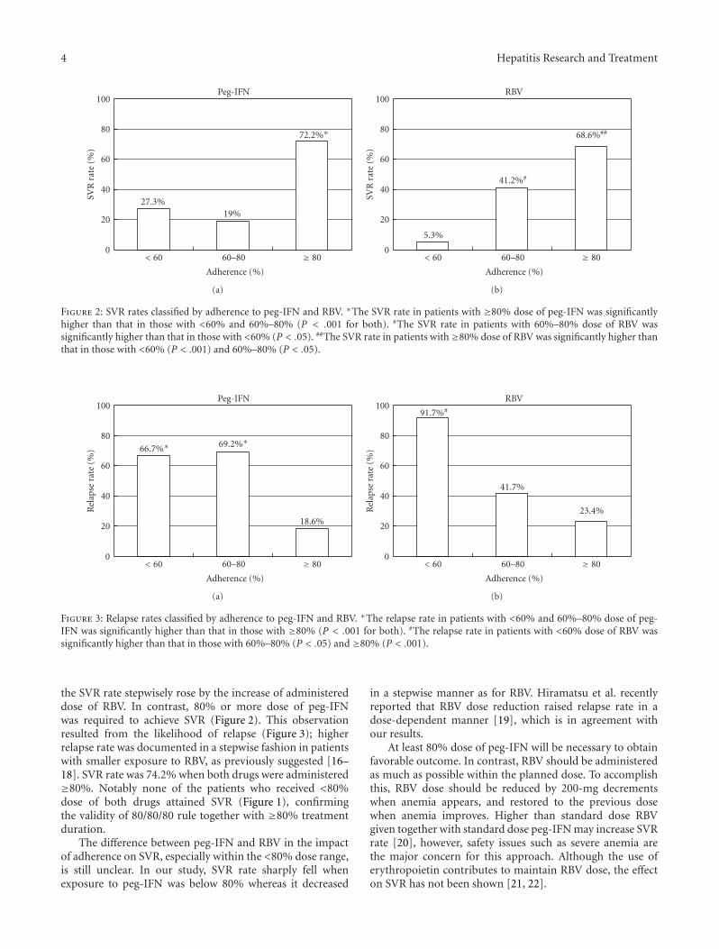
4 Hepatitis Research and Treatment
Peg-IFN
≥ 8060–80< 60
Adherence (%)
0
20
40
60
80
100SV
Rra
te(%
)
27.3%19%
72.2%∗
(a)
RBV
≥ 8060–80< 60
Adherence (%)
0
20
40
60
80
100
SVR
rate
(%)
5.3%
41.2%#
68.6%##
(b)
Figure 2: SVR rates classified by adherence to peg-IFN and RBV. ∗The SVR rate in patients with ≥80% dose of peg-IFN was significantlyhigher than that in those with <60% and 60%–80% (P < .001 for both). #The SVR rate in patients with 60%–80% dose of RBV wassignificantly higher than that in those with <60% (P < .05). ##The SVR rate in patients with≥80% dose of RBV was significantly higher thanthat in those with <60% (P < .001) and 60%–80% (P < .05).
Peg-IFN
≥ 8060–80< 60
Adherence (%)
0
20
40
60
80
100
Rel
apse
rate
(%) 66.7%∗ 69.2%∗
18.6%
(a)
RBV
≥ 8060–80< 60
Adherence (%)
0
20
40
60
80
100
Rel
apse
rate
(%)
91.7%#
41.7%
23.4%
(b)
Figure 3: Relapse rates classified by adherence to peg-IFN and RBV. ∗The relapse rate in patients with <60% and 60%–80% dose of peg-IFN was significantly higher than that in those with ≥80% (P < .001 for both). #The relapse rate in patients with <60% dose of RBV wassignificantly higher than that in those with 60%–80% (P < .05) and ≥80% (P < .001).
the SVR rate stepwisely rose by the increase of administereddose of RBV. In contrast, 80% or more dose of peg-IFNwas required to achieve SVR (Figure 2). This observationresulted from the likelihood of relapse (Figure 3); higherrelapse rate was documented in a stepwise fashion in patientswith smaller exposure to RBV, as previously suggested [16–18]. SVR rate was 74.2% when both drugs were administered≥80%. Notably none of the patients who received <80%dose of both drugs attained SVR (Figure 1), confirmingthe validity of 80/80/80 rule together with ≥80% treatmentduration.
The difference between peg-IFN and RBV in the impactof adherence on SVR, especially within the <80% dose range,is still unclear. In our study, SVR rate sharply fell whenexposure to peg-IFN was below 80% whereas it decreased
in a stepwise manner as for RBV. Hiramatsu et al. recentlyreported that RBV dose reduction raised relapse rate in adose-dependent manner [19], which is in agreement withour results.
At least 80% dose of peg-IFN will be necessary to obtainfavorable outcome. In contrast, RBV should be administeredas much as possible within the planned dose. To accomplishthis, RBV dose should be reduced by 200-mg decrementswhen anemia appears, and restored to the previous dosewhen anemia improves. Higher than standard dose RBVgiven together with standard dose peg-IFN may increase SVRrate [20], however, safety issues such as severe anemia arethe major concern for this approach. Although the use oferythropoietin contributes to maintain RBV dose, the effecton SVR has not been shown [21, 22].
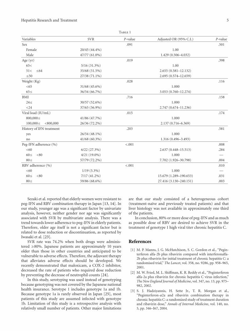
Hepatitis Research and Treatment 5
Table 1
Variables SVR P-value Adjusted OR (95% C.I.) P-value
Sex .091 .501
Female 20/45 (44.4%) 1.00
Male 47/77 (61.0%) 1.429 (0.506–4.032)
Age (yr) .019 .398
65< 5/16 (31.3%) 1.00
51< ≤64 35/68 (51.5%) 2.655 (0.581–12.132)
≤50 27/38 (71.1%) 2.695 (0.574–12.659)
Weight (Kg) .028 .116
<65 31/68 (45.6%) 1.000
65≤ 36/54 (66.7%) 3.053 (0.760–12.274)
BMI .716 .158
24≤ 30/57 (52.6%) 1.000
<24 37/65 (56.9%) 2.747 (0.674–11.236)
Viral load (IU/mL) .015 .174
800,000≤ 41/86 (47.7%) 1.000
100,000≤ <800,000 26/36 (72.2%) 2.137 (0.716–6.369)
History of IFN treatment .203 .581
yes 26/54 (48.1%) 1.000
no 41/68 (60.3%) 1.316 (0.496–3.493)
Peg-IFN adherence (%) <.001 .008
<60 6/22 (27.3%) 2.637 (0.448–15.513) .284
60≤ <80 4/21 (19.0%) 1.000 —
80≤ 57/79 (72.2%) 7.702 (1.926–30.798) .004
RBV adherence (%) <.001 .010
<60 1/19 (5.3%) 1.000 —
60≤ <80 7/17 (41.2%) 15.679 (1.289–190.653) .031
80≤ 59/86 (68.6%) 27.416 (3.130–240.151) .003
Sezaki et al. reported that elderly women were resistant topeg-IFN and RBV combination therapy in Japan [13, 14]. Inour study, younger age was a significant factor by univariateanalysis, however, neither gender nor age was significantlyassociated with SVR by multivariate analysis. There was atrend towards lower adherence to peg-IFN in elderly patients.Therefore, older age itself is not a significant factor but isrelated to dose reduction or discontinuation, as reported byIwasaki et al. [23].
SVR rate was 74.2% when both drugs were adminis-tered ≥80%. Japanese patients are approximately 10 yearsolder than those in other countries and anticipated to bevulnerable to adverse effects. Therefore, the adjuvant therapythat alleviates adverse effects should be developed. Werecently demonstrated that maloxicam, a COX-2 inhibitor,decreased the rate of patients who required dose reductionby preventing the decrease of neutrophil counts [24].
In this study, serotyping was used instead of genotypingbecause genotyping was not covered by the Japanese nationalhealth insurance. Serotype 1 includes genotype 1a and 1b.Because genotype 1a is rarely observed in Japan [25], mostpatients of this study are assumed infected with genotype1b. Limitation of this study is a retrospective analysis withrelatively small number of patients. Other major limitations
are that our study consisted of a heterogeneous cohort(treatment-naıve and previously treated patients) and thatliver histology was not available in approximately one thirdof the patients.
In conclusion, 80% or more dose of peg-IFN and as muchas possible dose of RBV are desired to achieve SVR in thetreatment of genotype 1 high viral titer chronic hepatitis C.
References
[1] M. P. Manns, J. G. McHutchison, S. C. Gordon et al., “Pegin-terferon alfa-2b plus ribavirin compared with interferonalfa-2b plus ribavirin for initial treatment of chronic hepatitis C: arandomised trial,” The Lancet, vol. 358, no. 9286, pp. 958–965,2001.
[2] M. W. Fried, M. L. Shiffman, K. R. Reddy et al., “Peginterferonalfa-2a plus ribavirin for chronic hepatitis C virus infection,”The New England Journal of Medicine, vol. 347, no. 13, pp. 975–982, 2002.
[3] S. J. Hadziyannis, H. Sette Jr., T. R. Morgan et al.,“Peginterferon-α2a and ribavirin combination therapy inchronic hepatitis C: a randomized study of treatment durationand ribavirin dose,” Annals of Internal Medicine, vol. 140, no.5, pp. 346–I67, 2004.

6 Hepatitis Research and Treatment
[4] J. G. McHutchison, M. Manns, K. Patel et al., “Adher-ence to combination therapy enhances sustained responsein genotype-1-infected patients with chronic hepatitis C,”Gastroenterology, vol. 123, no. 4, pp. 1061–1069, 2002.
[5] M. L. Shiffman, M. G. Ghany, T. R. Morgan et al., “Impactof reducing peginterferon alfa-2a and ribavirin dose duringretreatment in patients with chronic hepatitis C,” Gastroen-terology, vol. 132, no. 1, pp. 103–112, 2007.
[6] K. R. Reddy, M. L. Shiffman, T. R. Morgan et al., “Impactof ribavirin dose reductions in hepatitis C virus genotype1 patients completing peginterferon alfa-2a/ribavirin treat-mentnt,” Clinical Gastroenterology and Hepatology, vol. 5, no.1, pp. 124–129, 2007.
[7] H. S. Conjeevaram, M. W. Fried, L. J. Jeffers et al., “Pegin-terferon and ribavirin treatment in African American andCaucasian American patients with hepatitis C genotype 1,”Gastroenterology, vol. 131, no. 2, pp. 470–477, 2006.
[8] M. Romero-Gomez, M. Del Mar Viloria, R. J. Andrade etal., “Insulin resistance impairs sustained response rate topeginterferon plus ribavirin in chronic hepatitis C patients,”Gastroenterology, vol. 128, no. 3, pp. 636–641, 2005.
[9] A. J. Muir, J. D. Bornstein, and P. G. Killenberg, “Peginterferonalfa-2b and ribavirin for the treatment of chronic hepatitis Cin blacks and non-Hispanic whites,” The New England Journalof Medicine, vol. 350, no. 22, pp. 2265–2271, 2004.
[10] D. Ge, J. Fellay, A. J. Thompson et al., “Genetic variation inIL28B predicts hepatitis C treatment-induced viral clearance,”Nature, vol. 461, no. 7262, pp. 399–401, 2009.
[11] Y. Tanaka, N. Nishida, M. Sugiyama et al., “Genome-wideassociation of IL28B with response to pegylated interferon-αand ribavirin therapy for chronic hepatitis C,” Nature Genetics,vol. 41, no. 10, pp. 1105–1109, 2009.
[12] V. Suppiah, M. Moldovan, G. Ahlenstiel et al., “IL28B isassociated with response to chronic hepatitis C interferon-αand ribavirin therapy,” Nature Genetics, vol. 41, no. 10, pp.1100–1104, 2009.
[13] H. Sezaki, F. Suzuki, Y. Kawamura et al., “Poor response topegylated interferon and ribavirin in older women infectedwith hepatitis C virus of genotype 1b in high viral loads,”Digestive Diseases and Sciences, vol. 54, no. 6, pp. 1317–1324,2009.
[14] S. Watanabe, N. Enomoto, K. Koike et al., “Prolonged treat-ment with pegylated interferon α 2b plus ribavirin improvessustained virological response in chronic hepatitis C genotype1 patients with late response in a clinical real-life setting inJapan,” Hepatology Research, vol. 40, no. 2, pp. 135–144, 2010.
[15] T. Tanaka, K. Tsukiyama-Kohara, K. Yamaguchi et al., “Signif-icance of specific antibody assay for genotyping of hepatitis Cvirus,” Hepatology, vol. 19, no. 6, pp. 1347–1353, 1994.
[16] T. Poynard, P. Marcellin, S. S. Lee et al., “Randomised trialof interferon α2b plus ribavirin for 48 weeks or for 24 weeksversus interferon α2b plus placebo for 48 weeks for treatmentof chronic infection with hepatitis C virus,” The Lancet, vol.352, no. 9138, pp. 1426–1432, 1998.
[17] J. G. Mchutchison, S. C. Gordon, E. R. Schiff et al., “Interferonalfa-2b alone or in combination with ribavirin as initialtreatment for chronic hepatitis C,” The New England Journalof Medicine, vol. 339, no. 21, pp. 1485–1492, 1998.
[18] G. L. Davis, R. Esteban-Mur, V. Rustgi, et al., “Interferon alfa-2b alone or in combination with ribavirin for the treatmentof relapse of chronic hepatitis C. International HepatitisInterventional Therapy Group,” The New England Journal ofMedicine, vol. 339, pp. 1493–1499, 1998.
[19] N. Hiramatsu, T. Oze, T. Yakushijin et al., “Ribavirin dosereduction raises relapse rate dose-dependently in genotype 1patients with hepatitis C responding to pegylated interferonalpha-2b plus ribavirin,” Journal of Viral Hepatitis, vol. 16, no.8, pp. 586–594, 2009.
[20] K. Lindahl, L. Stahle, A. Bruchfeld, and R. Schvarcz, “High-dose ribavirin in combination with standard dose peginter-feron for treatment of patients with chronic hepatitis C,”Hepatology, vol. 41, no. 2, pp. 275–279, 2005.
[21] N. H. Afdhal, D. T. Dieterich, P. J. Pockros et al., “Epo-etin alfa maintains ribavirin dose in HCV-infected patients:a prospective, double-blind, randomized controlled study,”Gastroenterology, vol. 126, no. 5, pp. 1302–1311, 2004.
[22] M. L. Shiffman, J. Salvatore, S. Hubbard et al., “Treatmentof chronic hepatitis C virus genotype 1 with peginterferon,ribavirin, and epoetin alpha,” Hepatology, vol. 46, no. 2, pp.371–379, 2007.
[23] Y. Iwasaki, H. Ikeda, Y. Araki et al., “Limitation of combinationtherapy of interferon and ribavirin for older patients withchronic hepatitis C,” Hepatology, vol. 43, no. 1, pp. 54–63,2006.
[24] T. Kagawa, H. Shiozawa, S.-I. Kojima et al., “Eight-weekoral administration of meloxicam, a non-steroidal anti-inflammatory drug, prevents dose reduction of pegylatedinterferon α-2a in the treatment of chronic hepatitis C,”Hepatology Research, vol. 38, no. 3, pp. 259–266, 2008.
[25] E. Tanaka, K. Kiyosawa, T. Matsushima et al., “Egidemiologyof genotypes of hepatitis C virus in Japanese patients with typeC chronic liver diseases: a multi-institution analysis,” Journalof Gastroenterology and Hepatology, vol. 10, no. 5, pp. 538–545,1995.

Hindawi Publishing CorporationHepatitis Research and TreatmentVolume 2010, Article ID 537827, 5 pagesdoi:10.1155/2010/537827
Clinical Study
Retreatment of Patients Nonresponsive to Pegylated Interferonand Ribavirin with Daily High-Dose Consensus Interferon
Douglas F. Meyer,1 Hillel Tobias,2 Albert D. Min,1 Arathi Rajendra,1
Ivanka Zic,1 Edward Brettholz,1 David J. Clain,1 Franklin Klion,3 David Bernstein,4
and Henry C. Bodenheimer Jr.1
1 Division of Digestive Diseases, Beth Israel Medical Center and Albert Einstein, College of Medicine, New York, NY 10003, USA2 Division of Gastroenterology, New York University Medical Center, New York, NY 10016, USA3 Division of Liver Diseases, Mount Sinai School of Medicine, New York, NY 10029, USA4 Division of Gastroenterology, Hepatology and Nutrition, North Shore University Hospital, Manhasset, NY 11030, USA
Correspondence should be addressed to Henry C. Bodenheimer Jr., [email protected]
Received 17 April 2010; Revised 21 July 2010; Accepted 4 August 2010
Academic Editor: Tatehiro Kagawa
Copyright © 2010 Douglas F. Meyer et al. This is an open access article distributed under the Creative Commons AttributionLicense, which permits unrestricted use, distribution, and reproduction in any medium, provided the original work is properlycited.
Background. Current treatment of chronic hepatitis C with pegylated interferon and ribavirin has the ability to eliminate viralinfection in about half of the patients treated. Therapeutic options, for those with remaining chronic hepatitis, will remain limiteduntil novel antivirals become available in the future. Consensus interferon is currently available and has demonstrated clinicalefficacy with superior invitro antiviral activity, but the maximum tolerated dose is not defined. Methods. We assessed the efficacyof daily high-dose (24 ug) consensus interferon with weight-based (1000–1200 mg daily) ribavirin in HCV genotype 1-infectednon-responder patients. Results. Six adverse events were documented in five patients, and the trial was terminated with no subjectachieving viral clearance. Conclusions. The occurrence of serious adverse events effectively defined the upper limit of acceptabledose, while also revealing that this dose did not offer enhanced sustained viral clearance.
1. Introduction
The most commonly employed treatment of patients withhepatitis C virus (HCV) infection who are treatment-naıve consists of pegylated interferon-alfa and ribavirin withsustained viral response (SVR) rates in genotype 1 patientsof up to 52% [1, 2]. Since the FDA approval of pegylatedinterferon and ribavirin, there are an ever-increasing numberof hepatitis C patients who have failed to respond to thiscombination therapy. There is no standard of care for thetreatment of patients who have failed to respond to pegylatedinterferon and ribavirin [3]. Consensus interferon (CIFN), incombination with ribavirin, has recently been approved forthis use. Patients with advanced fibrosis may not have timeto await development of future antiviral agents and may needto utilize currently available therapies.
CIFN has more potent antiviral invitro activity comparedto other interferons [4, 5]. Currently, CIFN is approved for
initial or retreatment of persons infected with HCV. Recentclinical trials report reasonable SVR rates (23% to 37%) inthe treatment of nonresponders to standard or pegylatedinterferon and ribavirin using different regimens of dailyCIFN and weight-based ribavirin dosage [6–8]. Cornberget al. found that an eight-week induction phase with CIFNdoses of 18μg followed by 9μg for 40 weeks did not resultin a higher SVR compared to 9μg for 48 weeks, presumablydue to dose modifications in subjects receiving the inductiondose of CIFN. When the subgroup of patients receiving thehigher dose of CIFN, without early dose modification, wasevaluated, 40% achieved SVR [8]. There was one multicenterrandomized control trial, the DIRECT trial, evaluating theefficacy of daily CIFN at doses of 9μg or 15μg with weight-based ribavirin administration in the treatment of patientsunresponsive to prior pegylated interferon and ribavirintreatment. Viral clearance was achieved in a dose-dependentmanner with greatest loss of HCV in the 15μg group [9].
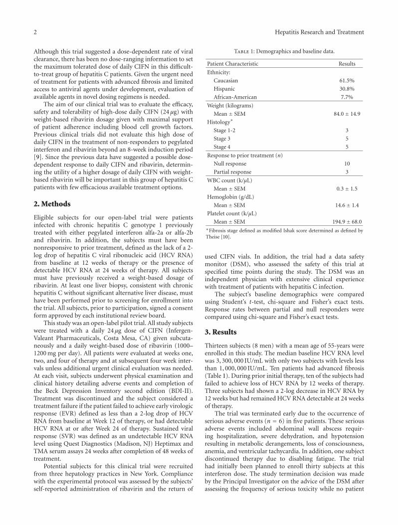
2 Hepatitis Research and Treatment
Although this trial suggested a dose-dependent rate of viralclearance, there has been no dose-ranging information to setthe maximum tolerated dose of daily CIFN in this difficult-to-treat group of hepatitis C patients. Given the urgent needof treatment for patients with advanced fibrosis and limitedaccess to antiviral agents under development, evaluation ofavailable agents in novel dosing regimens is needed.
The aim of our clinical trial was to evaluate the efficacy,safety and tolerability of high-dose daily CIFN (24 μg) withweight-based ribavirin dosage given with maximal supportof patient adherence including blood cell growth factors.Previous clinical trials did not evaluate this high dose ofdaily CIFN in the treatment of non-responders to pegylatedinterferon and ribavirin beyond an 8-week induction period[9]. Since the previous data have suggested a possible dose-dependent response to daily CIFN and ribavirin, determin-ing the utility of a higher dosage of daily CIFN with weight-based ribavirin will be important in this group of hepatitis Cpatients with few efficacious available treatment options.
2. Methods
Eligible subjects for our open-label trial were patientsinfected with chronic hepatitis C genotype 1 previouslytreated with either pegylated interferon alfa-2a or alfa-2band ribavirin. In addition, the subjects must have beennonresponsive to prior treatment, defined as the lack of a 2-log drop of hepatitis C viral ribonucleic acid (HCV RNA)from baseline at 12 weeks of therapy or the presence ofdetectable HCV RNA at 24 weeks of therapy. All subjectsmust have previously received a weight-based dosage ofribavirin. At least one liver biopsy, consistent with chronichepatitis C without significant alternative liver disease, musthave been performed prior to screening for enrollment intothe trial. All subjects, prior to participation, signed a consentform approved by each institutional review board.
This study was an open-label pilot trial. All study subjectswere treated with a daily 24μg dose of CIFN (Infergen-Valeant Pharmaceuticals, Costa Mesa, CA) given subcuta-neously and a daily weight-based dose of ribavirin (1000–1200 mg per day). All patients were evaluated at weeks one,two, and four of therapy and at subsequent four week inter-vals unless additional urgent clinical evaluation was needed.At each visit, subjects underwent physical examination andclinical history detailing adverse events and completion ofthe Beck Depression Inventory second edition (BDI-II).Treatment was discontinued and the subject considered atreatment failure if the patient failed to achieve early virologicresponse (EVR) defined as less than a 2-log drop of HCVRNA from baseline at Week 12 of therapy, or had detectableHCV RNA at or after Week 24 of therapy. Sustained viralresponse (SVR) was defined as an undetectable HCV RNAlevel using Quest Diagnostics (Madison, NJ) Heptimax andTMA serum assays 24 weeks after completion of 48 weeks oftreatment.
Potential subjects for this clinical trial were recruitedfrom three hepatology practices in New York. Compliancewith the experimental protocol was assessed by the subjects’self-reported administration of ribavirin and the return of
Table 1: Demographics and baseline data.
Patient Characteristic Results
Ethnicity:
Caucasian 61.5%
Hispanic 30.8%
African-American 7.7%
Weight (kilograms)
Mean ± SEM 84.0± 14.9
Histology∗
Stage 1-2 3
Stage 3 5
Stage 4 5
Response to prior treatment (n)
Null response 10
Partial response 3
WBC count (k/μL)
Mean ± SEM 0.3± 1.5
Hemoglobin (g/dL)
Mean± SEM 14.6± 1.4
Platelet count (k/μL)
Mean± SEM 194.9± 68.0∗Fibrosis stage defined as modified Ishak score determined as defined byTheise [10].
used CIFN vials. In addition, the trial had a data safetymonitor (DSM), who assessed the safety of this trial atspecified time points during the study. The DSM was anindependent physician with extensive clinical experiencewith treatment of patients with hepatitis C infection.
The subject’s baseline demographics were comparedusing Student’s t-test, chi-square and Fisher’s exact tests.Response rates between partial and null responders werecompared using chi-square and Fisher’s exact tests.
3. Results
Thirteen subjects (8 men) with a mean age of 55-years wereenrolled in this study. The median baseline HCV RNA levelwas 3, 300, 000 IU/mL with only two subjects with levels lessthan 1, 000, 000 IU/mL. Ten patients had advanced fibrosis(Table 1). During prior initial therapy, ten of the subjects hadfailed to achieve loss of HCV RNA by 12 weeks of therapy.Three subjects had shown a 2-log decrease in HCV RNA by12 weeks but had remained HCV RNA detectable at 24 weeksof therapy.
The trial was terminated early due to the occurrence ofserious adverse events (n = 6) in five patients. These seriousadverse events included abdominal wall abscess requir-ing hospitalization, severe dehydration, and hypotensionresulting in metabolic derangements, loss of consciousness,anemia, and ventricular tachycardia. In addition, one subjectdiscontinued therapy due to disabling fatigue. The trialhad initially been planned to enroll thirty subjects at thisinterferon dose. The study termination decision was madeby the Principal Investigator on the advice of the DSM afterassessing the frequency of serious toxicity while no patient

Hepatitis Research and Treatment 3
Time (weeks)
1234567
8910111213
00
1
2
3
4
5
6
7
8
1 2 4 8 12 16 20 24
HC
VR
NA
(IU
/mg)
Figure 1: The graph shows the viral kinetics of the 13 subjectsduring treatment with CIFN and weight-based ribavirin. None ofthe subjects with an initial drop in HCV RNA, including subject11 who had transient undetectable HCV RNA at week 20, failed toachieved a sustained loss of HCV RNA.
had achieved a sustained loss of HCV RNA. The severityand frequency, of serious adverse events, are evidence thatthe high-dose aggressive treatment regimen of this studydefined an upper dose limit of tolerability for CIFN-ribavirintherapy.
Subjects were discontinued from treatment, according toprotocol, when therapy was judged to be futile or for adverseevents. Only four subjects received 24 weeks of treatment.The median maximal decrease of HCV RNA from baselineon treatment was 3, 135, 000 IU/ml. Nine subjects (69%) hadgreater than a 2-log drop from baseline HCV RNA whileon treatment. One of these had transient loss of HCV butsubsequently experienced a viral breakthrough. The foursubjects who received 24 weeks of therapy all had partialviral response after 12 weeks of therapy, but had detectablehepatitis C virus at week 24 and therapy was discontinued asthey were deemed non-responders (Figure 1).
Three subjects with partial viral response prematurelydiscontinued therapy due to serious adverse events. Therewere three subjects still on therapy when the trial was halted.One subject had greater than a 2-log drop in HCV RNAlevel from baseline by week 8 of therapy, but the HCV RNAlevel at the time of termination had markedly increased, andthere was no longer a 2-log drop in the HCV RNA levelfrom baseline at that point. The second subject did have agreater than 2-log decrease from baseline HCV RNA levelafter 16 weeks of therapy, but by 20 weeks the hepatitis C virallevel had increased over 1-log from the nadir level. The thirdsubject did not have a 2-log decline from baseline viral levelafter 12 weeks of therapy and was deemed a non-responder.The final three subjects were non-responders to the study
medications at the time of discontinuation of therapy dueto adverse events.
There were three subjects with greater than a 2-logdrop in viral level by four weeks of therapy. One of thesesubjects could not tolerate therapy and did not complete12 weeks of therapy. Another was a nonresponder at 24weeks of therapy. The third subject was still on studymedication when the clinical trial was halted, however, whileon therapy there was marked increase in HCV RNA level. Allthree subjects who had greater than a 2-log drop from thebaseline viral level after four weeks of therapy were partialresponders to previous pegylated interferon and ribavirintherapy compared to none of the prior null responders(P < .04).
The most common adverse events were fatigue (n = 12)and flu-like symptoms (n = 12), anorexia (n = 10),anxiety/depression (n = 7), and insomnia (n = 7). The meandecrease in body weight during the experimental protocolwas 8.1± 5.8 kilograms. The median increase in BDI-II scorewas 9 ± 10.1. Serious adverse events (n = 6) occurred infive patients and included abdominal wall abscess, severedehydration, hypotension, and ventricular tachycardia allrequiring hospitalization and loss of consciousness andanemia. These events occurred within the first six to 16weeks of therapy. Two subjects (15%) had their dose ofCIFN reduced during the trial from 24μg to 15μg daily. Fivesubjects (38.5%) had a dose reduction of ribavirin and oneof the five subjects had ribavirin discontinued. The meandecrease in hemoglobin level from baseline to nadir was3.6 ± 1.6 g/dL with eight subjects receiving epoetin alphainjections.
No subject had a decrease in absolute neutrophil countbelow 500/μL. The mean decrease in WBC level frombaseline to nadir was 4, 200 ± 1, 300/μL. The mean decreasein platelet count from baseline was 104, 000± 5, 200/μL.
4. Discussion
Almost half the patients with HCV genotype 1 infectionare left with residual infection following treatment withpegylated interferon and ribavirin. Evaluation of currentlyavailable agents to optimize therapeutic efficacy is important,particularly for patients with advanced hepatic fibrosis.
Previous studies demonstrate promising SVR rates of23% to 37% using daily CIFN and ribavirin in the retreat-ment of non-responders to the combination of standardor pegylated interferon and ribavirin [6–8]. HCV clearanceby CIFN appears to be a dose-related response [8, 9, 11].The efficacy of high-dose CIFN, however, may be limited bytolerability and dose reductions for adverse events. There isno study defining the dose-limiting toxicity of CIFN usedwith ribavirin. One study, by Bocher et al., compared theefficacy of CIFN and ribavirin with high-dose induction(9μg daily for 24 weeks followed by 9μg three times per weekfor 24 weeks) to lower-dose treatment (CIFN 18μg threetimes per week for 12 weeks, followed by 9μg three timesper week for 36 weeks) in those patients who had previouslyfailed treatment with interferon and ribavirin therapy. Theweek 12 and 24 responses were superior in the high- versus

4 Hepatitis Research and Treatment
low-dose group (P < .05), but the SVR rates were identicalat 26%. This suggests that the initial positive effect of the24 weeks of high-dose daily induction was lost after dosereduction to three times per week treatment [12].
Kaiser etal. from Germany evaluated an induction doseof daily CIFN of 27μg for four weeks followed by 18μg foreight weeks versus 18μg for 12 weeks in 120 patients infectedwith HCV who were previous non-responders to pegylatedinterferon and weight-based ribavirin [6]. No ribavirin wasgiven during the induction time period. The SVR rate was44% in the high-dose group compared to 39% in the lower-dose group. The rate of discontinuation of therapy was 10%in the higher dose arm, and CIFN dose was reduced in 17%.Since this study required significant dose reductions withoutblood cell growth factor support, the ability to assess theefficacy potential of high-dose daily CIFN was limited.
Another study by Cornberg and colleagues investigatedthe efficacy of CIFN plus ribavirin in HCV patients who werenon-responders to standard interferon and ribavirin [8].Consensus interferon dosing with 18μg for the first 8-weeksof treatment resulted in an enhanced first-phase HCV-RNAdecay suggesting higher antiviral efficacy of a higher dose ofCIFN, but this did not translate to a better SVR, presumablydue to dose modifications [8]. Based on the suggestion of adose-dependent response and a superior response to high-dose CIFN, our trial sought to determine the upper limit oftolerability of CIFN dose in these difficult-to-treat patients.
Previous partial responders to pegylated interferon andweight-based ribavirin as compared to null responders hadbetter viral kinetic responses with all three partial respondershaving greater than a 2-log drop in HCV RNA from baselineviral level by week four. This response, however, did nottranslate into a higher overall efficacy since these subjectscould not tolerate continued therapy and viral response didnot decline to undetectable levels.
This trial used a sustained high daily dose (24 mg)of CIFN, a treatment regimen which showed promisingearly results in achieving a decline in HCV RNA levelsin a population of subjects resistant to prior therapy. Themajor factor leading to termination of this study was theoccurrence of serious adverse events. Although each eventwas not unique in patients receiving interferon ribavirintreatment, the occurrence of frequent serious adverse eventsin a small patient population raised significant concern.The abdominal wall abscess was related to injection siteinfection. The development of dehydration and hypotensionwas related to anorexia and severe lassitude. The episode ofloss of consciousness may have been related to anorexia andanemia. The observed adverse events, even including cardiacarrhythmias, have been reported previously in patientsreceiving interferon alfa and ribavirin. The frequency andseverity of adverse events, however, effectively determinedthe upper limit of tolerability of daily consensus interferon tobe less than the 24μg daily dose when used in combinationwith weight-based ribavirin. This observation is importantsince the efficacy results of the prior studies left open thepossibility of greater efficacy of high-dose CIFN.
Despite use of blood cell growth factors and maximalsupport of experienced investigators, this high-dose of daily
CIFN therapy in combination with a weight-based ribavirindose was not tolerated due to serious adverse events. Mostof the subjects required epoetin alfa injections for therapy-associated anemia. Even in the subjects who could toleratehigh-dose CIFN therapy with weight-based ribavirin, sus-tained loss of HCV was not achieved by 24 weeks.
We cannot recommend this higher daily 24μg dose ofCIFN in combination with weight-based ribavirin. Our studyeffectively defines the upper limit of tolerability of dailyCIFN dosage when used in combination with ribavirin.Furthermore, this high dosage of CIFN and weight-basedribavirin combination treatment was ineffective in erad-icating hepatitis C virus in patients who failed previouspegylated interferon and ribavirin therapy.
Abbreviations
Cifn: consensus interferonHCV: hepatitis C virusSVR: sustained viral responseHCV RNA: hepatitis C viral ribonucleic acidDSM: data safety monitorWBC: white blood cellBDI-II: Beck Depression Inventory second edition.
Author Disclosures
Dr. Henry C. Bodenheimer, Jr consults for Vertex andNovartis and is on an advisory board for Roche Genentech.Ivanka Zic, R. N., MSN was employed by Beth Israel MedicalCenter at the time of this research study. She is now employedby Merck.
Acknowledgment
This study was conducted using funds from Inter-Mune(Brisbane, Calif., USA) given to Beth Israel Medical Center,NY, as an unrestricted research grant.
References
[1] S. J. Hadziyannis, H. Sette Jr., T. R. Morgan et al.,“Peginterferon-α2a and ribavirin combination therapy inchronic hepatitis C: a randomized study of treatment durationand ribavirin dose,” Annals of Internal Medicine, vol. 140, no.5, pp. 346–355, 2004.
[2] J. G. McHutchison, E. J. Lawitz, M. L. Shiffman et al.,“Peginterferon alfa-2b or alfa-2a with ribavirin for treatmentof hepatitis C infection,” New England Journal of Medicine, vol.361, no. 6, pp. 580–593, 2009.
[3] M. G. Ghany, D. B. Strader, D. L. Thomas, and L. B. Seeff,“Diagnosis, management, and treatment of hepatitis C: anupdate,” Hepatology, vol. 49, no. 4, pp. 1335–1374, 2009.
[4] E. J. Heathcote, E. B. Keeffe, S. S. Lee et al., “Re-treatmentof chronic hepatitis C with consensus interferon,” Hepatology,vol. 27, no. 4, pp. 1136–1143, 1998.
[5] E. B. Melian and G. L. Plosker, “Interferon alfacon-1: a reviewof its pharmacology and therapeutic efficacy in the treatment
Hepatitis Research and Treatment 5
of chronic hepatitis C,” Drugs, vol. 61, no. 11, pp. 1661–1691,2001.
[6] S. Kaiser, H. Hass, M. Gregor, et al., “Successful retreatmentof chronic hepatitis C patients with a using daily consensusinterferon and ribavirin,” Hepatology, vol. 40, no. 4, article240A, 2004.
[7] C. B. Leevy, “Consensus interferon and ribavirin in patientswith chronic hepatitis C who were nonresponders to pegy-lated interferon alfa-2b and ribavirin,” Digestive Diseases andSciences, vol. 53, no. 7, pp. 1961–1966, 2008.
[8] M. Cornberg, J. Hadem, E. Herrmann et al., “Treatmentwith daily consensus interferon (CIFN) plus ribavirin in non-responder patients with chronic hepatitis C: a randomizedopen-label pilot study,” Journal of Hepatology, vol. 44, no. 2,pp. 291–301, 2006.
[9] B. R. Bacon, M. L. Shiffman, F. Mendes et al., “Retreatingchronic hepatitis C with daily interferon alfacon-1/ribavirinafter nonresponse to pegylated interferon/ribavirin: DIRECTresults,” Hepatology, vol. 49, no. 6, pp. 1838–1846, 2009.
[10] N. D. Theise, “Liver biopsy assessment in chronic viralhepatitis: a personal, practical approach,” Modern Pathology,vol. 20, no. 1, pp. S3–S14, 2007.
[11] M. H. Sjogren, R. Sjogren, K. Holtzmuller et al., “Interferonalfacon-1 and ribavirin versus interferon α-2b and ribavirin inthe treatment of chronic hepatitis C,” Digestive Diseases andSciences, vol. 50, no. 4, pp. 727–732, 2005.
[12] W. O. Bocher, M. Schuchmann, R. Link et al., “Consensusinterferon and ribavirin for patients with chronic hepatitis Cand failure of previous interferon-α therapy,” Liver Interna-tional, vol. 26, no. 3, pp. 319–325, 2006.

Hindawi Publishing CorporationHepatitis Research and TreatmentVolume 2010, Article ID 961359, 4 pagesdoi:10.1155/2010/961359
Review Article
Hepatitis C in Haematological Patients
Y. Y. Hwang and R. H. S. Liang
Division of Haematology, Department of Medicine, Queen Mary Hospital, The University of Hong Kong, Pokfulam, Hong Kong
Correspondence should be addressed to R. H. S. Liang, [email protected]
Received 19 April 2010; Revised 8 July 2010; Accepted 6 August 2010
Academic Editor: Tatehiro Kagawa
Copyright © 2010 Y. Y. Hwang and R. H. S. Liang. This is an open access article distributed under the Creative CommonsAttribution License, which permits unrestricted use, distribution, and reproduction in any medium, provided the original work isproperly cited.
There is no consensus guideline concerning the management of chronic hepatitis C patients during chemotherapy, andimmunosuppression. However, there are some suggestions in literature that hepatitis C viral load increases during chemotherapyand there is a risk of rebound immunity against hepatitis C after discontinuation of immunosuppression with a consequentliver injury. A close monitoring of liver function of these patients is prudent during treatment of haematological malignancy.Antiviral treatment is deferred after the completion of chemotherapy and recovery of patients’ immunity to minimize the toxicityof treatment. A combination of pegylated interferon and ribavirin is the standard therapy in hepatitis C infected haematologicalpatients.
1. Introduction
It is well known that reactivation of hepatitis B is apotential lethal complication of chemotherapy and pro-phylactic antiviral drugs are prudent during treatment ofhaematological malignancies. However, on the contrary, itis not certain how chronic hepatitis C infection wouldaffect the outcome of haematological malignancies and bonemarrow transplantation patients. There are case reports ofsevere flare up of chronic hepatitis C in patients undergoingchemotherapy [1, 2] but case series review found suchcomplications uncommon [3]. On the other hand, long-term survivors of bone marrow transplantation recipientsare more prone to the complications of hepatitis C infection[4] and treatment in this group of patients seems warranted.However, there is scanty data on management of chronichepatitis C in haematology patients.
Hepatitis C is associated with development of haemato-logical diseases ranging from immune thrombocytopenia tolymphoma. The underlying pathogenesis and its impact ontreatment are discussed.
2. Hepatitis C
There are six major genotypes of hepatitis C and more than50 subtypes. Genotype 1b is the most common subtype
worldwide. About 170 million people have chronic hepatitisand the estimated annual incidence of new case of hepatitisC is 3 to 4 millions [5]. In a study in Europe, the approximateprevalence of chronic hepatitis C among bone marrowtransplantation recipients is 6% [6].
Sixty to eighty percent of patients develop chronichepatitis C after acute infection. About twenty percent ofthese patients will be complicated by cirrhosis in twenty tothirty years, and some of them may develop hepatocellularcarcinoma [5, 7]. It is believed that successful control of viralreplication by effective antiviral treatment would preventsuch complications in these patients [8, 9].
3. Status of Hepatitis C during Chemotherapyand Immunosuppression
The level of hepatitis C viral RNA in blood has been shownto increase during chemotherapy and immunosuppression.At the same time, for those with pre-existing liver dys-function, the transaminase levels often normalize duringimmunosuppression. Upon the withdrawal of chemotherapyor immunosuppressants, the hepatitis C viral RNA decreaseswith a concomitant rise in transaminase levels. In a studyof ten chronic hepatitis C patients, the alanine transam-inase level decreased in eight patients while all of them

2 Hepatitis Research and Treatment
demonstrated a rise in hepatitis C RNA during a seven-week course of prednisone whereas there was a rebound ofalanine transaminase upon the withdrawal of steroid in sevenof them [10]. None of these patients developed fulminanthepatitis. Similarly, in another study of lymphoma patientswho received rituximab with combination chemotherapy,the hepatitis C RNA increased during chemotherapy anddeclined after completion of treatment. The use of rituximabin patients without hepatitis seldom leads to hepatotoxicity.However, in this cohort, one patient developed significanthepatotoxicity during rituximab chemotherapy [11].
This fluctuation in hepatitis C viral load and liverenzymes during and after chemotherapy or immunosuppres-sion is explained by the suppression of immunity duringchemotherapy and a rebound of reaction towards hepatitisC upon its withdrawal.
However, despite the above observations, the clinicalimpact of chronic hepatitis C infection on patients under-going bone marrow transplantation or chemotherapy is notwell characterized. In a study in United States, among thirtythree chronic hepatitis C patients, only eighteen of them(55%) developed mild to modest elevation of liver enzymesduring chemotherapy [3]. None of them developed severeflare up that required cessation of chemotherapy. On thecontrary, a study in Europe reported that up to 65% ofthe patients with chronic hepatitis C infection developedsignificant hepatotoxicity during chemotherapy [12]. Thishigh incidence of hepatotoxicity led to interruptions inchemotherapy and jeopardized the clinical outcome of thesepatients. In another study of 132 patients, five patientshad to discontinue chemotherapy because of severe hepaticdysfunction during treatment [13]. In a recently publishedreview of 160 chronic hepatitis C patients with non-Hodgkin lymphoma, twenty-four (15%) of them developedsignificant liver toxicity during chemotherapy [14]. Theoverall survival of chronic hepatitis C patients is also shownto be significantly worse than those without hepaitits Cinfection. At a median followup of two years of patientsdiagnosed with diffuse large B cell lymphoma in Grouped’Etude des Lymphomes de l’Adulte (GELA) programs, theoverall survival was 56% among chronic hepatitis C patientsversus 80% in those without (P = .02) [12].
The reported incidence of hepatotoxicity in chronichepatitis C patients undergoing chemotherapy varies greatlyamong these studies. In addition, because of the bleedingtendency commonly seen in haematology patients, all thesestudies only monitored liver enzymes and hepatitis C RNAlevel but none of them was based on histological evidence.Therefore, there is yet concrete data on the effect of chronichepatitis C infection in patients receiving chemotherapy andmore studies are needed in this aspect.
4. Hepatitis C in Hematopoietic StemCell Transplantation
Hepatitis C infection is associated with an increased risk ofveno-occlusive disease (VOD) and graft-versus-host disease(GVHD) of liver. The reported incidence of VOD was 14%among those with chronic hepatitis C infection while it
was only 8% in transplant recipients who were negativefor hepatitis C. The chronic inflammation in hepatitis C-infected liver causes endothelial changes in the hepaticsinusoids and this may predispose the patients to VODduring bone marrow transplant [15].
Although significant liver dysfunction during or imme-diately posttransplant is uncommon, bone marrow trans-plantation recipients with chronic hepatitis C infection havea significantly worse long-term outcome. The estimatedincidence of cirrhosis at twenty years post bone marrowtransplantation is 24% in those with chronic hepatitis C[16]. Moreover, there is evidence that the annual fibrosisprogression rate is significantly higher in posttransplantationpatients than those chronic hepatitis C patients withouttransplantation [4]. In fact, chronic hepatitis C infectionranked the third as a cause of late mortality, after infectionsand GVHD in posttransplant patients [16]. Once theydeveloped cirrhosis, their survival outcomes are markedlycompromised.
5. Treatment of Hepatitis C inHaematological Patients
The current standard of treatment of chronic hepatitisC infection is a combination of pegylated interferon andribavirin. There is currently no specific guideline for treat-ment of the infection in haematological malignancy patients.In these patients, including bone marrow transplantationrecipients, hepatitis C treatment is deferred until patients’immunity and bone marrow recover. In particular, forallogeneic stem cell transplantation patients, hepatitis Ctreatment should be withheld until all immunosuppressantsare tailed off and GVHD is completely resolved. Interferonis known to suppress bone marrow hematopoiesis and thiswould aggravate the cytopenia frequently seen in postbonemarrow transplantation patients. Moreover, it is reportedthat the use of interferon in allogeneic bone marrowtransplantation patients might trigger GVHD, and its use instem cell recipients should be cautious [17]. Ribavirin maysuppress erythropoiesis and this may aggravate the degreeof anemia in hematology patients. The use of erythropoietinhowever, is shown to reduce the transfusion requirement ofthese patients.
Combination therapy of pegylated interferon and rib-avirin is shown to produce a sustained virological response,that is, undetectable hepatitis C RNA six months offtherapy, in 20% of chronic hepatitis C patients after bonemarrow transplantation. This response rate is comparedunfavourably with that reported for the general population.Moreover, 30% of the studied patients did not receive thistreatment because of the presence of contraindications [18].The poor tolerability among hematology patients and theirinferior treatment outcome warrant more research in thisparticular group of patients.
6. Hepatitis C and Autoimmune Cytopenia
Chronic hepatitis C infection is associated with thrombo-cytopenia, and the cause is multifactorial in most patients.

Hepatitis Research and Treatment 3
There are various possible explanations, which includeincreased sequestration and destruction of platelets inpatients with hypersplenism, reduced thrombopoiesis as aresult of decreased production of endogenous thrombopoi-etin by diseased liver, direct marrow suppression by hepatitisC virus as well as dysregulation of immunity leading toautoimmune thrombocytopenia. One study demonstratedthe presence of increased level of antiplatelet glycoproteinsin serum of chronic hepatitis C infected patients withthrombocytopenia [19]. A significant proportion of thesepatients responded to standard immune thrombocytopeniatherapy such as steroids and intravenous immunoglob-ulin. In two of the studied patients, the platelet countnormalized after receiving pegylated interferon plus rib-avirin therapy. The rise of platelet count in one of thesepatients coincided with the disappearance of hepatitis CRNA in blood [19]. Although it is well known thatinterferon causes cytopenia, its administration in hepati-tis C infected patients with cytopenia is not necessarilycontraindicated but should be evaluated on an individualbasis.
7. Hepatitis C in Lymphomagenesis
It is well known that hepatitis C is a lymphotropic virusand is able to infect mononuclear cells in peripheral blood.Chronic hepatitis C infection is associated with various lym-phoproliferative disorders and the most commonly reportedassociation is mixed essential cryoglobulinemia. More than95% of patients with mixed cryoglobulinemia have evidenceof exposure to hepatitis C virus [20, 21].
B-cell clonality and t(14;18) translocation are bothprevalent in the peripheral blood mononuclear cells ofhepatitis C infected patients [22]. It has been shown thatchronic hepatitis C infection is significantly associated withthe development of non-Hodgkin lymphoma. The reportedodds ratio ranged from 2 to 4, with the risk being moreevident in area with a higher prevalence of hepatitis Cinfection [23]. The most commonly reported subtypes ofnon-Hodgkin lymphoma in chronic hepatitis C infectedpatients are marginal zone lymphoma and lymphoplasma-cytic lymphoma. There are also reports on an increasedprevalence of high grade B-cell lymphoma in hepatitis Cinfected patients but majority of them were arising from anunderlying low-grade lymphoma.
It is found that CD81 is a hepatitis C coreceptor andit is expressed in B lymphocytes [24]. The engagement ofCD81, which is part of the CD81/CD19/CD21 membranecomplex, activate B lymphocytes and subsequently leadto their proliferation. As a result, this chronic antigenicstimulation may predispose to the development of B-celllymphoproliferative disorders [25]. On the other hand, thereare suggestions that chronic hepatitis C infection inducespoint mutations in both immunoglobulin and nonim-munoglobulin genes of infected B lymphocytes [26, 27]. Itis, however, still controversial whether hepatitis C virus has adirect oncogenic effect. More studies are warranted before adefinitive conclusion can be made.
8. Role of Antiviral Treatment in HepatitisC-Associated Lymphoproliferative Diseases
As hepatitis C plays an important role in lymphomagenesis,it is postulated that eradication of virus may produce aresponse in its haematological manifestation as well. In areview of eighteen hepatitis C infected patients with indolentB-cell lymphoma, an overall response rate of the lymphomaafter antiviral treatment alone was 63% and 80%, respec-tively, in the group receiving interferon plus ribavirin andpegylated interferon plus ribavirin, respectively [28]. Therewas, however, persistent presence of cryoglobulin in majorityof the cases despite the absence of detectable tumour. Inanother study of nine hepatitis C infected patients withsplenic marginal zone lymphoma, seven of them had sus-tained virological response after antiviral treatment (whichconsisted of interferon as first line therapy with ribavirinadded if unsatisfactory response). All seven patients hada concomitant haematological response with a decrease insplenic size and disappearance of villous lymphocytes fromperipheral blood [28]. In the remaining two patients whohad persistent detectable hepatitis C RNA in blood, therewas no significant clinical haematological response. Theimmunoglobulin gene rearrangement observed at diagnosisin these patients was still detectable in patients achieving acomplete clinical remission. Although antiviral treatment hasa therapeutic role in the treatment of hepatitis C-associatedlymphoproliferative diseases, most of the patients still havedetectable residual diseases by molecular methods. Antiviraltherapy alone is unable to achieve a complete remission oftheir haematological diseases in these patients.
9. Conclusion
There are different opinions on the risk of hepatic dys-function during chemotherapy for haematological patientswith chronic hepatitis C infection. No consensus guidelinesconcerning the management of these patients are currentlyavailable and more studies on this issue are warranted.
The role of hepatitis C infection in lymphomagenesisis intriguing and a better understanding may help ourmanagement of hepatitis C-associated lymphoproliferativediseases. Although antiviral treatment may not be ableto eradiate hepatitis C-associated lymphoma, it shouldbe considered as a treatment option for tumour control,especially for those who may not be able to tolerate cytotoxicchemotherapy.
References
[1] H. Kanamori, H. Fukawa, A. Maruta et al., “Case report:fulminant hepatitis C viral infection after allogeneic bonemarrow transplantation,” American Journal of the MedicalSciences, vol. 303, no. 2, pp. 109–111, 1992.
[2] S. Vento, F. Cainelli, F. Mirandola et al., “Fulminant hepatitison withdrawal of chemotherapy in carriers of hepatitis Cvirus,” The Lancet, vol. 347, no. 8994, pp. 92–93, 1996.
[3] E. Zuckerman, T. Zuckerman, D. Douer, D. Qian, andA. M. Levine, “Liver dysfunction in patients infected with

4 Hepatitis Research and Treatment
hepatitis C virus undergoing chemotherapy for hematologicmalignancies,” Cancer, vol. 83, no. 6, pp. 1224–1230, 1998.
[4] C. A. P. Ivantes, H. Amarante, S. O. Ioshii, and R. Pasquini,“Hepatitis C virus in long-term bone marrow transplantsurvivors,” Bone Marrow Transplantation, vol. 33, no. 12, pp.1181–1185, 2004.
[5] World Health Organization, “Hepatitis C. Fact sheet N164,”October 2000, http://www.who.int/mediacentre/factsheets/fs164/en.
[6] A. Locasciulli, M. Testa, M. G. Valsecchi et al., “The role ofhepatitis C and B virus infections as risk factors for severeliver complications following allogeneic BMT: a prospectivestudy by the Infectious Disease Working Party of the EuropeanBlood and Marrow Transplantation Group,” Transplantation,vol. 68, no. 10, pp. 1486–1491, 1999.
[7] J. H. Jou and A. J. Muir, “In the clinic. Hepatitis C,” Annals ofInternal Medicine, vol. 148, pp. ITC6-1–ITC6-16, 2008.
[8] S. Bruno, T. Stroffolini, M. Colombo et al., “Italian Associationof the Study of the Liver Disease. Sustained virologicalresponse to interferon-α is with improved outcome in HCV-related cirrhosis: a retrospective study,” Hepatology, vol. 45, no.3, pp. 579–587, 2007.
[9] C.-H. Hung, C.-M. Lee, S.-N. Lu et al., “Long-term effectof interferon alpha-2b plus ribavirin therapy on incidence ofhepatocellular carcinoma in patients with hepatitis C virus-related cirrhosis,” Journal of Viral Hepatitis, vol. 13, no. 6, pp.409–414, 2006.
[10] T.-L. Fong, B. Valinluck, S. Govindarajan, F. Charboneau, R.H. Adkins, and A. G. Redeker, “Short-term prednisone therapyaffects aminotransferase activity and hepatitis C virus RNAlevels in chronic hepatitis C,” Gastroenterology, vol. 107, no.1, pp. 196–199, 1994.
[11] D. Ennishi, Y. Terui, M. Yokoyama et al., “Monitoring serumhepatitis C virus (HCV) RNA in patients with HCV-infectedCD20-positive B-cell lymphoma undergoing rituximab com-bination chemotherapy,” American Journal of Hematology, vol.83, no. 1, pp. 59–62, 2008.
[12] C. Besson, D. Cantoni, E. Lepage et al., “Characteristics andoutcome of diffuse large B-cell lymphoma in hepatitis C virus-positive patients in LNH 93 and LNH 98 groupe d’etudedes lymphomes de l’adulte programs,” Journal of ClinicalOncology, vol. 24, no. 6, pp. 953–960, 2006.
[13] C. Visco, L. Arcaini, E. Brusamolino et al., “Distinctivenatural history in hepatitis C virus positive diffuse large B-cell lymphoma: analysis of 156 patients from northern Italy,”Annals of Oncology, vol. 17, no. 9, pp. 1434–1440, 2006.
[14] L. Arcaini, M. Merli, F. Passamonti et al., “Impact oftreatment-related liver toxicity on the outcome of HCV-positive non-Hodgkin’s lymphomas,” American Journal ofHematology, vol. 85, no. 1, pp. 46–50, 2010.
[15] A. Ribas and R. P. Gale, “Should people with hepatitis C virusinfection receive a bone marrow transplant?” Bone MarrowTransplantation, vol. 19, no. 2, pp. 97–99, 1997.
[16] D. L. Peffault, V. Levy, T. Asselah et al., “Long-term outcomeof hepatitis C infection after bone marrow transplantation,”Blood, vol. 103, no. 5, pp. 1618–1624, 2004.
[17] C. Giardini, M. Galimberti, G. Lucarelli et al., “α-interferontreatment of chronic hepatitis C after bone marrow trans-plantation for homozygous β-thalassemia,” Bone MarrowTransplantation, vol. 20, no. 9, pp. 767–772, 1997.
[18] R. P. de Latour, T. Asselah, V. Levy et al., “Treatment ofchronic hepatitis C virus in allogeneic bone marrow transplantrecipients,” Bone Marrow Transplantation, vol. 36, no. 8, pp.709–713, 2005.
[19] A. J. de Almeida, M. Campos-de-Magalhaes, C. L. Antoniettiet al., “Autoimmune thrombocytopenia related to chronichepatitis C virus infection,” Hematology, vol. 14, no. 1, pp. 49–58, 2009.
[20] S. C. Gordon, “Extrahepatic manifestations of hepatitis C,”Digestive Diseases, vol. 14, no. 3, pp. 157–168, 1996.
[21] F. Lunel, L. Musset, P. Cacoub et al., “Cryoglobulinemia inchronic liver diseases: role of hepatitis C virus and liverdamage,” Gastroenterology, vol. 106, no. 5, pp. 1291–1300,1994.
[22] A. L. Zignego, C. Ferri, F. Giannelli et al., “Prevalence of bcl-2rearrangement in patients with hepatitis C virus-related mixedcryoglobulinemia with or without B-cell lymphomas,” Annalsof Internal Medicine, vol. 137, no. 7, pp. 571–580, 2002.
[23] E. Negri, D. Little, M. Boiocchi, C. La Vecchia, and S.Franceschi, “B-cell non-Hodgkin’s lymphoma and hepatitis Cvirus infection: a systematic review,” International Journal ofCancer, vol. 111, no. 1, pp. 1–8, 2004.
[24] P. Pileri, Y. Uematsu, S. Campagnoli et al., “Binding ofhepatitis C virus to CD81,” Science, vol. 282, no. 5390, pp. 938–941, 1998.
[25] D. S. Viswanatha and A. Dogan, “Hepatitis C virus andlymphoma,” Journal of Clinical Pathology, vol. 60, no. 12, pp.1378–1383, 2007.
[26] K. Machida, K. T.-N. Cheng, V. M.-H. Sung et al., “HepatitisC virus induces a mutator phenotype: enhanced mutationsof immunoglobulin and protooncogenes,” Proceedings of theNational Academy of Sciences of the United States of America,vol. 101, no. 12, pp. 4262–4267, 2004.
[27] K. Machida, K. T.-H. Cheng, N. Pavio, V. M.-H. Sung,and M. M. C. Lai, “Hepatitis C virus E2-CD81 interactioninduces hypermutation of the immunoglobulin gene in Bcells,” Journal of Virology, vol. 79, no. 13, pp. 8079–8089, 2005.
[28] C. Mazzaro, V. de Re, M. Spina et al., “Pegylated-interferonplus ribavirin for HCV-positive indolent non-Hodgkin lym-phomas,” British Journal of Haematology, vol. 145, no. 2, pp.255–257, 2009.

Hindawi Publishing CorporationHepatitis Research and TreatmentVolume 2010, Article ID 267412, 6 pagesdoi:10.1155/2010/267412
Research Article
Treatment of Chronic Hepatitis C Virus Infection inDialysis Patients: An Update
Hugo Weclawiak,1 Nassim Kamar,1, 2 Abdellatif Ould-Mohamed,1
Isabelle Cardeau-Desangles,1 Jacques Izopet,3, 4 and Lionel Rostaing1, 4
1 Department of Nephrology, Dialysis, and Organ Transplantation, Toulouse University Hospital, CHU Rangueil,1 avenue Jean Poulhes, TSA 50032- 31059- Toulouse Cedex 9, France
2 INSERM U858, Toulouse University Hospital, CHU Rangueil, IFR 31, 1 avenue Jean Poulhes, TSA 50032,31059 Toulouse Cedex 9, France
3 Laboratory of Virology, Toulouse University Hospital, CHU Purpan, 330 avenue de Grande-Bretagne, TSA 40031,31059 Toulouse Cedex 9, France
4 INSERM U563, Toulouse University Hospital, CHU Purpan, IFR 30, 330 avenue de Grande-Bretagne, TSA 40031,31059 Toulouse Cedex 9, France
Correspondence should be addressed to Hugo Weclawiak, [email protected]
Received 27 May 2010; Revised 19 August 2010; Accepted 24 August 2010
Academic Editor: Tatehiro Kagawa
Copyright © 2010 Hugo Weclawiak et al. This is an open access article distributed under the Creative Commons AttributionLicense, which permits unrestricted use, distribution, and reproduction in any medium, provided the original work is properlycited.
Hepatitis C virus (HCV) infection is a blood-borne infection and its prevalence used to be elevated in hemodialysis (HD) patients.Its main mode of contamination relies on nosocomial transmission. HCV infection is frequently associated in HD patients withnormal liver enzymes whereas liver histology can display some degree of HCV-related lesions. The assessment of HCV-relatedlesions, even in HD dialysis patients, can be done via noninvasive tests. After kidney transplantation, HCV-related lesions canworsen; however, in this setting antiviral treatment harbors the risk of acute rejection. Therefore, it is recommended to implementantiviral treatment while the patient is receiving dialysis therapy. In this setting, the rate of viral clearance is usually high. In caseof sustained virological response, no relapse occurs after kidney transplantation, despite heavy immunosuppression.
1. Introduction
The most important forms of liver disease in dialysispatients are viral hepatitis B (HBV) and C (HCV). Thevast majority of literature on dialysis for hepatitis refers tohemodialysis (HD). Individuals receiving peritoneal dialysis(PD) are at less risk of acquiring blood-borne infectionsfor several reasons, including an absence of extracorporealblood manipulation, a lack of intravascular access, as well asa lower requirement for blood transfusions. Also, PD takesplace in the patient’s home, where there is no exposure toother patients.
An accurate assessment of the natural history of HCVin dialysis patients is not easy to obtain. HCV infectionin dialysis patients is often asymptomatic with an apparentindolent course. HCV infection extends over decades rather
than years whereas chronic kidney-disease (CKD) patientsgenerally have higher morbidity and mortality rates thanthose of the general population, due to age and comorbidityconditions [1]. This makes the long-term consequences ofHCV difficult to establish. Additional factors also modifythe course of liver disease, including HBV/HCV coinfection,coinfection with human immunodeficiency virus, or alcoholabuse.
Because of the wide use of antiviral drugs and becauseposttransfusional hepatitis no longer occurs, future natural-history studies on chronic HCV will become less possible[2]. Accurate evaluation of HCV infection in the CKDpopulation is further complicated by the observation thataminotransferase values are typically lower in dialysis thannonuremic populations [3]. However, dialysis patients thatdo show detectable HCV RNA have aminotransferase levels

2 Hepatitis Research and Treatment
greater than those who do not, although values are typicallywithin the “normal” range [4, 5]. Therefore, if one wantsto assess the impact of chronic HCV infection in CKDpatients, a liver biopsy is usually performed [2]. However,a liver biopsy may be replaced by noninvasive tests, suchas a FibroTest or a FibroScan [6, 7], and these tests areof particular interest when there is a possibility of kidneytransplantation to treat CKD.
2. HCV-Related Outcomes inthe CKD Population
A recent meta-analysis on the impact of HCV on mortalityin 11,589 maintenance-dialysis patients, from seven observa-tional studies, concluded that the estimated adjusted relativerisk (aRR: all cause mortality) was 1.34 (1.13−1.59; P < .01)[8]. The cause of death as hepatocellular carcinoma and theincidence of liver cirrhosis, were significantly more frequentamong anti-HCV-positive than anti-HCV-negative dialysispatients in all seven studies. The unadjusted summaryestimate for liver-related mortality was 5.89 (1.93−17.99; P <.001) according to a random-effects model [8].
Recently, Kalantar-Zadeh et al. [9] evaluated a databaseof 13,664 chronic HD patients in the United States who hadundergone HCV serology. They observed that the mortality-hazard ratio was strongly associated with HCV infection:that is, it was 1.25 (1.12−1.39; P < .001). Thus, when HCV-positive CKD patients undergo kidney transplantation, it ispossible that the natural course of chronic HCV infection isaltered by the use of chronic immunosuppression. Indeed,two studies have shown that survival was significantlyimproved in HCV-positive patients who had benefited froma kidney transplant compared to those who remained on akidney waiting list [10, 11].
After kidney transplantation, within the first 5 years post-transplant, patient survival is similar in both HCV-positiveand HCV-negative patients [12–14]. However, when 10-yearsurvival rates are examined, HCV then appears to be adetrimental effect [12–14]. A meta-analysis of observationalstudies identified eight clinical trials (6,365 unique patients)in which the presence of anti-HCV antibodies in the serumwas an independent and significant risk factor for death andgraft failure after kidney transplantation. The estimates forrelative risk (RR) were 1.79 (1.57−2.03) and 1.56 (1.35−1.80),respectively [15].
The adverse impact of HCV on survival after kidneytransplantation has been linked to liver dysfunction. Fur-thermore, a positive anti-HCV serology in kidney-transplantpatients has been implicated in the development of denovo glomerulopathy [19], an increased incidence of seriousinfections [20], and new-onset diabetes mellitus [21]. Inaddition, in kidney-transplant patients, the use of alpha-interferon (αIFN) to treat HCV infection has been associatedwith (i) a poor response to antiviral therapy and (ii) theoccurrence of a high rate of acute rejection, that is, up to50% in some series [22, 23]. The latter were mainly humoral(sub)acute rejections [24]. Conversely, in kidney-transplantpatients, the use of pegylated alpha-interferon (pegαIFN),although more limited, has been rarely associated with acute
allograft rejection [25]. Because of the above concerns, itseems reasonable to treat HCV infection while the patientis on dialysis, that is, before they are placed on a kidney-transplant waiting list.
3. HCV Treatment in the General Population
At the moment, the best treatment for chronic HCVinfection in patients with normal renal function relies onthe combined use of pegαIFN and ribavirin (RBV). Thus,Hartwell and Shepherd recently performed a meta-analysisthat included ten randomized, controlled trials (RCTs) inwhich treatment was based on pegαIFN/RBV or αIFN/RBV[26]. pegαIFN/RBV therapy resulted in significantly highersustained virological response (SVR) rates than treatmentwith the combined αIFN/RBV therapy. Treatment for 48weeks with pegαIFN/RBV was significantly more effectivethan the same treatment for 24 weeks. Significantly higherSVR rates were seen with combined αIFN/RBV comparedto either an αIFN monotherapy or to no treatment. In thismeta-analysis (four αIFN trials), the relative risk of notexperiencing an SVR was 0.59 (95% CI, 0.51−0.69) andwas highly statistically significant (P < .00001). SVRs werehigher for patients with genotype non-1 compared withgenotype 1 for both pegαIFN/RBV and IFN/RBV treatments[26].
4. HCV Treatment in the CKD Population
The AASLD (American Association for the Study of LiverDiseases) has published guidelines for the CKD populationstating that “when HCV infection is identified in personswith CKD, interferon-based antiviral treatment must beconsidered, but the regimen will vary depending of thekidney disease. . .The decision to treat must take into accountthe competing severities of the CKD and the chronic liverdisease, the risks of the treatment itself, . . ., and whetherthere are comorbid conditions that may affect morbidity andmortality, such as cardiovascular disease.” [27].
The kidneys play a major role in the catabolism andfiltration of both interferon and ribavirin; thus, theirclearances may be affected in subjects with CKD [28,29]. The clearance of pegylated interferon is affected inthose with CKD, although hemodialysis does not affect itsclearance [30]. Hence, the AASLD guidelines recommendsubcutaneous weekly doses of 1 μg/kg of peginterferon alpha-2b and of 135 μg of peginterferon alpha-2a [27] to patientswith stage 3−5 CKD. Because ribavirin is eliminated by thekidney, and if overdosed might result in dramatic anemia[31], ribavirin therapy is contraindicated when creatinineclearance is <50 mL/min. Hence, most data regarding HCVtreatment in the CKD population deal with the use of eitherstandard α-interferon or α-pegylated interferon.
5. Treatment of Chronic HCV Infection inCKD Patients
With regards to end-stage kidney-disease (ESKD) patientswho are chronically treated by dialysis, Casanovas-Taltavull

Hepatitis Research and Treatment 3
Table 1: Treatment with alpha-interferon or pegylated-alpha interferon in dialysis HCV positive patients: results from 3 meta-analyses.
Meta-analysis 1(Fabrizi et al. [16])
Meta-analysis 2(Gordon et al. [17])
Meta-analysis 3(Alavian and Tabatabaei; [18])
Number of studies 28 25 33
Number of patients 645 459 770
Overall SVR (standardIFN/Peg-INF) %
39/41 41/37 39.1/39.3
Genotype 1(%) 33 Not reported Not reported
Treatment discontinuation(standard IFN/Peg-IFN/Placebo) %
19/27/ not reported 26/28/22 22.6/29.7/not reported
SVR: sustained virological response; IFN: alpha-interferon; Peg-IFN: pegylated alpha-interferon.
et al. reviewed two meta-analyses (Meta-1 and Meta-2) pub-lished in 2008 (Table 1). From these, they analyzed the SVRs,any adverse effects, and the reasons for discontinuing αIFNtreatment in dialysis patients [32]. The Meta-1 study ana-lyzed results obtained from 645 patients; the Meta-2 studyused data from 459 patients (19 studies were duplicated).Overall, the SVR was 40%; SVR in genotype 1 was 33%,with pegylated interferon providing few additional benefitsover conventional alpha-interferon. Adverse events, suchas typical flu-like syndrome occurred in 41% of patients,requiring withdrawal of antiviral treatment in 11% of them.A high rate of anemia was also documented, althoughthe use of recombinant erythropoietin, intravenous ironadministration, or transfusions was not generally reported.A typical flu-like syndrome occurred in 41% of patients,which required withdrawal of antiviral treatment in 11%.Severe adverse events were divided into the following groups:hormonal (thyroid), bone pain, cytopenia, gastrointestinal,immunological (prior graft rejection), central nervous sys-tem, cardiovascular, and infectious problems. The reviewersof these meta-analyses pointed out any bias in the selectioncriteria of candidates for treatment, limitations related to thenumber and type of adverse effects (as well as their clinicalevaluation), and discrepancies in cases of discontinuation oftreatment or loss to follow-up.
With regards to Meta-1, the primary outcome was aSVR (as a measure of efficacy); the secondary outcomewas the drop-out rate (as a measure of tolerability) [16].They identified 13 prospective studies, which were controlledclinical trials that included 539 unique patients, of whom10 (76.9%) patients were receiving maintenance dialysis.Pooling of these studies’ results showed a significant increasein viral response of patients treated with antiviral therapycompared to patients who did not receive any therapy(controls). The pooled odds ratio (OR) of failure to obtain aSVR was 0.081 (95% confidence intervals (CI), 0.029−0.230),P = .0001. The pooled OR of drop-out rate was significantlyincreased in treated versus control patients, OR = 0.389 (95%CI, 0.155−0.957), P = .04. The studies were heterogeneouswith regard to viral response and drop-out rate. In the subsetof clinical trials (n = 6) involving only dialysis patientsreceiving α-IFN monotherapy for chronic HCV, there was asignificant difference in the risk of failure to obtain a SVR(study versus control patients), OR = 0.054 (95% CI, 0.019;0.150), P = .0001. No difference in the drop-out rate between
study and control patients was shown (OR = 0.920 (95% CI,0.367; 2.311), NS). Meta-1 showed that viral response wasgreater in patients with chronic kidney disease who receivedantiviral therapy than in controls. No differences in the drop-out rates between study and control patients occurred in thesubgroup of dialysis patients on α-IFN monotherapy [16].
With regards to Meta-2, the authors took into accountthose chronic dialysis patients with chronic HCV infectionwho were either treated with αIFN or pegαIFN, withor without ribavirin [17]. They searched on MEDLINEfor indexed studies since 1966, and only selected studieswith a sample size greater than 10. They looked for thefollowing parameters: SVR at 6 months after treatment,rate of treatment discontinuation caused by adverse events,and factors associated with these outcomes. They analyzed20 studies that contained 459 αIFN-treated patients, threestudies that contained 38 pegαIFN-treated patients, and twostudies that contained 49 pegαIFN and ribavirin-treatedpatients. The overall SVR rate was 41% (95% confidenceinterval [CI], 33 to 49) for αIFN and 37% (95% CI, 9 to77) for pegαIFN. Treatment-discontinuation rates were 26%(95% CI, 20 to 34) for αIFN and 28% (95% CI, 12 to 53)for pegαIFN. SVR was higher, with 3 million units (MU)or higher of αIFN at three times weekly, with lower meanamounts of HCV RNA, lower rates of cirrhosis, a HCVgenotype 1, or elevated transaminase, though these findingswere not statistically significant.
Treatment-discontinuation rates were greater in studiesusing larger doses. Hence, side-effects from alpha-interferonwere numerous, particularly in the ESKD population. Themain side effects were fatigue/weakness and loss of appetite,which may lead to weight loss and, thus, fluid overload ifdry weight is not adapted accordingly. Many patients alsodeveloped anemia, which often requires commencement orincreased treatment with erythropoietin-stimulating agents;in addition, seizures can occur if there is fluid overloadand hypertension. The limitations of these meta-analyseswere publication bias, there were few randomized controlledtrials, and there were limitations in generalizability of allhemodialysis patients. In conclusion, alpha-IFN treatmentof hemodialysis patients resulted in an SVR rate of 41%.Thus, a higher weekly dose of αIFN, a lower mean level ofpretreatment HCV RNA, a lower rate of cirrhosis, an HCVgenotype different from 1, and/or decreased transaminaselevels may all be associated with greater SVR rates [17].

4 Hepatitis Research and Treatment
A more recent meta-analysis has been published ona group of 770 hemodialysis patients with chronic HCVinfection (Table 1), in which the authors evaluated factorsthat were associated to SVR after α-pegylated or standard α-IFN monotherapy. Twenty-one studies on α-IFN-alfa2a or α-IFN-alfa2b (491 patients) and 12 on pegylated-IFN-alfa2a orPEG-IFN-alfa2b (279 patients) were evaluated. The pooledSVRs for standard and pegylated α-IFN monotherapy inrandom-effect models were 39.1% (95% CI, 32.1 to 46.1) and39.3% (95% CI, 26.5 to 52.1), respectively. Pooled dropoutrates were 22.6% (95% CI, 10.4 to 34.8) and 29.7% (95%CI, 21.7 to 37.7), respectively. Female gender, HCV-RNAcopies per mL, HCV genotype, alanine transaminase pattern,duration of infection, stage of liver fibrosis, and treatmentduration were not associated with SVR. Only an age less than40 years was significantly associated with SVR (odds ratio,2.17; 95% CI, 1.03 to 4.50) [18].
There are only few limited reports that describe thecombined use of (peg)alpha-interferon and ribavirin indialysis patients. With regard to this combined therapy,the AASLD guidelines state that “Ribavirin can be used incombination with interferon with a markedly reduced dailydose with careful monitoring for anemia and other adverseeffects.” [27]. The largest series published so far on thecombined use of peginterferon alpha-2a plus ribavirin inhemodialysis patients obtained a SVR rate of 97% (34/35) inthe treated patients (peginterferon alpha-2a plus ribavirin)versus 0% (0/35) in untreated controls [33]. These findingshave not been confirmed in further reports where the SVRrate ranges between 7% and 71% [1].
6. Treatment of Acute HCV Infection inCKD Patients
In the general population, with regard to the treatmentof acute HCV infection, the AASLD guidelines state that“Treatment can be delayed for 8 to 12 weeks after acute onsetof hepatitis to allow spontaneous resolution; . . .Althoughexcellent results were achieved using standard interferonmonotherapy, it is appropriate to consider the use ofpeginterferon. . .Until more information becomes available,no definitive recommendation can be made about theoptimal duration needed for treatment of acute hepatitis C;however, it is reasonable to treat for at least 12 weeks and 24weeks may be considered.” [27].
In dialysis patients, Liu et al. have very recently publishedtheir experience regarding the treatment of acute HCVinfection. They included 35 dialysis patients that had nospontaneous clearance of HCV at 16 weeks after acuteHCV infection. They were thus then given a course ofpeginterferon alpha 2a at 135 μg weekly for 24 weeks [34].They compared the results with those from a historicalseries of 36 hemodialysis patients who had acute hepatitisC, but had not received treatment. The rate of SVR in theirtreatment group was significantly higher than the rate ofspontaneous HCV clearance in the control historical seriesgroup (88.6% versus 16.7%). All but one patient had a rapidvirologic response (undetectable HCV RNA levels at 4 weeksof therapy), and all patients who received more than 12
weeks of therapy had early and end-of-treatment virologicresponses. All patients who had clearance of HCV by 16weeks had undetectable HCV RNA levels during and at theend of follow-up. Liu et al. conclude that “Pegylated IFNalfa-2a monotherapy is safe and efficacious for hemodialysispatients with acute hepatitis C. It is suggested that patientswithout spontaneous clearance of HCV by week 16 shouldreceive this therapy.” [34].
In addition, dialysis patients who were cleared of theHCV virus after antiviral therapy, and received kidney trans-plantation, did not present with HCV reactivation, despiteheavy immunosuppression [35]. Hence, 16 HCV seropos-itive/HCV RNA-positive hemodialysis patients who weretreated with IFN-alpha (9 MU/wk during 6 or 12 months)underwent kidney transplantation 38 months (range: 2 to57) after alpha-IFN therapy. At kidney transplantation, HCVviremia was negative in all patients. Immunosuppressionrelied on anticalcineurin agents with or without steroidsand/or antimetabolites; in addition, 12 of them receivedinduction therapy with antithymocyte globulins; at the lastfollow-up after kidney transplantation, that is, 22.5 months(range, 2 to 88), HCV viremia remained negative in allpatients [35]. Recently, we have assessed the persistence ofHCV infection in 26 HCV seropositive kidney-transplantpatients currently receiving immunosuppressants, and whowere formerly infected with HCV, that is, they had eliminatedHCV either spontaneously or after interferon-α therapywhile on hemodialysis [36]. No biochemical or virolog-ical relapse was seen during the median posttransplantfollow-up of 10.5 years (range: 2−16) in those patientswho received immunosuppressive therapy that includedcalcineurin inhibitors (100%), and/or steroids (62%), and/orantimetabolites (94%). At the last follow-up, all had unde-tectable HCV RNA according to the conventional tests thatwere repeated, on average, five times (range, 1−15). We alsolooked for residual HCV RNA in their plasma and peripheralblood-mononuclear cells (PBMCs) (stimulated or not inculture) with an ultrasensitive RT-PCR assay, followed bySouthern blotting for PBMCs: no HCV genomic RNA wasdetected in the plasma samples or in the unstimulated andstimulated PBMCs. Thus, an absence of a relapse of HCVin formerly HCV-infected immunocompromised patientssuggests complete eradication of HCV after its eliminationwhile on dialysis [36]. These findings highlight the factthat HCV-positive dialysis patients who have a SVR aftercompletion of alpha-(peg)interferon therapy are really curedof HCV.
We conclude that, because it is not always safe to treatHCV infection after kidney transplantation, antiviral treat-ment should be implemented before transplantation, that is,while the patient is on dialysis therapy. The evidence suggeststhat treatment might be based on alpha interferon (standardor pegylated), this results in a high rate of sustained viralclearance. In cases where there is no virological response, onecould add very low doses of ribavirin therapy to the alpha-interferon in order to maximise the virological response.However, one needs to be mindful of the risk of hemolyticanemia. Finally, dialysis HCV seropositive patients who havea sustained virological response after antiviral therapy do

Hepatitis Research and Treatment 5
not relapse after kidney transplantation despite powerfulimmunosuppressive therapy.
References
[1] F. Fabrizi, P. Messa, C. Basile, and P. Martin, “Hepatic disor-ders in chronic kidney disease,” Nature Reviews Nephrology,vol. 6, no. 7, pp. 395–403, 2010.
[2] Kidney Disease: Improving Global Outcomes, “KDIGO clin-ical practice guidelines for the prevention, diagnosis, evalua-tion, and treatment of hepatitis C in chronic kidney disease,”Kidney International. Supplement, vol. 73, supplement 109, pp.S1–S99, 2008.
[3] J.-Y. Guh, Y.-H. Lai, C.-Y. Yang et al., “Impact of decreasedserum transaminase levels on the evaluation of viral hepatitisin hemodialysis patients,” Nephron, vol. 69, no. 4, pp. 459–465,1995.
[4] G. Salama, L. Rostaing, K. Sandres, and J. Izopet, “HepatitisC virus infection in French hemodialysis units: a multicenterstudy,” Journal of Medical Virology, vol. 61, no. 1, pp. 44–51,2000.
[5] F. Fabrizi, G. Lunghi, S. Andrulli et al., “Influence of hep-atitis C virus (HCV) viraemia upon serum aminotransferaseactivity in chronic dialysis patients,” Nephrology DialysisTransplantation, vol. 12, no. 7, pp. 1394–1398, 1997.
[6] A. Varaut, H. Fontaine, J. Serpaggi et al., “Diagnostic accuracyof the fibrotest in hemodialysis and renal transplant patientswith chronic hepatitis C virus,” Transplantation, vol. 80, no.11, pp. 1550–1555, 2005.
[7] G. Sebastiani, P. Halfon, L. Castera et al., “SAFE biopsy: avalidated method for large-scale staging of liver fibrosis inchronic hepatitis C,” Hepatology, vol. 49, no. 6, pp. 1821–1827,2009.
[8] F. Fabrizi, B. Takkouche, G. Lunghi, V. Dixit, P. Messa, and P.Martin, “The impact of hepatitis C virus infection on survivalin dialysis patients: meta-analysis of observational studies,”Journal of Viral Hepatitis, vol. 14, no. 10, pp. 697–703, 2007.
[9] K. Kalantar-Zadeh, R. D. Kilpatrick, C. J. McAllister et al.,“Hepatitis C virus and death risk in hemodialysis patients,”Journal of the American Society of Nephrology, vol. 18, no. 5,pp. 1584–1593, 2007.
[10] G. A. Knoll, M. R. Tankersley, J. Y. Lee, B. A. Julian, and J.J. Curtis, “The impact of renal transplantation on survivalin hepatitis C-positive end-stage renal disease patients,”American Journal of Kidney Diseases, vol. 29, no. 4, pp. 608–614, 1997.
[11] B. J. G. Pereira and A. S. Levey, “Hepatitis C virus infection indialysis and renal transplantation,” Kidney International, vol.51, no. 4, pp. 981–999, 1997.
[12] T. Hanafusa, Y. Ichikawa, H. Kishikawa et al., “Retrospectivestudy on the impact of hepatitis C virus infection on kidneytransplant patients over 20 years,” Transplantation, vol. 66, no.4, pp. 471–476, 1998.
[13] CH. Legendre, V. Garrigue, C. Le Bihan et al., “Harmful long-term impact of hepatitis C infection in kidney transplantrecipients,” Transplantation, vol. 65, no. 5, pp. 667–670, 1998.
[14] P. Mathurin, C. Mouquet, T. Poynard et al., “Impact ofhepatitis B and C virus on kidney transplantation outcome,”Hepatology, vol. 29, no. 1, pp. 257–263, 1999.
[15] F. Fabrizi, P. Martin, V. Dixit, S. Bunnapradist, and G.Dulai, “Hepatitis C virus antibody status and survival afterrenal transplantation: meta-analysis of observational studies,”
American Journal of Transplantation, vol. 5, no. 6, pp. 1452–1461, 2005.
[16] F. Fabrizi, S. V. Ganeshan, G. Lunghi, P. Messa, and P. Martin,“Antiviral therapy of hepatitis C in chronic kidney diseases:meta-analysis of controlled clinical trials,” Journal of ViralHepatitis, vol. 15, no. 8, pp. 600–606, 2008.
[17] C. E. Gordon, K. Uhlig, J. Lau, C. H. Schmid, A. S. Levey, and J.B. Wong, “Interferon treatment in hemodialysis patients withchronic hepatitis C virus infection: a systematic review of theliterature and meta-analysis of treatment efficacy and harms,”American Journal of Kidney Diseases, vol. 51, no. 2, pp. 263–277, 2008.
[18] S. M. Alavian and S. V. Tabatabaei, “Meta-analysis of fac-tors associated with sustained viral response in patients onhemodialysis treated with standard or pegylated interferon forhepatitis C infection,” Iranian Journal of Kidney Diseases, vol.4, pp. 181–194, 2010.
[19] J. M. Morales, J. M. Campistol, and B. Dominguez-Gil,“Hepatitis C virus infection and kidney transplantation,”Seminars in Nephrology, vol. 22, no. 4, pp. 365–374, 2002.
[20] B. A. Bouthot, B. V. R. Murthy, C. H. Schmid, A. S. Levey,and B. J. G. Pereira, “Long-term follow-up of hepatitis C virusinfection among organ transplant recipients: implications forpolicies on organ procurement,” Transplantation, vol. 63, no.6, pp. 849–853, 1997.
[21] N. Kamar, C. Mariat, M. Delahousse et al., “Diabetes mellitusafter kidney transplantation: a French multicentre observa-tional study,” Nephrology Dialysis Transplantation, vol. 22, no.7, pp. 1986–1993, 2007.
[22] L. Rostaing, A. Modesto, E. Baron, J. M. Cisterne, M. H.Chabannier, and D. Durand, “Acute renal failure in kidneytransplant patients treated with interferon alpha 2b forchronic hepatitis C,” Nephron, vol. 74, no. 3, pp. 512–516,1996.
[23] E. Thervet, S. Pol, C. Legendre, M.-F. Gagnadoux, R. Cav-alcanti, and H. Kreis, “Low-dose recombinant leukocyteinterferon-α treatment of hepatitis C viral infection in renaltransplant recipients: a pilot study,” Transplantation, vol. 58,no. 5, pp. 625–628, 1994.
[24] S. Baid, N. Tolkoff-Rubin, S. Saidman et al., “Acute humoralrejection in hepatitis C-infected renal transplant recipientsreceiving antiviral therapy,” American Journal of Transplanta-tion, vol. 3, no. 1, pp. 74–78, 2003.
[25] G.-P. Pageaux, M.-N. Hilleret, V. Garrigues et al., “Pegylatedinterferon-α-based treatment for chronic hepatitis C in renaltransplant recipients: an open pilot study,” Transplant Interna-tional, vol. 22, no. 5, pp. 562–567, 2009.
[26] D. Hartwell and J. Shepherd, “Pegylated and non-pegylatedinterferon-alfa and ribavirin for the treatment of mild chronichepatitis C: a systematic review and meta-analysis,” Interna-tional Journal of Technology Assessment in Health Care, vol. 25,no. 1, pp. 56–62, 2009.
[27] M. G. Ghany, D. B. Strader, D. L. Thomas, and L. B. Seeff,“Diagnosis, management, and treatment of hepatitis C: anupdate,” Hepatology, vol. 49, no. 4, pp. 1335–1374, 2009.
[28] V. Bocci, A. Pacini, and M. Muscettola, “Renal filtration,absorption and catabolism of human alpha interferon,” Jour-nal of Interferon Research, vol. 1, no. 3, pp. 347–352, 1981.
[29] P. Glue, “The clinical pharmacology of ribavirin,” Seminars inLiver Disease, vol. 19, no. 1, pp. 17–24, 1999.
[30] S. K. Gupta, A. L. Pittenger, S. K. Swan et al., “Single-dosepharmacokinetics and safety of pegylated interferon-α2b in
6 Hepatitis Research and Treatment
patients with chronic renal dysfunction,” Journal of ClinicalPharmacology, vol. 42, no. 10, pp. 1109–1115, 2002.
[31] E. Brochot, J. Castelain, G. Duverlie et al., “Ribavirin moni-toring in chronic hepatitis C therapy: anaemia versus efficacy,”Antiviral Therapy, vol. 15, no. 5, pp. 687–695, 2010.
[32] T. Casanovas Taltavull, C. Baliellas Comellas, and J. M.Cruzado Garrit, “Results of hepatitis C virus treatment inpatients on hemodialysis: data from published meta-analysesin 2008,” Transplantation Proceedings, vol. 41, no. 6, pp. 2082–2084, 2009.
[33] M. Rendina, A. Schena, N. M. Castellaneta et al., “Thetreatment of chronic hepatitis C with peginterferon alfa-2a (40kDa) plus ribavirin in haemodialysed patients awaiting renaltransplant,” Journal of Hepatology, vol. 46, no. 5, pp. 768–774,2007.
[34] C. H. Liu, C. C. Liang, C. J. Liu et al., “Pegylated interferonalfa-2a monotherapy for hemodialysis patients with acutehepatitis C,” Clinical Infectious Diseases, vol. 51, pp. 541–549,2010.
[35] N. Kamar, O. Toupance, M. Buchler et al., “Evidence thatclearance of hepatitis C virus RNA after α-interferon therapyin dialysis patients is sustained after renal transplantation,”Journal of the American Society of Nephrology, vol. 14, no. 8,pp. 2092–2098, 2003.
[36] F. Nicot, N. Kamar, B. Mariame, L. Rostaing, C. Pasquier,and J. Izopet, “No evidence of occult hepatitis C virus (HCV)infection in serum of HCV antibody-positive HCV RNA-negative kidney-transplant patients,” Transplant International,vol. 23, no. 6, pp. 594–601, 2010.

Hindawi Publishing CorporationHepatitis Research and TreatmentVolume 2010, Article ID 475746, 9 pagesdoi:10.1155/2010/475746
Review Article
Antiviral Treatment for Hepatitis C Virus Infection afterLiver Transplantation
Yasuhiko Sugawara, Sumihito Tamura, and Norihiro Kokudo
Artificial Organ and Transplantation Division, Department of Surgery, Graduate School of Medicine, University of Tokyo, 7-3-1 Hongo,Bunkyo-ku, Tokyo 113-8655, Japan
Correspondence should be addressed to Yasuhiko Sugawara, [email protected]
Received 21 May 2010; Revised 13 August 2010; Accepted 6 October 2010
Academic Editor: Tatehiro Kagawa
Copyright © 2010 Yasuhiko Sugawara et al. This is an open access article distributed under the Creative Commons AttributionLicense, which permits unrestricted use, distribution, and reproduction in any medium, provided the original work is properlycited.
A significant proportion of patients with chronic hepatitis C virus (HCV) infection develop liver cirrhosis and complicationsof end-stage liver disease over two to three decades and require liver transplantation, however , reinfection is common andleads to further adverse events under immunosuppression. Pretransplant antiviral or preemptive therapy is limited to mildlydecompensated patients due to poor tolerance. The mainstay of management represents directed antiviral therapy after evidenceof recurrence of chronic hepatitis C. Combined pegylated interferon and ribavirin therapy is the current standard treatment withsustained viral response rates of 25% to 45%. The rate is lower than that in the immunocompetent population, partly due tothe high prevalence of intolerability. To date, there is no general consensus regarding the antiviral treatment modality, timing,or dosing for HCV in patients with advanced liver disease and after liver transplantation. New anti-HCV drugs to delay diseaseprogression or to enhance viral clearance are necessary.
1. Introduction
According to the World Health Organization, 130 to 170million people are chronically infected with hepatitis C virus(HCV) and 3 to 4 million people are newly infected eachyear worldwide [1]. The median time to develop cirrhosisis estimated to be 30 years, and 33% of patients have anexpected median time to the development of cirrhosis ofless than 20 years. Although antiviral therapy is available,the number of patients with end-stage liver disease due toHCV will continue rise over the next 10 years [2]. HCV isthe major cause of chronic liver disease, cirrhosis, and livercancer in most developed countries [3], including Japan [4].It is the most common indication for liver transplantation indeveloped nations [5, 6].
Liver transplantation is an effective treatment to reducemorbidity and mortality in this population. Reinfection withHCV, however, is a critical complication with major effectson graft and patient survival. Indeed, the real challenge ofcontrolling HCV begins after liver transplantation under life-long immunosuppression, and mitigating the damage is a
crucial concern. In this paper, we focus on this challengingaspect, evaluating the available treatment options and strate-gies against HCV before and after transplantation to preventreinfection with the goal of eradicating recurrent infection.
2. Clinical Course after Liver Transplantation
Spontaneous clearance of HCV after transplantation is rare[7–11], and reinfection of the allograft is common [12, 13].Histologic progression of HCV under immunosuppressivetherapy is more rapid than that in nontransplant patients[14, 15]. HCV patients have a poorer prognosis after livertransplantation than those with other indications [16–19],a finding that remains unchanged in recent research [20].Large studies have demonstrated that recipients with HCVhave approximately 10% lower 5-year graft and patientsurvival rates than non-HCV controls [18, 19]. One studycomparing approximately 7500 HCV recipients and 20,000non-HCV recipients reported an overall 3-year patientsurvival rate of 79% in HCV patients and 81% in non-HCV patients [16]. Factors with a significant negative impact

2 Hepatitis Research and Treatment
on patient survival include a preoperative model for end-stage liver disease (MELD) score [21], fibrosis stage of 2 orgreater at 12-month biopsy, advanced donor age, history ofhepatocellular carcinoma (HCC), and early acute rejection[22].
HCV reinfection occurs during transplantation in thereperfusion phase of the graft, and acute hepatitis can usuallybe detected 1 to 3 months after transplantation [23]. Theclinical course following reinfection varies. In general, 8%to 30% of the recipients will present with severe progressivedisease within 5 years [15, 17, 24]. The median time tocirrhosis in the nontransplant patients was 30 years [25] andin the transplanted patients with HCV disease is expectedto be 10 years. The risk of decompensation is 50% within 1year after diagnosis in the absence of therapeutic intervention[15, 26]. A small percentage of recipients can develop an earlycholestatic hepatitis within the first year after transplantationwith a risk (2%–8%) of progressive liver dysfunction andrapid development of cirrhosis [12, 27]. Retransplantationin these patients is associated with poor outcomes and iscontroversial [28].
3. Risk Factors for Severe Recurrent HCV
The reported risk factors include advanced donor age [29,30], high viral load in the preoperative or early postoperativeperiods [31, 32], treatment of acute rejection [28], longduration between the antiviral therapy and transplantation[16, 18, 33], and baseline pre transplant liver function[34]. The expression of cytokeratin 19 and that of vimentinin liver biopsies without fibrotic changes (F0) [35] arealso considered risk factors for severe recurrent HCV.Postoperative insulin resistance diabetes mellitus [36, 37],metabolic syndrome [38], and lipid peroxidation (oxidativedegradation of lipids) [39] are reported to be associated withsevere HCV recurrence.
The role of corticosteroids in severe recurrent HCVis complicated. Steroid bolus injection as acute rejectiontherapy is one of the risk factors for severe HCV recurrenceand graft loss [19]. Rapid withdrawal of corticosteroidsmight cause graft fibrosis [40], but a corticoid-free regimenmay be promising [41]. A meta-analysis of randomized trialsusing a corticoid-free immunosuppression regimen showeda significant reduction of HCV recurrence in corticoid-freegroups [42].
The impact of other immunosuppressive agents,including mycophenolate mofetil, azathioprine [43], orinterleukin-2 inhibitors [44] on severe recurrent HCVremains controversial. OKT3 is associated with increasedgraft loss and increased mortality [12].
A viral quasispecies is a group of viruses related by asimilar mutation or mutations, competing within a highlymutagenic environment. After transplantation, diversifica-tion of hypervariable region 1 is decreased, and the viruspopulation becomes more homologous [45] although thechanges might be temporary [46]. A more complex HCVhypervariable region 1 quasispecies population was reportedto be associated with HCV recurrence after transplantation[47, 48].
4. Viral Kinetics
Powers et al. [49] confirmed that viral loads begin to rise 15hours after the anhepatic phase. In total, 19% hepatocytesare infected in an average of 37 days (range: 4–82 days) aftertransplantation. Schiano et al. [50] demonstrated acceleratedHCV kinetics in LDLT recipients (n = 11) compared todeceased donor liver transplantation (DDLT) recipients (n =15). In their study, HCV RNA levels rose more rapidly inLDLT patients; the differences in patient and graft survival,however, did not reach statistical significance.
Another study [51] focusing on the histologic aspectsof HCV recurrence with protocol biopsy reported moresevere progression of HCV disease in LDLT comparedto DDLT. Other studies [52, 53] have also demonstratedthat individuals with a high level of replication in theperioperative period develop more fibrosis in the allograft 1year after transplantation. When the graft develops fibroticor cirrhotic changes, the patient prognosis is dismal.
5. Pretransplant Antiviral Therapy
Antiviral treatment of patients with mildly decompensatedcirrhosis (model for end-stage liver disease, MELD > 18) is amore problematic approach than treatment after transplan-tation.
Suitable candidates for pretransplant therapy wouldbe treatment-naıve patients or prior relapsers to standardinterferon (IFN) and ribavirin (RBV) treatment becausethe chances for an on-treatment virologic response beforetransplantation are high [27]. Patients with predictabletiming of transplantation such as those with living donors orthose with HCC may be good candidates for pre-transplanttherapy. The use of growth factors is considered an optionto treat therapy-associated anemia and leucopenia as itcan improve quality of life and may decrease the need forantiviral therapy dose reduction [54].
Everson et al. [55] reported his experience in the treat-ment of 102 patients with chronic hepatitis and decompen-sated liver disease using a low accelerating dosage regimenof IFN alfa-2b plus RBV. Serum HCV RNA was cleared inapproximately 40% of patients on this treatment, and 22%achieved a sustained virologic response (SVR).
Forns et al. [56] administered antiviral therapy topatients with IFN alpha-2b 3 MU/day and RBV 800 mg/daywhen the expected time until transplantation was less than 4months. Of 30 patients enrolled, 9 (30%) achieved a virologicresponse and 21 did not respond to therapy. Of the nine,six remained free of infection after a median followup of46 weeks and HCV infection recurred in three patients aftertransplantation. In contrast, Smallwood et al. [57] reportedthat patients treated with IFN before transplantation have asignificantly earlier and more aggressive recurrence of HCV.Carrion et al. [58] performed a case control study comparing51 patients who underwent treatment with IFN and RBVand a control group (untreated 51 individuals awaitingtransplantation who were matched by age, Child-Pugh, andtime on the waiting list). There was a higher incidence ofbacterial infections after transplantation in treated patients,

Hepatitis Research and Treatment 3
particularly in Child-Pugh B-C individuals. Further data areneeded before a definitive conclusion can be drawn.
6. Early Posttransplant Antiviral(Preemptive) Therapy
6.1. Its Rationale and Use in LDLT Patients. Preemptivetreatment may be beneficial in potentially tolerant patients,such as patients with a lower natural MELD score undergoingliver transplantation for HCC within the Milan criteria, withan outcome comparable to that of non-HCC cases, and alsoin well-planned LDLT cases [59] with splenectomy [60].The preemptive approach may provide an oncologic benefit,which has been demonstrated in nontransplant patients withHCV who underwent liver resection for HCC [61].
The rationale for preemptive therapy is to strike ata time when the total HCV viral load is relatively lowafter liver transplantation [23, 49] and histologic damageis absent or minimal. Delay of histologic damage can beexpected [62, 63]. Garcia-Retortillo et al. [23] observed arapid decrease in the viral load during the anhepatic phasein 20 patients who underwent liver transplantation for HCV.The viral load continued to exponentially decrease after graftreperfusion with a progressive increase after the first weekof transplantation, reaching a plateau by the end of the firstmonth.
We examined the feasibility and efficacy of a preemptivecombination of IFN and RBV therapy against HCV in LDLTpatients [64]. Tolerance was a limiting factor, with 25% ofthe patients deviating from the planned protocol. Overall,among the 23 patients enrolled, 9 (39%) achieved SVR.The cumulative 3-year survival rate did not differ betweenthe enrolled patients and the HCV-negative patients (90%)during the study period [59]. The results of the studywere encouraging and demonstrated an optimal window fortreatment initiation and the optimal doses of IFN and RBVnecessary for effective viral eradication [65] after LDLT.
6.2. In DDLT. The overall efficacy and feasibility of pre-emptive therapy in DDLT is controversial. Randomizedstudies on a preemptive approach with IFN monotherapy[66, 67] or combined therapy [63] demonstrated a reducedor delayed incidence of hepatitis after liver transplantation,but not the prevention of viremia. Discontinuation of thetreatment was necessary in 30% of the subjects in the study.Mazzaferro et al. [68] treated 36 recipients with IFN-alpha-2b (3 million IU, 3 times/week) and RBV (10 mg/kg per day).The treatment was initiated at a median of 18 days after theoperation and continued for a year. After a median followupof 52 months, 5-year patient survival was 88%. SerumHCV RNA was cleared in 12 patients of the 36 recipients(33%) after a median of 37 days. These patients remainednegative for serum HCV RNA for a median of 36 additionalmonths without antiviral treatment. Dose reduction wasnecessary in 9 (25%) patients. Although these outcomes wereencouraging, the current understanding is that the adverseevents associated with treatment outweigh the theoreticalbenefits of preemptive therapy [12].
Shergill et al. [69] reported about the results of preemp-tive treatment. Only 51 (41%) of 124 transplant recipientswere eligible for preemptive treatment; eligible patients hadlower MELD and Child-Pugh scores pretransplantation.Dose reductions and discontinuations were required in 85%and 37% of patients, respectively, and 27% experiencedserious adverse events. Only 15% of patients were ableto achieve full-dose treatment during treatment. End-of-treatment rate and SVR were 14% and 9%, respectively.
7. Treatment for Established Infection
The recommendation of the International Liver Transplan-tation Society is to perform post-transplant surveillance andprotocol biopsies to detect recurrent HCV disease [70] andto start combined pegylated-(peg-) IFN and RBV therapy forstage II fibrosis [12]. Treatment of HCV recurrence is mostlymodeled after the strategy of treating HCV in nontransplantpatients [5, 6]: Studies of a noncontrolled series of patients[71–81] revealed an efficacy of 26% to 50% after peg-IFNand RBV therapy, a higher efficacy than that of conventionalIFN and RBV therapy.
These studies suggested that factors associated withhigher probabilities of a viral response were nongenotype1, low pretreatment HCV RNA levels, absence of advancedcirrhosis, early virologic response (≥ 2 log drop in HCVRNA from baseline at 3 months), and adherence to combi-nation therapy [71, 82–93] similar to that in nontransplantpatients. Other reports [90, 94] suggested that patientson cyclosporine had a higher SVR. Berenguer et al. [73]reported a higher SVR rate for nongenotype 1 (60%) versusgenotype 1 (31%) and a higher SVR rate with peg-IFNwith RBV compared to standard IFN with RBV (50% versus13%). Combined treatment with peg-IFN is suggested to beeffective among those who failed previous conventional IFNand RBV therapy, achieving an SVR in 30% with histologicimprovement [95]. These studies were, however, limited bysmall sample sizes and a lack of randomized controlled trials.
Carrion et al. [96] studied patients with mild HCVrecurrence (fibrosis stage F0-2) that were randomized intotwo groups; group A, no treatment; group B, treatedwith combined peg-IFN-alpha-2b (1.5 μg/kg/week) and RBV(adjusted for renal function, maximum dose 1200 mg/day)for 48 weeks. Median time to treatment was 14 months. AnSVR was achieved in 0 (0%) and 13 (48%), respectively. His-tologic stabilization or improvement, which correspondedwith improvement in the hepatic venous pressure gradient,was recognized with an SVR. Early virologic response was anindependent significant factor predicting SVR in the study, asdescribed previously [97–99]. Dose reduction was necessaryin 67% of cases, and interruption was necessary in 56%.An increased risk of rejection in the treatment groups wasnot substantiated. The study demonstrated the advantage ofantiviral treatment with peg-IFN and RBV, which achievespermanent viral clearance in a high proportion of patientswhen initiated at the stage of mild HCV recurrence.
One systematic review [100] of the efficacy of post-transplant treatment with standard IFN (IFN-RBV) [93,101–117] or peg-IFN in combination with RBV (peg-RBV)

4 Hepatitis Research and Treatment
[77–80, 118–120] given for 6 to 12 months included 38studies. Patients were predominantly men with a high rate ofgenotype 1 infection (> 80%). IFN-RBV was associated withan end-of-treatment virologic response rate of 34% and anSVR rate of 24%, while peg-RBV was associated with an end-of-treatment virologic response rate of 42% and an SVR rateof 27%. Pooled discontinuation rates were 24% with IFN-RBV and 26% with peg-RBV. Only 33% with IFN-RBV and21% with peg-RBV completed the intended protocol. Themajority of patients required dose reduction. The overall rateof acute graft rejection was 2% with IFN-RBV and 5% withpeg-RBV. The authors concluded that combination therapieshave similar tolerability and safety, but the advantage of peg-RBV in terms of viral response remains unclear, and furtherstudies are required.
Another systematic review [121] focused only on studiesusing peg-IFN in combination with RBV as post-transplanttreatment. The 19 reviewed studies showed a mean end-of-treatment response of 42% and a mean overall SVR of30%. A recent study [122] disclosed an SVR rate of 64% inDDLT patients. The antiviral treatment regimen comprisedpegylated-IFN (180 μg) every 2 weeks and RBV at a dose of200 to 400 mg every day. The treatment duration was flexibleand individualized, and it depended on the viral response totreatment. The dosage of tacrolimus was decreased gradually.
8. Nonresponders
When an SVR cannot be achieved, the next possible beststep is to prevent the progression of fibrosis. Peg-IFN may behistologically beneficial even when HCV eradication is notobtained [123, 124]. Studies of nonresponders to determinethe optimal dosage and duration of either IFN, RBV, orboth are lacking in terms of delayed disease progression.Administration of oral ursodeoxycholic acid may be bene-ficial in terms of improving the biochemical response, butnot histologic changes, as recently demonstrated in a large-scale, multicenter, double-blind trial in a nontransplantpopulation [125]. It has no clear benefits in liver transplantrecipients [126].
In a subset of patients with a progressive cholestaticvariant of HCV disease [127] despite combined IFN and RBVtherapy, indefinite continuation of antiviral therapy [128], ortemporary treatment with double-filtration plasmapheresis[129, 130], a novel option to decrease HCV viral load, incombination with antiviral therapy may be effective [130].Further studies focusing on the long-term risks and benefitsof various treatment modalities for patients nonresponsive toantiviral therapy are necessary.
9. Future Perspectives
Previous descriptions [131] as well as the descriptions aboveindicate the need for additional treatment modalities. Thetreatment goal should be eventual viral eradication. Intheory, this goal can be achieved by disrupting the steady-state HCV kinetics by reducing virion production, therebyallowing infected cells to be eliminated [132, 133]. Lang
[134] classifies the future drugs currently under investigationinto four categories: new IFNs, RBV alternatives, specificHCV inhibitors, and immunomodulators. Some of thesedrugs are in phase III trials awaiting further clinical evalu-ation.
As tolerability is the primary problem of the currentwidely applied combined IFN and RBV treatment after livertransplantation, specific HCV life-cycle inhibitors may have abeneficial role. Among the various drugs under investigation,protease inhibitors such as telaprevir and boceprevir appearto be safe and are currently undergoing clinical evaluation. Aclinical trial with triple combinations of peg-IFN, RBV, andtelaprevir has been performed [135] in patients with chronicHCV. The SVRs of the patients administered telaprevir for 24and 48 weeks were 61% and 67%, respectively. High SVR wasobtained in patients who had not had a sustained responseto the therapy with peg-IFN and RBV [136]. The SVR of thecontrol patients (peg-IFN and RBV for 48 weeks) was 41%.A phase-III clinical trial of boceprevir was begun in 2008[137–140]. None of the new anti-HCV drugs, however, arecurrently being evaluated in HCV-infected liver transplantrecipients.
10. Conclusions
HCV continues to be a major challenge in liver trans-plantation. Reinfection is common after transplantation,and progression of the disease leads to a dismal outcome.Pretransplant antiviral therapy with at least on-treatmentvirologic response at the time of transplantation would bedesirable, but the tolerability and risk of therapy limitsthe applicability to patients with compensated or mildlydecompensated liver disease (low MELD score). Preemptivetreatment is also limited by frequent complications in theearly post-transplant phase and a high rate of side effects. Theuse of combined peg-IFN and RBV therapy has increased therate of viral clearance, but its intolerability in the majorityof patients prevents its general application. The overall riskand benefit of the current strategy over the long term remainsto be evaluated. Additional modalities combining new drugs,such as protease inhibitors, should be pursued.
Abbreviations
DDLT: deceased donor liver transplantationHCC: hepatocellular carcinomaHCV: hepatitis C virusINF: interferonLDLT: living donor liver transplantationMELD: model for end stage liver diseasePeg-IFN: pegylated interferonSVR: sustained viral responseRBV: ribavirin.
Conflict of Interest
None declared.

Hepatitis Research and Treatment 5
Acknowledgment
The study was supported by a Grant-in-aid for ScientificResearch from the Ministry of Education, Culture, Sports,Science and Technology of Japan.
References
[1] S. Tamura and Y. Sugawara, “Treatment strategy for hepatitisC after liver transplantation,” Journal of Hepato-Biliary-Pancreatic Surgery, vol. 15, no. 2, pp. 111–123, 2008.
[2] D. Lavanchy, “The global burden of hepatitis C,” LiverInternational, vol. 29, no. 1, pp. 74–81, 2009.
[3] G. M. Lauer and B. D. Walker, “Hepatitis C virus infection,”The New England Journal of Medicine, vol. 345, no. 1, pp. 41–52, 2001.
[4] K. Kiyosawa, T. Umemura, T. Ichijo et al., “Hepatocellularcarcinoma: recent trends in Japan,” Gastroenterology, vol.127, no. 5, supplement 1, pp. S17–S26, 2004.
[5] J. H. Hoofnagle and L. B. Seeff, “Peginterferon and ribavirinfor chronic hepatitis C,” The New England Journal ofMedicine, vol. 355, no. 23, pp. 2444–2451, 2006.
[6] M. P. Manns, H. Wedemeyer, and M. Cornberg, “Treatingviral hepatitis C: efficacy, side effects, and complications,”Gut, vol. 55, no. 9, pp. 1350–1359, 2006.
[7] C. H. Dale, P. Burns, M. McCutcheon, R. Hernandez-Alejandro, and P. J. Marotta, “Spontaneous clearance ofhepatitis C after liver and renal transplantation,” CanadianJournal of Gastroenterology, vol. 23, no. 4, pp. 265–267, 2009.
[8] P. V. Suneetha, I. Mederacke, A. Heim et al., “Spontaneousclearance of chronic hepatitis C after liver transplantation:are hepatitis C virus-specific T cell responses the clue?” LiverTransplantation, vol. 14, no. 8, pp. 1225–1227, 2008.
[9] V. Bhagat, J. A. Foont, E. R. Schiff, and A. Regev, “Sponta-neous clearance of hepatitis C virus after liver transplantationin two patients coinfected with hepatitis C virus and humanimmunodeficiency virus,” Liver Transplantation, vol. 14, no.1, pp. 92–95, 2008.
[10] D. N. Samonakis, E. Cholongitas, C. K. Triantos et al.,“Sustained, spontaneous disappearance of serum HCV-RNAunder immunosuppression after liver transplantion for HCVcirrhosis,” Journal of Hepatology, vol. 43, no. 6, pp. 1091–1093, 2005.
[11] A. L. Doughty, A. Zekry, J. D. Spencer, S. Turhan, D. Painter,and G. W. McCaughan, “Spontaneous clearance of hepatitisC virus infection post-liver transplantation is associated withrapidly changing quasispecies: a single case report,” LiverTransplantation, vol. 6, no. 5, pp. 648–653, 2000.
[12] R. H. Wiesner, M. Sorrell, F. Villamil et al., “Report of thefirst International Liver Transplantation Society expert panelconsensus conference on liver transplantation and hepatitisC,” Liver Transplantation, vol. 9, no. 11, pp. S1–S9, 2003.
[13] M. Berenguer, F. X. Lopez-Labrador, and T. L. Wright, “Hep-atitis C and liver transplantation,” Journal of Hepatology, vol.35, no. 5, pp. 666–678, 2001.
[14] N. Yilmaz, M. L. Shiffman, R. T. Stravitz et al., “A prospectiveevaluation of fibrosis progression in patients with recurrenthepatitis C virus following liver transplantation,” LiverTransplantation, vol. 13, no. 7, pp. 975–983, 2007.
[15] M. Berenguer, L. Ferrell, J. Watson et al., “HCV-relatedfibrosis progression following liver transplantation: increasein recent years,” Journal of Hepatology, vol. 32, no. 4, pp. 673–684, 2000.
[16] P. J. Thuluvath, K. L. Krok, D. L. Segev, and H. Y. Yoo,“Trends in post-liver transplant survival in patients withhepatitis C between 1991 and 2001 in the United States,” LiverTransplantation, vol. 13, no. 5, pp. 719–724, 2007.
[17] D. Samuel, X. Forns, M. Berenguer et al., “Report of themonothematic EASL conference on liver transplantation forviral hepatitis (Paris, France, January 12–14, 2006),” Journalof Hepatology, vol. 45, no. 1, pp. 127–143, 2006.
[18] D. J. Mutimer, B. Gunson, J. Chen et al., “Impact ofdonor age and year of transplantation on graft and patientsurvival following liver transplantation for hepatitis C virus,”Transplantation, vol. 81, no. 1, pp. 7–14, 2006.
[19] L. M. Forman, J. D. Lewis, J. A. Berlin, H. I. Feldman,and M. R. Lucey, “The association between hepatitis Cinfection and survival after orthotopic liver transplantation,”Gastroenterology, vol. 122, no. 4, pp. 889–896, 2002.
[20] G. W. McCaughan, N. A. Shackel, S. I. Strasser, P. Dilworth,and P. Tang, “Minimal but significant improvement insurvival for non-hepatitis C-related adult liver transplantpatients beyond the one-year posttransplant mark,” LiverTransplantation, vol. 16, no. 2, pp. 130–137, 2010.
[21] R. J. Firpi, V. Clark, C. Soldevila-Pico et al., “The naturalhistory of hepatitis C cirrhosis after liver transplantation,”Liver Transplantation, vol. 15, no. 9, pp. 1063–1071, 2009.
[22] J. F. Gallegos-Orozco, A. Yosephy, B. Noble et al., “Naturalhistory of post-liver transplantation hepatitis C: a review offactors that may influence its course,” Liver Transplantation,vol. 15, no. 12, pp. 1872–1881, 2009.
[23] M. Garcia-Retortillo, X. Forns, A. Feliu et al., “Hepatitis Cvirus kinetics during and immediately after liver transplanta-tion,” Hepatology, vol. 35, no. 3, pp. 680–687, 2002.
[24] E. J. Gane, B. G. Portmann, N. V. Naoumov et al., “Long-termoutcome of hepatitis C infection after liver transplantation,”The New England Journal of Medicine, vol. 334, no. 13, pp.815–820, 1996.
[25] T. Poynard, P. Bedossa, and P. Opolon, “Natural history ofliver fibrosis progression in patients with chronic hepatitisC,” The Lancet, vol. 349, no. 9055, pp. 825–832, 1997.
[26] M. Berenguer, M. Prieto, J. M. Rayon et al., “Natural historyof clinically compensated hepatitis C virus-related graftcirrhosis after liver transplantation,” Hepatology, vol. 32, no.4, part 1, pp. 852–858, 2000.
[27] J. Peveling-Oberhag, S. Zeuzem, and W. P. Hofmann, “Antivi-ral therapy of chronic hepatitis C in patients with advancedliver disease and after liver transplantation,” Medical Micro-biology and Immunology, vol. 199, no. 1, pp. 1–10, 2010.
[28] G. W. McCaughan and A. Zekry, “Mechanisms of HCVreinfection and allograft damage after liver transplantation,”Journal of Hepatology, vol. 40, no. 3, pp. 368–374, 2004.
[29] U. P. Neumann, T. Berg, M. Bahra et al., “Fibrosis progressionafter liver transplantation in patients with recurrent hepatitisC,” Journal of Hepatology, vol. 41, no. 5, pp. 830–836, 2004.
[30] M. Berenguer, M. Prieto, F. S. Juan et al., “Contribution ofdonor age to the recent decrease in patient survival amongHCV-infected liver transplant recipients,” Hepatology, vol.36, no. 1, pp. 202–210, 2002.
[31] B. Roche and D. Samuel, “Risk factors for hepatitis C recur-rence after liver transplantation,” Journal of Viral Hepatitis,vol. 14, supplement 1, pp. 89–96, 2007.
[32] N. A. Shackel, J. Jamias, W. Rahman et al., “Early highpeak hepatitis C viral load levels independently predicthepatitis C-related liver failure post-liver transplantation,”Liver Transplantation, vol. 15, no. 7, pp. 709–718, 2009.

6 Hepatitis Research and Treatment
[33] L. S. Belli, A. K. Burroughs, P. Burra et al., “Liver trans-plantation for HCV cirrhosis: Improved survival in recentyears and increased severity of recurrent disease in femalerecipients: results of a long term retrospective study,” LiverTransplantation, vol. 13, no. 5, pp. 733–740, 2007.
[34] M. Berenguer, V. Aguilera, M. Prieto et al., “Worse recentefficacy of antiviral therapy in liver transplant recipientswith recurrent hepatitis C: impact of donor age and baselinecirrhosis,” Liver Transplantation, vol. 15, no. 7, pp. 738–746,2009.
[35] Z. Meriden, K. A. Forde, T. L. Pasha et al., “Histologicpredictors of fibrosis progression in liver allografts in patientswith hepatitis C virus infection,” Clinical Gastroenterologyand Hepatology, vol. 8, no. 3, pp. 289–296, 2010.
[36] B. J. Veldt, J. J. Poterucha, K. D. S. Watt et al., “Insulinresistance, serum adipokines and risk of fibrosis progressionin patients transplanted for hepatitis C,” American Journal ofTransplantation, vol. 9, no. 6, pp. 1406–1413, 2009.
[37] S. Sabharwal, A. Delgado-Borrego, and R. T. Chung, “Extra-hepatic hepatitis C virus after transplantation: diabetes andrenal dysfunction,” Liver Transplantation, vol. 14, supplement1, pp. S51–S57, 2008.
[38] I. A. Hanouneh, A. E. Feldstein, A. J. McCullough et al., “Thesignificance of metabolic syndrome in the setting of recurrenthepatitis C after liver transplantation,” Liver Transplantation,vol. 14, no. 9, pp. 1287–1293, 2008.
[39] J. Madill, B. M. Arendt, E. Aghdassi et al., “Hepatic lipidperoxidation and antioxidant micronutrients in hepatitisvirus C liver recipients with and without disease recurrence,”Transplantation Proceedings, vol. 41, no. 9, pp. 3800–3805,2009.
[40] M. Berenguer, A. Royuela, and J. Zamora, “Immunosuppres-sion with calcineurin inhibitors with respect to the outcomeof HCV recurrence after liver transplantation: results of ameta-analysis,” Liver Transplantation, vol. 13, no. 1, pp. 21–29, 2007.
[41] S. Marubashi, K. Dono, H. Nagano et al., “Steroid-free livingdonor liver transplantation in adults: impact on hepatitis Crecurrence,” Clinical Transplantation, vol. 23, no. 6, pp. 904–913, 2009.
[42] D. L. Segev, S. M. Sozio, E. J. Shin et al., “Steroid avoidancein liver transplantation: meta-analysis and meta-regressionof randomized trials,” Liver Transplantation, vol. 14, no. 4,pp. 512–525, 2008.
[43] P. Manousou, D. Samonakis, E. Cholongitas et al., “Outcomeof recurrent hepatitis C virus after liver transplantation ina randomized trial of tacrolimus monotherapy versus tripletherapy,” Liver Transplantation, vol. 15, no. 12, pp. 1783–1791, 2009.
[44] D. K. Moonka, D. Kim, A. Kapke, K. A. Brown, and A.Yoshida, “The influence of induction therapy on graft andpatient survival in patients with and without hepatitis C afterliver transplantation,” American Journal of Transplantation,vol. 10, no. 3, pp. 590–601, 2010.
[45] E. Schvoerer, E. Soulier, C. Royer et al., “Early evolution ofhepatitis C virus (HCV) quasispecies after liver transplant forHCV-related disease,” Journal of Infectious Diseases, vol. 196,no. 4, pp. 528–536, 2007.
[46] X. Fan and A. M. Di Bisceglie, “Diversification of hypervari-able region 1 of hepatitis C virus after liver transplantation,”Journal of Medical Virology, vol. 70, no. 2, pp. 212–218, 2003.
[47] A. C. Lyra, X. Fan, D. M. Lang et al., “Evolution ofhepatitis C viral quasispecies after liver transplantation,”Gastroenterology, vol. 123, no. 5, pp. 1485–1493, 2002.
[48] A. L. Doughty, D. M. Painter, and G. W. McCaughan, “Post-transplant quasispecies pattern remains stable over time inpatients with recurrent cholestatic hepatitis due to hepatitisC virus,” Journal of Hepatology, vol. 32, no. 1, pp. 126–134,2000.
[49] K. A. Powers, R. M. Ribeiro, K. Patel et al., “Kinetics ofhepatitis C virus reinfection after liver transplantation,” LiverTransplantation, vol. 12, no. 2, pp. 207–216, 2006.
[50] T. D. Schiano, J. A. Gutierrez, J. L. Walewski et al., “Accel-erated hepatitis C virus kinetics but similar survival rates inrecipients of liver grafts from living versus deceased donors,”Hepatology, vol. 42, no. 6, pp. 1420–1428, 2005.
[51] M. Garcia-Retortillo, X. Forns, J. M. Llovet et al., “HepatitisC recurrence is more severe after living donor compared tocadaveric liver transplantation,” Hepatology, vol. 40, no. 3, pp.699–707, 2004.
[52] A. Massaguer, S. Ramırez, J. A. Carrion, P. Gonzalez, J.M. Sanchez-Tapias, and X. Forns, “Evolution of the NS3and NS5B regions of the hepatitis C virus during diseaserecurrence after liver transplantation,” American Journal ofTransplantation, vol. 7, no. 9, pp. 2172–2179, 2007.
[53] R. Sreekumar, A. Gonzalez-Koch, Y. Maor-Kendler et al.,“Early identification of recipients with progressive histologicrecurrence of hepatitis C after liver transplantation,” Hepa-tology, vol. 32, no. 5, pp. 1125–1130, 2000.
[54] M. G. Ghany, D. B. Strader, D. L. Thomas, and L. B. Seeff,“Diagnosis, management, and treatment of hepatitis C: anupdate,” Hepatology, vol. 49, no. 4, pp. 1335–1374, 2009.
[55] G. T. Everson, “Treatment of patients with hepatitis C viruson the waiting list,” Liver Transplantation, vol. 9, no. 11, pp.S90–S94, 2003.
[56] X. Forns, M. Garcıa-Retortillo, T. Serrano et al., “Antiviraltherapy of patients with decompensated cirrhosis to preventrecurrence of hepatitis C after liver transplantation,” Journalof Hepatology, vol. 39, no. 3, pp. 389–396, 2003.
[57] G. A. Smallwood, R. Devine, C. Fasola, A. C. Stieber, andT. G. Heffron, “Does interferon use prior to liver transplantinfluence hepatitis C outcomes following transplantation?”Transplantation, vol. 86, no. 12, pp. 1795–1798, 2008.
[58] J. A. Carrion, E. Martınez-Bauer, G. Crespo et al., “Antiviraltherapy increases the risk of bacterial infections in HCV-infected cirrhotic patients awaiting liver transplantation: aretrospective study,” Journal of Hepatology, vol. 50, no. 4, pp.719–728, 2009.
[59] Y. Sugawara, M. Makuuchi, Y. Matsui et al., “Preemptivetherapy for hepatitis C virus after living-donor liver trans-plantation,” Transplantation, vol. 78, no. 9, pp. 1308–1311,2004.
[60] Y. Kishi, Y. Sugawara, N. Akamatsu et al., “Splenectomy andpreemptive interferon therapy for hepatitis C patients afterliving-donor liver transplantation,” Clinical Transplantation,vol. 19, no. 6, pp. 769–772, 2005.
[61] A. Marzano, P. Lampertico, V. Mazzaferro et al., “Prophylaxisof hepatitis B virus recurrence after liver transplantation incarriers of lamivudine-resistant mutants,” Liver Transplanta-tion, vol. 11, no. 5, pp. 532–538, 2005.
[62] N. Chalasani, C. Manzarbeitia, P. Ferenci et al., “Peginter-feron alfa-2a for hepatitis C after liver transplantation: tworandomized, controlled trials,” Hepatology, vol. 41, no. 2, pp.289–298, 2005.
[63] A. Kuo, V. Tan, B. Lan et al., “Long-term histological effectsof preemptive antiviral therapy in liver transplant recipientswith hepatitis C virus infection,” Liver Transplantation, vol.14, no. 10, pp. 1491–1497, 2008.

Hepatitis Research and Treatment 7
[64] S. Tamura, Y. Sugawara, N. Yamashiki, J. Kaneko, N. Kokudo,and M. Makuuchi, “Pre-emptive antiviral therapy in livingdonor liver transplantation for hepatitis C: observation basedon a single-center experience,” Transplant International, vol.23, no. 6, pp. 580–588, 2010.
[65] N. A. Terrault, “Prophylactic and preemptive therapies forhepatitis C virus-infected patients undergoing liver trans-plantation,” Liver Transplantation, vol. 9, no. 11, pp. S95–S100, 2003.
[66] P. A. Sheiner, P. Boros, F. M. Klion et al., “The efficacyof prophylactic interferon alfa-2b in preventing recurrenthepatitis C after liver transplantation,” Hepatology, vol. 28,no. 3, pp. 831–838, 1998.
[67] N. Singh, T. Gayowski, C. F. Wannstedt et al., “Interferon-α for prophylaxis of recurrent viral hepatitis C in livertransplant recipients: a prospective, randomized, controlledtrial,” Transplantation, vol. 65, no. 1, pp. 82–86, 1998.
[68] V. Mazzaferro, A. Taggerb, M. Schiavo et al., “Preventionof recurrent hepatitis C after liver transplantation withearly interferon and ribavirin treatment,” TransplantationProceedings, vol. 33, no. 1-2, pp. 1355–1357, 2001.
[69] A. K. Shergill, M. Khalili, S. Straley et al., “Applicability,tolerability and efficacy of preemptive antiviral therapy inhepatitis C-infected patients undergoing liver transplanta-tion,” American Journal of Transplantation, vol. 5, no. 1, pp.118–124, 2005.
[70] R. J. Firpi, M. F. Abdelmalek, C. Soldevila-Pico et al., “One-year protocol liver biopsy can stratify fibrosis progressionin liver transplant recipients with recurrent hepatitis Cinfection,” Liver Transplantation, vol. 10, no. 10, pp. 1240–1247, 2004.
[71] I. A. Hanouneh, C. Miller, F. N. Aucejo, R. Lopez, M.K. Quinn, and N. N. Zein, “Recurrent hepatitis C afterliver transplantation: on-treatment prediction of response topeginterferon/ribavirin therapy,” Liver Transplantation, vol.14, no. 1, pp. 53–58, 2008.
[72] I. Fernandez, J. C. Meneu, F. Colina et al., “Clinical andhistological efficacy of pegylated interferon and ribavirintherapy of recurrent hepatitis C after liver transplantation,”Liver Transplantation, vol. 12, no. 12, pp. 1805–1812, 2006.
[73] M. Berenguer, A. Palau, A. Fernandez et al., “Efficacy,predictors of response, and potential risks associated withantiviral therapy in liver transplant recipients with recurrenthepatitis C,” Liver Transplantation, vol. 12, no. 7, pp. 1067–1076, 2006.
[74] M. Biselli, P. Andreone, A. Gramenzi et al., “Pegylated inter-feron plus ribavirin for recurrent Hepatitis C infection afterliver transplantation in naıve and non-responder patients ona stable immunosuppressive regimen,” Digestive and LiverDisease, vol. 38, no. 1, pp. 27–32, 2006.
[75] E. Oton, R. Barcena, S. Garcia-Garzon et al., “Pegylatedinterferon and ribavirin for the recurrence of chronic hep-atitis C genotype 1 in transplant patients,” TransplantationProceedings, vol. 37, no. 9, pp. 3963–3964, 2005.
[76] S. Mukherjee, “Pegylated interferon alfa-2a and ribavirin forrecurrent hepatitis C after liver transplantation,” Transplan-tation Proceedings, vol. 37, no. 10, pp. 4403–4405, 2005.
[77] L. Castells, V. Vargas, H. Allende et al., “Combined treatmentwith pegylated interferon (α-2b) and ribavirin in the acutephase of hepatitis C virus recurrence after liver transplanta-tion,” Journal of Hepatology, vol. 43, no. 1, pp. 53–59, 2005.
[78] H. Rodriguez-Luna, A. Khatib, P. Sharma et al., “Treatmentof recurrent hepatitis C infection after liver transplantationwith combination of pegylated interferon α2b and ribavirin:
an open-label series,” Transplantation, vol. 77, no. 2, pp. 190–194, 2004.
[79] J. Dumortier, J.-Y. Scoazec, P. Chevallier, and O. Boillot,“Treatment of recurrent hepatitis C after liver transplan-tation: a pilot study of peginterferon alfa-2b and ribavirincombination,” Journal of Hepatology, vol. 40, no. 4, pp. 669–674, 2004.
[80] S. Mukherjee, J. Rogge, L. Weaver, and D. F. Schafer, “Pilotstudy of pegylated interferon alfa-2b and ribavirin for recur-rent hepatitis C after liver transplantation,” TransplantationProceedings, vol. 35, no. 8, pp. 3042–3044, 2003.
[81] Y. Ueda, Y. Takada, H. Marusawa, H. Egawa, S. Uemoto, andT. Chiba, “Individualized extension of pegylated interferonplus ribavirin therapy for recurrent hepatitis C genotype 1bafter living-donor liver transplantation,” Transplantation, vol.90, no. 6, pp. 661–665, 2010.
[82] S. J. Hadziyannis, H. Sette Jr., T. R. Morgan et al.,“Peginterferon-alpha-2a and ribavirin combination therapyin chronic hepatitis C: a randomized study of treatmentduration and ribavirin dose,” Annals of Internal Medicine, vol.140, no. 5, pp. 346–355, 2004.
[83] G. L. Davis, J. B. Wong, J. G. McHutchison, M. P. Manns, J.Harvey, and J. Albrecht, “Early virologic response to treat-ment with peginterferon alfa-2b plus ribavirin in patientswith chronic hepatitis C,” Hepatology, vol. 38, no. 3, pp. 645–652, 2003.
[84] J. G. McHutchison, M. Manns, K. Patel et al., “Adher-ence to combination therapy enhances sustained responsein genotype-1-infected patients with chronic hepatitis C,”Gastroenterology, vol. 123, no. 4, pp. 1061–1069, 2002.
[85] M. W. Fried, M. L. Shiffman, K. Rajender Reddy et al.,“Peginterferon alfa-2a plus ribavirin for chronic hepatitis Cvirus infection,” The New England Journal of Medicine, vol.347, no. 13, pp. 975–982, 2002.
[86] M. P. Manns, J. G. McHutchison, S. C. Gordon et al., “Pegin-terferon alfa-2b plus ribavirin compared with interferonalfa-2b plus ribavirin for initial treatment of chronic hepatitis C:a randomised trial,” The Lancet, vol. 358, no. 9286, pp. 958–965, 2001.
[87] S. C. Schmidt, M. Bahra, S. Bayraktar et al., “Antiviraltreatment of patients with recurrent hepatitis C after livertransplantation with pegylated interferon,” Digestive Diseasesand Sciences, vol. 55, no. 7, pp. 2063–2069, 2009.
[88] A. Jain, R. Sharma, C. Ryan et al., “Response to antiviraltherapy in liver transplant recipients with recurrent hepatitisC viral infection: a single center experience,” Clinical Trans-plantation, vol. 24, no. 1, pp. 104–111, 2010.
[89] S. Dinges, I. Morard, M. Heim et al., “Pegylated interferon-alpha2a/ribavirin treatment of recurrent hepatitis C afterliver transplantation,” Transplant Infectious Disease, vol. 11,no. 1, pp. 33–39, 2009.
[90] M. Cescon, G. L. Grazi, A. Cucchetti et al., “Predictors ofsustained virological response after antiviral treatment forhepatitis C recurrence following liver transplantation,” LiverTransplantation, vol. 15, no. 7, pp. 782–789, 2009.
[91] B. Raziorrouh, M.-C. Jung, C. A. Schirren et al., “Antiviraltherapy for recurrent hepatitis C after liver transplantation:sustained virologic response is related to genotype 2/3 andresponse at week 12,” European Journal of Gastroenterologyand Hepatology, vol. 20, no. 8, pp. 778–783, 2008.
[92] E. Hornfeldt, H. Gjertsen, and O. Weiland, “High adherencewith a low initial ribavirin dose in combination withpegylated-IFN alpha-2a for treatment of recurrent hepatitis

8 Hepatitis Research and Treatment
C after liver transplantation,” Scandinavian Journal of Infec-tious Diseases, vol. 40, no. 3, pp. 259–265, 2008.
[93] D. Samuel, T. Bizollon, C. Feray et al., “Interferon-α 2bplus ribavirin in patients with chronic hepatitis C after livertransplantation: a randomized study,” Gastroenterology, vol.124, no. 3, pp. 642–650, 2003.
[94] N. Selzner, E. L. Renner, M. Selzner et al., “Antiviraltreatment of recurrent Hepatitis C after liver transplantation:predictors of response and long-term outcome,” Transplan-tation, vol. 88, no. 10, pp. 1214–1221, 2009.
[95] T. Bizollon, P. Pradat, J.-Y. Mabrut et al., “Histologicalbenefit of retreatment by pegylated interferon alfa-2b andribavirin in patients with recurrent hepatitis C virus infectionposttransplantation,” American Journal of Transplantation,vol. 7, no. 2, pp. 448–453, 2007.
[96] J. A. Carrion, M. Navasa, M. Garcıa-Retortillo et al., “Efficacyof antiviral therapy on hepatitis C recurrence after liver trans-plantation: a randomized controlled study,” Gastroenterology,vol. 132, no. 5, pp. 1746–1756, 2007.
[97] F. Lodato, S. Berardi, A. Gramenzi et al., “Clinical trial:peg-interferon alfa-2b and ribavirin for the treatment ofgenotype-1 hepatitis C recurrence after liver transplanta-tion,” Alimentary Pharmacology and Therapeutics, vol. 28, no.4, pp. 450–457, 2008.
[98] P. Sharma, J. A. Marrero, R. J. Fontana et al., “Sustainedvirologic response to therapy of recurrent hepatitis C afterliver transplantation is related to early virologic response anddose adherence,” Liver Transplantation, vol. 13, no. 8, pp.1100–1108, 2007.
[99] E. Oton, R. Barcena, J. M. Moreno-Planas et al., “HepatitisC recurrence after liver transplantation: viral and histologicresponse to full-dose peg-interferon and ribavirin,” AmericanJournal of Transplantation, vol. 6, no. 10, pp. 2348–2355,2006.
[100] C. S. Wang, H. H. Ko, E. M. Yoshida, C. A. Marra, andK. Richardson, “Interferon-based combination anti-viraltherapy for hepatitis C virus after liver transplantation:a review and quantitative analysis,” American Journal ofTransplantation, vol. 6, no. 7, pp. 1586–1599, 2006.
[101] S. Yedibela, D. Schuppan, V. Muller et al., “Successfultreatment of hepatitis C reinfection with interferon-α2b andribavirin after liver transplantation. a long-term follow-up,”Liver International, vol. 25, no. 4, pp. 717–722, 2005.
[102] P. Toniutto, C. Fabris, E. Fumo et al., “Pegylated ver-sus standard interferon-α in antiviral regimens for post-transplant recurrent hepatitis C: comparison of tolerabilityand efficacy,” Journal of Gastroenterology and Hepatology, vol.20, no. 4, pp. 577–582, 2005.
[103] S. Mukherjee, E. Lyden, T. M. McCashland, and D. F. Schafer,“Interferon alpha 2b and ribavirin for the treatment ofrecurrent hepatitis C after liver transplantation: cohort studyof 38 patients,” Journal of Gastroenterology and Hepatology,vol. 20, no. 2, pp. 198–203, 2005.
[104] A. S. Ross, A. K. Bhan, M. Pascual, M. Thiim, A. B. Cosimi,and R. T. Chung, “Pegylated interferon α-2b plus ribavirin inthe treatment of post-liver transplant recurrent hepatitis C,”Clinical Transplantation, vol. 18, no. 2, pp. 166–173, 2004.
[105] E. Giostra, G. A. Kullak-Ublick, W. Keller et al., “Ribavirin/interferon-α sequential treatment of recurrent hepatitis Cafter liver transplantation,” Transplant International, vol. 17,no. 4, pp. 169–176, 2004.
[106] M. Berenguer, M. Prieto, A. Palau et al., “Recurrent hepatitisC genotype 1b following liver transplantation: treatment
with combination interferon-ribavirin therapy,” EuropeanJournal of Gastroenterology and Hepatology, vol. 16, no. 11,pp. 1207–1212, 2004.
[107] S. Nair, S. Khan, G. Loss et al., “Treatment of recurrenthepatitis C in liver transplant recipients: is there anyhistologic benefit?” Liver Transplantation, vol. 9, no. 4, pp.354–359, 2003.
[108] A. O. Shakil, B. McGuire, J. Crippin et al., “A pilot study ofinterferon alfa and ribavirin combination in liver transplantrecipients with recurrent hepatitis C,” Hepatology, vol. 36, no.5, pp. 1253–1258, 2002.
[109] K. V. Narayanan Menon, J. J. Poterucha, O. M. El-Amin etal., “Treatment of posttransplantation recurrence of hepatitisC with interferon and ribavirin: lessons on tolerability andefficacy,” Liver Transplantation, vol. 8, no. 7, pp. 623–629,2002.
[110] B. Lavezzo, A. Franchello, A. Smedile et al., “Treatment ofrecurrent hepatitis C in liver transplants: efficacy of a sixversus a twelve month course of interferon alfa 2b withribavirin,” Journal of Hepatology, vol. 37, no. 2, pp. 247–252,2002.
[111] R. J. Firpi, M. F. Abdelmalek, C. Soldevila-Pico et al.,“Combination of interferon alfa-2b and ribavirin in livertransplant recipients with histological recurrent hepatitis C,”Liver Transplantation, vol. 8, no. 11, pp. 1000–1006, 2002.
[112] J. Dumortier, J. Y. Scoazec, F. Berger, and O. Boillot,“Recurrence of hepatitis C after liver transplantation: follow-up and treatment,” Transplantation Proceedings, vol. 34, no.3, pp. 779–781, 2002.
[113] S. Targhetta, P. Burra, A. Popovic et al., “Natural α-IFN inHCV recurrence after liver transplantation,” TransplantationProceedings, vol. 33, no. 1-2, pp. 1457–1458, 2001.
[114] A. Kornberg, M. Hommann, A. Tannapfel et al., “Long-termcombination of interferon alfa-2b and ribavirin for hepatitisC recurrence in liver transplant patients,” American Journalof Transplantation, vol. 1, no. 4, pp. 350–355, 2001.
[115] M. E. De Vera, G. A. Smallwood, K. Rosado et al.,“Interferon-α and ribavirin for the treatment of recurrenthepatitis C after liver transplantation,” Transplantation, vol.71, no. 5, pp. 678–686, 2001.
[116] J. Ahmad, S. F. Dodson, A. J. Demetris, J. J. Fung, and A.O. Shakil, “Recurrent hepatitis C after liver transplantation:a nonrandomized trial of interferon alfa alone versus inter-feron alfa and ribavirin,” Liver Transplantation, vol. 7, no. 10,pp. 863–869, 2001.
[117] T. Bizollon, U. Palazzo, C. Ducerf et al., “Pilot study of thecombination of interferon alfa and ribavirin as therapy ofrecurrent hepatitis C after liver transplantation,” Hepatology,vol. 26, no. 2, pp. 500–504, 1997.
[118] J. M. Moreno Planas, E. Rubio Gonzalez, E. BoullosaGrana et al., “Peginterferon and ribavirin in patients withHCV cirrhosis after liver transplantation,” TransplantationProceedings, vol. 37, no. 5, pp. 2207–2208, 2005.
[119] M. Babatin, L. Schindel, and K. W. Burak, “Pegylated-interferon alpha 2b and ribavirin for recurrent hepatities Cafter liver liver transplantation: from a Canadian experienceto recommendations for therapy,” Canadian Journal ofGastroenterology, vol. 19, no. 6, pp. 359–365, 2005.
[120] G. W. Neff, M. Montalbano, C. B. O’Brien et al., “Treatmentof established recurrent hepatitis C in liver-transplant recipi-ents with pegylated interferon-alfa-2b and ribavirin therapy,”Transplantation, vol. 78, no. 9, pp. 1303–1307, 2004.
[121] M. Berenguer, “Systematic review of the treatment of estab-lished recurrent hepatitis C with pegylated interferon in
Hepatitis Research and Treatment 9
combination with ribavirin,” Journal of Hepatology, vol. 49,no. 2, pp. 274–287, 2008.
[122] W. C. Lee, T. J. Wu, H. S. Chou, C. F. Lee, K. M. Chan,and S. S. Cheng, “Flexible and individualized treatment toachieve sustained viral response for recurrent hepatitis C inliver transplant recipients,” Journal of Viral Hepatitis. In press.
[123] T. Bizollon, P. Pradat, J.-Y. Mabrut et al., “Benefit of sustainedvirological response to combination therapy on graft survivalof liver transplanted patients with recurrent chronic hepatitisC,” American Journal of Transplantation, vol. 5, no. 8, pp.1909–1913, 2005.
[124] T. Bizollon, S. N. S. Ahmed, S. Radenne et al., “Long term his-tological improvement and clearance of intrahepatic hepati-tis C virus RNA following sustained response to interferon-ribavirin combination therapy in liver transplanted patientswith hepatitis C virus recurrence,” Gut, vol. 52, no. 2, pp.283–287, 2003.
[125] M. Omata, H. Yoshida, J. Toyota et al., “A large-scale,multicentre, double-blind trial of ursodeoxycholic acid inpatients with chronic hepatitis C,” Gut, vol. 56, no. 12, pp.1747–1753, 2007.
[126] M. Berenguer, V. Aguilera, M. Prieto et al., “Delayed onsetof severe hepatitis C-related liver damage following livertransplantation: a matter of concern?” Liver Transplantation,vol. 9, no. 11, pp. 1152–1158, 2003.
[127] L. K. Schluger, P. A. Sheiner, S. N. Thung et al., “Severerecurrent cholestatic hepatitis C following orthotopic livertransplantation,” Hepatology, vol. 23, no. 5, pp. 971–976,1996.
[128] D. V. Gopal and H. R. Rosen, “Duration of antiviral therapyfor cholestatic HCV recurrence may need to be indefinite,”Liver Transplantation, vol. 9, no. 4, pp. 348–353, 2003.
[129] Y. Takada, T. Ito, Y. Ueda et al., “Effects of double-filtrationplasmapheresis combined with interferon plus ribavirin forrecurrent hepatitis C after living donor liver transplantation,”Liver Transplantation, vol. 14, no. 7, pp. 1044–1047, 2008.
[130] M. Taniguchi, H. Furukawa, T. Shimamura et al., “Impactof double-filtration plasmapheresis in combination withinterferon and ribavirin in living donor liver transplantrecipients with hepatitis C,” Transplantation, vol. 81, no. 12,pp. 1747–1749, 2006.
[131] S. Marubashi, K. Dono, A. Miyamoto et al., “Liver transplan-tation for hepatitis C,” Journal of Hepato-Biliary-PancreaticSurgery, vol. 13, no. 5, pp. 382–392, 2006.
[132] J.-M. Pawlotsky and R. G. Gish, “Future therapies forhepatitis C,” Antiviral Therapy, vol. 11, no. 4, pp. 397–408,2006.
[133] J. Pawlotsky, S. Chevaliez, and J. G. McHutchison, “Thehepatitis C virus life cycle as a target for new antiviraltherapies,” Gastroenterology, vol. 132, no. 5, pp. 1979–1998,2007.
[134] L. Lang, “Interim results presented at EASL from PROVE 1clinical trial of investigational drug telaprevir in patients withgenotype 1 hepatitis C,” Gastroenterology, vol. 132, no. 7, pp.2283–2284, 2007.
[135] J. G. McHutchison, G. T. Everson, S. C. Gordon et al.,“Telaprevir with peginterferon and ribavirin for chronicHCV genotype 1 infection,” The New England Journal ofMedicine, vol. 360, no. 18, pp. 1827–1838, 2009.
[136] J. G. McHutchison, M. P. Manns, A. J. Muir et al., “Telaprevirfor previously treated chronic HCV infection,” The NewEngland Journal of Medicine, vol. 362, no. 14, pp. 1292–1303,2010.
[137] C. Sarrazin, R. Rouzier, F. Wagner et al., “SCH 503034, anovel hepatitis C virus protease inhibitor, plus pegylatedinterferon alpha-2b for genotype 1 nonresponders,” Gas-troenterology, vol. 132, no. 4, pp. 1270–1278, 2007.
[138] S. L. Bogen, W. Pan, S. Ruan et al., “Toward the back-upof Boceprevir (SCH 503034): discovery of new extendedP4-capped ketoamide inhibitors of hepatitis C virus NS3serine protease with improved potency and pharmacokineticprofiles,” Journal of Medicinal Chemistry, vol. 52, no. 12, pp.3679–3688, 2009.
[139] I. Mederacke, H. Wedemeyer, and M. P. Manns, “Boceprevir,an NS3 serine protease inhibitor of hepatitis C virus,for the treatment of HCV infection,” Current Opinion inInvestigational Drugs, vol. 10, no. 2, pp. 181–189, 2009.
[140] K. Berman and P. Y. Kwo, “Boceprevir, an NS3 proteaseinhibitor of HCV,” Clinics in Liver Disease, vol. 13, no. 3, pp.429–439, 2009.