Embed Size (px)
Citation preview

Pediatric Knee Injuries Alfred A. Mansour, III, MD
Rod Turner, MS Shiraz Younas, MD
Lindsay Crawford, MD
Updated February 2016

Objectives
• Review Traumatic Pediatric Knee Injuries • Discuss workup and treatment options • Discuss complications associated with Pediatric Knee
Injuries and Surgical Treatment

Pediatric Knee Injuries
• Distal Femoral Physeal • Proximal Tibia Physeal • Tibial Tubercle • Tibial Eminence Fractures • Patellar Fractures • Osteochondral Fractures • Patella Dislocation • Meniscal Injuries • Ligament Injuries

Unique Pediatric Principles • Faster healing
– Less robust fixation is typically sufficient • Remodeling
– Extra-articular imperfect reductions are acceptable in many cases – Fractures closest to the physis, with deformity in the plane of motion have
highest remodeling potential • Lower chance of stiffness
– Casting/immobilizing limbs to augment fixation – Non-operative treatments using casting
• THE PHYSIS – “The gift that keeps on giving” – Injury to the physis (at the time of injury OR due to treatment) will continue to
present problems until skeletal maturity • RESPECT THE PHYSIS
– Limit manipulation of the physis to 7-10 days post-injury – When reducing – 90% of force in traction, 10% in translation
Distal Femoral Physis
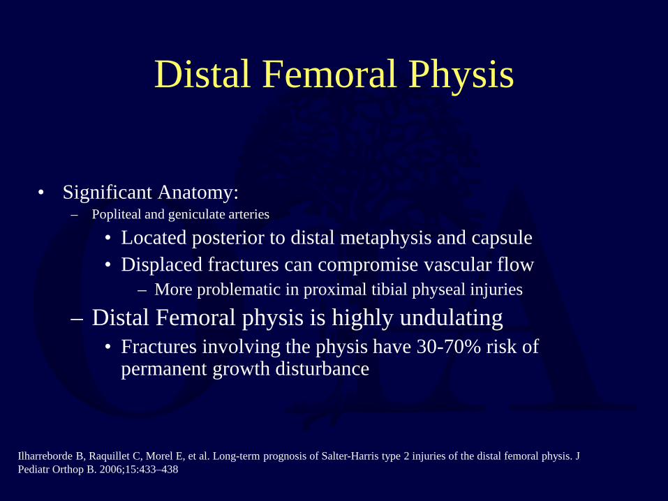
• Significant Anatomy: – Popliteal and geniculate arteries
• Located posterior to distal metaphysis and capsule • Displaced fractures can compromise vascular flow
– More problematic in proximal tibial physeal injuries
– Distal Femoral physis is highly undulating • Fractures involving the physis have 30-70% risk of
permanent growth disturbance Ilharreborde B, Raquillet C, Morel E, et al. Long-term prognosis of Salter-Harris type 2 injuries of the distal femoral physis. J
Pediatr Orthop B. 2006;15:433–438

Distal Femoral Physeal Fractures
• Fracture Epidemiology: – Rare, only accounts for <1% of fractures – Mechanism:
• High energy trauma • Sports injuries account for 2/3 of distal femur fractures • Varus/ Valgus force • Hyperextension of knee • Physis typically fails under traumatic force before ligaments in children

Distal Femoral Physeal Fractures • Physical exam:
– Effusion – Soft tissue swelling – Tenderness over physis – as opposed to isolated medial tenderness for
MCL sprain – Anteriorly displaced or hyperextension injuries cause patella to become
more prominent and anterior skin often dimpled – Posterior displacement can cause the distal metaphyseal fragment to
become more prominent above the patella – Inability to WB
(Zionts JAAOS 2002)

Distal Femoral Physeal Fractures • Always consider vascular
compromise • Knee dislocation equivalent • Perform AND document
– Peripheral pulses – Compartment evaluation – AAIs (Ankle-ankle Index) or
ABIs (Ankle-Brachial Index) • Reduce emergently if
vascular compromise – Reassess after reduction –
CTA if needed • Monitor for swelling

Distal Femoral Physeal Fractures • Associated injuries
– Ligamentous – Vascular – Nerve (peroneal if anteromedial displacement)
• Radiographs – AP & Lateral – Oblique View – Contralateral comparison – Stress X-ray – rarely utilized due to pain – CT – helpful in evaluating fracture complexity
• Surgical planning for fixation of metaphyseal fragment with screws – MRI
• For occult injuries or ruling out concomitant ligamentous/meniscal injuries
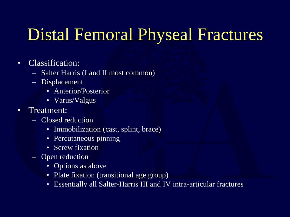
Distal Femoral Physeal Fractures • Classification:
– Salter Harris (I and II most common) – Displacement
• Anterior/Posterior • Varus/Valgus
• Treatment: – Closed reduction
• Immobilization (cast, splint, brace) • Percutaneous pinning • Screw fixation
– Open reduction • Options as above • Plate fixation (transitional age group) • Essentially all Salter-Harris III and IV intra-articular fractures

Distal Femoral Physeal Fractures • Closed reduction and casting:
– Non-displaced/stable fractures – Remodeling best in the flexion/extension plane – Do NOT manipulate after 7-10 days
• Early and rapid healing of physis • Delayed manipulation risks iatrogenic physeal injury
– Splint in slight knee flexion – Partial weight bearing at 3-4 weeks
• Closed reduction and internal fixation: – Reduction performed with traction and angular correction – Fixation should avoid physis if possible or cross with small diameter
smooth pins – Splint/Cast x4 weeks with pins – Almost always supplement reduction with fixation
• Prevent recurrent displacement
(Thomson J. JPO 1995)

Salter Harris I

Salter Harris I

Salter Harris I
• Treatment???

Salter Harris I -CRPP
After provisional urgent reduction and reassessment of NV status

Salter Harris I -CRPP
• Options – Antegrade percutaneous pin fixation
• Avoids pin placement into the knee joint • Decreases risk of septic arthritis
– Retrograde percutaneous pin fixation • Easier to place pins (more superficial starting point • Recommend burying to decrease infection risk • Removal at 6 weeks (if buried), 4 weeks if exposed
• Always supplement pin fixation with a splint/cast

Distal Femoral Physis
• Open Reduction – Indications
• Fractures that cannot be reduced closed – Interposed periosteum
• Open and displaced fractures • Floating knees
– Pre-operative CT can assist with surgical planning • Define plane of metaphyseal spike to plan screw trajectory
– Technical tip –
• The metaphyseal spike side will have intact periosteum covering in – open the fracture on the OPPOSITE side to remove interposed periosteum.

Salter Harris II
• 13 y.o male s/p football injury
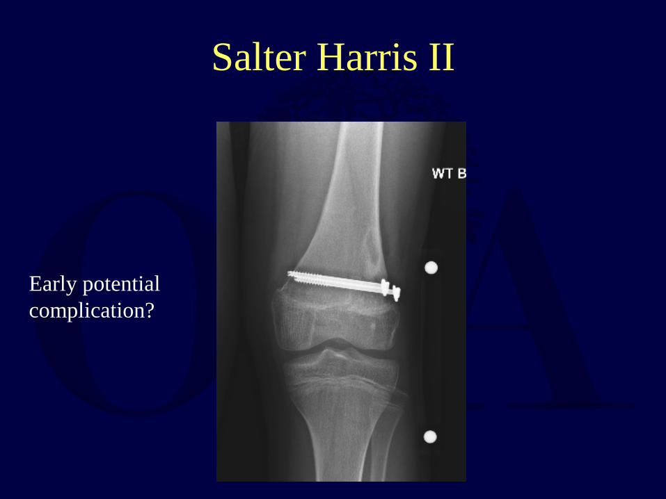
Salter Harris II
Early potential complication?

Salter Harris II
15 y.o soccer player injured during a game. Eight day delay in treatment due to being told it was probably an “ACL tear”
Salter Harris II
• Options for treatment?

Salter Harris II- ORIF
Treated with ORIF with plate and screw construct as patient was near skeletal maturity and to allow immediate unrestricted motion and decrease risk of stiffness.

Salter-Harris III
• 15 y.o male s/p football injury – valgus force to lateral left knee

Salter-Harris III
Technical tip: Hardware should remain anterior to Blumensaat’s line when passing intercondylar notch on AP view to remain out of the joint. Obtain notch view of distal femur to confirm intraosseous placement.
Open reduction and internal fixation

Distal Femoral Physeal Fractures • Outcomes:
– Risk of damage to growth plate & growth disturbance – Growth disturbance likely to occur in younger patients with
fractures that are displaced more than ½ the diameter of the shaft (Thomson JPO 1995)
– Check leg length, alignment, gait at 6 months (follow for 24 months) (Zionts JAAOS 2002)
– Leg length inequalities: » <2 cm at skeletal maturity nonsurgical » 2-5 cm appropriately timed epiphysiodesis of contralateral
leg » >5 cm leg lengthening should be considered
– Angular deformities managed by osteotomies or hemiepiphysiodesis

Major Complication - Growth Arrest
Initial injury after closed reduction attempt

Healed Notice early physeal closure

Growth Arrest – 6 months
Progressive limb length discrepancy

11 y.o M with right knee pain immediate after being tackled in football Minimally displaced SH1 distal femur fracture missed by ED and radiology
Salter Harris 1- Subtle Injury

Fracture treated closed, did not require reduction At follow up, physeal arrest noted
Expect a significant leg length discrepancy - 5 years of growth rema

Proximal Tibial Physeal Fractures
• Fracture epidemiology: – Rare, injury <1% of pediatric injuries – Mechanism:
• High energy trauma • Varus/ Valgus force • Hyperextension of the knee
• Physical Exam: – Pain – Knee effusion/ hemarthrosis – Tenderness at physis (circumferential tenderness/swelling) – Limb deformity – Record neurovascular exam before and after reduction
• Similar concern for vascular injury • AAI/ABI

Proximal Tibial Physeal Fractures
Can present with and develop significant swelling

Proximal Tibial Physeal Fractures • Associated Injuries:
– Ligamentous – Vascular
• Popliteal w/ posterior displacement of the metaphysis
– Compartment syndrome • Frequently reassess
• Radiographs – AP & Lateral X- rays – Stress X-rays (rarely used) – CT – MRI

Proximal Tibial Physeal Fractures
• Treatment: – Closed reduction
• Immobilization – Typically for nondisplaced fractures
• Fixation – Percutaneous pins
» Younger patients » Transphyseal
– Internal fixation » Epiphyseal fragments (screws) – for Salter-Harris III and IV » Metaphyseal fragments (screws and/or plates) – for Salter-
Harris II and IV – Open Reduction
• Similar fixation options

Proximal Tibial Physeal Fractures
• Closed reduction and casting: – Non-displaced/ stable fractures – Neurovascular exam post-reduction – Splint in slight knee flexion for 4-6 weeks
• Closed reduction and internal fixation – Unstable fractures (essentially all fractures requiring reduction) – Fixation parallel to physis or smooth pins if transphyseal necessary – Splint in slight knee flexion – Splint/Cast x4 weeks with pins

Salter Harris II Proximal Tibia and Fibula
14 y.o basketball player landing from a jump *Note the step-off posteriorly from epiphysis to metaphysis

Salter Harris II- CRPP Developed compartment syndrome and underwent emergent fasciotomy and CRPP.

Proximal Tibial Physeal Fractures
• ORIF: – Non anatomical reductions (Intra-articular fracture extension) – Internal fixation with screws parallel to physis – K-wires crossed and traversing the physis – Splint with slight knee flexion for 4 weeks – Typical postoperative progression
• Splint/Cast x4 weeks with pins • Remove pins and continue immobilization x 2 weeks versus
gentle motion • WBAT in cast/brace locked in extension, then progress

Case Example 10 yo M who was playing football and knee hyperextended when he was tackled
Concerns???

Developed Compartment Syndrome
He underwent CRPP and 4-compartment fasciotomy for compartment syndrome

Complications He developed proximal tibial physeal bar

Proximal Tibial Physeal Fractures
• Outcomes: (Zionts JAAOS 2002) – Prognosis good in most cases – Shortening and angular deformities less common because these
fractures occur in older children and because the proximal tibial epiphysis contributes less to growth than femur
– Open injuries coincide with poorer prognosis and more likely to have angular/shortening deformities

Tibial Tubercle Avulsion
• Anatomy: Epiphyseal development – Cartilaginous stage through 9-10 yrs – Apophyseal stage: ossification center (8-14 yo) – Epiphyseal stage: ossification center of tubercle and epiphysis
merge (10-17 yo) – Bony stage: physis is closed between tubercle and metaphysis
• Fracture Epidemiology:(Zionts JAAOS 2002) – 98% males, 12-17 yo – Mechanism:
• Active quadriceps extension with knee flexed • Jumping and sprinting

Tibial Tubercle Avulsion • Physical exam:
– Anterior prox. tibial swelling and tenderness – Palpable bony fragment – Patella alta possible – Hemarthrosis (with type 2/3 injuries) – Extensor lag/deficiency (with type 2/3 injuries)
• Associated Injuries: – Ligamentous – Meniscal – Extensor deficiency – Tibial plateau fracture
• Skin Blanching or compartment syndrome are surgical emergencies to prevent significant complications.

Tibial Tubercle Avulsion • Radiographs:
– AP & Lateral X-rays – Slight internal rotation on lateral may aid with tubercle
visualization – Differentiated from Osgood-Schlatters by acute fracture line
through physis
• Advanced Imaging – CT/MRI – Aids in surgical planning
• screw trajectory based on fracture line • concomitant injuries • Intercalary fragments
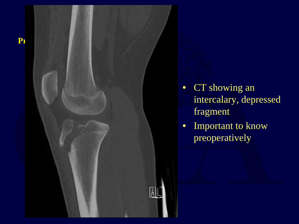
Preop CT
• CT showing an intercalary, depressed fragment
• Important to know preoperatively

Tibial Tubercle Avulsion
• Classification: Watson-Jones, with modifications of Ogden, Ryu, and Inoue – Type I: Fracture through the tubercle apophysis – Type II: Fracture through the apophysis that extends between
ossification centers of apophysis and epiphysis – Type III: Fracture through apophysis extends across epiphysis – Type IV: Fracture through apophysis extends posteriorly at level of
tibial physis – Type V: Avulsion of patellar tendon off tubercle physis (sleeve
fracture)

Tibial Tubercle Avulsion • Treatment:
– Closed reduction and casting – ORIF
• Closed reduction and casting: – Reduction with knee in extension – Cast molding above patella important for maintaining reduction – Cast in full extension for 6 weeks
• ORIF – Screw/pin fixation protected by soft tissue repair – In type 3 injuries the meniscus should be evaluated – Cylindrical cast for 6 weeks

Tibial Tubercle Avulsion
15 yo M was playing basketball and landed awkwardly on left leg from a jump shot

MRI MRI knee T1 and T2 images showing tibial tubercle avulsion fracture with patellar tendon avulsion off of the fracture fragment
Treatment options????
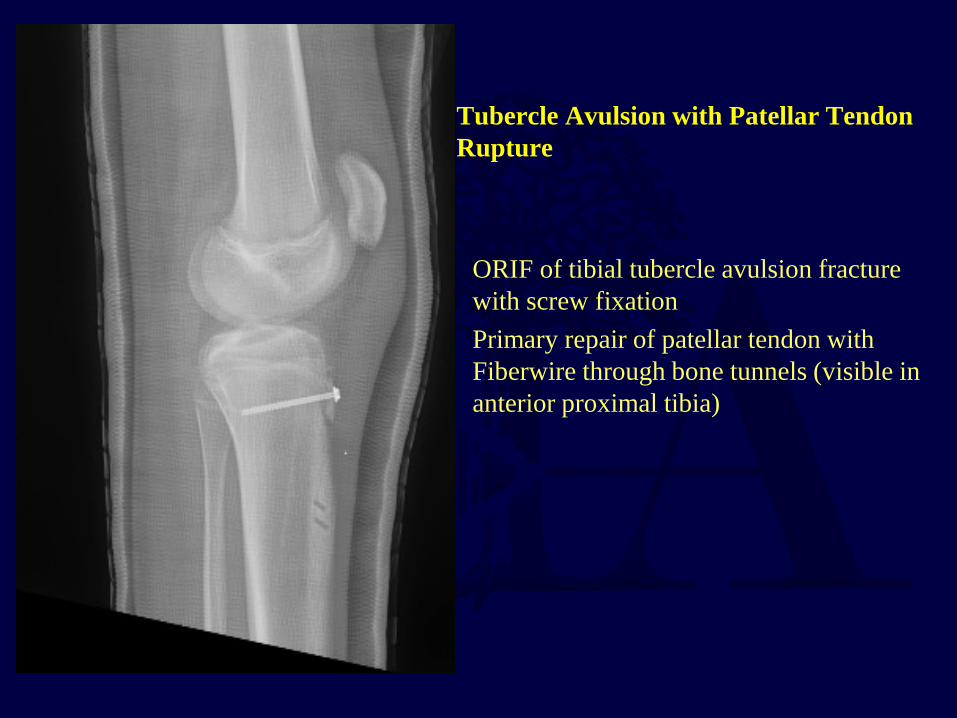
Tubercle Avulsion with Patellar Tendon Rupture
ORIF of tibial tubercle avulsion fracture with screw fixation Primary repair of patellar tendon with Fiberwire through bone tunnels (visible in anterior proximal tibia)

13 y.o male with pain after planting leg to throw football
Provisionally extend leg and immobilize in extension to reduce skin

CT for Surgical Planning

ORIF

Type 4 Tibial Tubercle 15 y.o football player with severe pain after planting leg decelerating to change directions
Note the posterior metaphyseal extension – complete articular instability

Type 4 Tibial Tubercle
Plating was utilized due to fracture configuration and posterior metaphyseal extension

Tibial Tubercle Avulsion
• Outcomes: – Good prognosis – Possible bursitis over prominent screws remove screws (Wiss
JOT 1991) – Possible growth disturbance – Possible loss of flexion secondary to stiffness

Patella Fracture
• Fracture Epidemiology – Rarely occur in children because patella is mostly cartilaginous
and has greater mobility than adults – Ossification occurs at 3-5 yo – Mechanism:
• Avulsion patella fractures more likely in children • Eccentric contraction • Comminuted fracture secondary to direct trauma

Patella Fracture
6 y.o female s/p fall directly onto knee

Patella Fracture
• Physical exam: – Painful/swollen knee – Lack of active knee extension – Inability to bear weight – Hemarthrosis – Patella alta
• Radiographs: – AP & Lateral x-rays – Sagittal plane fractures seen on sunrise view – Comparison contralateral
Patella Fracture
• Classification: (Grogan JPO 1990) – Primary osseous fractures – Avulsion Fractures
• Superior, inferior, medial (often w/ acute lateral dislocation of patella), lateral (chronic stress from repetitive pull from vastus lateralis)
– Sleeve fractures • Through cartilage on inferior or superior pole of patella • Easily overlooked - “Little amount of bone, Large amount of
cartilage” • Assess for palpable defect at the affected patellar pole • Loss of knee extension

Patella Fracture
• Treatment: – Closed treatment with casting – ORIF
• Closed treatment with casting: – Extensor mechanism intact – No significant displacement
• <2-3 mm at articular surface

Patella Fracture • ORIF: (AO tension band, circumferential wire/suture loop,
interfragmentary screws) – >3mm displacement at articular surface – Sutures alone good enough for sleeve fractures – Repair retinaculum – Splint 4-6 weeks
• Outcomes: (Zionts JAAOS 2002) – Good prognosis – Complication if patella not accurately reduced:
• Patella alta • Extensor lag • Quadriceps muscle atrophy

Patella Fracture -ORIF Tension band technique with braided nonabsorbable high-tension

INTRA-ARTICULAR INJURIES

Hemarthrosis
• ~50% chance traumatic hemarthrosis is ACL tear
• Can be due to tearing any vascularized intra-articular structure – Osteochondral – Meniscus – ACL/PCL – Patella
• If xrays negative, consider MRI as next di i

Tibial Eminence Fractures • Fracture epidemiology:(Zionts JAAOS 2002)
– 8-14 y.o children – Before ossification is complete the surface of the tibial spine is
cartilaginous – With excessive forces applied to the ACL, the spine offers less
resistance than the ligament • leads to a fracture through cancellous bone beneath tibial
spine – Mechanism:
• Rapid deceleration or hyperextension of the knee • Forces that would lead to ACL tear in adults lead to tibial spine
fractures in children
Tibial Eminence Fractures • Physical Exam:
– Pain – Effusion – Positive Lachman
• Associated injuries: – Meniscal injury – Collateral ligament injury – Capsular damage – Osteochondral fracture
• Recommend MRI to assess for associated injuries in all displaced tibial eminence fractures (Mitchell JPO2015)

Tibial Eminence Fractures
• Classification: Meyers-Mckeever (Meyers JBJS 1970) – Type I- non-displaced – Type II- minimally displaced with intact posterior hinge – Type III- complete, displaced, and may be rotated
• Treatment: – Reduction with evacuation of hemarthrosis – Above knee immobilization with knee in slight flexion
• Some suggest greater flexion to relax ACL – (Meyers JBJS 1970)
– Operative when extension is blocked, displacement is present or meniscus is entrapped
Tibial Eminence Fractures
• Outcomes: (Smith JPO 1984, Baxter JBJS 1988, Willis JPO 1993) – Short term prognosis is good, long-term remains unclear – Some report ACL laxity and loss of full extension despite healing
in anatomic position • Attributed to interstitial tearing of ACL that occurs before fragment
avulses • Laxity more common in type 2/3 fractures

Tibial Eminence Fractures
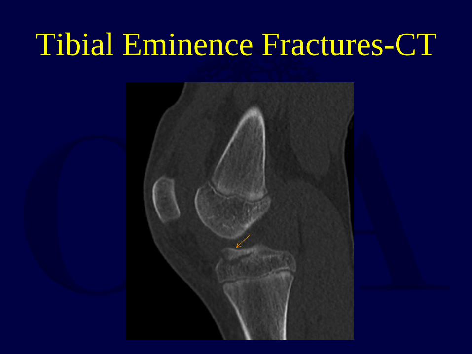
Tibial Eminence Fractures-CT

Intermeniscal Ligament Blocking Reduction

Arthroscopic Treatment • Screw or suture fixation options • Images pre- and post- reduction with suture
fixation
Pre Post
Osteochondral Fractures • Fracture epidemiology:(Rorabeck JBJS 1976)
– Occur in 5% of all acute patella dislocations – Mechanism:(Zionts JAAOS 2002)
• Direct blow to a flexed knee • Shearing forces associated with an acute dislocation or the patella
– 3 fracture patterns following dislocation (Rorabeck JBJS 1976) » Inferomedial fracture of patella » Fracture of lateral femoral condyle » Combination of the two
• Assume the osteochondral fracture is always present unless you prove it is not with careful MRI review – Will hide in plain sight….

Osteochondral Fractures
• Physical exam: – Painful/swollen joint – Flexion/extension resisted – Hemarthrosis with fat globules on knee aspiration
• Radiographs:(Zionts JAAOS 2002) – Hard to visualize on AP & Lateral – Oblique, skyline, and notch views – CT – MRI

Osteochondral Fractures
• Treatment: – Surgical excision or reattachment
• Depends on size/origin • Large weight bearing pieces should be reattached
• Outcomes:(Zionts JAAOS 2002) – Good prognosis for small weight bearing pieces – Prognosis less certain for larger weight bearing pieces – If secondary to patellar dislocation the patient may develop
recurrent subluxation or dislocation of the patella • More prevalent if the initial dislocation is in the early teenage years

Osteochondral Fractures-Lateral Femoral Condyle
Thin osteochondral fragment Resulting chondral defect

Treated with ORIF
Osteochondral fragment visualized
After fixation

Treated with ORIF
Postoperative images Planned full ROM, nonweightbearing for 3 months until subsequent screw removal

3 months Postop • Healing seen at time of hardware removal

Patellar Dislocation • Majority are lateral • Most reduce with knee extension and present with hemarthrosis • Rx: Immobilization in extension for 4 weeks, then PT for progressive
strengthening (especially hip abductors and VMO) • Factors leading to increased recurrence
– ligamentous laxity – genu valgum – torsional malalignment – trochlear dysplasia
• Surgical treatment considered for failed rehab, or recurrent dislocations

MRI
• Indirect evidence of patellar dislocation
• Osseous contusion medial aspect of patella (shown in image)
• Corresponding contusion lateral femoral condyle
• Osteochondral fracture

Patellar Dislocation • Surgical treatment
– Risk physeal injury with standard MPFL reconstruction
• Use fluoroscopic imaging to place femoral attachment point distal to the medial distal femoral physis
• Various techniques – Guided growth – hemiepiphysiodesis – should
be considered as initial option to resolve underlying mechanical malalignment
• May obviate need for further treatment of instability • Obtain longstanding hips to ankles x-rays on
patients once full extension achieved to evaluate alignment
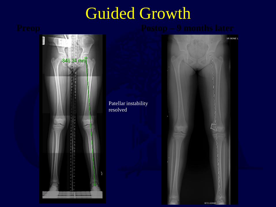
Guided Growth Preop Postop – 9 months later
Patellar instability resolved

Meniscal Injuries • Epidemiology:
– Common tears: bucket handles, flap, and radial – Often associated with ACL injuries – Mechanism:
• Squatting with a twisting motion at the knee • Direct trauma • Degenerative tears in older individuals
– Physical exam: (inconsistent) • Joint line tenderness • Stiffness and swelling • Catching or locking of your knee • Knee “giving out” • McMurray’s test – pop and pain with loaded flexed rotation of tibia on
femoral condyle

Meniscal Injuries
• Imaging: – MRI
• In children, high signal lines in the meniscus can be normal vascular ingrowth and not true tears
• Treatment: – Non-operative: small, stable, non-displaced, on the peripheral
region (<1 cm) – Partial meniscectomy: complex tears, central tears (white-white
zone), degenerative changes (less common in kids) – Meniscal repair: tears located in the middle and peripheral part of
the meniscus (red-red and white-red zone)

ACL and Bucket Tear Meniscus
Double-PCL sign = meniscus flipped into the intercondylar notch

Bucket Handle Medial Meniscus
Displaced anteriorly After Inside-Out repair

Ligament Injuries • Epidemiology:
– Teenage children in sports – ACL tears
• Clues – Fairly rapid hemarthrosis – Inability to return to game
– Mechanism: • Lateral blow to the leg • Cutting maneuvers while running
• Treatment: – Non-operative:
• Incomplete tears of ACL/PCL • Isolated collateral ligament injuries
– Operative: • Complete ACL/PCL tears
Proximal ACL tear with open physes

Knee Dislocations • Epidemiology:
– Rare, 0.02% of all orthopaedic injuries (Rihn JAAOS 2004) – Incidence of injury to popliteal injury ranges from 1.6-30%(Sill
JTACS 2014, Stannard JBJS 2004) – Even more rare in children
• Physis/bone fail prior to ligament failing – Usually associated with multiple ligamentous injuries
• Physical Exam: – Pain, swelling – Ligamentous instability – May have obvious deformity – If capsule disrupted, may present with only mild effusion – DOCUMENT Pulses, AAIs/ABIs
Knee Dislocations
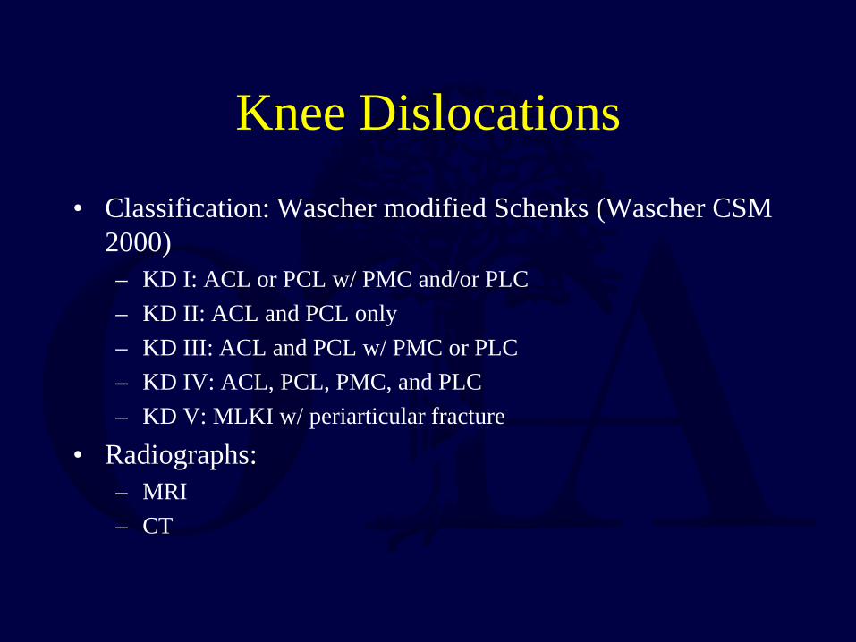
• Classification: Wascher modified Schenks (Wascher CSM 2000) – KD I: ACL or PCL w/ PMC and/or PLC – KD II: ACL and PCL only – KD III: ACL and PCL w/ PMC or PLC – KD IV: ACL, PCL, PMC, and PLC – KD V: MLKI w/ periarticular fracture
• Radiographs: – MRI – CT

Knee Dislocations
• Treatment: – Reduction with neurovascular exam before and after – Knee immobilizer in extension – Operative if there is an unstable knee with ligamentous injury – External fixation for stability if vascular repair is required

Summary • Pediatric Knee injuries present unique challenges due to
the physis • Monitor for neurovascular injuries, skin compromise,
and compartment syndrome with knee injuries (despite benign-appearing radiographs)
• Pediatric patients have a lower chance of stiffness so fixation can be supplemented with immobilization
• Articular injuries in kids still require anatomic reduction • Avoid crossing the physis with fixation unless near
skeletal maturity or using small-diameter smooth provisional pins

References • Baxter MP, Wiley JJ: Fractures of the tibial spine in children: An evaluation of knee stability. J
Bone Joint Surg Br 1988;70:228-230. • Grogan DP, Carey TP, Leffers D, Ogden JA: Avulsion fractures of the patella. J Pediatr
Orthop 1990;10:721-730. • Ilharreborde B, Raquillet C, Morel E, et al. Long-term prognosis of Salter-Harris type 2
injuries of the distal femoral physis. J Pediatr Orthop B. 2006;15:433–438. • Meyers MH, McKeever FM: Fracture of the intercondylar eminence of the tibia. J Bone Joint
Surg Am 1970;52:1677-1684. • Mitchell JJ, Sjostrom R, Mansour AA, Irion B, Hotchkiss M, Terhune EB, Carry P, Stewart
JR, Vidal AF, Rhodes JT. Incidence of meniscal injury and chondral pathology in anterior tibial spine fractures of children. J Pediatr Orthop. 2015 Mar;35(2):130-5.
• Rihn JA, Groff YJ, Harner CD, Cha PS: The acutely dislocated knee: Evaluation and management. J Am Acad Orthop Surg 2004;12(5):334-346.
• Rorabeck CH, Bobechko WP: Acute dislocation of the patella with osteochondral fracture: A review of eighteen cases. J Bone Joint Surg Br 1976;58: 237-240.
• Sillanpää PJ, Kannus P, Niemi ST, Rolf C, Felländer-Tsai L, Mattila VM: Incidence of knee dislocation and concomitant vascular injury requiring surgery: A nationwide study. J Trauma Acute Care Surg 2014;76 (3):715-719.

References • Smith JB: Knee instability after fractures of the intercondylar eminence of the tibia. J
Pediatr Orthop 1984;4:462-464. • Stannard JP, Sheils TM, Lopez-Ben RR, McGwin G Jr, Robinson JT, Volgas DA:
Vascular injuries in knee dislocations: The role of physical examination in determining the need for arteriography. J Bone Joint Surg Am 2004;86(5):910-915.
• Thomson JD, Stricker SJ, Williams MM: Fractures of the distal femoral epiphyseal plate. J Pediatr Orthop 1995;15:474-478.
• Wascher DC: High-velocity knee dislocation with vascular injury: Treatment principles. Clin Sports Med 2000;19(3):457-477.
• Willis RB, Blokker C, Stoll TM, Paterson DC, Galpin RD: Long-term follow-up of anterior tibial eminence fractures. J Pediatr Orthop 1993;13:361-364.
• Wiss DA, Schilz JL, Zionts L: Type III fractures of the tibial tubercle in adolescents. J Orthop Trauma 1991;5:475-479.
• Zionts, LE: Fractures around the knee in children. J Am Acad Orthop Surg 2002;10: 345-355

• For questions or comments, please send to [email protected]


![Tachdjian's Pediatric Orthopaedics [Chapter 20] · 2018-04-17 · CHAPTER 20 Disorders of the Knee Osteochondritis Dissecans, Patellofemoral Instability, Plica Syndrome, 81 0 Bipartite](https://img.dokumen.tips/doc/110x75/5b31b3ea7f8b9a744a8c0137/tachdjians-pediatric-orthopaedics-chapter-20-2018-04-17-chapter-20-disorders.jpg)