Embed Size (px)
Citation preview
https://d0887-21(http://c
DepartmCin
AddressRadE-m
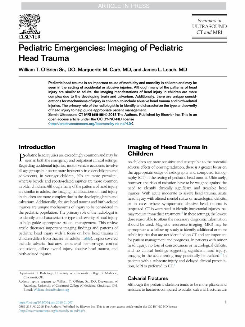
Pediatric Emergencies: Imaging of PediatricHead TraumaWilliam T. O’Brien Sr., DO, Marguerite M. Caré, MD, and James L. Leach, MD
oi.org/10.105371/& 2018 Threativecommon
ent of Radiocinnati, OH.reprint reque
iology, Universail: William.ob
Pediatric head trauma is an important cause of morbidity andmortality in children and may beseen in the setting of accidental or abusive injuries. Although many of the patterns of headinjury are similar to adults, the imaging manifestations of head injury in children are morecomplex due to the developing brain and calvarium. Additionally, there are unique consid-erations for mechanisms of injury in children, to include abusive head trauma and birth-relatedinjuries. The primary role of the radiologist is to identify and characterize the type and severityof head injury to help guide appropriate patient management.Semin Ultrasound CT MRI ]:]]]-]]] C 2018 The Authors. Published by Elsevier Inc. This is anopen access article under the CC BY-NC-ND license(http://creativecommons.org/licenses/by-nc-nd/4.0/).
Introduction
Pediatric head injuries are exceedingly common andmay beseen in both the emergency andoutpatient clinical settings.
Regarding accidental injuries, motor vehicle accidents involveall age groups but occur more frequently in older children andadolescents. In younger children, falls are more prevalent,whereas bicycle and sports-related injuries are more commonin older children. Althoughmany of the patterns of head injuryare similar to adults, the imaging manifestations of head injuryin children are more complex due to the developing brain andcalvarium. Additionally, abusive head trauma and birth-relatedinjuries are unique mechanisms of injury to be considered inthe pediatric population. The primary role of the radiologist isto identify and characterize the type and severity of head injuryto help guide appropriate patient management. This reviewarticle discusses important imaging findings and patterns ofpediatric head injury with a focus on how head trauma inchildren differs from that seen in adults (Table). Topics coveredinclude calvarial fractures, extra-axial hemorrhage, corticalcontusions, diffuse axonal injury, abusive head trauma, andbirth-related injuries.
/j.sult.2018.01.007e Authors. Published by Elsevier Inc. This is an open access as.org/licenses/by-nc-nd/4.0/).
logy, University of Cincinnati College of Medicine,
sts to William T. O’Brien, Sr., DO, Department ofity of Cincinnati College of Medicine, Cincinnati, [email protected]
Imaging of Head Trauma inChildrenAs children are more sensitive and susceptible to the potentialadverse effects of ionizing radiation, there is a greater focus onthe appropriate usage of radiographs and computed tomog-raphy (CT) in the setting of pediatric head trauma. Ultimately,however, the risks of radiation have to be weighed against theneed to identify clinically significant and treatable headinjuries. With acute moderate to severe head trauma, acutehead injury with altered mental status or neurological deficits,or in cases where symptomatic abusive head trauma issuspected, CT is warranted to identify intracranial injuries thatmay require immediate treatment.1 In these settings, the lowestdose reasonable to attain the necessary diagnostic informationshould be used. Magnetic resonance imaging (MRI) may beappropriate as a follow-up study to identify additional or moresubtle injuries that are not identified on CT and are importantfor patient management and prognosis. In patients with minorhead injury, no loss of consciousness or neurological deficits,and no clinical findings suggesting significant head injury,imaging in the acute setting may potentially be avoided.2 Inpatients with a subacute injury and delayed clinical presenta-tion, MRI is preferred to CT.1
Calvarial FracturesAlthough the pediatric skeleton tends to be more pliable andresistant to fractures compared to adults, calvarial fractures are
1rticle under the CC BY-NC-ND license
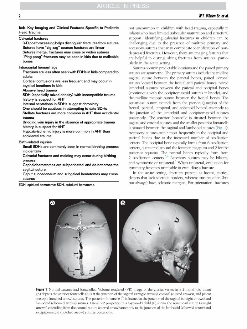
Table Key Imaging and Clinical Features Specific to PediatricHead Trauma
Calvarial fractures3-D postprocessing helps distinguish fractures fromsuturesSutures have “zig-zag” course; fractures are linearSutures merge; fractures may cross or widen sutures“Ping pong” fractures may be seen in kids due to malleablebones
Intracranial hemorrhageFractures are less often seenwith EDHs in kids compared toadultsCortical contusions are less frequent and may occur inatypical locations in kidsAbusive head traumaSDH (especially mixed density) with incompatible traumahistory is suspect for AHTInternal septations in SDHs suggest chronicityOne should be cautious in attempting to date SDHsStellate fractures are more common in AHT than accidentaltraumaBridging vein injury in the absence of appropriate traumahistory is suspect for AHTHypoxic-ischemic injury is more common in AHT thanaccidental trauma
Birth-related injuriesSmall SDHs are commonly seen in normal birthing processincidentallyCalvarial fractures and molding may occur during birthingprocessCephalohematomas are subperiosteal and do not cross thesagittal sutureCaput succedaneum and subgaleal hematomas may crosssutures
EDH, epidural hematoma; SDH, subdural hematoma.
A B
Figure 1 Normal sutures and fontanelles. Volume rendered (V(A) depicts the anterior fontanelle (AF) at the junction of the sagmetopic (notched arrow) sutures. The posterior fontanelle (*) islambdoid (elbowed arrows) sutures. Lateral VR projection in aarrows) extending from the coronal suture (curved arrow) anteroccipitomastoid (notched arrow) sutures posteriorly.
W.T. O’Brien Sr. et al.2
not uncommon in children with head trauma, especially ininfants who have limited trabecular maturation and structuralsupport. Identifying calvarial fractures in children can bechallenging due to the presence of multiple primary andaccessory sutures that may complicate identification of non-depressed fractures. However, there are imaging features thatare helpful in distinguishing fractures from sutures, partic-ularly in the acute setting.Sutures occur in predictable locations and the pairedprimary
sutures are symmetric. The primary sutures include themidlinesagittal suture between the parietal bones, paired coronalsutures located between the frontal and parietal bones, pairedlambdoid sutures between the parietal and occipital bones(continuous with the occipitomastoid sutures inferiorly), andthe midline metopic suture between the frontal bones. Thesquamosal suture extends from the pterion (junction of thefrontal, parietal, temporal, and sphenoid bones) anteriorly tothe junction of the lambdoid and occipitomastoid suturesposteriorly. The anterior fontanelle is situated between thesagittal and coronal sutures, and the smaller posterior fontanelleis situated between the sagittal and lambdoid sutures (Fig. 1).Accessory sutures occur most frequently in the occipital andparietal bones due to the increased number of ossificationcenters. The occipital bone typically forms from 6 ossificationcenters, 4 centered around the foramen magnum and 2 for theposterior squama. The parietal bones typically form from2 ossification centers.3,4 Accessory sutures may be bilateraland symmetric or unilateral.5 When unilateral, evaluation forsymmetry becomes unreliable in excluding a fracture.In the acute setting, fractures present as lucent, cortical
defects that lack sclerotic borders, whereas sutures often (butnot always) have sclerotic margins. For orientation, fractures
R) image of the cranial vertex in a 2-month-old infantittal (straight arrows), coronal (curved arrows), and patentlocated at the junction of the sagittal (straight arrows) and4-year-old child (B) shows the squamosal suture (straightiorly to the junction of the lambdoid (elbowed arrow) and
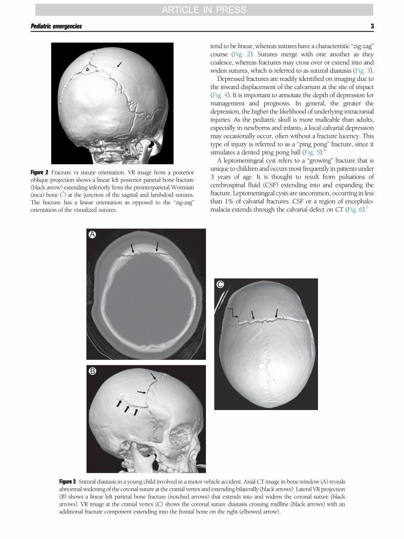
Figure 2 Fracture vs suture orientation. VR image from a posterioroblique projection shows a linear left posterior parietal bone fracture(black arrow) extending inferiorly from the preinterparietal Wormian(inca) bone (*) at the junction of the sagittal and lambdoid sutures.The fracture has a linear orientation as opposed to the “zig-zag”orientation of the visualized sutures.
A
B
Figure 3 Sutural diastasis in a young child involved in amotor veabnormalwidening of the coronal suture at the cranial vertex and(B) shows a linear left parietal bone fracture (notched arrows)arrows). VR image at the cranial vertex (C) shows the coronaladditional fracture component extending into the frontal bone
Pediatric emergencies 3
tend to be linear, whereas sutures have a characteristic “zig-zag”course (Fig. 2). Sutures merge with one another as theycoalesce, whereas fractures may cross over or extend into andwiden sutures, which is referred to as sutural diastasis (Fig. 3).Depressed fractures are readily identified on imaging due to
the inward displacement of the calvarium at the site of impact(Fig. 4). It is important to annotate the depth of depression formanagement and prognosis. In general, the greater thedepression, the higher the likelihood of underlying intracranialinjuries. As the pediatric skull is more malleable than adults,especially in newborns and infants, a focal calvarial depressionmay occasionally occur, often without a fracture lucency. Thistype of injury is referred to as a “ping pong” fracture, since itsimulates a dented ping pong ball (Fig. 5).6
A leptomeningeal cyst refers to a “growing” fracture that isunique to children andoccursmost frequently in patients under3 years of age. It is thought to result from pulsations ofcerebrospinal fluid (CSF) extending into and expanding thefracture. Leptomeningeal cysts are uncommon, occurring in lessthan 1% of calvarial fractures. CSF or a region of encephalo-malacia extends through the calvarial defect on CT (Fig. 6).7
C
hicle accident. Axial CT image in bonewindow (A) revealsextending bilaterally (black arrows). Lateral VRprojectionthat extends into and widens the coronal suture (blacksuture diastasis crossing midline (black arrows) with anon the right (elbowed arrow).

A B
C D
Figure 4 Depressed calvarial fracture with intracranial injuries. Axial (A) and sagittal reformatted (B) CT images in bonewindow show a comminuted depressed right parietal bone fracture with inward angulation. Corresponding images inbrain window (C and D) show associated intracranial hemorrhage (*) and a superficial scalp hematoma (arrows).
W.T. O’Brien Sr. et al.4
On CT, fractures that are nondisplaced and oriented similarto the plane of imaging can easily be overlooked. For thesereasons, it is important to review the scout image, havereformatted images available in at least 2 planes (commonlyaxial and coronal or all three planes in the setting of trauma), or
A B
Figure 5 “Ping pong” fracture in an infant post-trauma. Axial Cinvolving the left parietal bone without a discernible fracture l(B) illustrates how the depression is similar to a dented ping po
use 3-dimensional reconstructions to appropriately evaluatethe calvarium (Fig. 7).Skull base fractures can be subtle and difficult to identify on
imaging, especially in the presence of multiple accessorysutures. When fractures extend into pneumatized and aerated
T image in bone window (A) reveals a focal depressionucency (arrow). VR image in a lateral oblique projectionng ball.

A B
C D
Figure 6 Leptomeningeal cyst (“growing” fracture) in a child after amotor vehicle accident. Axial CT image in bonewindow(A) shows a mildly displaced right parietal bone fracture (white arrow). Corresponding axial image in brain window(B) reveals underlying extra-axial (notched arrow) and subarachnoid (elbowed arrows) hemorrhage, as well as corticalcontusions with surrounding edema. Follow-up CT image in bone window 6 months later (C) shows widening of thecalvarial fracture (white arrow). Corresponding image in brain window (D) reveals cerebrospinal fluid extending into thefracture lucency (black arrow) with underlying encephalomalacia.
Pediatric emergencies 5
cavities, such as the paranasal sinuses, mastoid air cells, ormiddle ear cavities, secondary findings of layering hyperdensefluid (hemorrhage) within the aerated cavities should promptcloser evaluation for an underlying fracture (Fig. 8). Pneumo-cephalus may also be seen as the fracture leads to communi-cation of the aerated cavity with the cranial vault. Anterior orcentral skull base fractures may have additional orbital or facialbone fractures. Central skull base fractures may involve thecarotid canal, jugular foramen, or dural venous sinus channels,resulting in vascular injury or occlusion (Fig. 9).Secondary findings commonly associated with calvarial
fractures include superficial soft tissue swelling or underlyingextra-axial hemorrhage, pneumocephalus, and parenchymalcontusions. Diffuse axonal injury may also be seen. Withdelayed presentation, as may be seen in some cases of abusivehead trauma, secondary findings may no longer be present atthe time of imaging; therefore, their absence does not excludeprior trauma.
Extra-Axial HemorrhageExtra-axial hemorrhages are common in the setting of headtrauma and are classified as epidural, subdural, or subarach-noid in location based upon the compartment in which theyoccur. At times, it may be difficult on imaging to distinguishbetween epidural and subdural hematomas, especially whensmall and not located in midline or along a suture.In the acute setting, extra-axial hemorrhages are typically
hyperdense compared to gray matter on CT. In the subacutestage, they become isodense to underlying gray matter, atwhich point they may be overlooked if not looking carefully atthe peripheral cortical boundaries. In the chronic stage, extra-axial hemorrhage is hypodense to gray matter (Fig. 10). Oneimaging pitfall in the CT imaging appearance of an extra-axialhemorrhage occurs in the hyperacute stage where portions ofthe hemorrhage may appear isodense or even slightly hypo-dense compared to gray matter due to unclotted bloodproducts.8 The imaging appearance and typical evolution
A
C
B
Figure 7 Subtlety of fractures in the plane of imaging. Axial CT image in bonewindow through the area of injury (A) revealsno discernible fracture. Corresponding image in the coronal plane (B) shows a minimally displaced right parietal bonefracture (arrow)with overlying scalp soft tissue swelling. VR image from a lateral projection (C) best depicts the extent of thefracture involving the mid to posterior portion of the parietal bone (white arrow) and extending to the lambdoid suture(black arrow).
W.T. O’Brien Sr. et al.6
pattern of hemorrhagic blood products may be altered withactive hemorrhage, when there are hemorrhages of differentages, in patients with underlying anemia or coagulopathy, orwith intermixing of CSF within the regions of hemorrhage.
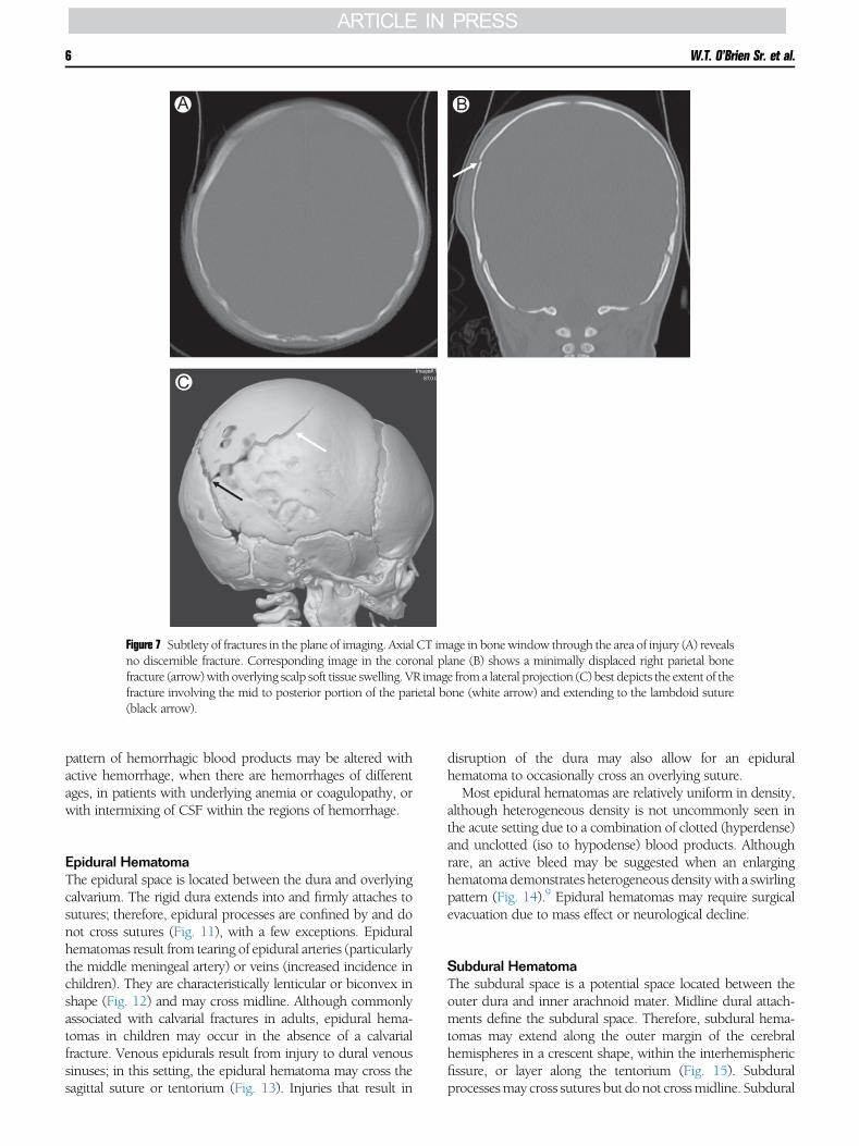
Epidural HematomaThe epidural space is located between the dura and overlyingcalvarium. The rigid dura extends into and firmly attaches tosutures; therefore, epidural processes are confined by and donot cross sutures (Fig. 11), with a few exceptions. Epiduralhematomas result from tearing of epidural arteries (particularlythe middle meningeal artery) or veins (increased incidence inchildren). They are characteristically lenticular or biconvex inshape (Fig. 12) and may cross midline. Although commonlyassociated with calvarial fractures in adults, epidural hema-tomas in children may occur in the absence of a calvarialfracture. Venous epidurals result from injury to dural venoussinuses; in this setting, the epidural hematoma may cross thesagittal suture or tentorium (Fig. 13). Injuries that result in
disruption of the dura may also allow for an epiduralhematoma to occasionally cross an overlying suture.Most epidural hematomas are relatively uniform in density,
although heterogeneous density is not uncommonly seen inthe acute setting due to a combination of clotted (hyperdense)and unclotted (iso to hypodense) blood products. Althoughrare, an active bleed may be suggested when an enlarginghematomademonstrates heterogeneous densitywith a swirlingpattern (Fig. 14).9 Epidural hematomas may require surgicalevacuation due to mass effect or neurological decline.
Subdural HematomaThe subdural space is a potential space located between theouter dura and inner arachnoid mater. Midline dural attach-ments define the subdural space. Therefore, subdural hema-tomas may extend along the outer margin of the cerebralhemispheres in a crescent shape, within the interhemisphericfissure, or layer along the tentorium (Fig. 15). Subduralprocessesmay cross sutures but donot crossmidline. Subdural

A
B
Figure 8 Hyperdense fluid within pneumatized spaces as a secondaryfinding of skull base fracture. Axial CT image in soft tissue window(A) demonstrates hyperdense fluid layering within the left sphenoidsinus (*), which prompted a second review of bone windows. AxialCT image in bonewindow (B), a few slices superior to image A, showsa subtle skull base fracture extending through the walls of the leftsphenoid sinus (white arrows), as well as involvement of the carotidcanal (black arrow).
A
B
C
Pediatric emergencies 7
hematomas result from injury to cortical bridging veins as theytraverse the subdural space.As discussed previously are typically hyperdense in the
acute stage, isodense in the subacute stage, and hypodensein the chronic stage compared to underlying gray matter.The attenuation may be more heterogeneous due to
A
B
Figure 9 Vascular injury associated with skull base fractures. Refor-matted coronal CT image in bone window (A) shows a skull basefracture on the right that involves the carotid canal (black arrows).There is complete opacification of the right middle ear cavity (*). Axialimage from a follow-up CT angiogram (B) demonstrates absentcontrast enhancement with occlusion of the distal petrous segment ofthe right internal carotid artery (white arrow).
Figure 10 Stages of hemorrhage in a 14-year-old boy post-trauma.Initial axial noncontrast head CT image (A) shows a hyperdensesubdural hemorrhage overlying the left cerebral convexity (arrows).On a follow-up study 1week later (B), the hemorrhage is decreased insize and relatively isodense to underlying gray matter (arrows).Additional follow-up examination 1month later (C) shows continueddecrease in size of the subdural hemorrhage which is now hypodensecompared to brain parenchyma (arrows).
intermixing of CSF within the subdural collections (hema-tohygroma) or in the setting of repeat or active hemorrhage(Fig. 16). Treatment often depends upon the size of thehematoma, degree of mass effect, and neurological status ofthe patient.

A B
Figure 11 Epidural hemorrhage bounded by sutures. Axial noncontrast CT image in brain window (A) shows a hyperdenselenticular epidural hemorrhage overlying the left frontal lobe (*). Corresponding axial CT image in bonewindow (B) showsthat the posterior margin of the hemorrhage in bounded by the coronal suture (arrow).
W.T. O’Brien Sr. et al.8
Subarachnoid HemorrhageThe subarachnoid space is located between the overlyingarachnoid mater and underlying pial covering of the brainparenchyma. Subarachnoid hemorrhage is common in thesetting of head trauma, whereas a ruptured aneurysm repre-sents the most common nontraumatic etiology of subarach-noid hemorrhage.Subarachnoid hemorrhage appears as hyperdense foci
typically located dependently within the cerebral sulci orCSF cisterns (Fig. 17). It is important to properly window
A B
Figure 12 Epidural hemorrhagemorphology. Axial noncontrast Chyperdense epidural hemorrhage (*) overlying the left frontaCorresponding CT image in bone window (B) shows a minima
and level each study to optimally evaluate for foci ofhemorrhage. Commonly overlooked locations for subtle sub-arachnoid hemorrhage include the interpeduncular fossa andsylvian fissures.10
Pseudosubarachnoid HemorrhageIn the setting of diffuse cerebral edema, relative hyperdensitywithin the basal cisterns may be noted in the absence of a truebleed (Fig. 18). This hyperdensity results from vascularengorgement secondary to increased intracranial pressures,
T image in brainwindow (A) reveals a lenticular/biconvexl lobe. There is a prominent scalp hematoma (arrows).lly displaced left frontal bone fracture (arrow).
A B
C D
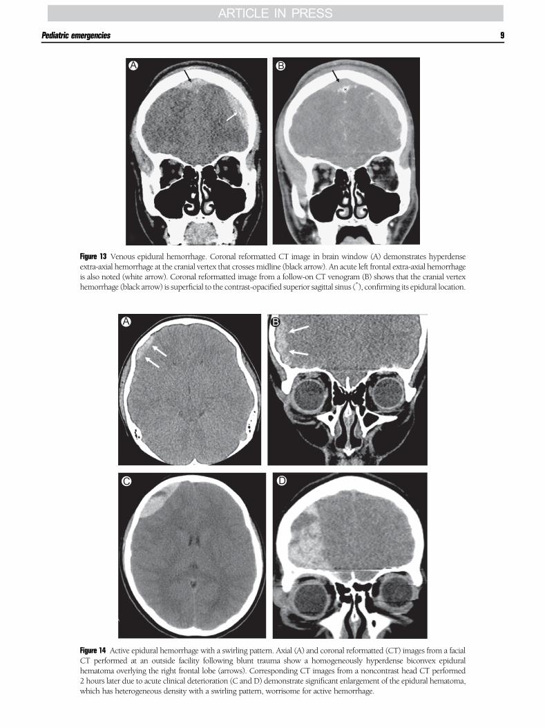
Figure 14 Active epidural hemorrhage with a swirling pattern. Axial (A) and coronal reformatted (CT) images from a facialCT performed at an outside facility following blunt trauma show a homogeneously hyperdense biconvex epiduralhematoma overlying the right frontal lobe (arrows). Corresponding CT images from a noncontrast head CT performed2 hours later due to acute clinical deterioration (C and D) demonstrate significant enlargement of the epidural hematoma,which has heterogeneous density with a swirling pattern, worrisome for active hemorrhage.
A B
Figure 13 Venous epidural hemorrhage. Coronal reformatted CT image in brain window (A) demonstrates hyperdenseextra-axial hemorrhage at the cranial vertex that crosses midline (black arrow). An acute left frontal extra-axial hemorrhageis also noted (white arrow). Coronal reformatted image from a follow-on CT venogram (B) shows that the cranial vertexhemorrhage (black arrow) is superficial to the contrast-opacified superior sagittal sinus (*), confirming its epidural location.
Pediatric emergencies 9

A B
Figure 15 Subdural hemorrhage morphology and distribution. Axial noncontrast CT image in brain window (A) shows ahyperdense crescent-shaped subdural hemorrhage overlying the left cerebral hemisphere (black arrows) with extensionalong the interhemispheric fissure (white arrows). Coronal reformatted CT image in brain window (B) again demonstratesthe subdural hemorrhage along the left cerebral convexity (black arrows) and interhemispheric fissure (white arrows), aswell as subdural hemorrhage extending along the tentorium on the left (notched arrows).
W.T. O’Brien Sr. et al.10
along with effacement of CSF and cortical edema (hypoden-sity) within adjacent brain parenchyma. The attenuationassociated with pseudosubarachnoid hemorrhage (30 Houns-field units [HU]) is less than that of subarachnoid hemorrhage(55-70 HU).11
Cortical ContusionsCortical contusions result from impact of the superficial brainparenchyma against a rigid surface, such as the innermargin ofthe calvarium or falx cerebri. Often times they occur at (coup)or opposite (contra-coup) the site of impact. Commonlocations include the inferior frontal lobes along the anteriorcranial fossa, anteroinferior temporal lobes within the middlecranial fossae, and parasagittal locations adjacent to the falx. As
A B
Figure 16 Heterogeneous subdural hemorrhage. Axial (A) andwindow show a large mixed density subdural hemorrhage overleffacement of the right lateral ventricle, and midline shift withpresumably attributed to a combination of unclotted hemorrha
the calvarium in children is less rigid than adults, corticalcontusions occur less frequently in children and may occur inatypical locations.12-14
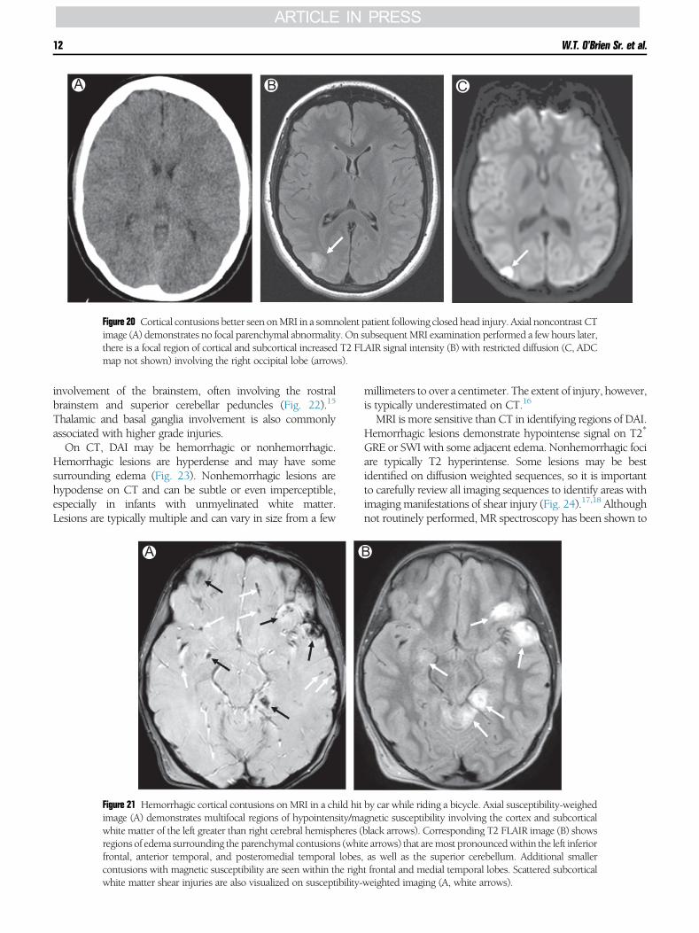
On CT, cortical contusions present as regions of hypodenseedema or hyperdense foci of hemorrhage (Fig. 19). Extensioninto the subcortical white matter is not uncommonwith largercontusions. Initially, these injuries may be subtle and becomemore conspicuous on short-term follow-up imaging over thenext several hours or few days. As contusions may be multipleand occur bilaterally, care should be taken to identify addi-tional regions of parenchymal injury. MRI has a greatersensitivity than CT in identifying cortical contusions(Fig. 20). When hemorrhagic, contusions will demonstratefoci of hemorrhagic susceptibility (hypointensity on T2*
gradient echo [GRE] or susceptibility-weighted imaging
coronal reformatted (B) noncontrast CT images in brainying the right cerebral convexity with markedmass effect,subfalcine herniation. The mixed density in this case isge and intermixing of cerebrospinal fluid.

A B
Figure 17 Subarachnoid hemorrhage following a motor vehicle accident. Axial noncontrast CT image through the level ofthe lateral ventricles (A) demonstrates hyperdense subarachnoid hemorrhage within the posterior aspect of the sylvianfissures (arrows) andwithin several sulci. A small amount of hemorrhage is seenwithin the occipital horn of the both lateralventricles (notched arrows). There is a small focus of pneumocephalus overlying the left frontal lobe (*). Axial noncontrastCT image at the level of the basal cisterns (B) reveals a small amount of subarachnoid hemorrhage within theinterpeduncular cistern (arrow).
Pediatric emergencies 11
[SWI]) surrounded by T2 hyperintense edema (Fig. 21). Bloodproducts may have regions of diffusion restriction.
Diffuse Axonal InjuryIn the setting of rotational acceleration and decelerationinjuries, stretching or shearing injuries may occur within theaxonal white matter, which is referred to as diffuse axonalinjury (DAI). Due to the mechanism of injury, DAI is classifiedbased upon the location of injury with higher grade injuries
Figure 18 Pseudosubarachnoid hemorrhage in an infant with diffusehypoxic-ischemic injury post-trauma. Axial noncontrast CT imagethrough the basal cisterns reveals extra-axial hyperdensity mimickingsubarachnoid hemorrhage due to vascular engorgement, CSF efface-ment, and adjacent parenchymal hypodensity.
portending worsened neurological deficits and prognosis.Often times patients withDAI experience loss of consciousnessat the time of injury and later display neurological deficitsbeyondwhat would be expected based upon the initial injuriesidentified on CT. Grade I injuries occur within the lobar whitematter near the gray-white matter junction. Grade II injuriesinclude the lobar white matter plus involvement of the corpuscallosum with preferential involvement of the body andsplenium of the corpus callosum. Grade III injuries involvethe lobar white matter and corpus callosum with additional
Figure 19 Cortical contusion on CT following a fall. Axial noncontrastCT image shows a large hyperdense cortical contusion involving theright middle frontal gyrus (*). Foci of subarachnoid hemorrhage areseen within several sulci (arrows). Scalp soft tissue swelling isidentified over the right frontal impact site.
A B C
Figure 20 Cortical contusions better seen onMRI in a somnolent patient following closed head injury. Axial noncontrast CTimage (A) demonstrates no focal parenchymal abnormality. On subsequent MRI examination performed a few hours later,there is a focal region of cortical and subcortical increased T2 FLAIR signal intensity (B) with restricted diffusion (C, ADCmap not shown) involving the right occipital lobe (arrows).
W.T. O’Brien Sr. et al.12
involvement of the brainstem, often involving the rostralbrainstem and superior cerebellar peduncles (Fig. 22).15
Thalamic and basal ganglia involvement is also commonlyassociated with higher grade injuries.On CT, DAI may be hemorrhagic or nonhemorrhagic.
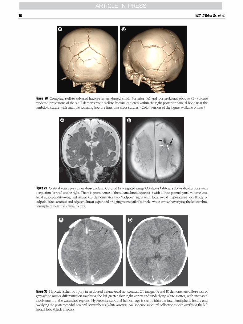
Hemorrhagic lesions are hyperdense and may have somesurrounding edema (Fig. 23). Nonhemorrhagic lesions arehypodense on CT and can be subtle or even imperceptible,especially in infants with unmyelinated white matter.Lesions are typically multiple and can vary in size from a few
A
Figure 21 Hemorrhagic cortical contusions on MRI in a child hitimage (A) demonstrates multifocal regions of hypointensity/mawhite matter of the left greater than right cerebral hemispheres (regions of edema surrounding the parenchymal contusions (whifrontal, anterior temporal, and posteromedial temporal lobes,contusions with magnetic susceptibility are seen within the righwhite matter shear injuries are also visualized on susceptibility-
millimeters to over a centimeter. The extent of injury, however,is typically underestimated on CT.16
MRI is more sensitive than CT in identifying regions of DAI.Hemorrhagic lesions demonstrate hypointense signal on T2*
GRE or SWI with some adjacent edema. Nonhemorrhagic fociare typically T2 hyperintense. Some lesions may be bestidentified on diffusion weighted sequences, so it is importantto carefully review all imaging sequences to identify areas withimaging manifestations of shear injury (Fig. 24).17,18 Althoughnot routinely performed, MR spectroscopy has been shown to
B
by car while riding a bicycle. Axial susceptibility-weighedgnetic susceptibility involving the cortex and subcorticalblack arrows). Corresponding T2 FLAIR image (B) showste arrows) that aremost pronouncedwithin the left inferioras well as the superior cerebellum. Additional smallert frontal and medial temporal lobes. Scattered subcorticalweighted imaging (A, white arrows).

A B C
D
Figure 22 Patterns of diffuse axonal injury (DAI). Axial susceptibility-weighted images (A-D) show regions of hypointensityor magnetic susceptibility involving the subcortical white matter (A-D, grade 1), corpus callosum (B and C, arrows, grade2), and brainstemormidbrain (D, notched arrow, grade 3). ImageC also shows involvement of the superior portions of thebasal ganglia and thalami.
Pediatric emergencies 13
identify regions of axonal injury that are not visualized onconventional MR sequences.19
Craniocervical Junction and CervicalSpine InjuriesDue to the large head size relative to a child’s body, as well asundeveloped neck musculature, children—especially thoseunder 8 years of age—are prone to injuries at the craniocervicaljunction and upper cervical spine in association with headtrauma. For these reasons, it is important to include and closelyreview the craniocervical junction and portions of the cervicalspine down to at least the C2 level on trauma head CTs.Although a detailed description of craniocervical and cervicalspine injuries is beyond the scope of this review, commoninjuries include skull base fractures, atlanto-occipital disloca-tion, atlantoaxial instability, rotatory subluxation, and fracturesand ligamentous injury of the upper cervical spine. Comparedto adults, ligamentous injuries without associated fractures are
more common in children. Therefore, in patients with neuro-logical deficits despite the absence of fractures on cervical spineradiographs or CT, MRI should be obtained to look forligamentous, cord, and soft tissue injuries.
Abusive Head TraumaThe role of imaging in the evaluation of suspected cases ofabusive head trauma (AHT) is both important and complex. Acomprehensive discussion of the neurological imaging man-ifestations of AHT is beyond the scope of this review;however, important and characteristic considerations will bediscussed.Head injury in association with AHT is unfortunately
common and is a leading cause of morbidity and mortalityin abused children. The pattern and extent of injury dependupon the underlying mechanisms and severity of trauma,which is often incompletely disclosed by caregivers. Commonmechanisms of abusive head trauma include direct blows,
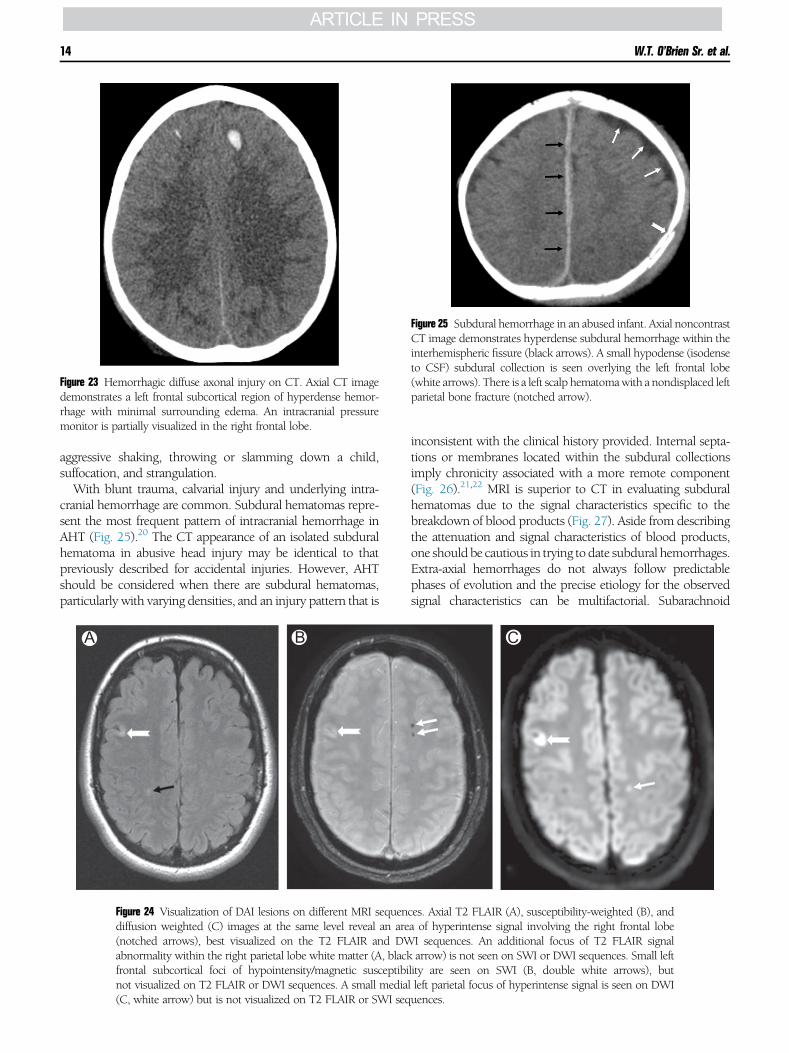
Figure 23 Hemorrhagic diffuse axonal injury on CT. Axial CT imagedemonstrates a left frontal subcortical region of hyperdense hemor-rhage with minimal surrounding edema. An intracranial pressuremonitor is partially visualized in the right frontal lobe.
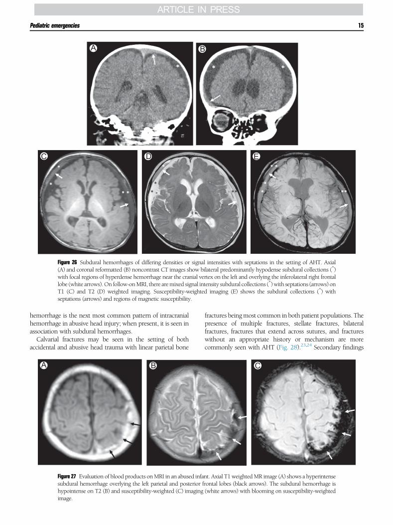
Figure 25 Subdural hemorrhage in an abused infant. Axial noncontrastCT image demonstrates hyperdense subdural hemorrhage within theinterhemispheric fissure (black arrows). A small hypodense (isodenseto CSF) subdural collection is seen overlying the left frontal lobe(white arrows). There is a left scalp hematomawith a nondisplaced leftparietal bone fracture (notched arrow).
W.T. O’Brien Sr. et al.14
aggressive shaking, throwing or slamming down a child,suffocation, and strangulation.With blunt trauma, calvarial injury and underlying intra-
cranial hemorrhage are common. Subdural hematomas repre-sent the most frequent pattern of intracranial hemorrhage inAHT (Fig. 25).20 The CT appearance of an isolated subduralhematoma in abusive head injury may be identical to thatpreviously described for accidental injuries. However, AHTshould be considered when there are subdural hematomas,particularly with varying densities, and an injury pattern that is
A B
Figure 24 Visualization of DAI lesions on different MRI sequendiffusion weighted (C) images at the same level reveal an are(notched arrows), best visualized on the T2 FLAIR and DWabnormality within the right parietal lobe white matter (A, blackfrontal subcortical foci of hypointensity/magnetic susceptibinot visualized on T2 FLAIR or DWI sequences. A small media(C, white arrow) but is not visualized on T2 FLAIR or SWI seq
inconsistent with the clinical history provided. Internal septa-tions or membranes located within the subdural collectionsimply chronicity associated with a more remote component(Fig. 26).21,22 MRI is superior to CT in evaluating subduralhematomas due to the signal characteristics specific to thebreakdown of blood products (Fig. 27). Aside from describingthe attenuation and signal characteristics of blood products,one should be cautious in trying todate subdural hemorrhages.Extra-axial hemorrhages do not always follow predictablephases of evolution and the precise etiology for the observedsignal characteristics can be multifactorial. Subarachnoid
C
ces. Axial T2 FLAIR (A), susceptibility-weighted (B), anda of hyperintense signal involving the right frontal lobeI sequences. An additional focus of T2 FLAIR signalarrow) is not seen on SWI or DWI sequences. Small left
lity are seen on SWI (B, double white arrows), butl left parietal focus of hyperintense signal is seen on DWIuences.
A B
C D E
Figure 26 Subdural hemorrhages of differing densities or signal intensities with septations in the setting of AHT. Axial(A) and coronal reformatted (B) noncontrast CT images show bilateral predominantly hypodense subdural collections (*)with focal regions of hyperdense hemorrhage near the cranial vertex on the left and overlying the inferolateral right frontallobe (white arrows).On follow-onMRI, there aremixed signal intensity subdural collections (*)with septations (arrows) onT1 (C) and T2 (D) weighted imaging. Susceptibility-weighted imaging (E) shows the subdural collections (*) withseptations (arrows) and regions of magnetic susceptibility.
Pediatric emergencies 15
hemorrhage is the next most common pattern of intracranialhemorrhage in abusive head injury; when present, it is seen inassociation with subdural hemorrhages.Calvarial fractures may be seen in the setting of both
accidental and abusive head trauma with linear parietal bone
A B
Figure 27 Evaluation of blood products onMRI in an abused infasubdural hemorrhage overlying the left parietal and posterior fhypointense on T2 (B) and susceptibility-weighted (C) imagingimage.
fractures beingmost common in both patient populations. Thepresence of multiple fractures, stellate fractures, bilateralfractures, fractures that extend across sutures, and fractureswithout an appropriate history or mechanism are morecommonly seen with AHT (Fig. 28).23,24 Secondary findings
C
nt. Axial T1weightedMR image (A) shows a hyperintenserontal lobes (black arrows). The subdural hemorrhage is(white arrows) with blooming on susceptibility-weighted
A B
Figure 29 Cortical vein injury in an abused infant. Coronal T2weighted image (A) shows bilateral subdural collections witha septation (arrow) on the right. There is prominence of the subarachnoid spaces (*) with diffuse parenchymal volume loss.Axial susceptibility-weighted image (B) demonstrates two “tadpole” signs with focal ovoid hypointense foci (body oftadpole, black arrows) and adjacent linear expanded bridging veins (tail of tadpole, white arrows) overlying the left cerebralhemisphere near the cranial vertex.
A B
Figure 28 Complex, stellate calvarial fracture in an abused child. Posterior (A) and posterolateral oblique (B) volumerendered projections of the skull demonstrate a stellate fracture centered within the right posterior parietal bone near thelambdoid suture with multiple radiating fracture lines that cross sutures. (Color version of the figure available online.)
A B
Figure 30 Hypoxic-ischemic injury in an abused infant. Axial noncontrast CT images (A and B) demonstrate diffuse loss ofgray-white matter differentiation involving the left greater than right cortex and underlying white matter, with increasedinvolvement in the watershed regions. Hyperdense subdural hemorrhage is seen within the interhemispheric fissure andoverlying the posteromedial cerebral hemispheres (white arrows). An isodense subdural collection is seen overlying the leftfrontal lobe (black arrows).
W.T. O’Brien Sr. et al.16

A B
Figure 31 Sequela of hypoxic-ischemic injury in an abused child. Axial noncontrast head CT at the level of the deep graymatter nuclei (A) demonstrates a small left and tiny right predominantly hypodense subdural collections (white arrows),with hyperdense hemorrhage extending into the interhemispheric fissure (black arrow). There is loss of gray-white matterdifferentiation involving the occipital lobes (**). Follow-up noncontrast head CT at the level of the lateral ventricles severalmonths later (B) shows diffuse parenchymal volume loss with occipital lobe encephalomalacia, left great than right (**).There are moderate-sized bilateral hypodense subdural collections.
Pediatric emergencies 17
of associated soft tissue swelling may or may not be present,even in the acute setting. With a delayed presentation, thesesecondary findings have typically resolved by the time ofimaging.“Shaken baby syndrome” refers to the mechanism of injury
where a child is shaken vigorously in the anterior-posteriorplane. With this mechanism, the brain parenchyma andcalvarium move at different rates of acceleration and deceler-ation, which when combined with relatively large subarach-noid spaces, predisposes the child to tearing of bridging veinswith resultant subdural hemorrhage. Retinal hemorrhages are
Figure 32 Craniocervical junction injury in an abused infant. Midlinesagittal reformatted CT image demonstrates hyperdense subduralhemorrhage within the interhemispheric fissure (black arrows), alongthe tentorium (white arrow), and posterior to the cerebellar vermis(double white arrow). At the craniocervical junction, there is a smallamount of hemorrhage along the posterior margin of the dens(notched black arrow) and a large extra-axial hemorrhage posteriorly(**), resulting in effacement of CSF at the foramen magnum.
commonly seen clinically. The presence of superficial bridgingvein injury or thrombosis in the absence of an appropriatetrauma history has been shown to be a positive indicator ofAHT. Bridging vein injury is best depicted on susceptibility-weighted imaging and commonly results in the “tadpole” sign,as demonstrated by a focal round clot caused by bridging veindisruption (body of the tadpole) with an expanded throm-bosed bridging vein (tail of the tadpole) (Fig. 29).25 Withsevere trauma, hypoxic-ischemic injury (HII) may be seen inassociation with extra-axial hemorrhage and is more commonin the setting of AHT compared to accidental injuries (Figs. 30and 31). Common imaging patterns of HII include diffuseischemic injury, watershed distribution injury, and, lesscommonly, focal injury or venous infarction.26 Often theamount of subdural hemorrhage is small in relation to theextent of HII, but may serve as a potential marker of trauma.Despite the mechanism of injury in this population, diffuseaxonal injury is relatively uncommon in AHT. Injuries to thecraniocervical junction and cervical spinemay also be seen dueto the relatively large head size, orientation of facets, andunderdeveloped musculature in children (Fig. 32).23 Extrac-ranial injuries with a high specificity for AHT include classicmetaphyseal lesions and posterior rib fractures.27
Suffocation results in cerebral edema that is proportional tothe severity and duration of the injury. Strangulation results insimilar intracranial findings and may also result in vascularinjuries that further exasperate the degree of intracranial edema.
Birth-Related InjuriesIntracranial InjuriesBirth-related injuries refer to those encounteredduring deliveryand are more common with vaginal deliveries compared tocesarean section. Imaging of birth-related head trauma typi-cally beginswith ultrasound (US), followed byCT, if necessary.

A B
Figure 33 Birth-related subdural hemorrhage. Sagittal T1 (A) and T2 (B) weighted images in a newborn demonstrate a smallamount of T1 hyperintense and T2 hypointense subdural hemorrhage at the junction of the falx and tentorium andextending inferiorly within the posterior fossa along the occiput (arrows). A scalp hematoma is also noted at the cranialvertex.
W.T. O’Brien Sr. et al.18
Although MRI is more sensitive in identification of hemor-rhagic lesions not well seen onCT, the availability and speed ofCT is preferred in the acute settingwhenUS evaluation alone isinsufficient.In the normal birthing process, subdural hematomas are
commonly seen, typically small, and of no clinical signifi-cance.28,29 Although they may occur anywhere, including theposterior fossa, theymost often occur at the junction of the falxcerebri and tentoriumcerebelli (Fig. 33).Most are notwell seenon US and are better characterized on CT or MRI. Beyond theacute setting, MRI not only has increased sensitivity forhemorrhagic lesions, but can also identify traumatic andischemic white matter injuries that are not well visualized onCT.30 Epidural and subarachnoid hemorrhage are less com-mon in the setting of birth-related injuries, but when present,
A B
Figure 34 Birth-related calvarial remodeling or molding. Newboruse of instrumentation (forceps). Axial CT image (A) shows inwrendered projection (B) better depicts the extent of calvarial de(Color version of the figure available online.)
are often seen in association with subdural hematomas.Epidural hematomas have a higher incidence when associatedwith calvarial fractures and cephalohematomas,31,32 often inthe setting of a difficult birth requiring instrumentation.
Calvarial FracturesAs is seenwith blunt head injury describedpreviously, calvarialfractures may occur during a vaginal birth as the skullcompresses against the mother’s pelvis and undergoes remod-eling or with the use of instrumentation (Fig. 34). Nonde-pressed fractures in this setting are of no clinical significancewhen seen in isolation and without secondary injuries.33
Depressed fractures are less commonly seen but have a higherincidence of associated intracranial injuries.
n after a prolonged and difficult vaginal delivery with theard depression of the left parietal bone (arrow). Volumepression (arrow) without an associated fracture lucency.

A B
Figure 35 Birth-related scalp hemorrhage. Coronal reformatted CT (A) and T2 MR (B) images in a newborn child showhemorrhage and edema within all 3 compartments of the scalp. The cephalohematoma is subperiosteal in location and ishyperdense on CT and hypointense on T2 (*); it does not cross the sagittal suture. The subgaleal hemorrhage is locatedbetween the superficial subgaleal aponeurosis and the subjacent periosteum; it is hypodense onCT andhyperintense onT2(5-point stars) and crossesmidline. The caput succedaneum is superficial to the galeal aponeurosis and ismixed hyper andhypodense on CT and hyperintense on T2 (arrows); it crosses midline.
Pediatric emergencies 19
Extracranial InjuriesIn addition to simple scalp hematomas, there are 3 primarytypes of superficial extracranial hemorrhage: caput succeda-neum, subgaleal hemorrhage, and cephalohematoma. Thedistinction between the types of hemorrhage is based uponthe compartment in which the hemorrhage occurs (Fig. 35).Caput succedaneum refers to scalp hemorrhage superficial
to the galeal aponeurosis (Fig. 35). It may spread throughoutthe scalp and cross midline since it is not bounded byperiosteum or an aponeurosis. Most cases are self-limited.A subgaleal hemorrhage occurs between the periosteum and
overlying galeal aponeurosis, can also cross the midline andmay extend into the soft tissues of the neck when large(Fig. 35). It most often results from instrumentation during adifficult birth with rupture of emissary veins. Although mostcases are self-limited and resolve within a few weeks, largehemorrhages may be extensive and symptomatic.
A
Figure 36 Calcified cephalohematoma. Patient with known pricontour deformity. Axial (A) and coronal reformatted (B) CT imasuperior portion of the left parietal bone, consistent with a calc
A cephalohematoma refers to a subperiosteal hemorrhagethat occurs between the underlying calvarium and overlyingperiosteum (Fig. 35). There is an increased incidence withprolonged labor and utilization of instrumentation. As thehemorrhage is subperiosteal in location, the hemorrhage isconfined by sutures (cannot cross midline sagittal suture atcranial vertex) and is often firm. The hemorrhage can persistformonths and rarelymay calcify, forming a persistent calvarialcontour abnormality (Fig. 36).34
SummaryCross-sectional imaging plays an integral role in identifyingand characterizing pediatric head trauma, which remains animportant cause of morbidity and mortality in children.Understanding the patterns and appearances of head injury
B
or birth-related scalp hematoma and persistent calvarialges show a focal region of cortical thickening involving theified cephalohematoma.
W.T. O’Brien Sr. et al.20
on imaging studies and how they differ in children comparedto adults is essential to the imaging evaluation. The keydifferences in children stem from the complexities of thedeveloping brain and calvarium, as well as some uniquemechanisms of injury, to include abusive head trauma andbirth-related injuries. Knowledge of the intricacies of pediatrichead trauma will assist the radiologist in appropriatelyevaluating the full spectrum of injuries and help guideappropriate patient management and follow-up.
References1. American College of Radiology Appropriateness Criteria American
College of RadiologyAppropriatenessCriteria; for head trauma in childrenfor head trauma in children, 2014. https://acsearch.acr.org/docs/3083021/Narrative/. Accessed September 20, 2017.
2. PECARN Kuppermann N, Holmes JF, Dayan PS, et al: Identification ofchildren at very low risk of clinically important brain injuries after headtrauma: A prospective cohort study. Lancet 374(9696):1160-1170, 2009
3. Weir P, Suttner NJ, Flynn P, McAuley D: Normal skull suture variantmimicking intentional injury. Br Med J 332(7548):1020-1021, 2006
4. SanchezT, Stewart D,WalvickM, et al: Skull fracture vs. accessory sutures:How can we tell the difference? Emerg Radiol 17(5):413-418, 2010
5. Idriz S, Patel J, Renani SA, et al: CT of normal developmental and variantanatomy of the pediatric skull: Distinguishing trauma from normality.RadioGraphics 35(5):1585-1601, 2015
6. Zergham Z, Morris AM, Paw R: Ping-pong fracture. Emerg Med J 24(10):731, 2007
7. Liu XS, You C, Lu M, et al: Growing skull fracture stages and treatmentstrategy. J Neurosurg Pediatr 9(6):670-675, 2012
8. Parizel PM, Makkat S, Van Miert E, et al: Intracranial hemorrhage:Principles of CT and MRI interpretation. Eur Radiol 11(9):1170-1183,2001
9. Al-Nakshabandi NA: The swirl sign. Radiology 218(2):433, 200110. Strub WM, Leach JL, Tomsick T, et al: Overnight preliminary head CT
interpretations by residents: Locations of misidentified intracranialhemorrhage. Am J Neuroradiol 28(9):1679-1682, 2007
11. Given CA, Burdette JH, Elster AD, et al: Pseudo-subarachnoid hemor-rhage: A potential imaging pitfall associated with diffuse cerebral edema.Am J Neuroradiol 24(2):254-256, 2003
12. Woodcock RJ, Davis PC, Hopkins KL: Imaging of head trauma in infancyand childhood. Semin Ultrasound CT MR 22(2):162-182, 2001
13. Poussaint TY, Moeller KK: Imaging of pediatric head trauma. Neuro-imaging Clin N Am 12(2):271-294, 2002
14. Gentry LR: Imaging of closed head injury. Radiology 191(1):1-17, 199415. Adams JH, Doyle D, Ford I, et al: Diffuse axonal injury in head injury:
Definition, diagnosis and grading. Histopathology 15(1):49-59, 198916. Mittl RL, Grossman RI, Hiehle JF, et al: Prevalence of MR evidence of
diffuse axonal injury in patients with mild head injury and normal headCT findings. Am J Neuroradiol 15(8):1583-1589, 1994
17. Tong KA, Ashwal S, Holshouser BA, et al: Hemorrhagic shearinglesions in children and adolescents with posttraumatic diffuse axonalinjury: Improved detection and initial results. Radiology 227(2):332-339,2003
18. Hergan K, Schaefer PW, Sorensen AG, et al: Diffusion-weightedMRI in diffuse axonal injury of the brain. Eur Radiol 12(10):2536-2541,2002
19. Holshouser BA, Tong KA, Ashwal S: Proton MR spectroscopic imagingdepicts diffuse axonal injury in childrenwith traumatic brain injury. AJNRAm J Neuroradiol 26(5):1276-1285, 2005
20. Barnes PD, Robson CD: CT findings in hyperacute nonaccidental braininjury. Pediatr Radiol 30(2):74-81, 2000
21. Hedlund GL: Subdural hemorrhage in abusive head trauma: Imagingchallenges and controversies. J Am Osteopath Coll Radiol 1(1):23-30,2012
22. Cramer JA, Rassner UA, Hedlund GL: Limitations of T2*-gradientrecalled-echo and susceptibility-weighted imaging in characterizingchronic subdural hemorrhage in infant survivors of abusive head trauma.Am J Neuroradiol 37(9):1752-1756, 2016
23. Hsieh KLC, Zimmerman RA, Kao HW, et al: Revisiting neuroimagingof abusive head trauma in infants and young children. Am J Roentgenol204(5):944-952, 2015
24. Meservy CJ, Towbin R,McLaurin RL, et al: Radiographic characteristics ofskull fractures resulting from child abuse. AJR Am J Roentgenol 149(1):173-175, 1987
25. Hahnemann ML, Kinner S, Schweiger B, et al: Imaging of bridging veinthrombosis in infants with abusive head trauma: the “Tadpole Sign.” EurRadiol 25(2):299-305, 2015
26. Zimmerman RA, Bilaniuk LT, Farina L: Non-accidental brain trauma ininfants: diffusion imaging, contributions to understanding the injuryprocess. J Neuroradiol 34(2):109-114, 2007
27. Lonergan G, Baker A, Morey M, et al: Child abuse: radiologic-pathologiccorrelation. RadioGraphics 23(4):811-845, 2003
28. Rooks VJ, Eaton JP, Ruess L, et al: Prevalence and evolution of intracranialhemorrhage in asymptomatic term infants. Am J Neuroradiol 29(6):1082-1089, 2008
29. Tavani F, Zimmerman R, Clancy R, et al: Incidental intracranialhemorrhage after uncomplicated birth: MRI before and after neonatalheart surgery. Neuroradiology 45(4):253-258, 2003
30. Kelly AB, Zimmerman R, Snow RB, et al: Head trauma: comparisonof MR and CT – experience in 100 patients. Am J Neuroradiol 9(4):699-708, 1988
31. Choux M, Grisoli F, Peragut J: Extradural hematomas in children: 104cases. Childs Brain 1(6):337-347, 1975
32. TangHT, LimCCT: Imaging of accidental paediatric head trauma. PediatrRadiol 39(5):438-446, 2009
33. Rabelo NN, Matushita H, Cardeal DD: Traumatic brain lesions innewborns. Arq Neuropsiquiatr 75(3):180-188, 2017
34. Glass RBJ, Fernbach SK, Norton KI, et al: The infant skull: a vault ofinformation. RadioGraphics 24(2):507-522, 2004