Embed Size (px)
Citation preview

Pediatric
Assessment-Jason Haag, CCEMT-P

Pediatric Resuscitation:Facts and Stats
Children fare worse than adults in the out-of-hospital phase of resuscitation
70% of all pediatric trauma deaths occur in the field
Survival rate for out-of-hospital cardiac arrest is half that of adults
Failure rate for resuscitative interventions in field is 2x that of adults
Failure rates for prehospital ET intubation for injured children is near 50%

Pediatric Resuscitation:Facts and Stats
Injury: leading cause of death 1-14 8% of kids die prior to EMS arrival 10% of all calls for kids Optimal pre-hospital management is of major importance to
reduce M & M
Most common errors in peds management:
Failure to manage the Airway
Failure to provide fluid resuscitation
Failure to recognize and treat internal bleeding

Overview
Trauma is leading cause of death and
disability in children
Blunt injury represents 80-90%
Penetrating injury less common

Mechanisms of Injury
Most common for peds:
Falls
MVCs
Car –vs- ped
Drownings and near-drownings
Burns
Physical abuse

Concerns due to MOI
Shock
Musculoskeletal Injuries
Blunt Trauma
Burns Thermal
Electrical
Chemical

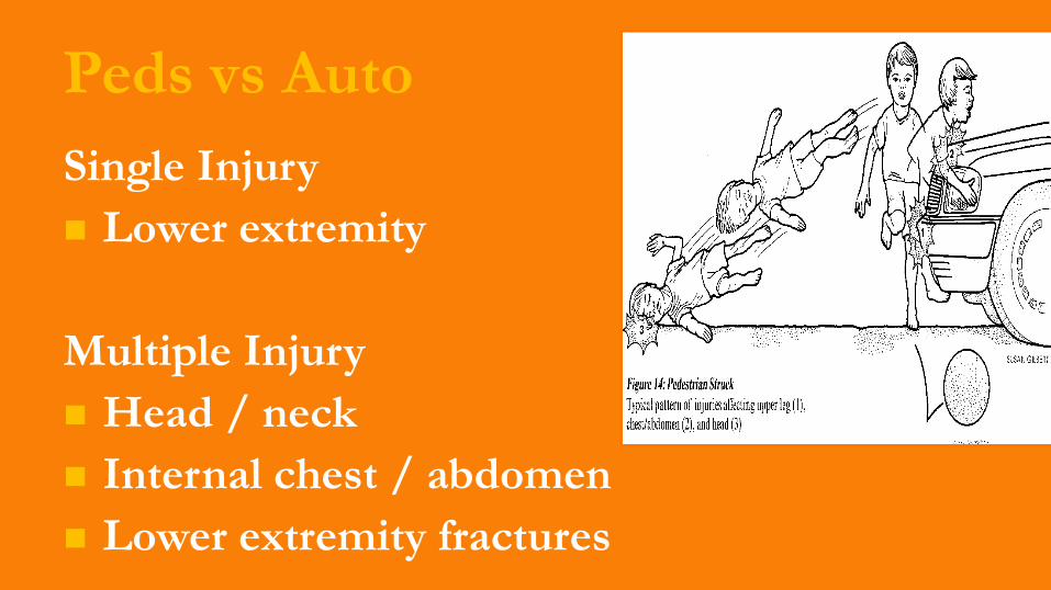
Peds vs Auto
Single Injury
Lower extremity
Multiple Injury
Head / neck
Internal chest / abdomen
Lower extremity fractures
Blunt Trauma
Number one cause of death in infants and
children
50 % of pediatric deaths occur within the first hour
Most injuries result from blunt trauma
Thinner body walls allow greater energy transfer
Higher incidence of penetrating injuries in urban
areas

Falls
Low height
Upper extremity fracture
Medium height
Head/neck injury
Face/scalp injury
Upper extremity fracture

Falls
High height
Head / neck injury
Scalp / facial laceration
Internal chest / abdominal injury
Upper / lower extremity fracture

Bicycle Injuries
Helmeted
Upper extremity fractures
Unhelmeted
Head/neck injuries
Scalp/facial lacerations
Upper extremity fractures
Handlebar
Internal abdominal injury


Motor Vehicle CrashesOccupant
Restrained
Internal abdominal Injuries
Lower spine fractures
Especially if restraints are not size appropriate
Unrestrained
Head/neck injuries
Scalp/facial lacerations

Spinal Injuries

Specific Injuries: Head
Head and brain
Involved in 60% of blunt injuries Head, face and neck
Head injuries: #1 cause of trauma death in peds
Soft skull
60-70% of pediatric cervical fractures occur
at C1-C3


Head and Neck Injuries
CREATED FOR TACOMA FIRE DEPARTMENT OTEP

Concussion
Mechanics: direct blow to head/face/neck or indirect force transmission (body blow)
Timecourse: rapid onset, short-lived impairment, spontaneous resolution
Pathophysiology: function > structure
Symptoms: graded syndromes, may or may not include LOC, sequential resolution

Postconcussion Symptom Scale Headache Nausea Vomiting Balance problems Dizziness Fatigue Trouble falling asleep Sleeping more than usual Sleeping less than usual Drowsiness Sensitivity to light
Sensitivity to noise Irritability Sadness Nervousness Feeling more emotional Numbness or tingling Feeling slowed down Feeling mentally “foggy” Difficulty concentrating Difficulty remembering Visual problems

Clinical Signs of Concussion
Consciousness (LOC) – not required
Memory – post-traumatic/retrograde
amnesia
Cognition
Neurological (physical)
Personality (emotional)
Specific Injuries: Chest
Softer, more flexible ribs
Soft, pliant airways
Greater mobility of heart and great vessels

Specific Injuries: Abdomen
Small abdominal cavity size concentrates injury
forces
Softer, more flexible ribs allow upper abdominal
organs to be injured
Thinner muscles of abdominal wall transmit injury
forces directly to internal organs
Internal organs- Involved in 10% of blunt injuries
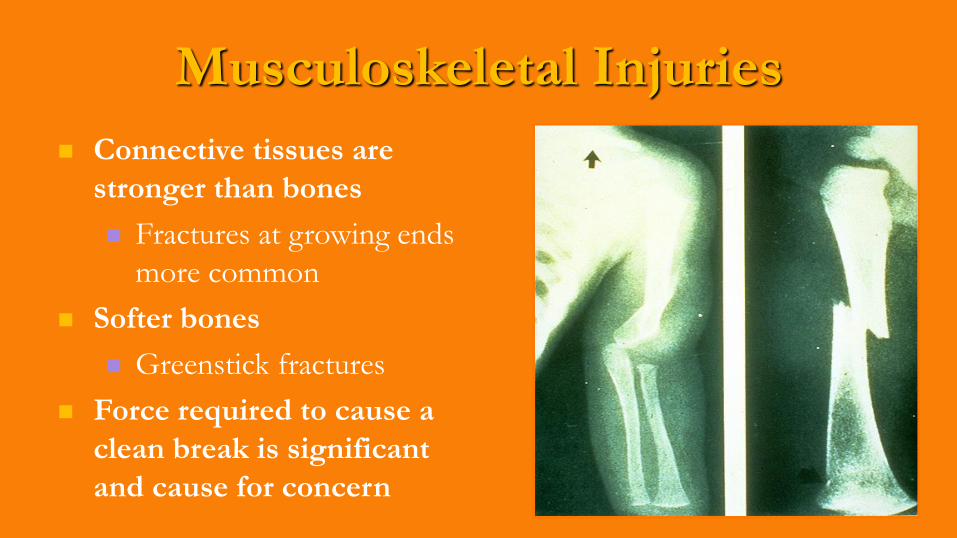
Musculoskeletal Injuries
Connective tissues are
stronger than bones
Fractures at growing ends
more common
Softer bones
Greenstick fractures
Force required to cause a
clean break is significant
and cause for concern

Forearm Fracture

Greenstick Fracture

Give Comfort…Give Pain Meds


General Pediatric Assessment
Scene Assessment
Initial Assessment General Impression
Transport Decision
Additional Assessment Focused History and Exam
Detailed Physical Exam
Ongoing Assessment

Pediatric Assessment Triangle
Work of BreathingAppearance
Circulation to Skin
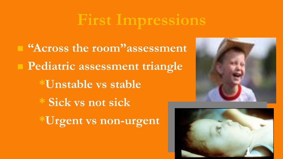
First Impressions
“Across the room”assessment
Pediatric assessment triangle
*Unstable vs stable
* Sick vs not sick
*Urgent vs non-urgent

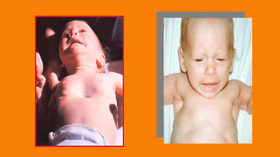
PAT….Appearance
TICLS…..pronounced Tickles
Tone
Interactivity
Consolability
Look
Speech

PAT….Breathing
Body position
Visible movement of
chest and abdomen
Respiratory rate and effort
Audible airway sounds

Assessment: Breathing
Increased rate and effort
Ensure oxygenation and ventilation
Hypoxia can cause hypoperfusion
Chest trauma can cause obstructive shock
Treat any cause of respiratory distress

PAT….Circulation
Skin Temperature
Pulse strength
Capillary refill time


Primary Survey
Assessment and
management occur
simultaneously
Determine any life-
threatening conditions

A - Airway
B - Breathing
C - Circulation
D - Da Brain
E - Exposure
Primary Survey

Any Airway Red Flags ?
Vocalization
Drooling
Abnormal airway sounds
Preferred posture
Tongue obstruction
Loose teeth or foreign objects
Bleeding/vomitus

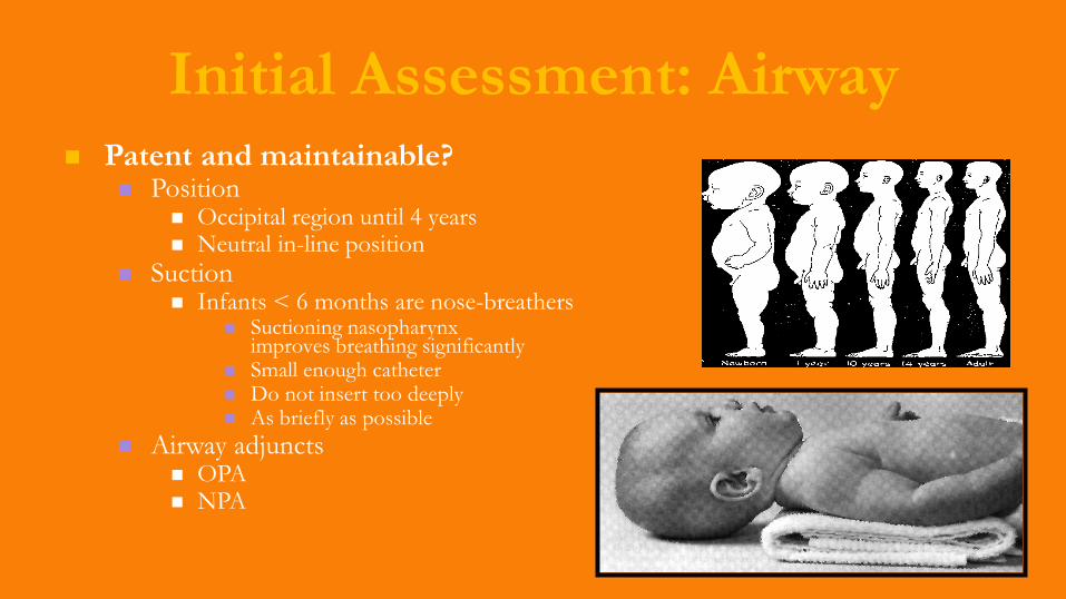
Initial Assessment: Airway Patent and maintainable?
Position Occipital region until 4 years Neutral in-line position
Suction Infants < 6 months are nose-breathers
Suctioning nasopharynx improves breathing significantly
Small enough catheter Do not insert too deeply As briefly as possible
Airway adjuncts OPA NPA


Any Breathing Red Flags ?
Level of consciousness
Rate and depth of respirations
Breath sounds
Symmetric chest rise and fall
Work of breathing Nasal flaring Head bobbing
Retractions Grunting
Accessory muscle use
Any Circulation Red Flags ?
Central and peripheral pulse rate and quality
Skin color, temperature, and moisture
(most reliable indicator of perfusion)
Capillary refill <2
Mental status
External bleeding
Hypoperfusion
Second major cause of pediatric
cardiopulmonary arrest
Unusual because of efficient pediatric vessel
constriction
Fast decompensation

Causes
Heat loss (newborns, neonates)
Dehydration
Infection, sepsis, anaphylaxis
Trauma
Blood loss
EMS care focuses on suspecting shock before it develop
Bleeding and Shock
The total blood volume is smaller (80 mL/kg)
1 y/o ~ 10 kg: Blood volume
800 mL = 27 oz = 2 cans of soda
6 y/o ~ 20 kg: Blood volume
1,600 mL = 54 oz = 4.5 cans of soda
Child’s loss is proportionally greater

Compensated Shock
Irritability or anxiety
Tachycardia
Tachypnea
Weak peripheral pulses, full central pulses
Delayed capillary refill (>2 sec in <6 y/o)
Cool, pale extremities
Systolic BP within normal limits
Decreased urinary output
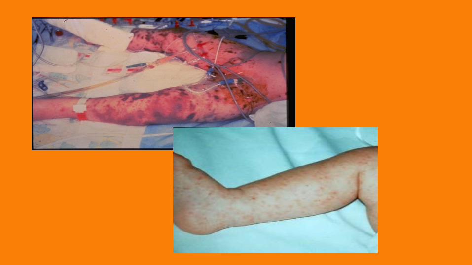
Decompensated Shock
Lethargy or coma
Marked tachycardia or bradycardia
Absent peripheral pulses, weak central pulses
Markedly delayed capillary refill
Cool, pale, dusky, mottled extremities
Hypotension
Markedly decreased urinary output
Absence of tears

Reminder…… First Impression
Ill-appearing children are in decompensated
shock
Compensated shock presents with more
subtle findings

Assessment: Mental Status
Key indicator of
perfusion

Treatment Plan
High flow O2
Aggressive airway/ventilation management
Volume replacement (20 ml/kg boluses)
Rapid transport to definitive care
Secondary Survey
F – Full set of vitals
and family
G – Give comfort
H – Head to toe and
history
I – Inspect
“It ain’t over until the
patient is over”

Memorize their vitals
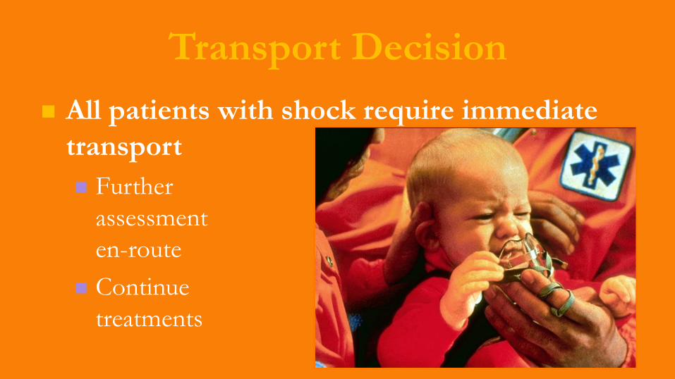
Transport Decision
All patients with shock require immediate
transport
Further
assessment
en-route
Continue
treatments

Immobilization and Transport
Immobilize small children C-collar
Layer of padding
KED
Other devices
Keep warm Blankets
Head coverings
Cold weather
Near drowning
Summary
Maintain Airway, Breathing, and C-spine
control
Give high-flow, high concentration O2
Assist ventilations if child demonstrates
altered mental status or respiratory distress

Summary
Consider length-based resuscitation tape
Immobilize as appropriate
Transport to pediatric tertiary care if
possible
Keep the child warm

And finally……………
Boys will be boys

CREATED FOR TACOMA FIRE DEPARTMENT OTEP


