Embed Size (px)
Citation preview

5/12/2015
1
Peter H. Kehoe, OD, FAAODiplomate – American Board of Optometry
COPE Course ID: 36509-GO
VEP AND ERG:PRIMARY CARE ELECTRODIAGNOSTICS
BIOGRAPHY
• Principal in 2 practices in Galesburg, and Galva, IL
• Past-President, of the American Optometric Association
• Optometric contractor to Illinois Department of Corrections
• ICO Class of 1984
• Fellow, American Academy of Optometry
• Doctor of Science in Optometry – ICO 2009
• Diplomate – American Board of Optometry - 2011
DISCLOSURES
• Affiliated with Transitions Optical as a consultant for professional development
• Non-executive board member of Optos, Plc based in Scotland
• Consultant to Diopsys, Inc.
• Administrator for Vision Source in Illinois
Phototransduction
Conversion of light into electricity
Neuro-Physiology
ISCEV
International Society for Clinical Electrophysiology of Vision
http://www.iscev.org/
ISCEV
http://www.iscev.org/
Mission
To promote and extend the knowledge of clinical electrophysiology of vision.
To promote co‐operation and communication among workers in the field of clinical and basic electrophysiology of vision.

5/12/2015
2
ISCEV
http://www.iscev.org/
Documenta Ophthalmologicahttp://www.springer.com/medicine/ophthalmology/journal/10633
ISCEVStandards, Recommendations and Guidelines
VISUAL ELECTRODIAGNOSTICS A Guide To Procedures
Confirmation of Neurological or Ophthalmological DiseaseUnexplained Visual LossPediatric NeurologyOpacities in Media
Monitoring Health ‐ ToxicityDetection of the Disease or Carrier States of Inherited Visual Disorders
Quantitative Assessment of Visual DiseaseAssessment of Retinal and Optic Nerve Function Following Trauma
Infants with questionable vision
http://www.iscev.org/standards/proceduresguide.html
ISCEVStandards, Recommendations and Guidelines
VISUAL ELECTRODIAGNOSTICS A Guide To Procedures
Vascular Disease of the Eye Including DiabetesOpaque MediaRetrobulbar NeuritisUnexplained Visual LossPediatric Cases AlbinismToxic and Nutritional Eye DiseaseIntracranial Lesion
http://www.iscev.org/standards/proceduresguide.html
AAOAmerican Academy of Ophthalmology
Preferred Practice Patterns
http://one.aao.org/CE/PracticeGuidelines/PPP.aspx
AOAAmerican optometric Association
Clinical Practice Guidelines
http://www.aoa.org/x4813.xml
VEP
Photoreceptor
Ganglion cell axon
Relay neuron
Visual cortex neuron
Bipolar
Light
PhototransductionConversion of light into electricity
Mid-retinal Layers
Electricity
Optic Chiasm/Tract
LGN
Neuro-Physiology
• Electrocardiogram
• Electromyography
• Auditory Evoked Potential
• Electroencephalogram
Electrophysiology

5/12/2015
3
VISUAL EVOKED POTENTIAL (VEP)
• Electric signal registered at the occipital region in response to a visual stimuli.
• VEP
• Visual – patient observes a visual stimulus
• Evoked – generates electrical energy at the retina
• Potential – measure the electrical activity in the visual cortex
• Measure the function of the entire vision system; no patient response required – OBJECTIVE TEST
Previous Limitations• Test time was approximately 45
minutes
• Required highly trained operators
• Required highly trained
interpretation (subjective)
• Limited to large research
institutions
VEP
Advantages of Current Technology
• Test time is approximately 1 minute
• Does not require highly trained operators
• Does not require highly trained interpretation
•Similar to interpreting VF or OCT
• Currently installed in about 2000 offices (one
company) 2 or three other companies - limited
VEP
Reference Ground Active
VEP Electrodes
• Flash
• Pattern
Reversal
Pattern-onset
Transient
Steady State
VEP Stimulus
• Pattern
Contrast Sensitivity
Visual Acuity
Color
VEP Stimulus

5/12/2015
4
N75
P100
N135
VEP Components
AMPLITUDE
Microvolts (µv)
1 Volt = 1 000 000 Microvolts
LATENCY
Milliseconds (ms)
1 second = 1000 milliseconds
VEP
LATENCY (ms)
AMPLITUDE (µv)
• Amplitude usually translates to the amount of axons conducting along the visual pathway
• Latency usually translates to the myelin status of the visual pathway
VEP
TIME
Am
plitu
de
ERG mERG pERG EOG mVEP
ElectroretinogramMultifocal ElectroretinogramPattern ElectroretinogramElectrooculogramMultifocal Visual Evoked Potential
Other Electrophysiological Tests
• Psychophysics• VF• GDx• HRT• OCT
VEP and other Ophthalmic Diagnostic Tests

5/12/2015
5
Visual Acuity Test
Psychophysics of Vision
Psychophysics of Vision
Contrast Sensitivity Test
Visual Field Test
Psychophysics of Vision
OPTIONS FOR CUSTOMIZED VEP TESTING
• User-Defined Protocol
• LKC
• Diagnosys
• Diopsys® NOVA-TR
• Customize testing parameters specific to each patient and pathology
• Pattern Type & Size, Contrast level, Eye
• Testing times are flexible and depend upon the customized settings
EXAMPLE:2 DIFFERENT SPATIAL FREQUENCIES
16 X 16 64 X 64
BUILDING PROTOCOLS

5/12/2015
6
OPTIONS
BUILDING OPTION SETS
RESPONSE TO TREATMENT EXAMPLE
OU with Lens
OU without Lens
MULTIPLE SCLEROSIS EXAMPLE
Expected P100 timingActual P100 timing
PATIENT D.G. 35 Y.O. MS PATIENT
• Multi-Contrast Stimuli
• LKC Requires User to Create Fixed
• Diopsys® NOVA-LX
• Easy to follow fixed protocol guides the technician through the test procedure.
• Testing time takes 38-53 seconds per eye on Diopsys, or about 5 minutes total – about 1/3 of LKC
ABILITY TO USE FIXED PROTOCOLS

5/12/2015
7
VEP AND GLAUCOMA:WELL DEFINED SCIENCE
THE VISUAL EVOKED POTENTIAL IN GLAUCOMA AND OCULAR HYPERTENSION: EFFECTS OF CHECK SIZE, FIELD SIZE, AND STIMULATION RATE
INVEST OPHTHALMOL VIS SCI 24:175-183, 1983
“Increased pattern VEP latency was significantly correlated with both the severity and location of visual field defects and the degree of cupping and pallor of the optic disc.” The
authors of this paper are world recognized electrophysiology specialist form New England Medical Center and University of Chicago
“Glaucoma has also been reported to affect the VEP by causing both reductions in amplitude and increases in latency.”
“The finding that is of clinical importance is the presence of abnormally long VEP latencies in some patients with ocular hypertension. The abnormal prolongation of VEP latency in these eyes may reflect subclinical optic nerve lesions that have not been uncovered with other techniques.”
WHY VEP?
• Many optic nerve diseases are asymptomatic because central vision is not affected until late in the disease1
• Diagnosis and management of optic nerve disorders are often based on structural or subjective visual field tests2
1 Glaucoma. American Optometric Association. www.aoa.org2 Prata, Tiago MD, G. De Moraes MD, J. Liebmann MD, R. Ritch, C. Tello MD. (2009). Diagnostic Ability of Fast Transient Visual Evoked Potential
for Glaucoma Assessment [Poster & Abstract] American Academy of Ophthalmology. 128
VEP is an objective, functional test that can help discriminate between healthy and glaucomatous eyes2
CLINICAL APPLICATIONS
• Clinicians may objectively test all patients suspected of optic nerve disease with VEP testing (Diopsys® NOVA-LX) to assess the function of the entire vision system
• The NOVA-LX VEP Vision Testing System:
• Has the sensitivity to help detect optic nerve disorders earlier, allowing treatment to start sooner
• Allows tracking of disease progression
• Normative database created over 3 years of testing at New York Eye & Ear Infirmary
5/12/2015
8
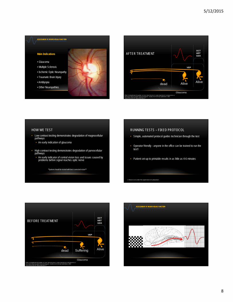
Main Indications
• Glaucoma
• Multiple Sclerosis
• Ischemic Optic Neuropathy
• Traumatic Brain Injury
• Amblyopia
• Other Neuropathies
ASSESSMENT OF NEURO-VISUAL FUNCTIONASSESSMENT OF NEURO-VISUAL FUNCTIONASSESSMENT OF NEURO-VISUAL FUNCTION
HOW WE TEST
• Low contrast testing demonstrates degradation of magnocellular pathways
• An early indication of glaucoma
• High contrast testing demonstrates degradation of parvocellular pathways
• An early indicator of central vision loss and issues caused by problems before signal reaches optic nerve
**patient should be tested with best corrected vision**
dead Suffering Alive
Glaucoma
VEP
OCTHRTGDX
BEFORE TREATMENT
Effect of epigallocatechin-gallate on inner retinal function in ocular hypertension and glaucoma: a short-term study by pattern electroretinogram. Graefes Arch Clin Exp Ophthalmol. 2009 Sep;247(9):1223-33. Epub 2009 Mar 17.
Alive
Glaucoma
VEP
OCTHRTGDX
Alivedead
AFTER TREATMENT
Effect of epigallocatechin-gallate on inner retinal function in ocular hypertension and glaucoma: a short-term study by pattern electroretinogram. Graefes Arch Clin Exp Ophthalmol. 2009 Sep;247(9):1223-33. Epub 2009 Mar 17.
RUNNING TESTS – FIXED PROTOCOL
• Simple, automated protocol guides technician through the test
• Operator friendly - anyone in the office can be trained to run the test1
• Patient set-up to printable results in as little as 4-6 minutes
1 Tests to run under the supervision of a physician.
ASSESSMENT OF NEURO-VISUAL FUNCTIONASSESSMENT OF NEURO-VISUAL FUNCTIONASSESSMENT OF NEURO-VISUAL FUNCTION

5/12/2015
9
Diopsys® VEP Report
ASSESSMENT OF NEURO-VISUAL FUNCTIONASSESSMENT OF NEURO-VISUAL FUNCTIONASSESSMENT OF NEURO-VISUAL FUNCTION
Diopsys® VEP Report
ASSESSMENT OF NEURO-VISUAL FUNCTIONASSESSMENT OF NEURO-VISUAL FUNCTIONASSESSMENT OF NEURO-VISUAL FUNCTION
Diopsys® VEP Report
ASSESSMENT OF NEURO-VISUAL FUNCTIONASSESSMENT OF NEURO-VISUAL FUNCTIONASSESSMENT OF NEURO-VISUAL FUNCTION
Diopsys® VEP Report
ASSESSMENT OF NEURO-VISUAL FUNCTIONASSESSMENT OF NEURO-VISUAL FUNCTIONASSESSMENT OF NEURO-VISUAL FUNCTION
Diopsys® VEP Report
ASSESSMENT OF NEURO-VISUAL FUNCTIONASSESSMENT OF NEURO-VISUAL FUNCTIONASSESSMENT OF NEURO-VISUAL FUNCTION
Diopsys® VEP Report
ASSESSMENT OF NEURO-VISUAL FUNCTIONASSESSMENT OF NEURO-VISUAL FUNCTIONASSESSMENT OF NEURO-VISUAL FUNCTION

5/12/2015
10
Diopsys® VEP Report
ASSESSMENT OF NEURO-VISUAL FUNCTIONASSESSMENT OF NEURO-VISUAL FUNCTIONASSESSMENT OF NEURO-VISUAL FUNCTION
READING THE RESULTS
• Quickly interpret results to enhance medical decision making and treatment planning
• Easy-to-read reports allow clinician to demonstrate therapeutic results and monitor disease progression
READING RESULTS:NORMAL
READING RESULTS:ABNORMAL
Nathan Lighthizer, O.D., F.A.A.OAssistant Professor, NSUOCO
Chief of Specialty Care Clinics
Chief of Electrodiagnostics Clinic
MULTI-FOCAL ERG THANKS TO:

5/12/2015
11
Photopic ERG of the central retina Tests the central retinal function***
35-40 degrees of central retina

5/12/2015
12
Trace Array
Step 4 from the full-field ERG
Trace Array
Step 4 from the full-field ERG
Ring Ratios R1/R2 = 1.943 R1/R3 = 3.161 R1/R4 = 4.613 R1/R5 = 5.46
Ring Ratios
Normal ring ratios Elevated ring ratios
Multifocal Electroretinogram
✤ Modern Available Equipment
✤ Diagnosys mfERG & VEP testing equipment
✤ Industry leader for 30+ years
✤ Dawson, Trick, Litzkow (DTL) Electrode
✤ New industry standard, non-invasive
5/12/2015
13
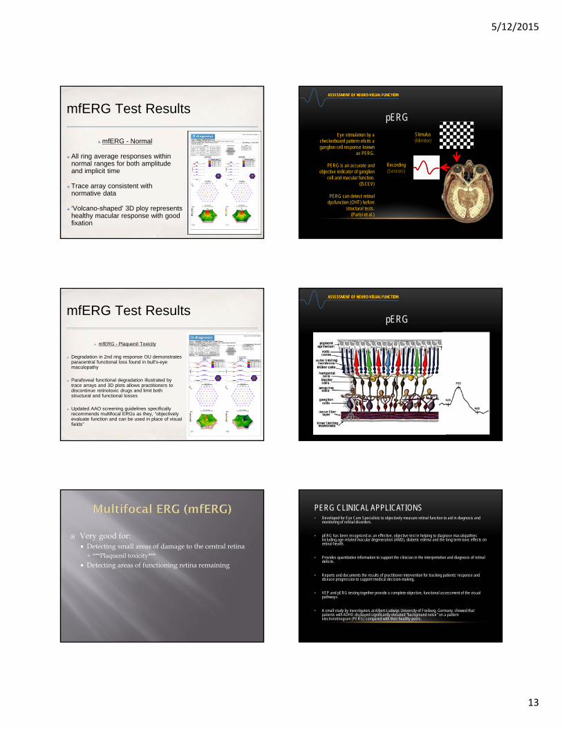
mfERG Test Results
✤ mfERG - Normal
✤ All ring average responses within normal ranges for both amplitude and implicit time
✤ Trace array consistent with normative data
✤ ‘Volcano-shaped’ 3D ploy represents healthy macular response with good fixation
mfERG Test Results
✤ mfERG - Plaquenil Toxicity
✤ Degradation in 2nd ring response OU demonstrates paracentral functional loss found in bull’s-eye maculopathy
✤ Parafoveal functional degradation illustrated by trace arrays and 3D plots allows practitioners to discontinue retinotoxic drugs and limit both structural and functional losses
✤ Updated AAO screening guidelines specifically recommends multifocal ERGs as they, “objectively evaluate function and can be used in place of visual fields”
Very good for: Detecting small areas of damage to the central retina ***Plaquenil toxicity***
Detecting areas of functioning retina remaining
ASSESSMENT OF NEURO-VISUAL FUNCTIONASSESSMENT OF NEURO-VISUAL FUNCTIONASSESSMENT OF NEURO-VISUAL FUNCTION
Stimulus(Monitor)
Recording(Sensors)
Eye stimulation by a checkerboard pattern elicits a ganglion cell response known
as PERG.
PERG is an accurate and objective indicator of ganglion
cell and macular function.(ISCEV)
PERG can detect retinal dysfunction (OHT) before
structural tests.(Parisi et al.)
pERG
ASSESSMENT OF NEURO-VISUAL FUNCTIONASSESSMENT OF NEURO-VISUAL FUNCTIONASSESSMENT OF NEURO-VISUAL FUNCTION
pERG
PERG CLINICAL APPLICATIONS• Developed for Eye Care Specialists to objectively measure retinal function to aid in diagnosis and
monitoring of retinal disorders.
• pERG has been recognized as an effective, objective test in helping to diagnose maculopathies including age-related macular degeneration (AMD), diabetic edema and the long term toxic effects on retinal health.
• Provides quantitative information to support the clinician in the interpretation and diagnosis of retinal deficits.
• Reports and documents the results of practitioner intervention for tracking patients’ response and disease progression to support medical decision-making.
• VEP and pERG testing together provide a complete objective, functional assessment of the visual pathways.
• A small study by investigators at Albert-Ludwigs University of Freiburg, Germany, showed that patients with ADHD displayed significantly elevated "background noise" on a pattern electroretinogram (PERG) compared with their healthy peers.

5/12/2015
14
INTERNATIONAL PERG STANDARD
• Time is measured in milliseconds (ms)
• Amplitude is measured in microvolts (uV)
• N35-P50-N95 Complex
• N35: Negative Pulse around 35ms
• P50: Positive pulse around 50ms
• N95: Negative pulse around 95ms
ASSESSMENT OF NEURO-VISUAL FUNCTIONASSESSMENT OF NEURO-VISUAL FUNCTIONASSESSMENT OF NEURO-VISUAL FUNCTION
pERG Electrodes
ReferenceGround
Active
ERG Electrodes
Comfort Convenience Quality
ASSESSMENT OF NEURO-VISUAL FUNCTIONASSESSMENT OF NEURO-VISUAL FUNCTIONASSESSMENT OF NEURO-VISUAL FUNCTION
ASSESSMENT OF NEURO-VISUAL FUNCTIONASSESSMENT OF NEURO-VISUAL FUNCTIONASSESSMENT OF NEURO-VISUAL FUNCTION
pERG
Extensive and growing literature supports the clinical usage of PERG for the early
diagnosis and tracking of Glaucoma.
PERG FUNCTION PRECEDES STRUCTURE GLC-S
ASSESSMENT OF NEURO-VISUAL FUNCTIONASSESSMENT OF NEURO-VISUAL FUNCTIONASSESSMENT OF NEURO-VISUAL FUNCTION
Main Indications
• Maculopathies
•Glaucoma
pERG

5/12/2015
15
STEADY STATE – PATTERN ERG CONTRAST AND CONCENTRIC STIMULUS FIELD TESTING• BCVA
• Patient should be tested with best corrected vision
• 85% contrast – or High 85% and Low 15% Contrast
• 24” testing distance
• Right Eye (OD) then Left Eye (OS)
• 8 second “warm up”
• 20 seconds at 24⁰ - Used for Hc and Lc
• 20 seconds at 16⁰
ERG PROTOCOL SELECTION
Chronic Open Angle GlaucomaDiabetic Retinopathy
AMDDiabetic RetinopathyDiabetic Macular EdemaToxic Maculopathies
Healthy
Asymptomatic Symptomatic
VF
Glaucoma
Ellish NJ, Higginbotham EJ. Evaluating a visual field screening test for glaucoma: how the choice of the gold standard affects the validity of the test. Ophthalmic Epidemiol. 2001 Dec;8(5):297-307.
ASSESSMENT OF NEURO-VISUAL FUNCTIONASSESSMENT OF NEURO-VISUAL FUNCTIONASSESSMENT OF NEURO-VISUAL FUNCTION
Healthy
Asymptomatic Symptomatic
VF
Glaucoma
OCT
Documented structural damageNon documented structural damage
Schuman JS, Hee MR, Arya AV, Pedut‐Kloizman T, Puliafito CA, Fujimoto JG, Swanson EA. Optical coherence tomography: a new tool for glaucoma diagnosis. Curr Opin Ophthalmol. 1995 Apr;6(2):89‐95.
ASSESSMENT OF NEURO-VISUAL FUNCTIONASSESSMENT OF NEURO-VISUAL FUNCTIONASSESSMENT OF NEURO-VISUAL FUNCTION
Healthy
VF
Glaucoma
OCT
Documented structural damageNon documented structural damage
PERG/VEP
Documented functional damageNon documented functional damage
Parisi V, Miglior S, Manni G, Centofanti M, Bucci MG. Clinical ability of pattern-electroretinograms and visual evoked potentials in detecting visual dysfunction in ocular hypertension and glaucoma. Ophthalmology. 2006 Feb;113(2):216-28.
ASSESSMENT OF NEURO-VISUAL FUNCTIONASSESSMENT OF NEURO-VISUAL FUNCTIONASSESSMENT OF NEURO-VISUAL FUNCTION
OHT
Normal Functioning Ganglion Cell
Ganglion Cell Under Stress (OHT)
Normal Functioning Ganglion CellAfter treatment
Falsini B, Marangoni D, Salgarello T, et al. Effect of epigallocatechin-gallate on inner retinal function in ocular hypertension and glaucoma: A short-term study by pattern electroretinogram. Graefes Arch Clin Exp Ophthalmol (2009) 247:1223–1233
ASSESSMENT OF NEURO-VISUAL FUNCTIONASSESSMENT OF NEURO-VISUAL FUNCTIONASSESSMENT OF NEURO-VISUAL FUNCTION
As measured by PERG,progressive loss of RGC
function in early glaucomais hindered after IOP
lowering.

5/12/2015
16
VEP (Function)
EyeBrainLGN
Stress (OHT)
OCTHRT (Structure)GDX
Yucel YH, Zhang Q, Weinreb RN, Kaufman PL, Gupta1N. Atrophy of Relay Neurons in Magno- and Parvocellular Layers in the Lateral Geniculate Nucleus in Experimental Glaucoma. Invest Ophthalmol Vis Sci. 2001;42:3216–3222.
Glaucomatous Brain Damage
ASSESSMENT OF NEURO-VISUAL FUNCTIONASSESSMENT OF NEURO-VISUAL FUNCTIONASSESSMENT OF NEURO-VISUAL FUNCTION
Normal Concentric Stimulus Fields Normal Contrast Sensitivity
Abnormal Concentric - ARMD Abnormal Contrast - Glaucoma
OUR GALESBURG PRACTICE
• 4 optometrists – two FT equivalent
• 6 exam lanes
• Pre-Test
• FDT - AR/AK - LM – Topograher - Optos
• Special Testing Room #1
• HVF - HRT
• Special Testing Room #2
• Heidelberg Spectralis – PHP - QuantifEYE – Diopsys VEP
• Vision Therapy Specialty – Dr. Carter
If it is good for the patient…..
BASIC PREMISE OF OUR PRACTICE…..
It will be good for the practice!

5/12/2015
17
TECHNOLOGY CONSIDERATIONS TODAY
• Does it do something our other technology doesn’t?
• Will it provide clinical information that will impact the treatment of our patients?
• Can it be incorporated into our office?
• Space – Patient Flow - Staff
• Is it “standard of care” or “leading edge”?
• Is it “patient friendly”?
• Will it be profitable and/or Practice Builder?
• Efficiency – Billable - Referrals
VEP + ERG IS GOOD FOR THE PATIENT….
• Technology has always been a highlight of our practice
• Glaucoma went away……but came back in 1994 with TPA’s
• Visual Fields traditionally the only measure of “function”
• Very subjective and patients don’t like the test
• But now VEP can be incorporated in any practice
• NOT subjective – and patients like the test
• For structure, we use OCT and HRT
• Objective and able to detect subtle changes
VEP/ERG –DOES IT DO SOMETHING DIFFERENT?
• Absolutely! – However, not the research based, school based systems that may or may not have been at your school
• VEP results are a representation of the functional integrity of all levels of the visual pathway including anterior seg, retina, optic nerve, LGN and visual cortex
• ERG measures function at the ganglion cell level
• An objective way to measure “function” for a variety of conditions
• Glaucoma – MS – Amblyopia – Stroke – TBI
• Maculopathies
• InfantSEE®
DOES THE VEP + ERG IMPACT TREATMENT?
• ABSOLUTELY!
• Glaucoma
• Adjunct to visual fields (especially low reliability)
• We now have 2 measures of “function” to go with 2 measures of “structure”
• Developmental Disabled Patients – unable to do VF and even OCT/HRT
• Amblyopia
• Predictor of success and monitoring therapy
• Maculopathies
• Monitor function along with structure
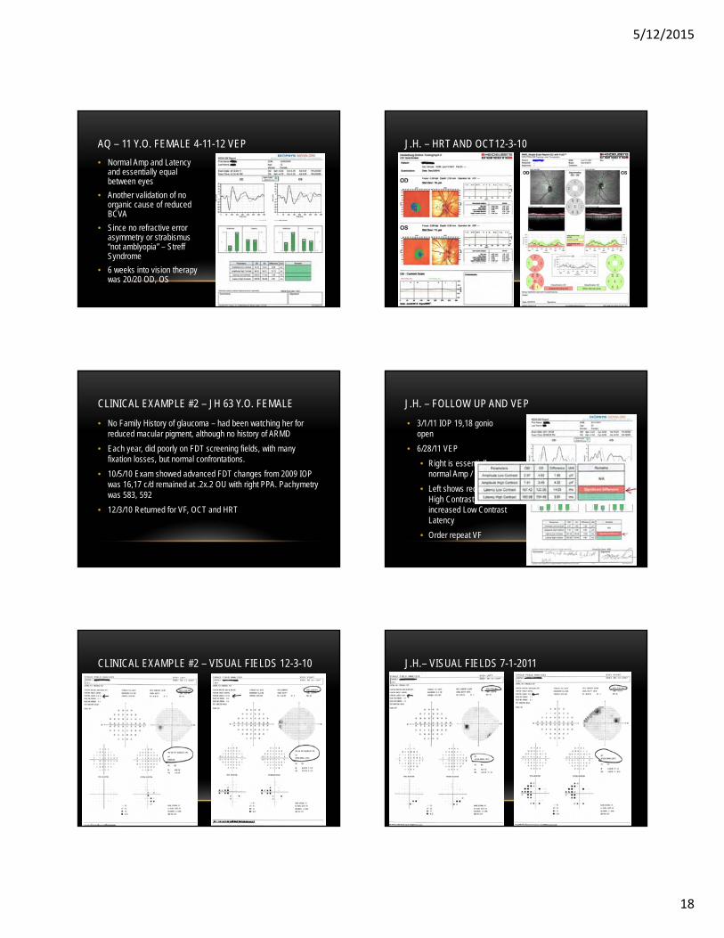
CLINICAL EXAMPLE #1 - AQ 11 Y.O. FEMALE
• Patient since 2008 in combination with IA City Ophthalmology
• Progressively more near sighted each year with good BCVA
• 4/2/2012 presented complaining of daily HA’s for 2 months and vision “not clear”. Full work ups at IA City found a seizure disorder with EEG and Johns Hopkins diagnosed malingering
• BCVA on 4/2/2012 was 20/150 OD, OS - cyclopleged
• Ocular health otherwise normal with c/d’s of .7x7 OU
• Ordered VEP and HVF
AQ – 11 Y.O. FEMALE 4-11-12 VISUAL FIELDS
5/12/2015
18
• Normal Amp and Latency and essentially equal between eyes
• Another validation of no organic cause of reduced BCVA
• Since no refractive error asymmetry or strabismus “not amblyopia” – StreffSyndrome
• 6 weeks into vision therapy was 20/20 OD, OS
AQ – 11 Y.O. FEMALE 4-11-12 VEP
CLINICAL EXAMPLE #2 – JH 63 Y.O. FEMALE
• No Family History of glaucoma – had been watching her for reduced macular pigment, although no history of ARMD
• Each year, did poorly on FDT screening fields, with many fixation losses, but normal confrontations.
• 10/5/10 Exam showed advanced FDT changes from 2009 IOP was 16,17 c/d remained at .2x.2 OU with right PPA. Pachymetrywas 583, 592
• 12/3/10 Returned for VF, OCT and HRT
CLINICAL EXAMPLE #2 – VISUAL FIELDS 12-3-10
J.H. – HRT AND OCT12-3-10
• 3/1/11 IOP 19,18 gonioopen
• 6/28/11 VEP
• Right is essentially normal Amp / Latency
• Left shows reduced High Contrast Amp but increased Low Contrast Latency
• Order repeat VF
J.H. – FOLLOW UP AND VEP
J.H.– VISUAL FIELDS 7-1-2011

5/12/2015
19
J.H. VF 7-9-13
J.H. HRT AND OCT 7-9-13
PATIENT J.H. – PRE CAT SXVEP’S 2-25-13 & 7-17-13
PATIENT – J.H. ERG’S ON 7-17-13
PATIENT J.H. – S/P CATARACTS 5-19-14
PATIENT – J.H. 12-9-2014 (IOP ABOUT 13 ON TX)

5/12/2015
20
PATIENT JH 3-3-15 (NO TX FOR 3 MONTHS – IOP 17)
PATIENT JH – 3-3-15 (NO TX FOR 3 MONTHS – IOP 17)
JH – “WHY WE PURCHASED THIS TECHNOLOGY”
• The initial VEP prompted more testing and starting treatment early
• The VEP and ultimately the pERG allowed to watch the nerves progressively under more stress
• Removing the cataracts ultimately lowered pressures and allowed nerves to return to health
• Sensitivity allowed removal of treatment
• And ultimately back on treatment
• WE PREVENTED STRUCTURAL DAMAGE
INCORPORATING IN OUR PRACTICE
• Space – 4 feet by 7 feet in a corner
• Patient Flow
• We schedule 15 minutes – usually 4-8 total
• Incorporating in glaucoma care - coming
• Staff
• Trained all paraoptometrics
• We use primarily a “special test” para
• More efficient and more consistent results
OUR GLAUCOMA PROTOCOL –INCLUDING VEP/ERG
• Annual exam – include photos - dilated
• 3 or 4 month visit – non dilated
• IOP – Gonio (UBM) – VEP (95930) ERG (92275)
• Next 3 or 4 month visit – dilated
• HVF – HRT – OCT
• Initially did many VEP with this visit at the beginning to get initial data on our patients. Slows the flow in our system because it is preferred to do VEP un-dilated
OUR ARMD/DIABETIC PROTOCOL –INCLUDING ERG
• Annual exam – include photos - dilated
• We Do Macula Risk on all patients
• Helps determine frequency of visits
• Helps guide proper supplement prescribing
• Typical “Quarterly” Q4Months, Once a Year Etc. (Dilated)
• pERG (concentric) – PHP - OCT
• For Diabetic Retinopathy (must have retinopathy)
• OCT and pERG (concentric)

5/12/2015
21
LEADING EDGE
• VEP + ERG data for glaucoma is decades old, just didn’t have a system that was truly office-based until Diopsys NOVA and now other companies
• Normative data comparison makes it much easier as a clinician to interpret the information and implement clinically
• We are no longer the only practice in the area that offers VEP -ERG
• Because of the multiple uses – our practice continues to be recognized as “the most high-tech” in the area
• Referrals from patients, pediatricians, neurologists
OFFICE-BASED VEP + ERG IS PATIENT FRIENDLY
• Even on a “great hair day” – skilled technicians can attach the leads without much disruption
• Patients appreciate the simplicity – no stress when taking the test
• Easily understood report of findings for the patient – excellent patient education
• Relatively quick and easily incorporated with an office visit
• Patients tell their friends about the test – it is very accepted
PROFITABILITY
• We have had TWO coverage issues with any insurers including Illinois Medicaid CPT code – 95930/92275
• Medicare Allowable in “rest of IL” reimburses $119.16 / $137.88
• OCT (92133) = $42.17 ($40.12)
• Fundus Photos (92250) = $73.83 ($63.67)
• HVF (92083) = $60.70 ($55.45)
• In Illinois there are no diagnosis codes associated/limited to CPT code – 95930 or 92275
• No frequency limitations – VEP usually annually ERG can be potentially quarterly or more frequent
OUR MOST COMMON DIAGNOSIS CODES
• 377.14 – Glaucomatous Atrophy (cupping) of optic nerve
• 368.4X – Visual Field Defect (abnormal VF – screening FDT)
• 368.0X – Amblyopia
• 377.11 – Primary Optic Atrophy
• 377.00 - Papilledema
• 368.12 – Transient Visual Loss
• 362.xx (now for pERG
• 365.xx (pERG and in IL for VEP)
• LCD’s list over 80 diagnosis codes
YOU DO YOUR OWN MATH
• How many 365.xx patients do you have?
• How many 368.xx patients do you have?
• How many 377.xx patients do you have?
• How many 362.xx patients do you have
• If you do a screening FDT or other visual field – how many of them do you currently bring back for a full visual field – you should now consider adding a VEP to the battery of tests
• How many patients each year come in with “unspecified visual disturbance or transient visual loss”?
REFERRALS AND MARKETING = MORE PROFITS
• Neurologists have recognized my partner as very neuro oriented and now that we have Diopsys NOVA-VEP they are referring to our practice.
• We’ve successfully treated 2 “malingering” or “Streff Syndrome” patients and because the VEP was an additional test to rule out organic cause – pediatricians are referring patients for testing AND vision therapy
• Patients are telling friends that “their doctor has a VEP” and it is converting to new patients – children and glaucoma

5/12/2015
22
IN SUMMARY – VEP + ERG
• Good for The Patient
• Patients accept and understand the technology
• Objective data with no patient stress
• Good for The Practice
• Valuable clinical data for a variety of diagnoses
• Easily incorporated into practice flow
• A source of professional and patient referrals
• One of the highest reimbursed procedures in the practice
QUESTIONS?