Embed Size (px)
Citation preview
Crit Care Clin 21 (2005) 563–587
Hemostatic Defects in End Stage Liver Disease
Jody L. Kujovich, MD
Division of Hematology and Medical Oncology, Mail Code: L-586,
Oregon Health & Science University, 3181 Sam Jackson Park Road, Portland,
OR 97239-3098, USA
The liver has a pivotal role in hemostasis by synthesizing all clotting factors
(except von Willebrands factor) and coagulation inhibitors as well as several
fibrinolytic proteins. The hepatic reticuloendothelial system clears activated
clotting factors, proteolytic enzyme/inhibitor complexes, and fibrin and fibrino-
gen degradation products. End stage liver disease (ESLD) results in a complex
and variably severe failure of hemostasis that may predispose to abnormal
bleeding. The diverse spectrum of hemostatic defects includes impaired synthesis
of clotting factors, excessive fibrinolysis, disseminated intravascular coagulation,
thrombocytopenia, and platelet dysfunction (Table 1). This article reviews the
hemostatic defects that occur in ESLD with a focus on laboratory diagnosis
and treatment.
Coagulation defects
The progressive loss of hepatic parenchymal cells results in clotting factor
deficiencies. The importance of the coagulopathy is underscored by the in-
corporation of coagulation parameters into prognostic scores for fulminant
hepatic failure and cirrhosis, and their use for assessing bleeding risk [1]. Clot-
ting factor deficiencies primarily reflect impaired hepatic synthetic function,
although increased consumption and extravascular re-distribution may also
contribute. The number and degree of clotting factor deficiencies parallels the
severity of liver damage [2]. Several studies demonstrated the progressive loss of
hepatocytes expressing several clotting factors with more advanced liver disease
0749-0704/05/$ – see front matter D 2005 Elsevier Inc. All rights reserved.
doi:10.1016/j.ccc.2005.03.002 criticalcare.theclinics.com
E-mail address: [email protected]
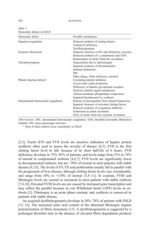
Table 1
Hemostatic defects in ESLD
Hemostatic defect Possible mechanisms
Impaired coagulation Reduced synthesis of clotting factors
Vitamin K deficiency
Dysfibrinogenemia
Systemic fibrinolysis Impaired clearance of tPA and fibrinolytic enzymes
Reduced synthesis of a2-antiplasmin and TAFI
Reabsorption of ascitic fluid into circulation
Thrombocytopenia Sequestration due to splenomegaly
Impaired synthesis of thrombopoietin
Immune destruction
DIC
Other (drugs, folate deficiency, alcohol)
Platelet function defectsa Circulating platelet inhibitors
Excess nitric oxide production
Deficiency of platelet glycoprotein receptors
Defective platelet signal transduction
Altered membrane phospholipid composition
Impaired thromboxand A2 synthesis
Disseminated intravascular coagulation Release of procoagulants from injured hepatocytes
Impaired clearance of activated clotting factors
Reduced synthesis of coagulation inhibitors
Endotoxins in portal circulation
Entry of ascitic fluid into systemic circulation
Abbreviations: DIC, disseminated intravascular coagulation; TAFI, thrombin activatable fibrinolysis
inhibitor; tPA, tissue plasmingen activator.a None of these defects occur consistently in ESLD.
kujovich564
[2,3]. Factor (F)V and FVII levels are sensitive indicators of hepatic protein
synthesis often used to assess the severity of disease [4,5]. FVII is the first
clotting factor level to fall, because of its short half-life of 6 hours. FVII
deficiency develops in 75%–85% of patients, and levels range from 23% to 74%
of normal in compensated cirrhosis [4,6,7]. FVII levels are significantly lower
in decompensated cirrhosis, but are N30% of normal in most patients with stable
disease [4,5,8]. The levels of FV, FX and prothrombin usually fall in parallel with
the progression of liver disease, although clotting factor levels vary considerably,
and range from 10% to N110% of normal [5,8–11]. In contrast, FVIII and
fibrinogen levels are normal or increased in most patients with stable cirrhosis
[7,8,10]. Elevated FVIII levels are not caused by increased gene transcription and
may reflect the parallel increase in von Willebrand factor (vWF) levels in cir-
rhosis [3]. Fibrinogen is an acute phase reactant, and synthesis is conserved in
patients with stable disease.
An acquired dysfibrinogenemia develops in 50%–78% of patients with ESLD
[12–14]. The increased sialic acid content of the abnormal fibrinogen impairs
polymerization of fibrin monomers [15]. A dysfibrinogenemia is suggested by a
prolonged thrombin time in the absence of elevated fibrin degradation products
hemostatic defects in liver disease 565
(FDP) or D-dimer levels. The level of clottable fibrinogen may be low with a
functional assay, but the fibrinogen protein level will be normal using an
immunologic assay. The clinical significance of the dysfibrinogenemia of ESLD
is unclear, but unlikely to cause bleeding in most patients.
Vitamin K is a required cofactor for gamma-carboxylation of glutamic acid
residues on prothrombin, FVII, FIX, FX, protein C, and protein S, a modi-
fication required for binding to phospholipid surfaces. Vitamin K deficiency
occurs commonly in ESLD as a result of poor nutrition, malabsorption of fat
soluble vitamins, or biliary tract obstruction. Cirrhotic patients also develop an
acquired defect of gamma carboxylation unresponsive to vitamin K, which is
reflected by increased levels of hypocarboxylated vitamin K- dependent clotting
factors [16].
The risk of bleeding associated with the coagulopathy of ESLD depends on
the number and severity of clotting factor deficiencies. Individual clotting factor
levels often remain in a hemostatically effective range in patients with stable
disease. In contrast, patients with the more severe and complex hemostatic
defects characteristic of decompensated cirrhosis are at risk for spontaneous and
procedure-related bleeding. However, individual clotting factor levels do not
reliably predict bleeding or survival [17,18].
Systemic fibrinolysis
Laboratory evidence of low grade systemic fibrinolysis is found in 30% – 46%
of patients with ESLD [19–21]. Fibrinolytic acitivity increases with the
progression of liver disease, but varies considerably between individuals
[18,19,21–23]. Accelerated fibrinolysis results from the impaired clearance of
tissue plasminogen activator (tPA) and other fibrinolytic enzymes by the diseased
liver, without an appropriate increase in plasminogen activator inhibitors
[21,22,24]. Impaired hepatic synthesis of fibrinolytic inhibitors (a2 anti-plasmin
and thrombin-activatable fibrinolysis inhibitor [TAFI]) contributes to the increase
in circulating plasmin. TAFI removes C-terminal lysine residues on fibrin, which
serve as binding sites for the activation of plasminogen. TAFI levels are markedly
reduced in patients with cirrhosis and correlate with the severity of disease
[25–27]. Reabsorption of ascitic fluid into the systemic circulation may con-
tribute to accelerated fibrinolyisis in some cases [28].
The clinical significance of systemic fibrinolysis varies between individuals
and with the severity of liver disease. Low grade fibrinolysis probably does
not markedly increase the bleeding risk in most patients with stable disease. Up
to 30% of patients with compensated cirrhosis have laboratory evidence of
accelerated fibrinolysis without clinically significant bleeding [19,23]. However,
patients with liver disease often have an exaggerated fibrinolytic response to
physiologic and iatrogenic stresses, especially surgery [24,29–31]. Major surgery
may stimulate the release of large amounts of tPA from injured tissues, tem-
kujovich566
porarily overwhelming antifibrinolytic mechanisms and resulting in a ‘‘burst’’ of
fibrinolysis and bleeding [32]. Premature lysis of hemostatic plugs at vascular
injury sites may provoke or exacerbate bleeding. Systemic fibrinolysis was
associated with soft tissue, variceal and surgical bleeding in some [20,21,23,
32–34] but not all studies [18,34]. Bleeding can occur anywhere, but is par-
ticularly prominent at sites of trauma or surgery. Affected patients often develop
generalized oozing from surgical incisions and venipuncture sites, occasionally
severe and refractory to therapy [35].
Systemic fibrinolysis should be suspected in patients with persistent bleed-
ing despite optimal clotting factor and platelet replacement. The diagnosis is
suggested by a shortened euglobulin clot lysis time, which reflects circulating
fibrinolytic enzymes. An elevated D-dimer level indicates plasmin lysis of cross-
linked fibrin when fibrinolysis is secondary to the activation of coagulation.
Severe fibrinolysis often results in a substantial fall in fibrinogen and a2anti-
plasmin levels, due to consumption as well as impaired synthesis.
Thrombocytopnia
Mild to moderate thrombocytopenia occurs in 49% –64% of patients with
ESLD [36,37]. However, the platelet count is rarely less than 30, 000 to 40,000
and spontaneous bleeding is uncommon. The etiology of thrombocytopenia
is multifactorial, and includes sequestration of platelets in an enlarged spleen,
impaired platelet production, and immune and non-immune mediated platelet
destruction. Other causes such as folate deficiency, alcohol, sepsis, disseminated
intravascular coagulation (DIC), and drugs may also contribute in individ-
ual cases.
Radiolabled platelet studies demonstrate splenic sequestration in ESLD. The
spleen normally contains approximately one-third of the total platelet mass,
exchanging with circulating platelets [38]. In contrast, a markedly enlarged
spleen may sequester up to 90% of the total platelet mass [38–40]. Platelet counts
correlated inversely with spleen size in some [41–44] although not all studies
[45–48]. ‘‘Hypersplenism’’ is thought to reflect an exaggeration of the normal
function of splenic macrophages to remove senescent cells. In one study, platelet
counts correlated inversely with the number of phagocytically active splenic
macrophages, supporting their role in the pathogenesis of thrombocytopenia [49].
There is conflicting data on platelet lifespan in ESLD, with decreased survival
reported in some [40,45,46] but not all studies [38,39].
Although it contributes in some cases, other evidence argues against splenic
sequestration as the primary cause of thrombocytopenia in ESLD. Portal decom-
pression procedures and splenectomy do not consistently improve platelet counts
[37,50–55]. Thrombocytopenia occurs in nearly 25% of cirrhotic patients with
normal spleen size, suggesting other mechanisms must be involved [42,48,56].
Conversely, 19%–29% of cirrhotic patients with splenomegaly maintain normal
platelet counts, suggesting splenic pooling alone is not sufficient to cause
hemostatic defects in liver disease 567
thrombocytopenia [42,56]. Flow cytometric analysis of young reticulated plate-
lets in peripheral blood is a useful indicator of platelet production. Reticulated
platelet counts are significantly lower in cirrhotic patients with thrombocytopenia
than in those with normal platelet counts, implicating impaired thrombopoiesis
[57,58].
Thrombopoietin (TPO), the priniciple physiologic regulator of platelet pro-
duction, is synthesized constitutively in the liver [59]. There is accumulating
evidence that impaired hepatic synthesis of TPO is a major cause of thrombo-
cytopenia in liver disease. Thrombocytopenic patients with ESLD have inappro-
priately low TPO levels [43,44,57,60–63]. TPO levels are significantly lower in
cirrhotic patients with thrombocytopenia than in those with normal platelet
counts [41,47,58]. Patients with splenomegaly and normal platelet counts have
significantly higher TPO levels than those with thrombocytopenia, which
suggests that higher TPO levels result in a compensatory increase in platelet
production [42]. Serum TPO levels correlate inversely with the severity of liver
disease reflected by the degree of fibrosis, Child Pugh class, and sensitive mea-
sures of liver function [41–43,48]. TPO mRNA levels are significantly reduced in
cirrhotic liver tissue [60,61]. Replacement of a cirrhotic liver with a functional
graft restores TPO production and corrects thrombocytopenia [47,60,62–64]. The
consistent increase in TPO levels and platelet count after orthotopic liver
transplantation strongly implicates impaired TPO production as a primary cause
of thrombocytopenia [47,62,63,65].
Elevated levels of platelet-associated IgG are found in 55%–88% of patients
with chronic liver disease [44,66–70], and correlates inversely with the platelet
count in some [44,68,70] but not all studies [66]. However, platelet associated
IgG does not confirm immune-mediated platelet destruction, since high levels
occur in liver disease patients with normal platelet counts and non-immune
thrombocytopenia [66,69]. More specific autoantibodies directed against the most
common platelet glycoprotein antigenic targets in idiopathic thrombocytopenic
purpura (ITP) are found in 38%–64% of patients with liver disease [66,71]. There
is some evidence that immune mediated thrombocytopenia is more frequent in
patients with hepatitis C-associated liver disease [68,71,72]. Anti-platelet
glycoprotein autoantibodies are more common in patients with chronic hepati-
tis C than those with ESLD due to other etiologies [71,72]. Patients with chronic
hepatitis C have an increased frequency of ITP, consistent with the propensity for
autoimmune complications [72–75]. Reports of response to standard immunologic
therapies (corticosteroids, intravenous immunoglobulin [IVIG]) support immune-
mediated thrombocytopenia in some cases [72–75].
The available evidence suggests impaired TPO production caused by the
progressive loss of functioning hepatocytes, is a primary cause of thrombo-
cytopenia in ESLD. However, the widely variable extent of splenic sequestration,
TPO levels, platelet survival, and unpredictable effect of portal decompression
procedures, suggest different mechanisms predominate in different patients
[40,46,50,53]. It is unknown whether the etiology of thrombocytopenia affects
the risk of bleeding.

kujovich568
Platelet function defects
Qualitative platelet abnormalities occur in patients with ESLD, reflected by
a prolonged bleeding time, and impaired platelet aggregation responses to multi-
ple agonists. The bleeding time is prolonged in 40% of patients with cirrhosis
and correlates with the severity of disease, reflected by Child-Pugh class and
laboratory parameters [76–79]. Abnormal bleeding times are only partly ex-
plained by thrombocytopenia, since they occur in 12%–25% of cirrhotic patients
with normal platelet counts [76–78]. The platelet function analyzer (PFA-100) is
an in vitro system for evaluation of platelet-dependent primary hemostasis.
Although abnormal PFA-100 results occur in patients with ESLD, its use in this
population has not been well studied [80].
Impaired platelet aggregation in vitro is found in 46% of patients with cirrho-
sis, although the pattern of abnormalities is not consistent [80–82]. Abnormal
platelet function has been attributed to circulating platelet inhibitors (FDP and D-
dimers), plasmin degradation of platelet receptors, dysfibrinogenemia, and excess
nitric oxide synthesis [77,78,81–84]. Nitric oxide is a powerful vasodilator and
inhibitor of platelet adhesion and aggregation produced by vascular endothelial
cells. Inhibition of nitric oxide production normalized prolonged bleeding times
in a rat model of cirrhosis [83]. A variety of intrinsic platelet defects are also
reported including a deficiency of platelet GPIb receptors, defective signal
transduction, impaired thromboxane A2 synthesis, altered membrane phospho-
lipid composition, and acquired storage pool deficiency [85–87]. However, none
of these defects occur consistently, which suggests a multifactorial etiology in-
volving both intrinsic and extrinsic factors.
Disseminated intravascular coagulation
Patients with ESLD often have evidence of chronic low grade DIC. Fibrino-
gen survival is reduced and elevated levels of various markers of coagulation
activation are found in plasma [40]. Multiple studies demonstrated elevated levels
of the prothrombin activation fragment F1 + 2, Fibrinopeptide A, D-dimer, and
thrombin-antithrombin complexes, although values vary markedly between pa-
tients [88–93]. The infusion of heparin lowered the level of coagulation activation
markers and prolonged fibrinogen survival, confirming accelerated intravascular
coagulation [40,91]. The frequency and severity of DIC correlate with the stage
of liver disease in most studies [88,89,93,94]. Elevated levels of D-dimers and
F1 + 2 are found in up to 93%–100% of patients with advanced stage cirrhosis
and complications such as ascites [28,89]. In contrast, laboratory evidence of
DIC is found in a minority of patients with compensated cirrhosis [93,95,96].
The mechanisms triggering DIC in ESLD are complex and include release of
procoagulants from injured hepatocytes, impaired clearance of activated clotting
factors, reduced synthesis of coagulation inhibitors, and entry of endotoxins into
the portal circulation. The severity of DIC is inversely related to antithrombin and
hemostatic defects in liver disease 569
protein C levels, and antithrombin replacement prolongs fibrinogen survival
[88,90,97]. Plasma levels of tissue factor are elevated in patients with advanced
cirrhosis and correlate with the severity of disease [98]. Endotoxinemia is
strongly associated with high plasma levels of F1 + 2 and D-dimer, implicating it
as a trigger of coagulation activation [89]. The entry of procoagulant-rich ascitic
fluid into the systemic circulation may also contribute, explaining the develop-
ment of DIC after peritoneal-venous shunt placement.
Because of the similar hemostatic defects and pattern of laboratory abnor-
malities, the diagnosis of DIC in ESLD is difficult, and its clinical significance is
often unclear. DIC is more likely in patients with coexisting conditions inde-
pendently associated with accelerated intravascular coagulation, such as sepsis, or
trauma. Classic laboratory tests for DIC are often abnormal in ESLD, but may
reflect other hemostatic defects. However, the pattern of testing is often useful for
identifying superimposed DIC. D-dimer (and FDP) levels are usually much
higher in DIC. An elevated D-dimer level is a more specific marker of DIC since
it indicates activation of both coagulation and fibrinolyisis. In contrast, high FDP
levels may reflect fibrinogen degradation products or cross-reacting dysfunc-
tional fibrinogen, both common in ESLD [96]. Because fibrinogen synthesis is
conserved in compensated cirrhosis, low levels (b 100–120 mg/dL) suggest DIC.
Serial testing demonstrating declining FVIII and fibrinogen levels with an
elevated D-dimer level in an appropriate clinical setting provides strong pre-
sumptive evidence for DIC.
Thrombosis
The balance between the levels of procoagulant and anticoagulant proteins
determines the overall effect on hemostasis and resulting risk of hemorrhage and
thrombosis. Although bleeding occurs more frequently, the hemostatic imbalance
in ESLD occasionally favors hypercoagulability, predisposing to thrombosis.
Deficiencies of anticoagulation proteins are common, and correlate with the
severity of disease [13,18,26,88,90,99]. Antithrombin, protein C and protein S
levels range from 30% to 65% of normal, similar to the range of values found in
patients with inherited deficiencies [18,26,95,99]. The high levels of several
procoagulant factors in cirrhosis (FVIII, vWF and fibrinogen) may contribute to
hypercoagulability [100]. Cirrhotic patients with prothrombotic risk factors
superimposed on an already activated coagulation system are at risk for throm-
botic complications. There is some evidence that thrombophilic disorders in-
crease the risk of thrombosis in patients with ESLD [99,101]. Cirrhotic patients
with the prothrombin gene mutation have a nearly sixfold increased risk of portal
vein thrombosis [101]. Cirrhotic patients with a prolonged PT are not necessarily
‘‘auto-anticoagulated’’ and therefore protected from thrombosis. Therapeutic
warfarin lowers the levels of all four vitamin K-dependent clotting factors (FII,
FVII, FIX, FX) to 15%–30% of normal. In contrast, patients with ESLD often
have low FVII levels with relatively higher FIX, FX and prothrombin levels in

kujovich570
the 40%–60% range [7,9,11,102]. Because lower levels of prothrombin and FX
are required for an effective anti-thrombotic effect, the coagulopathy of ESLD
does not necessarily prevent thrombosis [103].
A variety of thrombotic complications occur in patients with ESLD. An
autopsy series found thrombi in one or multiple organs in 54% and 22% of
patients with cirrhosis, respectively [104]. Hepatic vein thrombosis is common in
cirrhosis and is implicated in disease progression [105]. Thrombotic risk factors
are independently associated with the extent of hepatic fibrosis, which sug-
gests that vascular obstruction may accelerate its development [106]. Portal vein
thrombosis occurs in 9% – 20% of patients with cirrhosis, and is more frequent
in those with advanced disease [101,104,107,108]. Cirrhotic patients have bio-
chemical evidence of hypercoagulability in the portal circulation, which may
predispose to thrombosis in this particular location [109].
Laboratory tests
Standard screening tests are used to assess the hemostatic derangement in
patients with liver disease who are actively bleeding or require invasive
procedures (Table 2). Initial testing should include a PT/INR, aPTT, platelet
count, and fibrinogen level. These tests provide a measure of liver disease
severity and serve as a baseline for monitoring blood product replacement. In
selected patients, a D-dimer, euglobulin clot lysis time, thrombin time, and PFA-
100 (or bleeding time) may provide additional useful information (Table 2).
Clotting times typically remain in the normal range until clotting factor levels
Table 2
Typical laboratory results in ESLD
Condition Laboratory findings
Compensated ESLD PT/INR prolonged
aPTT prolonged
Fibrinogen normal or decreased
Platelet count normal or decreased
Systemic fibrinolysis Fibrinogen normal or decreased
Euglobulin Clot Lysis Time shortened
D-dimer increased
a2-antiplasmin decreased
Dysfibrinogenemia Clottable fibrinogen decreaseda
Fibrinogen antigen normalb
Thrombin Time prolonged
Disseminated intravascular coagulation Fibrinogen normal or decreasedc
Platelet count decreased
D-dimer increased
FVIII normal or decreasedc
a Using a functional assay of clottable fibrinogen.b Using an immunologic assay of fibrinogen protein.c Declining levels on serial testing suggests DIC.
hemostatic defects in liver disease 571
are less than 30%–40% of normal. A prolonged PT/INR with a normal aPTT
occurs in mild liver disease, indicating an isolated deficiency of FVII, which
affects only the PT. With disease progression the PT/INR and aPTT are both
prolonged, reflecting deficiencies of multiple clotting factors. However, prolon-
gation of the aPTT may be blunted by the high FVIII levels common in com-
pensated cirrhosis [110]. There is no definitive evidence that individual clotting
factor levels are more predictive of bleeding or prognosis, although they may be
useful in specific circumstances.
The fibrinogen level is normal or elevated in patients with stable chronic liver
disease. Severe hypofibrinogenemia (b100 mg/dL) is uncommon, but occurs in
decompensated cirrhosis or DIC. Fibrinogen levels b80 mg/dL markedly prolong
the PT and PTT, caused by the resulting inability to form a detectable fibrin
clot, which is the endpoint of these assays. The thrombin time is prolonged by
hypofibrinogenemia, dysfibrinogenemia, or elevated levels of fibrin/fibrinogen
degradation products and D-dimers.
The diagnosis of ITP is suggested by thrombocytopenia disproportionate to
the severity of liver disease, and is confirmed by a response to immunosup-
pression. An elevated peripheral blood reticulated platelet count occurs with ITP
and other causes of increased platelet destruction. A prolonged bleeding time
or abnormal PFA-100 out of proportion to thrombocytopenia suggests platelet
dysfunction, although neither test reliably predicts bleeding [79].
International normalized ratio in ESLD
The international normalized ratio (INR) system was developed to standardize
PT reporting for patients on stable oral anticoagulation. Its validity for reporting
PT values in ESLD has not been confirmed. Several studies demonstrated
significantly different INR values with different thromboplastin reagents in
patients with liver disease, especially at high INR values [9,111]. The variable
results with different thromboplastins suggest INR values may not accurately
reflect the coagulopathy in ESLD. Depending on the sensitivity of the throm-
boplastin, the INR may over- or underestimate the severity of liver disease, po-
tentially affecting clinical prognostic scores.
The INR is calculated from the PT ratio (patient PT/control PT) adjusted for
the international sensitivity index (ISI). The ISI reflects the sensitivity of a
particular thromboplastin to a reduction in vitamin K-dependent clotting factors,
and is derived from a cohort of patients on stable vitamin K antagonists. How-
ever, the INR in liver disease does not reflect the same pattern of clotting factor
deficiencies found in patients on oral anticoagulation. Patients with ESLD typi-
cally have lower FV and fibrinogen levels and higher FX and prothrombin levels
than patients on warfarin with similar INR values [5,9]. In one study, liver disease
patients had significantly lower FV and FVII levels for a given increment in INR
than warfarin-anticoagulated controls [5]. Thus, a particular INR value does not

kujovich572
necessarily reflect the same degree of ‘‘auto-anticoagulation’’ or bleeding risk in
liver disease as in patients receiving warfarin.
Treatment
Correction of hemostatic defects is required in patients who are actively
bleeding or who require surgery or other invasive procedures (Table 3). The
therapeutic approach should be tailored to the type, site, and severity of bleeding.
Since the most common cause of bleeding in ESLD is a localized anatomic
defect, the evaluation should focus on identifying the site of bleeding. Correction
of hemostatic defects should be coordinated with definitive therapy to the
bleeding site, such as sclerotherapy of bleeding varices. Serial hemostasis labo-
ratory tests are used to monitor the response to therapy. Therapy should be aimed
at achieving hemostatic competence rather than complete correction of abnormal
laboratory values.
Coagulopathy
Vitamin K
Coagulation screening tests do not distinguish between vitamin K deficiency
and clotting factor deficiencies caused by impaired hepatic synthesis. Thus, a
trial of vitamin K is useful, especially in patients with cholestatic liver dis-
ease. Although vitamin K deficiency is rarely the primary cause of a co-
agulopathy, a brief course of vitamin K (5 –10 mg/d for 3 days) will exclude it
as a contributing factor.
Plasma
Fresh frozen plasma (FFP) contains all coagulation proteins (except vWF) and
inhibitors present in circulating blood. FFP is administered to correct coagulation
Table 3
Treatment of hemostatic defects in actively bleeding patients with ESLD
Laboratory parametera Treatment
INR N2.0 (with normal aPTT) or aPTT N1.3 � control Fresh frozen plasma
?Recombinant factor VIIa
Fibrinogen b125 mg/dL Cryoprecipitate
Platelet count b50,000–75,000/mL Platelet transfusion
Prolonged bleeding time or prolonged closure time
on PFA-100b?DDAVP
Red cell transfusion if Hct b30%
Abbreviations: DDAVP, desmopressin; Hct, hematocrit; PFA-100, platelet function analyzer.a Serial testing used to monitor response to therapy.b See text for details.

hemostatic defects in liver disease 573
defects before invasive procedures and to control active bleeding. However,
correction of the coagulopathy of ESLD is difficult, due to the short half-life of
several clotting factors and the large volumes required. Several studies showed
that infusion of 2–6 units of FFP corrected a prolonged PT in only a minority
(12% – 36%) of patients with chronic liver disease [112–114]. Although larger
volumes are more effective, complete correction does not occur in at least 25%
of patients [114,115]. The duration of effect is transient; PT values return to
baseline within 24 hours in the majority of cases [115,116]. Thus, repeated
transfusions every 8 –12 hours are usually required to maintain a near normal PT.
FFP is variably effective in increasing individual clotting factor levels, depending
on the severity of coagulopathy and volume infused. Although hemostatically
effective levels are achieved in most cases, the effect is transient, especially for
FVII [112,113,115,116].
There are no controlled trials confirming the efficacy of FFP for prophylaxis
or treatment of bleeding in ESLD. There is currently no consensus on the severity
of coagulopahy or specific invasive procedures that mandate prophylactic FFP
infusions. The volume of FFP required to prevent or treat bleeding is also
unknown, and likely varies with the severity of coagulopathy and clinical setting.
Replacement therapy should be directed at achieving hemostatically effective
clotting factor levels rather than normalization of coagulation screening tests. For
example, an isolated mild FVII deficiency (reflected by a prolonged PT and
normal aPTT) is unlikely to cause bleeding in the absence of coexisting hemo-
static defects. Since FVII levels N15% of normal are adequate for hemostasis
in patients with an inherited deficiency, an isolated mildly prolonged PT/INR
does not require plasma infusions [117]. The minimum level of clotting factors
required for hemostasis in patients with multiple coagulation defects is not well-
defined. Large volumes of plasma are poorly tolerated by patients with ESLD,
who usually already have an expanded intravascular volume. Volume overload
may precipitate congestive heart failure or increase portal pressure, with the risk
of variceal rupture. Other potential adverse effects of FFP include transmission of
blood-borne infections, febrile or allergic reactions, and transfusion-related acute
lung injury.
Plasma exchange
Plasma exchange may be required to correct the coagulopathy or control
refractory bleeding in patients at risk for volume overload with FFP alone.
However the efficacy of plasma exchange in ESLD has not been demonstrated in
controlled trials. Plasma exchange is used primarily to prepare patients for liver
transplantation, or to manage patients with fulminant hepatic failure [118,119].
Other replacement therapy
Cryoprecipitate is a fraction of plasma rich in fibrinogen, FVIII, vWF and
factor XIII. Cryoprecipitate may be required in patients with a severe co-
kujovich574
agulopathy and hypofibrinogenemia (b 100 mg/dL). Prothrombin-complex
concentrates contain the vitamin K dependent clotting factors (FII, FVII, FIX
and FX) in high concentration. Despite several reports of the safe use of these
products, they should be avoided in patients with ESLD due to the risk of
thrombotic complications [120,121].
Recombinant factor VIIa
Recombinant FVIIa (rFVIIa), is a synthetic analog of the naturally occurring
serine protease enzyme, genetically engineered in baby hamster cells. In normal
physiologic concentrations, FVIIa is enzymatically active only after binding
tissue factor exposed at vascular injury sites. The tissue factor-FVIIa complex
activates factors IX and X, ultimately accelerating thrombin generation. In high
(pharmacologic) concentrations, FVIIa activates FX directly on the surface of
activated platelets, independent of tissue factor [122]. The requirement for tissue
factor or activated platelets localizes rFVIIa’s procoagulant activity to the vas-
cular injury site, thereby minimizing the risk of thrombotic complications.
Recombinant FVIIa is FDA approved for the treatment of bleeding in he-
mophilia patients with inhibitors. It has been used to correct the coagulopathy of
ESLD, although limited data support its use in this setting. Multiple studies
demonstrated prompt normalization of the PT in the majority of patients with
baseline prolonged values, including those refractory to plasma infusions
[11,102,123–128]. Higher doses achieve greater and more prolonged correction
of the PT, suggesting a ‘‘dose-response’’ effect [11,124]. However, the effect
is transient, even at higher doses, reflecting rFVIIa’s short half-life of approxi-
mately 2 hours.
The prompt predictable correction of the PT, provides a rationale for the pro-
phylactic and therapeutic use of rFVIIa. However, the current evidence sup-
porting its use in ESLD is limited to case reports and case series. Recombinant
FVIIa was reported to prevent bleeding at the time of liver biopsy, intracranial
pressure (ICP) monitor placement, pericardiocentesis, pancreatic aspiration,
injection of hepatocellular carcinoma and colon polypectomy, and to reduce
blood loss during orthotopic liver transplantation [123,124,126,129–131].
Prophylactic doses ranged from 5 to 120 mg/kg, with some patients receiving
additional doses during or after the procedure [124,126].
Recombinant FVIIa also controlled refractory epistaxis, bleeding after den-
tal extractions, hematuria, oozing from catheter sites, and gastrointestinal bleed-
ing in a small number of patients with ESLD [125,132,133]. A single dose
(50–110 mg/kg) achieved hemostasis in cirrhotic patients with variceal hemor-
rhage refractory to standard measures. However, early rebleeding occurred in
25% of cases, and there was a high mortality rate from bleeding-related causes
[102,134]. In another study, the majority of patients with persistent uncontrolled
bleeding after rFVIIa, had a complex coagulopathy due to liver disease, which
suggested uncertain efficacy in this particular population [135].
hemostatic defects in liver disease 575
Despite rFVIIa’s excellent overall safety record in hemophilia patients, its
safety in patients with other thrombotic risk factors is not established. Reports of
increased levels of several coagulation activation markers, especially after higher
doses, fuel a theoretical concern that a rFVIIa-induced ‘‘thrombin burst’’ could
exacerbate subclinical DIC [11,124,136]. However, none of the reported patients
developed clinical DIC, and no adverse effects occurred in the majority of other
studies [11,124]. There are a few anecdotal reports of DIC and thrombosis in
patients with ESLD receiving rFVIIa, although a causal connection to rFVIIa was
not confirmed [124,131].
Recombinant FVIIa has several advantages over plasma in ESLD, including
more rapid and predictable correction of the PT, a lower infection risk, and
effectiveness in small volumes [125,126]. However, transient shortening of the
PT does not guarantee hemostasis. Moreover, because of rFVIIa’s short half-life,
effective hemostasis may be followed by recurrent bleeding in the absence of
definitive therapy [102,133]. The optimal dose and dosing schedule for prophy-
laxis or treatment of bleeding are unknown, and may depend on the severity of the
coagulopathy and clinical indication. It is also unclear to what extent the
hemostatic effect of rFVIIa depends on the platelet count and other clotting factor
levels. Larger prospective controlled trials are required to confirm efficacy and
safety in ESLD and to define guidelines for its cost-effective use.
Thrombocytopenia
Platelet transfusions are indicated in actively bleeding patients with platelet
counts b50,000–75,000. Since platelets are suspended in plasma, a single
apheresis product provides a unit of plasma, also replacing clotting factors.
However, the plasma in platelets has a lower concentration of FV and FVIII than
FFP. Prophylactic platelet transfusions are often administered before invasive
procedures in patients with platelet counts b 50,000. Since thrombocytopenia is
typically mild in ESLD, they are rarely required in stable patients who do not
require invasive procedures. A platelet count of 10,000 is a common trigger for
prophylactic platelet transfusions in stable thrombocytopenic patients. A higher
threshold may be appropriate for patients with other hemostatic defects or risk
factors for bleeding. Post-transfusion platelet counts are used to assess recovery
and guide subsequent therapy. The platelet count increment will be blunted in
patients with splenomegaly because of sequestration of transfused platelets
[38,39]. Other causes of platelet refractoriness include DIC, infection, and
alloimmunization caused by platelet-specific and /or HLA antibodies. The
potential benefits of platelet transfusions must be weighed against the risk of
alloimmunization, an important consideration for patients awaiting liver trans-
plantation. The use of leukoreduced products will reduce the risk of alloim-
munization. Corticosteroids and IVIG are occasionally effective in patients with a
clinical presentation suggesting ITP [72,74,75]. The clinical use of recombinant

kujovich576
thrombopoietin is still investigational, and trials in patients with liver disease
have not yet been performed.
Platelet dysfunction
The indications for specific therapy to improve platelet function are unclear.
Since severe anemia may also impair platelet function, red cell transfusions
should be considered for patients with hematocrits b 30% [80]. Desmopression
(1-deamino-8-D-arginine vasopressin, DDAVP) is a synthetic analog of anti-
diuretic hormone which stimulates the release of vWF from endothelial cells.
DDAVP significantly shortens bleeding times in up to 60% of patients with cir-
rhosis [137–140]. The effect is transient, with maximal shortening 30–60 minutes
after a single dose, and does not depend on the platelet count [137]. Although
the mechanism is still not well understood, DDAVP appears to increase platelet
adhesiveness. There are no studies confirming its clinical efficacy for the pre-
vention or treatment of bleeding in ESLD. Because of the complexity of the
hemostatic derangement, a transient shortening of the bleeding time may not
significantly reduce the risk of bleeding. DDAVP did not reduce intra-operative
blood loss or improve control of active variceal hemorrhage in two randomized
trials [141,142]. However, a trial of DDAVP is reasonable in patients with
refractory bleeding and a prolonged bleeding time or abnormal PFA.
Disseminated intravascular coagulation
Although laboratory evidence of low grade DIC is common in ESLD, specific
therapy is rarely required. When DIC is suspected, therapeutic strategies should
focus on detection and reversal of potential precipitating factors, such as infec-
tion. Replacement of clotting factors, coagulation inhibitors, and platelets may be
necessary in patients with active bleeding, using laboratory tests to guide therapy.
Anticoagulation with heparin is not recommended because of the unacceptably
high risk of bleeding. Infusion of antithrombin concentrate normalized fibrinogen
survival in cirrhotic patients, but had no significant effect on molecular markers
of coagulation activation [97,143]. Currently, there is no evidence that anti-
thrombin replacement prevents the clinical complications of DIC or improves
outcome in ESLD.
Systemic fibrinolysis
Patients with mild systemic fibrinolysis who are not bleeding do not require
specific therapy. In contrast, those with severe fibrinolysis and serious bleeding
require replacement of hemostatic components. FFP replaces a2-antiplasmin as
well as clotting factors and cryoprecipitate contains fibrinogen and FVIII in high
hemostatic defects in liver disease 577
concentration. Antifibrinolytic agents (epsilon aminocaproic acid, tranexamic
acid) inhibit plasmin generation and may control diffuse bleeding [19,34,35].
However, because of the frequent simultaneous activation of coagulation, there
is a high risk of thrombotic complications in this particular population.
Antifibrinolytic agents may be considered in selected patients with fibrinolytic
bleeding unresponsive to plasma and platelet transfusions after exclusion of DIC.
Invasive procedures
Patients with ESLD and coagulopathy are assumed to have an increased risk
of bleeding with invasive procedures, although the magnitude of risk is not well
defined. The bleeding risk is likely higher in patients with multiple hemostatic
defects, renal failure, or a prior bleeding history [144,145].
Liver biopsy
Clinically significant bleeding is uncommon after percutaneous liver biopsy,
complicating 0.35%–0.7% of cases [146–149]. Severe hemostatic defects are
considered a contraindication to the procedure [150]. However, there is no
consensus on acceptable hemostatic parameters, which vary considerably among
practicing gastroenterologists and academic centers [151,152]. Peripheral blood
coagulation tests correlate poorly with the severity of bleeding from the liver
biopsy site observed at laparoscopy [146,153–155]. In most studies, a mildly
prolonged PT (within 4 seconds of control) did not increase the risk of bleed-
ing [146,153,155,156]. In contrast, a severe coagulopathy may predispose to
procedure-related bleeding [147,149,157]. Mild thrombocytopenia (platelet count
N50,000–100,000) does not appear to increase the risk of post-biopsy bleeding
[146,153,158]. More severe thrombocytopenia (b50,000) was associated with
bleeding complications in some [158] but not all studies [154,159]. A prolonged
bleeding time (N12 minutes) was reported to confer a fivefold higher risk of post-
biopsy bleeding [160]. However, the bleeding time is not a validated predictor
of bleeding after invasive procedures, and most centers do not include it in their
pre-biopsy testing [151].
The available data suggest that hemostasis screening tests do not reliably
predict bleeding after liver biopsy. Various technical aspects of the procedure,
including the number of punctures and underlying liver pathology, may also
affect the risk of bleeding. Although there are no universally accepted guidelines,
most authorities recommend a minimum platelet count of N50,000 to 80,000 and a
PT within 3–4 seconds of control values [110,151,161–164].) FFP and platelet
transfusions are recommended for a PT N 3 –4 seconds above control (INR N1.4),
and a platelet count b 50,000–60,000, respectively, although there is no evidence
that prophylactic therapy prevents bleeding complications [161,162,164]. Pre-
liminary studies suggest rFVIIa provides effective prophylaxis, although there
is insufficient data to support its routine use [124]. Alternative biopsy methods

kujovich578
(‘‘plugged’’ percutaneous or transjugular biopsy) may be safer for patients with
severe coagulation defects that cannot be corrected [154,157,163,165].
Intracranial pressure monitors
Patients with fulminant liver failure often undergo ICP monitor placement
for detection of cerebral edema. Bleeding complications occur in 3%–18% of
patients, depending on the particular monitoring technique [166]. Since there is
no consensus on ‘‘safe’’ coagulation parameters for monitor placement, accept-
able values are often defined by neurosurgical consultants. Plasma or platelets are
frequently transfused to correct hemostatic defects before the procedure.
However, the large volumes of plasma usually required have a theoretical risk
of exacerbating cerebral edema. Recombinant FVIIa transiently corrects the PT,
allowing ICP monitor placement, although the optimal dose and duration of
therapy is unknown [126].
Central venous catheters
Placement of central venous catheters in hemostatically compromised patients
with ESLD is associated with a low risk of bleeding, with major bleeding
complications reported in 0%–0.2% of cases, and minor bleeding complications
reported in 1%–12% of cases [144,167–170]. The infrequency of bleeding
suggests that routine administration of blood products is unnecessary before
catheter placement. Prompt application of pressure may prevent hematoma for-
mation, even in patients with coagulation defects.
Paracentesis and thoracentesis
Excessive bleeding occurs in 0%–3% of patients undergoing paracentesis; 0%
to 1.2% of procedures are complicated by major hemorrhage requiring trans-
fusion [145,171–175]. One study found no significant difference in the incidence
of bleeding between patients with and without hemostatic defects, or between
those who did and did not receive prophylactic plasma infusions before the
procedure [145]. In another large series of 1,100 paracenteses, there were no
significant bleeding complications in cirrhotic patients with platelet counts
b50,000 and/or INR values �1.5 [175]. Several consensus guidelines recommend
against routine administration of blood products before paracentesis, suggesting
coagulation defects are a contraindication only in patients with clinically overt
fibrinolysis or DIC [173,176,177].
The limited available evidence suggests bleeding is uncommon after
thoracentesis in patients with ESLD, even in the absence of prophylactic blood
products [145]. However, the American Thoracic Society and the American
College of Physicians identify a coagulopathy as a contraindication to
hemostatic defects in liver disease 579
thoracentesis and pleural biopsy, recommending a minimum platelet count of
50,000 [178,179]. Because of difficulty detecting bleeding and risk of hemo-
thorax, severe coagulation defects should be corrected.
Summary
Patients with ESLD develop multiple hemostatic defects, the severity of which
depends on the degree of hepatic injury. In addition, other coexisting
complications such as uremia and esophageal varices may predispose to
abnormal bleeding. The hemostatic derangement of ESLD has a multifactorial
etiology. Impaired hepatic synthesis of coagulation proteins and TPO is a major
cause of coagulation defects, fibrinolyisis, and thrombocytopenia. However, the
widely variable range of all hemostatic parameters suggests different mechanisms
predominate in individual patients. Although the hemostatic derangement of
ESLD usually results in a bleeding tendency, it occasionally predisposes to DIC
or thrombosis.
Hemostasis laboratory testing is used to assess disease severity and bleeding
risk, and to monitor the response to therapy. However, no particular coagulation
profile reliably predicts bleeding. The INR is valid for reporting PT results within
an institution, but does not standardize the PT in ESLD independent of the
particular thromboplastin used. FFP, cryoprecipitate and platelet transfusions
remain the mainstay of therapy for patients who are actively bleeding or require
invasive procedures. Until larger controlled trials confirm efficacy and safety in
ESLD, rFVIIa should be used with caution based on an individual risk/benefit
assessment. There is no consensus on acceptable coagulation laboratory parame-
ters for performance of invasive procedures. Nevertheless, most experts rec-
ommend correction of severe coagulation defects, especially before high-risk
procedures, in locations where bleeding is difficult to control. Decisions about
prophylaxis should be based on the type of procedure, severity of hemostatic
defects, co-existing risk factors, and the patient’s bleeding history.
References
[1] Pugh RN, Murray-Lyon IM, Dawson JL, et al. Transection of the oesophagus for bleeding
oesophageal varices. Br J Surg 1973;60:646–9.
[2] Rodriguez-Inigo E, Bartolome J, Quiroga JA, et al. Expression of factor VII in the liver of
patients with liver disease: correlations with the disease severity and impairment in the
hemostasis. Blood Coagul Fibrinolysis 2001;12:193–9.
[3] Hollestelle MJ, Geertzen HG, Straatsburg IH, et al. Factor VIII expression in liver disease.
Thromb Haemost 2004;91:267–75.
[4] Green G, Poller L, Thomson JM, et al. Factor VII as a marker of hepatocellular synthetic
function in liver disease. J Clin Pathol 1976;29:971–5.
[5] Deitcher SR. Interpretation of the international normalised ratio in patients with liver disease.
Lancet 2002;359:47–8.

kujovich580
[6] Hallen A, Nilsson IM. Coagulation studies in liver disease. Thromb Diath Haemorrh
1964;11:51–63.
[7] Kupfer HG, Gee W, Ewald AT, et al. Statistical correlation of liver function tests with
coagulation factor deficiencies in Laennec’s cirrhosis. Thromb Diath Haemorrh 1964;10:
317–31.
[8] Kerr R. New insights into haemostasis in liver failure. Blood Coagul Fibrinolysis 2003;
14(Suppl 1):S43–5.
[9] Kovacs MJ, Wong A, MacKinnon K, et al. Assessment of the validity of the INR system for
patients with liver impairment. Thromb Haemost 1994;71:727–30.
[10] Biland L, Duckert F, Prisender S, et al. Quantitative estimation of coagulation factors in liver
disease. The diagnostic and prognostic value of factor XIII, factor V and plasminogen. Thromb
Haemost 1978;39:646–56.
[11] Bernstein DE, Jeffers L, Erhardtsen E, et al. Recombinant factor VIIa corrects prothrombin
time in cirrhotic patients: a preliminary study. Gastroenterology 1997;113:1930–7.
[12] Green G, Thomson JM, Dymock IW, et al. Abnormal fibrin polymerization in liver disease.
Br J Haematol 1976;34:427–39.
[13] Kelly DA, Tuddenham EG. Haemostatic problems in liver disease. Gut 1986;27:339–49.
[14] Francis JL, Armstrong DJ. Acquired dysfibrinogenaemia in liver disease. J Clin Pathol 1982;
35:667–72.
[15] Roberts HR, Stinchcombe TE, Gabriel DA. The dysfibrinogenaemias. Br J Haematol 2001;114:
249–57.
[16] Blanchard RA, Furie BC, Jorgensen M, et al. Acquired vitamin K-dependent carboxylation
deficiency in liver disease. N Engl J Med 1981;305:242–8.
[17] Gazzard BG, Henderson JM, Williams R. Factor VII levels as a guide to prognosis in fulminant
hepatic failure. Gut 1976;17:489–91.
[18] Boks AL, Brommer EJ, Schalm SW, et al. Hemostasis and fibrinolysis in severe liver failure
and their relation to hemorrhage. Hepatology 1986;6:79–86.
[19] Hu KQ, Yu AS, Tiyyagura L, et al. Hyperfibrinolytic activity in hospitalized cirrhotic patients
in a referral liver unit. Am J Gastroenterol 2001;96:1581–6.
[20] Violi F, Ferro D, Basili S, et al. Hyperfibrinolysis increases the risk of gastrointestinal
hemorrhage in patients with advanced cirrhosis. Hepatology 1992;15:672–6.
[21] Violi F, Ferro D, Basili S, et al. Hyperfibrinolysis resulting from clotting activation in patients
with different degrees of cirrhosis. Hepatology 1993;17:78–83.
[22] Leebeek FW, Kluft C, Knot EA, et al. A shift in balance between profibrinolytic and
antifibrinolytic factors causes enhanced fibrinolysis in cirrhosis. Gastroenterology 1991;101:
1382–90.
[23] Violi F, Basili S, Ferro D, et al. Association between high values of D-dimer and tissue-
plasminogen activator activity and first gastrointestinal bleeding in cirrhotic patients. Thromb
Haemost 1996;76:177–83.
[24] Fletcher AP, Biederman O, Moore D, et al. Abnormal plasminogen-plasmin system activity
(fibrinolysis) in patients with hepatic cirrhosis: its cause and consequences. J Clin Invest 1964;
43:681–95.
[25] Lisman T, Leebeek FW, Mosnier LO, et al. Thrombin-activatable fibrinolysis inhibitor defi-
ciency in cirrhosis is not associated with increased plasma fibrinolysis. Gastroenterology 2001;
121:131–9.
[26] Colucci M, Binetti BM, Branca MG, et al. Deficiency of thrombin activatable fibrinolysis
inhibitor in cirrhosis is associated with increased plasma fibrinolysis. Hepatology 2003;38:
230–7.
[27] Van Thiel DH, George M, Fareed J. Low levels of thrombin activatable fibrinolysis inhibitor
(TAFI) in patients with chronic liver disease. Thromb Haemost 2001;85:667–70.
[28] Agarwal S, Joyner Jr KA, Swaim MW. Ascites fluid as a possible origin for hyperfibrinolysis
in advanced liver disease. Am J Gastroenterol 2000;95:3218–24.
[29] Porte RJ, Bontempo FA, Knot EA, et al. Tissue-type-plasminogen-activator-associated
fibrinolysis in orthotopic liver transplantation. Transplant Proc 1989;21:3542.
hemostatic defects in liver disease 581
[30] Harper PL, Luddington RJ, Jennings I, et al. Coagulation changes following hepatic
revascularization during liver transplantation. Transplantation 1989;48:603–7.
[31] Lewis JH, Bontempo FA, Awad SA, et al. Liver transplantation: intraoperative changes in
coagulation factors in 100 first transplants. Hepatology 1989;9:710–4.
[32] Steib A, Gengenwin N, Freys G, et al. Predictive factors of hyperfibrinolytic activity during
liver transplantation in cirrhotic patients. Br J Anaesth 1994;73:645–8.
[33] Gutierrez A, Sanchez-Paya J, Marco P, et al. Prognostic value of fibrinolytic tests for hospital
outcome in patients with acute upper gastrointestinal hemorrhage. J Clin Gastroenterol 2001;
32:315–8.
[34] Francis Jr RB, Feinstein DI. Clinical significance of accelerated fibrinolysis in liver disease.
Haemostasis 1984;14:460–5.
[35] Kahl BS, Schwartz BS, Mosher DF. Profound imbalance of pro-fibrinolytic and anti-fibrinolytic
factors (tissue plasminogen activator and plasminogen activator inhibitor type 1) and severe
bleeding diathesis in a patient with cirrhosis: correction by liver transplantation. Blood Coagul
Fibrinolysis 2003;14:741–4.
[36] Bashour FN, Teran JC, Mullen KD. Prevalence of peripheral blood cytopenias (hypersplenism)
in patients with nonalcoholic chronic liver disease. Am J Gastroenterol 2000;95:2936–9.
[37] Jabbour N, Zajko A, Orons P, et al. Does transjugular intrahepatic portosystemic shunt (TIPS)
resolve thrombocytopenia associated with cirrhosis? Dig Dis Sci 1998;43:2459–62.
[38] Aster RH. Pooling of platelets in the spleen: role in the pathogenesis of ‘‘hypersplenic’’
thrombocytopenia. J Clin Invest 1966;45:645–57.
[39] Harker LA, Finch CA. Thrombokinetics in man. J Clin Invest 1969;48:963–74.
[40] Stein SF, Harker LA. Kinetic and functional studies of platelets, fibrinogen, and plasminogen
in patients with hepatic cirrhosis. J Lab Clin Med 1982;99:217–30.
[41] Giannini E, Botta F, Borro P, et al. Relationship between thrombopoietin serum levels and liver
function in patients with chronic liver disease related to hepatitis C virus infection. Am J
Gastroenterol 2003;98:2516–20.
[42] Adinolfi LE, Giordano MG, Andreana A, et al. Hepatic fibrosis plays a central role in the
pathogenesis of thrombocytopenia in patients with chronic viral hepatitis. Br J Haematol
2001;113:590–5.
[43] Kawasaki T, Takeshita A, Souda K, et al. Serum thrombopoietin levels in patients with chronic
hepatitis and liver cirrhosis. Am J Gastroenterol 1999;94:1918–22.
[44] Sanjo A, Satoi J, Ohnishi A, et al. Role of elevated platelet-associated immunoglobulin G and
hypersplenism in thrombocytopenia of chronic liver diseases. J Gastroenterol Hepatol 2003;
18:638–44.
[45] Toghill PJ, Green S, Ferguson F. Platelet dynamics in chronic liver disease with special
reference to the role of the spleen. J Clin Pathol 1977;30:367–71.
[46] Toghill PJ, Green S. Platelet dynamics in chronic liver disease using the 111Indium oxine label.
Gut 1983;24:49–52.
[47] Goulis J, Chau TN, Jordan S, et al. Thrombopoietin concentrations are low in patients with
cirrhosis and thrombocytopenia and are restored after orthotopic liver transplantation. Gut
1999;44:754–8.
[48] Giannini E, Borro P, Botta F, et al. Serum thrombopoietin levels are linked to liver function in
untreated patients with hepatitis C virus-related chronic hepatitis. J Hepatol 2002;37:572–7.
[49] Yongxiang W, Zongfang L, Guowei L, et al. Effects of splenomegaly and splenic macrophage
activity in hypersplenism due to cirrhosis. Am J Med 2002;113:428–31.
[50] Sanyal AJ, Freedman AM, Purdum PP, et al. The hematologic consequences of transjugular
intrahepatic portosystemic shunts. Hepatology 1996;23:32–9.
[51] Karasu Z, Gurakar A, Kerwin B, et al. Effect of transjugular intrahepatic portosystemic shunt
on thrombocytopenia associated with cirrhosis. Dig Dis Sci 2000;45:1971–6.
[52] Mutchnick MG, Lerner E, Conn HO. Effect of portacaval anastomosis on hypersplenism. Dig
Dis Sci 1980;25:929–38.
[53] Sullivan Jr BH, Tumen HJ. The effect of portacaval shunt on thrombocytopenia associated
with portal hypertension. Ann Intern Med 1961;55:598–603.

kujovich582
[54] Felix Jr WR, Myerson RM, Sigel B, et al. The effect of portacaval shunt on hypersplenism.
Surg Gynecol Obstet 1974;139:899–904.
[55] Kamisasa I, Hidai K, Sugiura M, et al. Effects of splenectomy on blood coagulation and
fibrinolysis in patients with liver cirrhosis: possible role of the spleen in haemostasis. Thrombos.
Haemostas 1979;42:1529–35.
[56] Liangpunsakul S, Ulmer BJ, Chalasani N. Predictors and implications of severe hypersplenism
in patients with cirrhosis. Am J Med Sci 2003;326:111–6.
[57] Koike Y, Yoneyama A, Shirai J, et al. Evaluation of thrombopoiesis in thrombocytopenic
disorders by simultaneous measurement of reticulated platelets of whole blood and serum
thrombopoietin concentrations. Thromb Haemost 1998;79:1106–10.
[58] Panasiuk A, Prokopowicz D, Zak J, et al. Reticulated platelets as a marker of megakaryopoiesis
in liver cirrhosis; relation to thrombopoietin and hepatocyte growth factor serum concentration.
Hepatogastroenterology 2004;51:1124–8.
[59] Kuter DJ, Begley CG. Recombinant human thrombopoietin: basic biology and evaluation of
clinical studies. Blood 2002;100:3457–69.
[60] Martin III TG, Somberg KA, Meng YG, et al. Thrombopoietin levels in patients with cirrhosis
before and after orthotopic liver transplantation. Ann Intern Med 1997;127:285–8.
[61] Ishikawa T, Ichida T, Matsuda Y, et al. Reduced expression of thrombopoietin is involved in
thrombocytopenia in human and rat liver cirrhosis. J Gastroenterol Hepatol 1998;13:907–13.
[62] Peck-Radosavljevic M, Wichlas M, Zacherl J, et al. Thrombopoietin induces rapid resolution of
thrombocytopenia after orthotopic liver transplantation through increased platelet production.
Blood 2000;95:795–801.
[63] Peck-Radosavljevic M, Zacherl J, Meng YG, et al. Is inadequate thrombopoietin production
a major cause of thrombocytopenia in cirrhosis of the liver? J Hepatol 1997;27:127–31.
[64] Yanaga K, Tzakis AG, Shimada M, et al. Reversal of hypersplenism following orthotopic liver
transplantation. Ann Surg 1989;210:180–3.
[65] Peck-Radosavljevic M. Thrombocytopenia in liver disease. Can J Gastroenterol 2000;
14(Suppl D):60D–6D.
[66] Pereira J, Accatino L, Alfaro J, et al. Platelet autoantibodies in patients with chronic liver
disease. Am J Hematol 1995;50:173–8.
[67] Samuel H, Nardi M, Karpatkin M, et al. Differentiation of autoimmune thrombocytopenia from
thrombocytopenia associated with immune complex disease: systemic lupus erythematosus,
hepatitis-cirrhosis, and HIV-1 infection by platelet and serum immunological measurements. Br
J Haematol 1999;105:1086–91.
[68] Nagamine T, Ohtuka T, Takehara K, et al. Thrombocytopenia associated with hepatitis C viral
infection. J Hepatol 1996;24:135–40.
[69] Graber D, Giuliani D, Leevy CM, et al. Platelet-associated IgG in hepatitis and cirrhosis.
J Clin Immunol 1984;4:108–11.
[70] de Noronha R, Taylor BA, Wild G, et al. Inter-relationships between platelet count, platelet
IgG, serum IgG, immune complexes and severity of liver disease. Clin Lab Haematol 1991;
13:127–35.
[71] Kajihara M, Kato S, Okazaki Y, et al. A role of autoantibody-mediated platelet destruction
in thrombocytopenia in patients with cirrhosis. Hepatology 2003;37:1267–76.
[72] Pockros PJ, Duchini A, McMillan R, et al. Immune thrombocytopenic purpura in patients
with chronic hepatitis C virus infection. Am J Gastroenterol 2002;97:2040–5.
[73] Bauduer F, Marty F, Larrouy M, et al. Immunologic thrombocytopenic purpura as presenting
symptom of hepatitis C infection. Am J Hematol 1998;57:338–40.
[74] Ramos-Casals M, Garcia-Carrasco M, Lopez-Medrano F, et al. Severe autoimmune cytopenias
in treatment-naive hepatitis C virus infection: clinical description of 35 cases. Medicine
(Baltimore) 2003;82:87–96.
[75] Hernandez F, Blanquer A, Linares M, et al. Autoimmune thrombocytopenia associated with
hepatitis C virus infection. Acta Haematol 1998;99:217–20.
[76] Blake JC, Sprengers D, Grech P, et al. Bleeding time in patients with hepatic cirrhosis.
BMJ 1990;301:12–5.
hemostatic defects in liver disease 583
[77] Violi F, Leo R, Vezza E, et al. Bleeding time in patients with cirrhosis: relation with degree
of liver failure and clotting abnormalities. J Hepatol 1994;20:531–6.
[78] Hsu WC, Lee FY, Lee SD, et al. Prolonged bleeding time in cirrhotic patients: relationship
to peripheral vasodilation and severity of cirrhosis. J Gastroenterol Hepatol 1994;9:437–41.
[79] Basili S, Ferro D, Leo R, et al. Bleeding time does not predict gastrointestinal bleeding in
patients with cirrhosis. J Hepatol 1996;24:574–80.
[80] Escolar G, Cases A, Vinas M, et al. Evaluation of acquired platelet dysfunctions in uremic and
cirrhotic patients using the platelet function analyzer (PFA-100): influence of hematocrit
elevation. Haematologica 1999;84:614–9.
[81] Ballard HS, Marcus AJ. Platelet aggregation in portal cirrhosis. Arch Intern Med 1976;136:
316–9.
[82] Thomas DP, Ream VJ, Stuart RK. Platelet aggregation in patients with Laennec’s cirrhosis
of the liver. N Engl J Med 1967;276:1344–8.
[83] Albornoz L, Bandi JC, Otaso JC, et al. Prolonged bleeding time in experimental cirrhosis: role
of nitric oxide. J Hepatol 1999;30:456–60.
[84] Coller BS. Platelets and thrombolytic therapy. N Engl J Med 1990;322:33–42.
[85] Pantaleo P, Marra F, Vizzutti F, et al. Effects of dietary supplementation with arachidonic
acid on platelet and renal function in patients with cirrhosis. Clin Sci (Lond) 2004;106:
27–34.
[86] Sanchez-Roig MJ, Rivera J, Moraleda JM, et al. Quantitative defect of glycoprotein Ib in
severe cirrhotic patients. Am J Hematol 1994;45:10–5.
[87] Laffi G, Marra F, Gresele P, et al. Evidence for a storage pool defect in platelets from cirrhotic
patients with defective aggregation. Gastroenterology 1992;103:641–6.
[88] Vukovich T, Teufelsbauer H, Fritzer M, et al. Hemostasis activation in patients with liver
cirrhosis. Thromb Res 1995;77:271–8.
[89] Violi F, Ferro D, Basili S, et al. Prognostic value of clotting and fibrinolytic systems in a
follow-up of 165 liver cirrhotic patients. Hepatology 1995;22:96–100.
[90] Bakker CM, Knot EA, Stibbe J, et al. Disseminated intravascular coagulation in liver cirrhosis.
J Hepatol 1992;15:330–5.
[91] Coccheri S, Mannucci PM, Palareti G, et al. Significance of plasma fibrinopeptide A and high
molecular weight fibrinogen in patients with liver cirrhosis. Br J Haematol 1982;52:503–9.
[92] Takahashi H, Tatewaki W, Wada K, et al. Thrombin and plasmin generation in patients with
liver disease. Am J Hematol 1989;32:30–5.
[93] Mombelli G, Fiori G, Monotti R, et al. Fibrinopeptide A in liver cirrhosis: evidence against a
major contribution of disseminated intravascular coagulation to coagulopathy of chronic liver
disease. J Lab Clin Med 1992;121:83–90.
[94] Kemkes-Matthes B, Bleyl H, Matthes KJ. Coagulation activation in liver diseases. Thromb Res
1991;64:253–61.
[95] Ben-Ari Z, Osman E, Hutton RA, et al. Disseminated intravascular coagulation in liver
cirrhosis: fact or fiction? Am J Gastroenterol 1999;94:2977–82.
[96] vanDeWater L, Carr JM, Aronson D, et al. Analysis of elevated fibrin(ogen) degradation
product levels in patients with liver disease. Blood 1986;67:1468–73.
[97] Schipper HG, ten Cate JW. Antithrombin III transfusion in patients with hepatic cirrhosis. Br J
Haematol 1982;52:25–33.
[98] Tacke F, Schoffski P, Trautwein C, et al. Tissue factor and thrombomodulin levels are
correlated with stage of cirrhosis in patients with liver disease. Blood Coagul Fibrinolysis
2001;12:539–45.
[99] Romero Gomez M, Suarez Garcia E, Lopez Lacomba D, et al. Antiphospholipid antibodies
are related to portal vein thrombosis in patients with liver cirrhosis. J Clin Gastroenterol
2000;31:237–40.
[100] Koster T, Blann AD, Briet E, et al. Role of clotting factor VIII in effect of von Willebrand factor
on occurrence of deep-vein thrombosis. Lancet 1995;345:152–5.
[101] Amitrano L, Guardascione MA, Brancaccio V, et al. Risk factors and clinical presentation of
portal vein thrombosis in patients with liver cirrhosis. J Hepatol 2004;40:736–41.
kujovich584
[102] Ejlersen E, Melsen T, Ingerslev J, et al. Recombinant activated factor VII (rFVIIa) acutely
normalizes prothrombin time in patients with cirrhosis during bleeding from oesophageal
varices. Scand J Gastroenterol 2001;36:1081–5.
[103] Zivelin A, Rao LV, Rapaport SI. Mechanism of the anticoagulant effect of warfarin as evaluated
in rabbits by selective depression of individual procoagulant vitamin K-dependent clotting
factors. J Clin Invest 1993;92:2131–40.
[104] Oka K, Tanaka K. Intravascular coagulation in autopsy cases with liver diseases. Thromb
Haemost 1979;42:564–70.
[105] Wanless IR, Wong F, Blendis LM, et al. Hepatic and portal vein thrombosis in cirrhosis:
possible role in development of parenchymal extinction and portal hypertension. Hepatology
1995;21:1238–47.
[106] Papatheodoridis GV, Papakonstantinou E, Andrioti E, et al. Thrombotic risk factors and extent
of liver fibrosis in chronic viral hepatitis. Gut 2003;52:404–9.
[107] Yerdel MA, Gunson B, Mirza D, et al. Portal vein thrombosis in adults undergoing liver
transplantation: risk factors, screening, management, and outcome. Transplantation 2000;69:
1873–81.
[108] Belli L, Romani F, Sansalone CV, et al. Portal thrombosis in cirrhotics. A retrospective analysis.
Ann Surg 1986;203:286–91.
[109] Violi F, Ferro D, Basili S, et al. Ongoing prothrombotic state in the portal circulation of cirrhotic
patients. Thromb Haemost 1997;77:44–7.
[110] Rapaport SI. Coagulation problems in liver disease. Blood Coagul Fibrinolysis 2000;
11(Suppl 1):S69–74.
[111] Robert A, Chazouilleres O. Prothrombin time in liver failure: time, ratio, activity percentage,
or international normalized ratio? Hepatology 1996;24:1392–4.
[112] Gazzard BG, Henderson JM, Williams R. The use of fresh frozen plasma or a concentrate
of factor IX as replacement therapy before liver biopsy. Gut 1975;16:621–5.
[113] Mannucci PM, Franchi F, Dioguardi N. Correction of abnormal coagulation in chronic liver
disease by combined use of fresh-frozen plasma and prothrombin complex concentrates. Lancet
1976;2:542–5.
[114] Youssef WI, Salazar F, Dasarathy S, et al. Role of fresh frozen plasma infusion in correction of
coagulopathy of chronic liver disease: a dual phase study. Am J Gastroenterol 2003;98:1391–4.
[115] Spector I, Corn M, Ticktin HE. Effect of plasma transfusions on the prothrombin time and
clotting factors in liver disease. N Engl J Med 1966;275:1032–7.
[116] Williamson LM, Llewelyn CA, Fisher NC, et al. A randomized trial of solvent/detergent-treated
and standard fresh-frozen plasma in the coagulopathy of liver disease and liver transplantation.
Transfusion 1999;39:1227–34.
[117] Perry DJ. Factor VII deficiency. Br J Haematol 2002;118:689–700.
[118] Wang YJ, He NH, Wang ZW, et al. Assessment of the combined effect of plasma exchange
and plasma perfusion on patients with severe hepatitis awaiting orthotopic liver transplantation.
Int J Artif Organs 2004;27:40–4.
[119] Singer AL, Olthoff KM, Kim H, et al. Role of plasmapheresis in the management of acute
hepatic failure in children. Ann Surg 2001;234:418–24.
[120] Lorenz R, Kienast J, Otto U, et al. Efficacy and safety of a prothrombin complex concentrate
with two virus-inactivation steps in patients with severe liver damage. Eur J Gastroenterol
Hepatol 2003;15:15–20.
[121] Kohler M. Thrombogenicity of prothrombin complex concentrates. Thromb Res 1999;
95(Suppl. 1):S13–7.
[122] Monroe DM, Hoffman M, Oliver JA, et al. Platelet activity of high-dose factor VIIa is
independent of tissue factor. Br J Haematol 1997;99:542–7.
[123] Anantharaju A, Mehta K, Mindikoglu AL, et al. Use of activated recombinant human factor VII
(rhFVIIa) for colonic polypectomies in patients with cirrhosis and coagulopathy. Dig Dis Sci
2003;48:1414–24.
[124] Jeffers L, Chalasani N, Balart L, et al. Safety and efficacy of recombinant factor VIIa in patients
with liver disease undergoing laparoscopic liver biopsy. Gastroenterology 2002;123:118–26.

hemostatic defects in liver disease 585
[125] Brown JB, Emerick KM, Brown DL, et al. Recombinant factor VIIa improves coagulopathy
caused by liver failure. J Pediatr Gastroenterol Nutr 2003;37:268–72.
[126] Shami VM, Caldwell SH, Hespenheide EE, et al. Recombinant activated factor VII for
coagulopathy in fulminant hepatic failure compared with conventional therapy. Liver Transpl
2003;9:138–43.
[127] Surudo T, Wojcicki M, Milkiewicz P, et al. Rapid correction of prothrombin time after low-
dose recombinant factor VIIA in patients undergoing orthotopic liver transplantation.
Transplant Proc 2003;35:2323–5.
[128] Chuansumrit A, Treepongkaruna S, Phuapradit P. Combined fresh frozen plasma with recom-
binant factor VIIa in restoring hemostasis for invasive procedures in children with liver
diseases. Thromb Haemost 2001;85:748–9.
[129] Papatheodoridis GV, Chung S, Keshav S, et al. Correction of both prothrombin time and
primary haemostasis by recombinant factor VII during therapeutic alcohol injection of hepa-
tocellular cancer in liver cirrhosis. J Hepatol 1999;31:747–50.
[130] Mindikoglu AL, Anantharaju A, Villanueva J, et al. Pericardiocentesis and pancreatic
aspiration needle biopsy in coagulopathic and thrombocytopenic cirrhotic patient. Chest 2003;
123:956–8.
[131] Hendriks HG, Meijer K, de Wolf JT, et al. Reduced transfusion requirements by recombinant
factor VIIa in orthotopic liver transplantation: a pilot study. Transplantation 2001;71:402–5.
[132] Lucia JF, Aguilar C, Orna E, et al. Successful outcome of a cirrhotic patient with postoperative
haematuria treated with a single high dose of recombinant factor VIIa. Haemophilia
2001;7:600–2.
[133] Berthier AM, Guillygomarc’h A, Messner M, et al. Use of recombinant factor VIIa to treat
persistent bleeding following dental extractions in two cirrhotic patients. Vox Sang 2002;
82:119–21.
[134] Romero-Castro R, Jimenez-Saenz M, Pellicer-Bautista F, et al. Recombinant-activated factor
VII as hemostatic therapy in eight cases of severe hemorrhage from esophageal varices. Clin
Gastroenterol Hepatol 2004;2:78–84.
[135] O’Connell NM, Perry DJ, Hodgson AJ, et al. Recombinant FVIIa in the management of
uncontrolled hemorrhage. Transfusion 2003;43:1711–6.
[136] Caldwell SH, Chang C, Macik BG. Recombinant activated factor VII (rFVIIa) as a hemostatic
agent in liver disease: a break from convention in need of controlled trials. Hepatology
2004;39:592–8.
[137] Mannucci PM, Vicente V, Vianello L, et al. Controlled trial of desmopressin in liver cirrhosis
and other conditions associated with a prolonged bleeding time. Blood 1986;67:1148–53.
[138] Cattaneo M, Tenconi PM, Alberca I, et al. Subcutaneous desmopressin (DDAVP) shortens the
prolonged bleeding time in patients with liver cirrhosis. Thromb Haemost 1990;64:358–60.
[139] Burroughs AK, Matthews K, Qadiri M, et al. Desmopressin and bleeding time in patients with
cirrhosis. Br Med J (Clin Res Ed) 1985;291:1377–81.
[140] Agnelli G, Parise P, Levi M, et al. Effects of desmopressin on hemostasis in patients with liver
cirrhosis. Haemostasis 1995;25:241–7.
[141] de Franchis R, Arcidiacono PG, Carpinelli L, et al. Randomized controlled trial of
desmopressin plus terlipressin vs. terlipressin alone for the treatment of acute variceal hem-
orrhage in cirrhotic patients: a multicenter, double-blind study. Hepatology 1993;18:1102–7.
[142] Wong AY, Irwin MG, Hui TW, et al. Desmopressin does not decrease blood loss and transfusion
requirements in patients undergoing hepatectomy. Can J Anaesth 2003;50:14–20.
[143] Scherer R, Kabatnik M, Erhard J, et al. The influence of antithrombin III (AT III) substitution to
supranormal activities on systemic procoagulant turnover in patients with end-stage chronic
liver disease. Intensive Care Med 1997;23:1150–8.
[144] DeLoughery TG, Liebler JM, Simonds V, et al. Invasive line placement in critically ill patients:
do hemostatic defects matter? Transfusion 1996;36:827–31.
[145] McVay PA, Toy PT. Lack of increased bleeding after paracentesis and thoracentesis in
patients with mild coagulation abnormalities. Transfusion 1991;31:164–71.
kujovich586
[146] McGill DB, Rakela J, Zinsmeister AR, et al. A 21-year experience with major hemorrhage after
percutaneous liver biopsy. Gastroenterology 1990;99:1396–400.
[147] Gilmore IT, Burroughs A, Murray-Lyon IM, et al. Indications, methods, and outcomes of
percutaneous liver biopsy in England and Wales: an audit by the British Society of Gastro-
enterology and the Royal College of Physicians of London. Gut 1995;36:437–41.
[148] Piccinino F, Sagnelli E, Pasquale G, et al. Complications following percutaneous liver biopsy.
A multicentre retrospective study on 68,276 biopsies. J Hepatol 1986;2:165–73.
[149] Mahal AS, Knauer CM, Gregory PB. Bleeding after liver biopsy. West J Med 1981;134:11–4.
[150] Jacobs WH, Goldberg SB. Statement on outpatient percutaneous liver biopsy. Dig Dis Sci
1989;34:322–3.
[151] Sue M, Caldwell SH, Dickson RC, et al. Variation between centers in technique and guidelines
for liver biopsy. Liver 1996;16:267–70.
[152] Mayoral W, Lewis JH. Percutaneous liver biopsy: what is the current approach? Results of
a questionnaire survey. Dig Dis Sci 2001;46:118–27.
[153] McVay PA, Toy PT. Lack of increased bleeding after liver biopsy in patients with mild
hemostatic abnormalities. Am J Clin Pathol 1990;94:747–53.
[154] Ewe K. Bleeding after liver biopsy does not correlate with indices of peripheral coagulation.
Dig Dis Sci 1981;26:388–93.
[155] Dillon JF, Simpson KJ, Hayes PC. Liver biopsy bleeding time: an unpredictable event.
J Gastroenterol Hepatol 1994;9:269–71.
[156] Inabnet WB, Deziel DJ. Laparoscopic liver biopsy in patients with coagulopathy, portal
hypertension, and ascites. Am Surg 1995;61:603–6.
[157] Denzer U, Helmreich-Becker I, Galle PR, et al. Liver assessment and biopsy in patients with
marked coagulopathy: value of mini-laparoscopy and control of bleeding. Am J Gastroenterol
2003;98:893–900.
[158] Sharma P, McDonald GB, Banaji M. The risk of bleeding after percutaneous liver biopsy:
relation to platelet count. J Clin Gastroenterol 1982;4:451–3.
[159] Caturelli E, Squillante MM, Andriulli A, et al. Fine-needle liver biopsy in patients with severely
impaired coagulation. Liver 1993;13:270–3.
[160] Boberg KM, Brosstad F, Egeland T, et al. Is a prolonged bleeding time associated with an
increased risk of hemorrhage after liver biopsy? Thromb Haemost 1999;81:378–81.
[161] Grant A, Neuberger J. Guidelines on the use of liver biopsy in clinical practice. British Society
of Gastroenterology. Gut 1999;45(Suppl 4):IV1–11.
[162] Amitrano L, Guardascione MA, Brancaccio V, et al. Coagulation disorders in liver disease.
Semin Liver Dis 2002;22:83–96.
[163] Bravo AA, Sheth SG, Chopra S. Liver biopsy. N Engl J Med 2001;344:495–500.
[164] Martinez J, Barsigian C. Coagulopathy of liver failure and vitamin K deficiency. In: Loscalzo J,
Schafer AI, editors. Thrombosis and hemorrhage. New York7 Lippincott Williams & Wilkins;
2003. p. 803–15.
[165] Kamphuisen PW, Wiersma TG, Mulder CJ, et al. Plugged-percutaneous liver biopsy in patients
with impaired coagulation and ascites. Pathophysiol Haemost Thromb 2002;32:190–3.
[166] Blei AT, Olafsson S, Webster S, et al. Complications of intracranial pressure monitoring in
fulminant hepatic failure. Lancet 1993;341:157–8.
[167] Foster PF, Moore LR, Sankary HN, et al. Central venous catheterization in patients with
coagulopathy. Arch Surg 1992;127:273–5.
[168] Goldfarb G, Lebrec D. Percutaneous cannulation of the internal jugular vein in patients with
coagulopathies: an experience based on 1,000 attempts. Anesthesiology 1982;56:321–3.
[169] Doerfler ME, Kaufman B, Goldenberg AS. Central venous catheter placement in patients with
disorders of hemostasis. Chest 1996;110:185–8.
[170] Fisher NC, Mutimer DJ. Central venous cannulation in patients with liver disease and
coagulopathy–a prospective audit. Intensive Care Med 1999;25:481–5.
[171] Runyon BA. Paracentesis of ascitic fluid. a safe procedure. Arch Intern Med 1986;146:
2259–61.
hemostatic defects in liver disease 587
[172] Mallory A, Schaefer JW. Complications of diagnostic paracentesis in patients with liver disease.
JAMA 1978;239:628–30.
[173] Runyon BA. Management of adult patients with ascites caused by cirrhosis. Hepatology
1998;27:264–72.
[174] Friedman EW, Sussman II. Safety of invasive procedures in patients with the coagulopathy of
liver disease. Clin Lab Haematol 1989;11:199–204.
[175] Grabau CM, Crago SF, Hoff LK, et al. Performance standards for therapeutic abdominal
paracentesis. Hepatology 2004;40:484–8.
[176] Runyon BA. Management of adult patients with ascites due to cirrhosis. Hepatology 2004;39:
841–56.
[177] Moore KP, Wong F, Gines P, et al. The management of ascites in cirrhosis: report on the
consensus conference of the International Ascites Club. Hepatology 2003;38:258–66.
[178] Sokolowski Jr JW, Burgher LW, Jones Jr FL, et al. Guidelines for thoracentesis and needle
biopsy of the pleura. Am Rev Respir Dis 1989;140:257–8.
[179] Health and Public Policy Committee, American College of Physicians. Diagnostic tho-
racentesis and pleural biopsy in pleural effusions. Ann Intern Med 1985;103:799–802.