Embed Size (px)
Citation preview

PCI From The Radial Route In A Case Of Dual (TYPE IV) Left Anterior Descending Artery With Anomalous
Origin Of The Left Circumflex Artery From Right Coronary Artery
Dr. Harinder K Bali MD, DM - Director CardiologyDr Kapil Chattree MD DM Associate ConsultantDr. Kapil Chattree MD, DM – Associate Consultant
Dr. Harsh Batra MD – Consultant
Dr. Hiteshi K. C. Chauhan MD
Dr. C. P. Shukla CRA
Dr. Kumar Vikram PGDCC

The Case
Presentation : 60 year old hypertensive male Presentation : 60 year old hypertensive male – Crescendo angina X 10 days
O i d f l d i i h – One episode of prolonged rest pain with diaphoresis X 30 minutes.
EKG ST t l ti i th i f i EKG : ST – segment elevation in the inferior leads.
ECHO : LVEF -55%, Mild hypokinesia inferior wall.
Bali HK et al.

Left Coronary Angio in LAO cranial 60* showing Left Main, Small LAD, D1 and proximal LCx.
Bali HK et al.

Left Coronary Angio in LAO cranial 60* showing Left Main, Small LAD, D1 and proximal LCx.
Bali HK et al.

RCA Angio in RAO cranial 30* view showing anomalous LAD (Dual Type IV) and critical disease of mid RCA origin of PDA and PDA
Bali HK et al.

RCA Angio in RAO cranial 30* view showing anomalous LAD (Dual Type IV) and critical disease of mid RCA origin of PDA and PDA
Bali HK et al.

RCA Angio in RAO cranial 30* view showing anomalous LAD (Dual Type VI) and anomalous LCx arising from the proximal RCA
Bali HK et al.
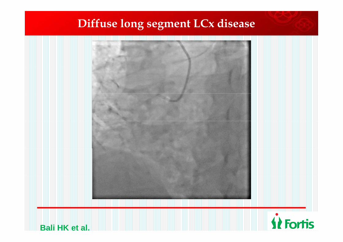
Diffuse long segment LCx disease
Bali HK et al.

Selective Angio of the disease –free Type IV LAD
Bali HK et al.

Selective Angio of the LCx
Bali HK et al.

Right Coronary Angio in RAO cranial 30*
Dominant diseased RCA, anomalous ,LAD going between aorta RVOT and
l LC ithanomalous LCx with its retro aortic course.
Bali HK et al.

CAG Findings
Bali HK et al.

Primary PCI of RCA
Right radial artery 6F sheath.g yAR 1, 6F guiding catheter to hook the RCA.Lesion crossed with BMW 0.014 coronary wire. Lesion crossed with BMW 0.014 coronary wire.
Bali HK et al.

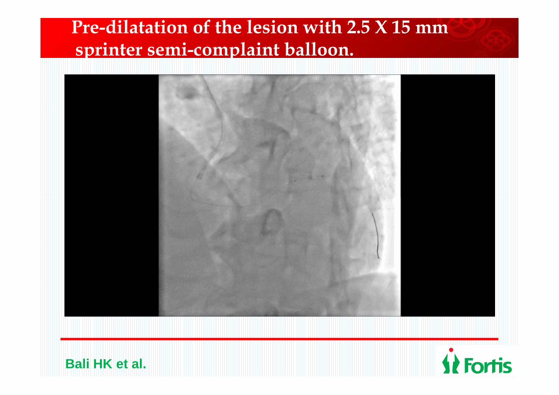
Pre-dilatation of mid-RCA and PDA ostial/distal lesions with 2.5 X 15 mm sprinter semi-complaint balloon
Bali HK et al.

Post – Ballooning Angiogram showing residual stenosis in mid RCA, proximal and distal PDA
Bali HK et al.

Stenting of distal ostium of PDA
Distal lesion stented with 2 5 X stented with 2.5 X 28 mm Xience V DES.Post-dilatation with 2.75 mm non-complaint balloon at 20@atm.
Bali HK et al.
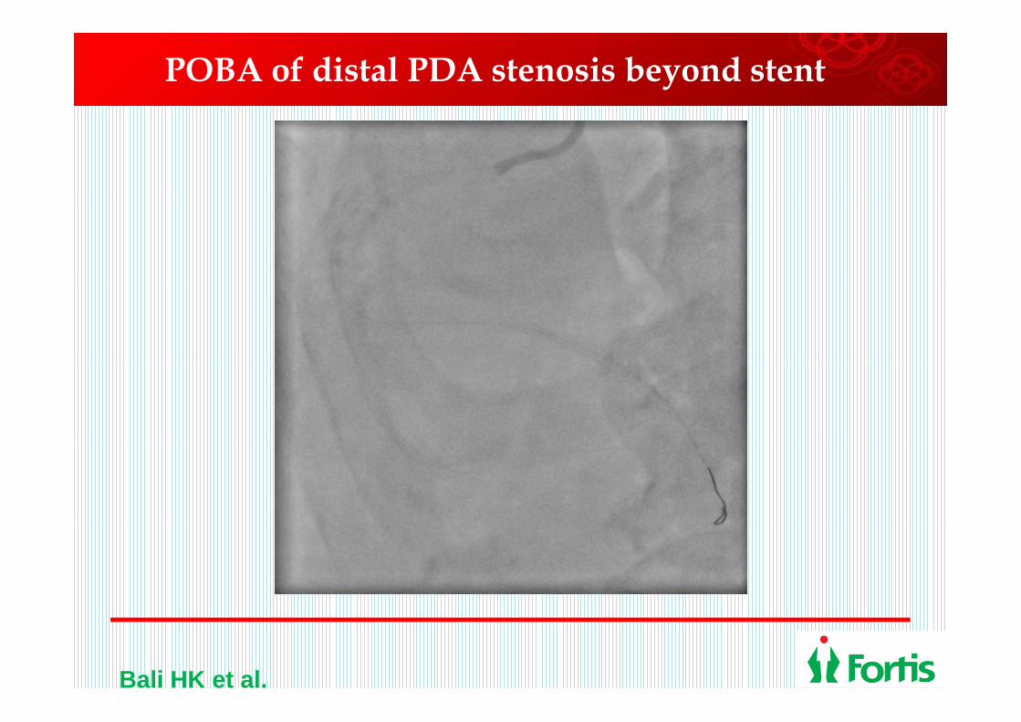
POBA of distal PDA stenosis beyond stent
Bali HK et al.

Post-stent Angiogram showing good flow in PDA
Bali HK et al.

Mid-RCA stenting
4 X 18 mm BMS Zeta deployed at nominal
i id pressure in mid – RCA.
Post-dilatation with 4 X 15 mm with 4 X 15 mm non-complaint balloon at 20@atm
Bali HK et al.

RAO Cranial view: Post-stenting RCA angiogram showing TIMI 3 flow with no residual stenosis.
Bali HK et al.
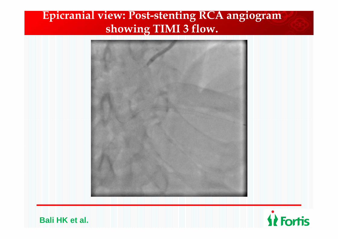
Epicranial view: Post-stenting RCA angiogram showing TIMI 3 flow.
Bali HK et al.

RAO Cranial view: Post-stenting Final RCA angiogram showing TIMI 3 flow.
Bali HK et al.

Lateral view: Post-stenting Final RCA angiogram showing TIMI 3 flow.
Bali HK et al.

CT Angio VRT reconstruction
Bali HK et al.

CT coronary angiography
Bali HK et al.

Staged PCI of LCx
h d l h hRight radial artery 6F sheath.AR 1, 6F guiding catheter to hook the RCA.g gLesion crossed with BMW 0.014 coronary wire.
Bali HK et al.

Selective coronary angiography of LCx showing diffuse disease maximum 90% stenosis.
Bali HK et al.
Pre-dilatation of the lesion with 2.5 X 15 mm sprinter semi-complaint balloon.
Bali HK et al.
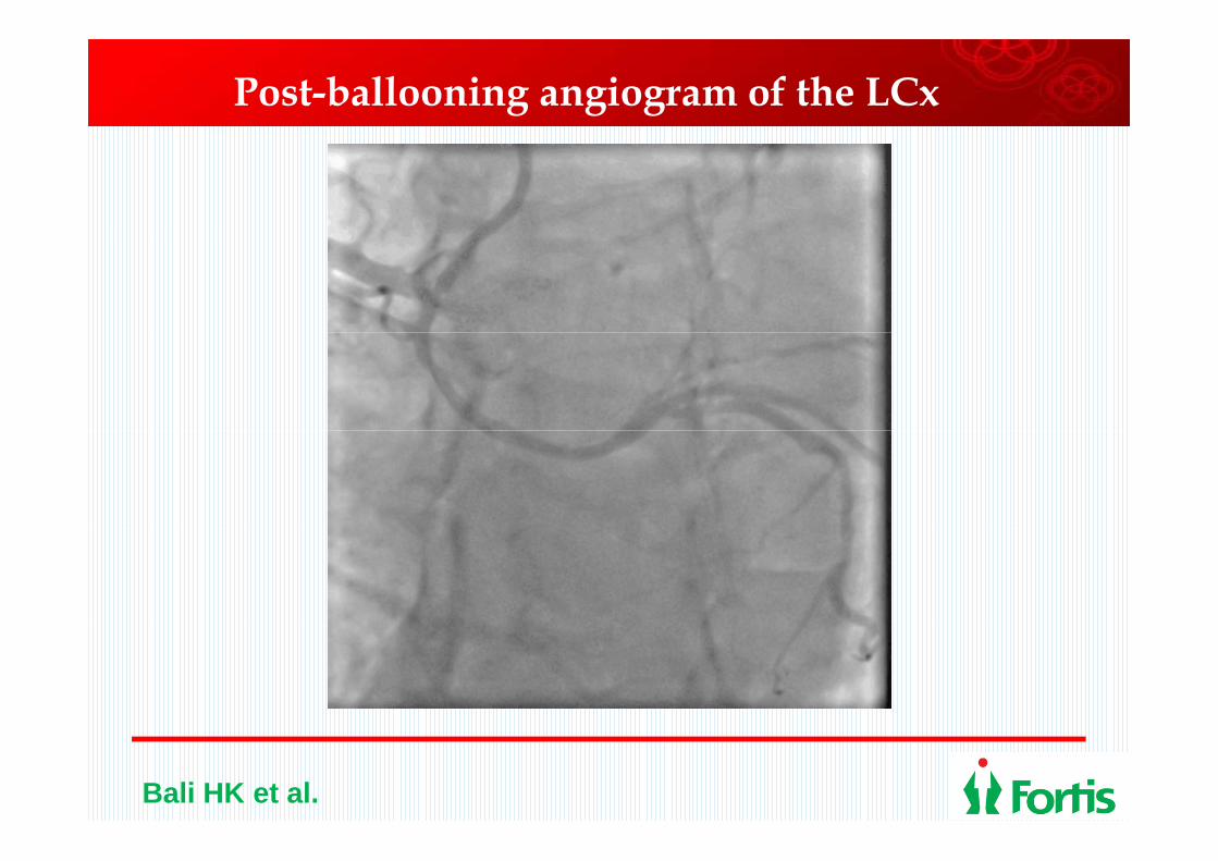
Post-ballooning angiogram of the LCx
Bali HK et al.

2 overlapping DES Xience V positioned – 2.5 X 38 mm distally and 2.75 X 33 mm proximally
Bali HK et al.

Post-Stent Angiogram
Bali HK et al.
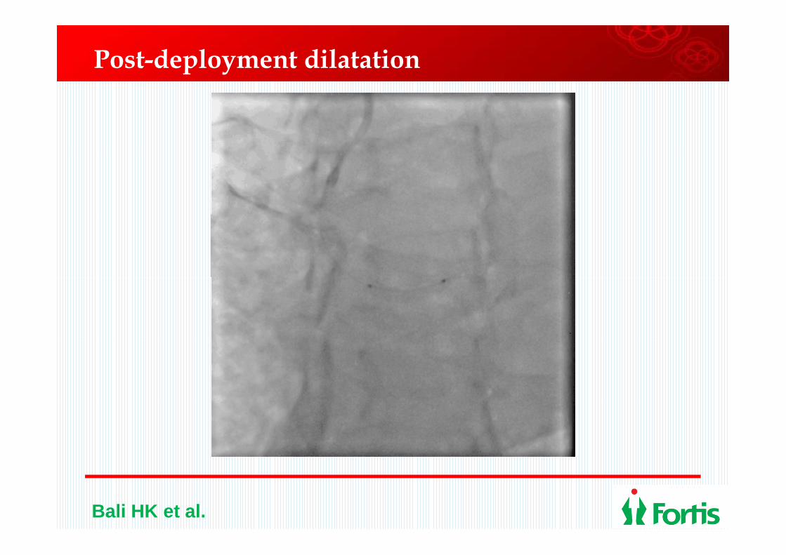
Post-deployment dilatation
Bali HK et al.

RA Caudal View : Angiogram of LCx showing TIMI III Flow
Bali HK et al.

Right Coronary Artery in RAO 30* cranial view for stenting showing stented RCA and anomalous LCx.
Bali HK et al.
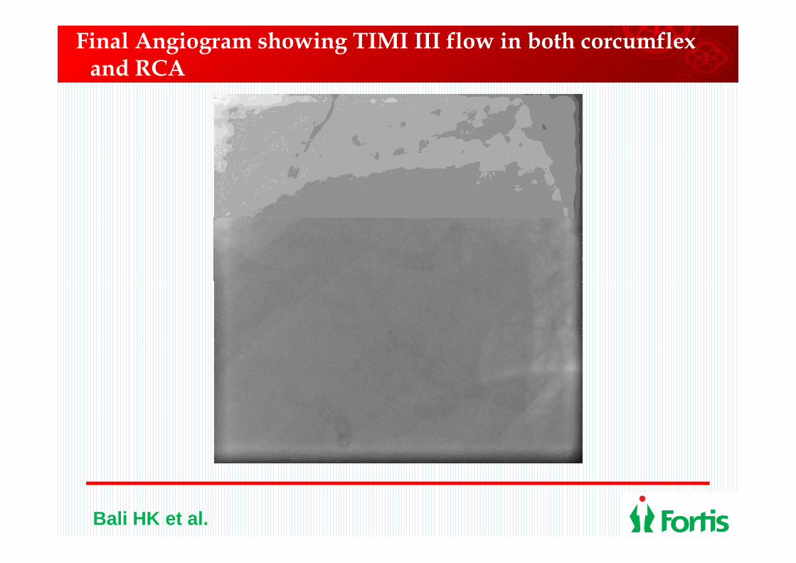
Final Angiogram showing TIMI III flow in both corcumflexand RCA
Bali HK et al.
Conclusion
We describe for the first time multivessel PCI We describe for the first time multivessel PCI through the radial route in an extremely uncommon case of multiple coronary artery uncommon case of multiple coronary artery anomalies – Type IV dual LAD with anomalous origin of the left circumflex from anomalous origin of the left circumflex from the RCA.
Bali HK et al.

Coronary artery anomalies
Bali HK et al.

Classification of Dual LAD
Bali HK et al.

Conclusion
Anomalous coronaries can be safely and Anomalous coronaries can be safely and successfully treated through the radial route after careful evaluation of the origin and after careful evaluation of the origin and course of the anomalous vessels.
CT coronary angiography is extremely useful i d li i h l d h iin delineating the vessel course and their relation to the great arteries.
Bali HK et al.