Embed Size (px)
Citation preview

Special thanks to Waleed Abu Lubbad for no specific reason
PBL 1
Done by: Ali Alabbadi
Corrected by: AbdelRahman Al
Orthopedics:
Means “straight child” in Greek
Deal with treating diseases of bones and muscles
Diseases that affect bones and muscles tend to fall into 3 different categories:
1. Congenital
a. Genetic, drugs, radiation
2. Developmental
a. Developmental dislocation of hip (DDH), tendon abnormalities,
compressive neuropathy (nerve having pressure applied abnormally)
3. Acquired
a. Trauma, infection, paralysis, arthritis, neoplasm
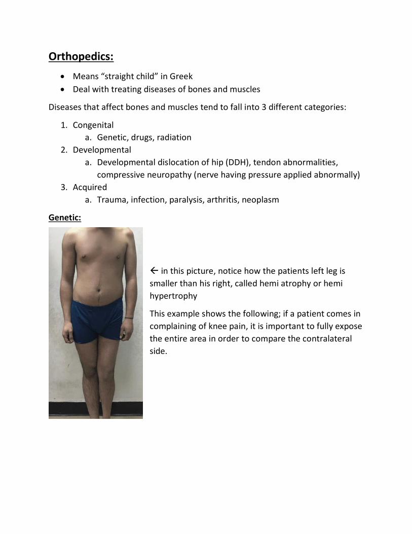
Genetic:
in this picture, notice how the patients left leg is
smaller than his right, called hemi atrophy or hemi
hypertrophy
This example shows the following; if a patient comes in
complaining of knee pain, it is important to fully expose
the entire area in order to compare the contralateral
side.

Here, we see that the patient is shorter than normal, and he
has a joint deformity that is called genu valgum. It also called
“knock knees”.
This example we all know, typical dwarfism caused by
achondroplasia. His trunk and head are normal, but his
limbs are short.
This is another form of dysplasia, called
macrodactyly. It means one digit is larger
compared to the rest.

This is called lobster feet, because the
feet look like lobster claws.
Other forms of genetic abnormalities involving the peripheries include syndactyly,
polydactyly, craniosynostosis, and blue sclera caused by OI. (remember patho)
Defects that affect the spine:
Scoliosis; the spine is deviated to the left or right (coronal or
AP view).
Kyphosis; the spine has an exaggerated hump

Here the patients’ shoulders are
pushed together, in a disease
called clavicle dysostosis, they
have no clavicle, allowing the
medial deviation of their
shoulders.
Drugs and Radiation:
In the 50s pregnant women were prescribed
thalidomide for morning sickness, this caused their
babies to be born with malformed limbs.
Developmental:
Carpal Tunnel syndrome, caused by pressure on the carpal
tunnel and is painful for patient while affecting distribution
of the median nerve in hand.

DDH is a dislocation of
the femoral head in
newborns.
Genu varum, opposite of genu valgum, in which the
knees are bowed outward.

Acquired:
Infection with sinus drainage. This
infection is iatrogenic, because it was
caused by improper aseptic technique by
hospital staff (notice surgical scar).
Claw hand, caused by damage to the ulnar nerve
in the hand, also called ulnar nerve palsy.
Here, you can see the muscles of the thumb (thenars)
are wasting, meaning this patient has his median nerve
compressed (carpal tunnel), although to reach this stage
it must be chronic.
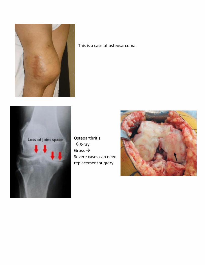
This is a case of osteosarcoma.
Osteoarthritis
X-ray
Gross
Severe cases can need
replacement surgery

Here is another case of arthritis,
but in the hip. Hip replacement
surgery may be required.
Trauma and Infection:
Can be soft tissue, bone or both, and it is the bread and butter of orthopedics.
Here is a simple cut wound. When it comes to
trauma of limbs, the most important things that
need to be worried about are nerves and vessels.
In trauma cases there is a very important list that we all need to know:
1. Life; make sure the patient will not die.
2. Limb; save the limb by saving vascularity, no vascularity = no limb
3. Wound; prevent infection
4. Fracture; treatment can be delayed

In the picture above, the trauma is not life threatening, so you should check on
the functionality of the nerves and vessels that may be damaged, such as the
radial nerve, and axillary artery.
Here we see a small puncture wound in the
patients arm, caused by a skewer. The patient
later came in with swelling, pain and a tingling
sensation.
From anatomy, we remember in the limbs there are some major structures; bone,
muscle, nerves, vessels, and fascia. In a wound such as this, the fascia is
penetrated, but not to a degree that allows free bleeding. This means that blood
will continue to pool in the compartment, causing even more swelling (other than
natural inflammation). If this swelling is ignored, it can cause extravascular
compression which can close major arteries and veins. This can lead to an
infarction and necrosis of large portions of the arm (no vascularity = no limb). This
also compresses nerves, which causes the pain and tingling sensation the patient
complained about. This is called compartment syndrome.
For major cases of compartment symptom, the
treatment is to drain the compartment, in this case
with an operation called a fasciotomy. This relieves the
pressure, saving the limb.

Another common form of acquired soft
tissue ailments is infection. Here we
see a large portion of the skin is
necrotic, which we call eschar.
Treatment for necrotic tissue is
debridement and drainage of the pus.
The most common pathology of bone
is fractures, here we have a fracture of
the diaphysis (a little patho review,
this is a transverse, displaced, simple
fracture).

Here is another fracture, but in this case, the
trauma also caused a dislocation of the
t tibiotalar joint. This trauma will most likely
have long term effects.
Treatment can vary from simple immobilization, to reduction (putting bone back
in place), to major surgeries requiring prostheses.
In cases with open fracture like this, your
main cause of concern is not the fracture
itself or any soft tissue damage, but
infection. Major traumas have a list of
treatment and support methods in order to
help the patient survive, recover limb
functionality, and be comfortable (relieve
pain).

This list is as follows:
Advanced Trauma Life Support (ATLS)
Analgesia
Antibiotics
ATS (??)
Adequate irrigation and splinting
ATLS has yet another checklist in order to deal with the trauma, abbreviated as
ABCDE:
1. A – airway
2. B – breathing
3. C – Circulation
4. D – disability (unconsciousness)
5. E – exposure (full physical examination of patient)
These were put in this specific order based on their mortality rates.
This patient has a fracture, and it
also caused compartment
syndrome, and the surgery he
had performed fixed the fracture,
as well as draining the
compartment.
The shoulder is the most mobile
joint in the body, meaning it is
also the least stable, and is
commonly dislocated. The
shoulder can be dislocated
anteriorly or posteriorly.

Posterior dislocations are much
less common, and happen in 2
situations; seizures or electric
shocks. The dislocation happens
due to a loss of control of the
muscles, allowing the stronger
internal rotators of the shoulder to
take control and pop the humerus
out of its socket.
The scary thing about shoulder dislocations is its proximity to the axillary nerve.
Some people are hyper lax, meaning they are very flexible and can voluntarily
dislocate joints in their body. There is pathologic hyper laxity which is usually
involved with genetic or developmental defects. You should not attempt to treat
these people.
Finally, the last case we are familiar with, and it is ACL injuries. The ACL is located
in the knee joint and normally it prevents anterior displacement of the tibia on
the humerus. This can happen due to being hit on the leg, or it can happen due to
pivoting of the leg when landing from a jump. Surgery is a must in this injury, and
it is performed by grafting a part of the hamstring in place of the ACL.
Hamstring graft