Embed Size (px)
Citation preview

A research report submitted to the Faculty of Oral Health Sciences, University of the
Witwatersrand, in partial fulfillment of the requirements for the degree of Master of
Science in Dentistry.
Supervisor: Prof. Ugo Ripamonti
School of Oral Health Sciences, Faculty of Health Sciences,
University of the Witwatersrand, Johannesburg, South Africa
2017
PATTERNS OF BONE INDUCTION BY CORAL-DERIVED
VS HIGHLY SINTERED CRYSTALLINE
HYDROXYAPATITE CONSTRUCTS
Assaedi B Alkonti
Student number: 1298238

ii
DECLARATION
I, Assaedi Alkonti declare that this research report is my own work. It is being
submitted for an MSc degree in Oral Medicine & Periodontology at the University of the
Witwatersrand, Johannesburg. It has not been submitted before for any degree or examination
at this or any other University, and all the sources I have used or quoted have been indicated
and acknowledged by complete references.
21th
November 2017.

iii
DEDICATION
To my mother, father, beloved wife, precious son and extended family for their support and
encouragement.

iv
ABSTRACT
Aim: To evaluate the morphological patterns of bone induction by coral-derived vs highly
sintered hydroxyapatite constructs.
Methods: Twenty-eight (28) histological slides of harvested tissue samples from the rectus
abdominis muscle of adult baboons of both coral-derived and highly sintered hydroxyapatite
specimens were histologically examined to describe the induction of bone formation along the
macroporous spaces of the hydroxyapatite (HA) substrata and the development of osteoblast-
like cells within the mesenchymal condensations.
Results: Two specific tissues characterized the tissue invasion within the macroporous spaces
of the coral-derived hydroxyapatite constructs, namely, 1: cellular connective tissue matrix
penetrated by sprouting capillaries and 2: mesenchymal collagenous condensations that
differentiated and attached to the surface of the coral-derived substratum. To the contrary, in
the highly sintered constructs, the mesenchymal cellular condensations could not be detected.
Fibrovascular tissue was richly invaded by blood vessels almost in direct contact with the
sintered hydroxyapatite interface. Elongated spindle-shaped-like cells localised within the
mesenchymal collagenous condensation.
The main histological feature that characterised the induction of bone formation by coral-
derived hydroxyapatite implant was the induction of mesenchymal collagenous condensation.

v
In all harvested specimens of highly sintered hydroxyapatite, the initiation of bone formation
formed directly against the hydroxyapatite interface without formation of mesenchymal
collagenous condensations at the sintered hydroxyapatite interface.
Conclusion: The results obtained from this study further support the concept of construction
of solid geometric calcium phosphate osteogenic devices in controlling the induction of bone
formation for the treatment of multiple periodontal osseous defects in human patients.

vi
ACKNOWLEDGMENTS
I would like to express my deepest gratitude to my supervisor, Prof. Ugo Ripamonti, who
taught and guided me all the way. Thank you for your excellent guidance, caring, patience,
monitoring and constant encouragement throughout the learning process – it is much
appreciated.
I would also like to thank:
The staff members of the Oral Medicine & Periodontology Department of the School
of Oral Health Science.
To my family and colleagues who have made this thesis possible and because of whom
my postgraduate experience has been one that I will appreciate forever.

vii
Table of Contents
DECLARATION ........................................................................................................................ ii
DEDICATION .......................................................................................................................... iii
ABSTRACT ............................................................................................................................... iv
ACKNOWLEDGMENTS .......................................................................................................... vi
LIST OF FIGURES .................................................................................................................... ix
LIST OF TABLES ...................................................................................................................... x
LIST OF ABBREVIATIONS .................................................................................................... xi
CHAPTER 1- INTRODUCTION & LITERATURE REVIEW ................................................. 1
1.1. Induction of Bone Formation during Embryonic Development ............................................. 1
1.1.1. Endochondral bone formation ......................................................................................... 1
1.1.2. Intramembranous bone formation .................................................................................. 3
1.2. Heterotopic Bone Induction .................................................................................................... 4
1.3. Induction of Bone Formation by Demineralized Bone Matrix (DBM) ................................... 6
1.4. Calcium Phosphate Hydroxyapatite and Bone Induction ...................................................... 7
1.5. Intrinsic Induction of Bone Formation .................................................................................... 9
1.6. Geometric Induction of Bone Formation .............................................................................. 10
1.7. Angiogenesis and the Induction of Bone Formation ............................................................ 11
CHAPTER 2- AIMS AND OBJECTIVES ............................................................................... 14
2.1. Aims ............................................................................................................................................ 14
2.2. Objectives ................................................................................................................................... 14
2.3. Rationale of the Study ................................................................................................................ 14
CHAPTER 3- METHODOLOGY ............................................................................................ 15
3.1. Study Design .......................................................................................................................... 15
3.2. Study Sample Size .................................................................................................................. 15
3.3. Data Collection and Analysis ................................................................................................. 15
3.4. Data Collection Tools ............................................................................................................. 16
CHAPTER 4- RESULTS .......................................................................................................... 19

viii
4.1 Tissue Induction and Morphogenesis by Coral-Derived Macroporous Constructs Implanted in
the Rectus Abdominis Muscle of Papio Ursinus and Harvested on Day 30 ..................................... 19
4.2 Tissue Induction and Morphogenesis by Highly Sintered Macroporous Constructs Implanted
in the Rectus Abdominis Muscle of Papio Ursinus and Harvested on Day 30 ................................. 20
4.3 Tissue Induction and Morphogenesis by Coral-Derived Macroporous Constructs Implanted in
the Rectus Abdominis Muscle of Papio Ursinus and Harvested on Day 90 ..................................... 21
4.4 Tissue Induction and Morphogenesis by Highly Sintered Macroporous Constructs Implanted
in the Rectus Abdominis Muscle of Papio Ursinus and Harvested on Day 90 ................................. 22
4.5. Morphological Differences of Patterns of Bone Formation by Coral-Derived vs Highly Sintered
Crystalline Hydroxyapatite Constructs ............................................................................................. 23
CHAPTER 5- DISCUSSION .................................................................................................... 40
CHAPTER 6- CONCLUSIONS ................................................................................................ 47
REFERENCES .......................................................................................................................... 49
APPENDICES ........................................................................................................................... 59
Appendix A: Title Approval ............................................................................................................... 59
Appendix B: Ethical Waiver ............................................................................................................... 60
Appendix C: Plagiarism Report.......................................................................................................... 61
ix
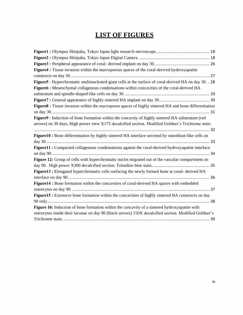
LIST OF FIGURES
Figure 1 : Olympus Shinjuku, Tokyo Japan light research microscope. ................................................ 18
Figure 2 : Olympus Shinjuku, Tokyo Japan Digital Camera. ................................................................ 18
Figure 3 : Peripheral appearance of coral- derived implant on day 30. ................................................. 26
Figure 4 : Tissue invasion within the macroporous spaces of the coral-derived hydroxyapatite
constructs on day 30. .............................................................................................................................. 27
Figure 5 : Hyperchromatic multinucleated giant cells at the surface of coral-derived HA on day 30. .. 28
Figure 6 : Mesenchymal collagenous condensations within concavities of the coral-derived HA
substratum and spindle-shaped-like cells on day 30. ............................................................................. 29
Figure 7 : General appearance of highly sintered HA implant on day 30. ............................................. 30
Figure 8 : Tissue invasion within the macroporus spaces of highly sintered HA and bone differentiation
on day 30. ............................................................................................................................................... 31
Figure 9 : Induction of bone formation within the concavity of highly sintered HA substratum (red
arrows) on 30 days. High power view X175 decalcified section. Modified Goldner’s Trichrome stain.
................................................................................................................................................................ 32
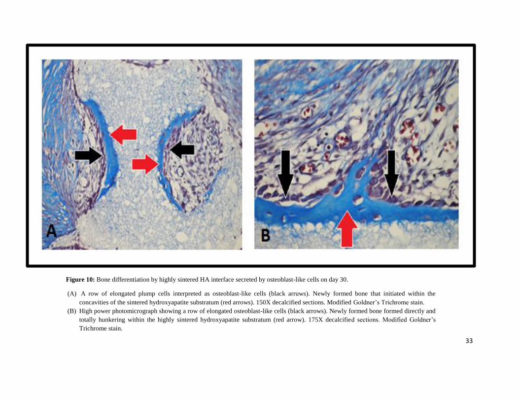
Figure 10 : Bone differentiation by highly sintered HA interface secreted by osteoblast-like cells on
day 30. .................................................................................................................................................... 33
Figure 11 : Compacted collagenous condensations against the coral-derived hydroxyapatite interface
on day 90. ............................................................................................................................................... 34
Figure 12: Group of cells with hyperchromatic nuclei migrated out of the vascular compartment on
day 90. High power X300 decalcified section. Toluidine blue stain. .................................................... 35
Figure 13 : Elongated hyperchromatic cells surfacing the newly formed bone at coral- derived HA
interface on day 90. ................................................................................................................................ 36
Figure 14 : Bone formation within the concavities of coral-derived HA spaces with embedded
osteocytes on day 90. ............................................................................................................................. 37
Figure 15 : Extensive bone formation within the concavities of highly sintered HA constructs on day
90 only. ................................................................................................................................................... 38
Figure 16: Induction of bone formation within the concavity of a sintered hydroxyapatite with
osteocytes inside their lacunae on day 90 (black arrows) 150X decalcified section. Modified Goldner’s
Trichrome stain. ..................................................................................................................................... 39

x
LIST OF TABLES
Table 1: Distribution of histological 7 slides according to time period of implantation ....................... 16
Table 2: Histological finding of coral-derived and highly sintered HA on day 30 ............................... 21
Table 3: Histological finding of coral-derived and highly sintered HA on day 90 ............................... 23
Table 4: Morphological differences of patterns of bone formation by coral-derived vs. highly sintered
HA .......................................................................................................................................................... 25

xi
LIST OF ABBREVIATIONS
Ihh: Indian hedgehog.
PTHrP: Parathyroid Hormone-Related Proteins.
ECM: Extracellular Matrix.
VEGF: Vascular Endothelial Growth Factor.
MMP9: Metalloproteinase 9.
BMPs/OPs: Bone Morphogenetic and Osteogenetic Proteins.
CBFA1: Core Binding Factor alpha 1.
TGF-β: Transforming Growth Factor-β.
DBM: Demineralized Bone Matrix.
HA: Hydroxyapatite.
TCP: Tricalcium Phosphate.
BRL: Bone Research Laboratory.
AESC: Animal Ethics Screening Committee.

1
CHAPTER 1- INTRODUCTION & LITERATURE REVIEW
1.1. Induction of Bone Formation during Embryonic Development
The induction of bone formation starts early during embryonic development by differentiation
of mesenchymal cells into bone cells in two ways: firstly, by the formation of a cartilage
anlage as in the induction of endochondral bone formation and secondly, by direct cellular
condensation with the later intramembranous bone formation as in the induction of the
calvaria skeleton (Hall, 1978).
1.1.1. Endochondral bone formation
The induction of endochondral bone formation during embryonic development starts when
the mesenchymal outgrowth from the embryo trunk gives rise to the limb buds. The central
mesenchymal cells of the limb bud divides rapidly, forming mesenchymal cell condensations.
Cells within the central core of mesenchymal condensations differentiate into chondroblasts
and chondrocytes forming a cartilage anlage, which acts as a strut upon which the induction of
bone formation will develop (Hall, 1978).
Cells at the margins of the cartilage anlage flatten and extend to form thick sheets of
connective tissue defined as perichondrium. Chondrocytes in the centre of the cartilage anlage
differentiate into hypertrophic chondrocytes (Hall, 1978).

2
A critical step is the vascular invasion of the mineralized cartilage and of the hypertrophic
chondrocytes, which is associated with chondrolysis or death of the cartilage and with
mesenchymal cell differentiation into osteoblasts. Osteoblasts in the perichondrium region
start secreting bone matrix around the hypertrophic area of cartilage, which is defined as the
bone collar (Trueta, 1963).
The induction of a cartilage anlage for the ideal long bone differentiation during embryonic
development enhances tri-dimensional growth resulting in development of the cartilaginous
growth plate. The cartilaginous growth plate acts as a master for tri-dimensional appositional
growth of the mammalian long bones (Ripamonti, 2006).
The development of the growth plate depends on the morphogenesis of the cartilage anlage
followed by vascular invasion of the hypertrophic cartilage by blood vessels and sprouting
capillaries resulting in the induction of bone formation in angiogenesis (Ripamonti, 2006).
Specific molecular signals regulate endochondral bone formation. Mesenchymal cells within
the centre of the condensations start to differentiate initially into chondrocytes which express
specific molecular markers such as aggrecan and α 1 (II) collagen (Karsenty and Wagner,
2002).
Expression of such molecular markers enhances chondrocyte proliferation and differentiation
into proliferating non-hypertrophic chondrocytes responsible for the cartilage anlage
formation. These molecular markers also play a significant role in regulating the rate of
cartilage hypertrophy (Karsenty and Wagner, 2002).
Hypertrophic chondrocytes consist of two types of cells. The first group is pre-hypertrophic
chondrocytes found under the proliferating chondrocytes, and they are larger than proliferating

3
chondrocytes. These kinds of cells arrest in growth and continue to express a limited amount
of α1 (II) collagen. It mainly expresses the Indian hedgehog gene (Ihh) which activates the
specific molecular factor parathyroid hormone-related proteins (PTHrP) on the surface of
hypertrophic chondrocytes. PTHrP enhances the differentiation of these cells into fully
differentiated hypertrophic chondrocytes, which is the second group of cells. Fully
differentiated hypertrophic chondrocytes do not express α 1 (II) collagen but express α1 (X)
collagen (Linsenmayer et al., 1991; Poole, 1991; St-Jacques et al., 1999; Karsenty and
Wagner, 2002).
Before the fully differentiated hypertrophic chondrocytes die by apoptosis, they become
surrounded by a calcified cartilage extracellular matrix (ECM). Fully differentiated
hypertrophic chondrocytes later express molecular markers such as the vascular endothelial
growth factor (VEGF). VEGF stimulates the vascular invasion of the calcified cartilage
extracellular matrix by blood vessels followed by chondroclast expression of
metalloproteinase 9 (MMP9), which degrades the cartilage (Vu et al., 1998; Karsenty and
Wagner., 2002).
Cells in contact with the basement membrane of invading capillaries differentiate into
osteoblasts secreting the bone extracellular matrix (ECM) rich in type I collagen under the
control of bone morphogenetic and osteogenetic proteins (BMPs/OPs) (Paralkar et al., 1990).
1.1.2. Intramembranous bone formation
Neural crest-derived mesenchymal cells proliferate to form mesenchymal connective tissue
condensations of high cell density. Some of these cells within the mesenchymal condensations

4
develop into capillaries, and other cells under the control of certain molecular markers alter
their shape to become osteoblast-like cells secreting bone matrix which undergo
mineralisation to form bone spicules without any cartilage anlage formation (Hall, 1988; Hall
& Miyake, 2000).
Some molecular markers of intramembranous bone formation during embryonic development
include the bone morphogenetic proteins (BMPs) and an activated transcription factor called
core binding factor alpha 1 (CBFA1). BMPs activate the CBFA1 gene in mesenchymal cells
to enable it to differentiate directly into osteoblasts secreting bone matrix without the cartilage
anlage formation to initiate and form the craniofacial skeleton and calvaria (Hall, 1988).
Extensive research has shown that the process of bone formation by induction in postnatal life
recapitulates events which occur in the normal course of embryonic bone development
(Wozney, et al., 1988; Reddi, 1994; Reddi, 1998). Postnatal bone induction is regulated by the
transforming growth factor-β (TGF-β) supergene family (Reddi, 1994; Reddi, 1998; Reddi,
2000; Ripamonti, 2003; Ripamonti, 2006).
1.2. Heterotopic Bone Induction
The heterotopic induction of bone formation is a fascinating phenomenon: cells are induced
and transformed into osteoblast-like cells by the osteogenic proteins of the transforming
growth factor-β (TGF-β) supergene family (Reddi, 1992; Wozney, 1992; Ripamonti, 2006;
Ripamonti, 2003).

5
The heterotopic induction of bone formation requires three key components (Reddi, 2000): an
osteoinductive soluble signal; an insoluble signal or substratum; and responding stem cells.
The osteoinductive soluble signal is a member of the osteogenic proteins of the TGF-β
supergene family (Ripamonti, 2003). These osteogenic proteins can initiate bone formation in
heterotopic extraskeletal sites of a variety of animal models (Reddi, 1992; Wozney, 1992;
Ripamonti, 2006).
The insoluble signal or substratum acts as a delivery system for the osteogenic soluble
molecular signals which initiate the induction of the bone formation. The combination of the
osteogenic signals with a substratum defines an osteoinductive biomaterial (Sampath & Reddi,
1983; Ripamonti & Reddi, 1995; Reddi, 2000; Ripamonti, et al. 2000; Ripamonti, 2009).
Host cells, including undifferentiated mesenchymal cells, start to move toward chemotactic
ingredients at the site of heterotopic implantation of the osteoinductive biomaterial. This is
followed by proliferation and differentiation of undifferentiated mesenchymal cells into
chondrocytes that form a cartilage anlage. The anlage undergoes chondrolysis upon vascular
invasion and cartilage death is replaced by bone marrow within the newly formed bone
(Reddi, 1981).
Undifferentiated mesenchymal cells can also directly differentiate into osteoblast-like cells
forming bone as in intramembranous bone formation by heterotopic implantation of calcium
phosphate-based biomaterials in the rectus abdominis muscle of adult baboons (Papio
Ursinus) (Ripamonti, 1990; Ripamonti, 1991).

6
1.3. Induction of Bone Formation by Demineralized Bone Matrix (DBM)
The classic work of Urist, (1965) Reddi and Huggins, (1972) has described that there are
substances capable of inducing heterotopic bone induction after heterotopic implantation of
intact demineralized bone matrix (DBM) in extraskeletal sites of different animal models.
Further experimental work has shown that the intact DBM could be dissociatively extracted
using chaotropic agents like urea or guanidinium hydrochloride into an inactive insoluble
collagenous bone matrix and a soluble protein extract (Sampath and Reddi, 1981; Sampath
and Reddi, 1983).
Heterotopic bone formation by induction can only initiate when both the insoluble collagenous
bone matrix and the solubilized osteogenic proteins are recombined and later implanted in the
rodent subcutaneous site (Sampath and Reddi, 1981; Sampath and Reddi, 1983; Reddi, 2000).
The osteogenic proteins which are delivered by the insoluble collagenous bone matrix and
initiate endochondral bone formation are the bone morphogenetic osteogenetic proteins
(BMPs/ Ops), members of the TGF-β supergene family (Reddi, 1992; Wozney, 1992; Reddi,
1994; Reddi, 2000; Ripamonti, 2003; Ripamonti, 2004; Ripamonti et al., 2004; Ripamonti et
al., 2005; Ripamonti, 2006).
The molecular and morphological event induced by BMPs/OPs during heterotopic induction
of bone formation in the extraskeletal site of animal models recapitulates events that occur
during embryonic bone development (Reddi, 1994; Reddi, 1997: Ripamonti, 2003).

7
1.4. Calcium Phosphate Hydroxyapatite and Bone Induction
Hydroxyapatite (HA) and tricalcium phosphate (TCP) are two types of calcium phosphate-
based biomaterials which are chemically similar. HA is a natural inorganic component of bone
tissue and several macroporous calcium phosphate-based constructs have been documented to
be safe and efficient biomaterials for bone formation (De Groot, 1980; Jarcho, 1981; Jarcho,
1986).
Calcium phosphate biomaterials are highly biocompatible synthetic substrates implanted in
hard and soft tissue causing neither tissue toxicity nor inflammatory response. HA has a high
affinity for binding BMPs/OPs (Sampath et al., 1987; Ripamonti, 1991; Ripamonti, 2006).
The heterotopic induction of bone formation by a variety of calcium phosphate-based
biomaterials is a very interesting phenomenon. Osteoinductive biomaterials initiate tissue
induction and transformation of osteoblast-like cells secreting bone matrix associated with
expression of the selected gene of the transforming growth factor-β (TGF-β) supergene family
(Ripamonti, 1991; Ripamonti et al, 1999; Ripamonti, et al., 2001; Klar et al, 2013).
Coral-derived hydroxyapatite is one of the calcium phosphate-based biomaterials of natural
origin which is obtained from the hydrothermal exchange of calcium carbonate exoskeleton of
marine coral converted into calcium phosphate hydroxyapatite (Roy and Linnehan, 1974).
Highly sintered crystalline hydroxyapatite is one of the synthetic calcium phosphate-based
biomaterials prepared when slurry preparations of hydroxyapatite are sintered to form a
macroporous construct with specific concavities within the macroporous spaces (Ripamonti et
al., 1999).

8
The morphogenesis of bone in porous bioceramics, when implanted in the heterotopic site,
was first described when implanting coral-derived hydroxyapatite constructs in the rectus
abdominis muscle of adult non-human primates (Papio Ursinus) (Ripamonti,
1990; Ripamonti, 1991).
The implantation of calcium carbonate coral-derived hydroxyapatite constructs in the rectus
abdominis muscle of adult baboons shows the differentiation of mesenchymal connective
tissue condensations that develop against the macroporous surfaces of the substratum. Such
condensations have been shown to differentiate into bone by the differentiation of osteoblast-
like cells within the mesenchymal condensations (Ripamonti, 1990; Ripamonti, 1991;
Ripamonti, et al., 1993).
The development of mesenchymal condensation is a critical phenomenon for the induction of
bone formation by coral-derived hydroxyapatite constructs (Ripamonti, 1990; Ripamonti,
1991; Ripamonti et al., 1993).
Implantation of coral-derived macroporous hydroxyapatite constructs has led to the
manufacture of highly sintered hydroxyapatite constructs (Ripamonti, et al., 1999).
The induction of bone formation by coral-derived macroporous constructs occurs between
thirty (30) and sixty (60) days of implantation in the muscle of adult baboons (Ripamonti, et
al., 1993). Similarly, bone formation by highly sintered macroporous construct occurs
between thirty (30) and sixty (60) days (Ripamonti, et al., 1999).
The induction of bone formation as initiated by such macroporous calcium phosphate-based
biomaterials is intramembranous in origin and the induction of cartilage has never been
reported (Ripamonti, 1991; Ripamonti et al., 1999; Yuan, et al., 2002).

9
1.5. Intrinsic Induction of Bone Formation
The heterotopic implantation of specific types of porous hydroxyapatite biomaterials has led to
spontaneous induction of bone formation due to their ability to express BMPs/OPs within
concavities of porous hydroxyapatite without exogenous addition of any osteogenic proteins
(Ripamonti et al., 1993; Ripamont et al. 1999).
An ability of some macroporous calcium phosphate-based hydroxyapatite constructs to initiate
the intrinsic induction of bone formation is the nano-topographical and geometrical
configuration of the substratum (Ripamonti, 1996; Ripamonti et al., 1999).
The perfect osteoinductive biomaterials for initiation of bone formation should be
biocompatible, carvable and adaptable to different shapes of osseous defects. It should
enhance sufficient vascular and connective tissue invasion within the macroporous spaces of
substratum in contact with BMPs/OPs (Ripamonti and Duneas, 1996). The calcium phosphate
hydroxyapatite biomaterials are considered the most common synthetic biomaterials for bone
construction therapy (Jarcho, 1981).
Several studies reported that the particular type of porous hydroxyapatite obtained from the
hydrothermal exchange of calcium carbonate exoskeleton of marine coral, converted into
calcium phosphate hydroxyapatite, can initiate the induction of bone formation when
implanted in the rectus abdominis muscle of adult baboons (Ripamonti, 1990; Ripamonti et
al., 1993; Ripamonti, 1996; van Eeden and Ripamonti, 1994).

10
1.6. Geometric Induction of Bone Formation
The influence of substratum shape on induction of bone formation was first described by
Reddi and Huggins (1973). They found that subcutaneous implantation of two substrata with
different geometric configurations blocked the bone induction cascade.
An open-end demineralized root can initiate the induction of bone formation while the
substratum with the dead-end tube could not initiate the induction of bone formation but rather
initiated the induction of chondrogenesis. They reported that the vascular invasion within the
open-end tube was more than that on the dead-end tube which provides more oxygen and
nutritional supply which enhances the induction of bone formation (Reddi and Huggins,
1973). Indeed, teeth with the apex intact generate cartilage within the dead-end of the
endodontic canal (Reddi and Huggins, 1973).
Many experimental studies have shown the effect of the geometric configuration of
demineralized bone matrix (DBM) when implanted in the heterotopic site in the induction of
endochondral bone formation, which can be changed by the geometry of the substratum
(Reddi, 1974; Sampath and Reddi, 1984).
Coarse powder (420 to 850 μm) of demineralized bone matrix implanted in the subcutaneous
space of the rodent, results in induction of endochondral bone formation (Sampath and Reddi,
1984). On the other hand, the implantation of demineralized matrix (fine powder 44 to 74 μm)
did not result in the induction of bone formation (Sampath and Reddi, 1984).

11
The implantation of coral-derived porous hydroxyapatite of both granular and disc
configuration in the subcutaneous space of rodents resulted only in the induction of bone
formation in porous hydroxyapatites of disc configuration (Sampath and Reddi, 1984).
The addition of exogenous osteogenic protein to porous hydroxyapatite of granular
configuration did not result in the induction of bone formation. The above experiments were a
clear cut determination of the effect of the geometry on the local induction of bone formation
(Sampath and Reddi, 1984; Ripamonti et al., 1992).
Intramuscular implantation of highly sintered crystalline hydroxyapatite with repetitive
sequences of concavities on both planar surfaces in Papio Ursinus was performed to evaluate
the geometrical effects of the substratum. This has shown the induction of bone formation
within the concavities of the sintered hydroxyapatite construct by day 30 after implantation.
This experimental work demonstrates the critical role of the geometry of the substratum in the
spontaneous induction of bone formation by controlling the expression of the soluble
molecular signals (Ripamonti et al., 1999).
1.7. Angiogenesis and the Induction of Bone Formation
Blood vessels are essential for the induction of bone formation (Trueta, 1963). The invading
blood vessels and perivascular hyperchromatic stem cells were noted to be within
mesenchymal condensations.

12
Vascular endothelial and pericytic stem cells, together with the basement membrane of
invading blood vessels, act as a source of molecular markers that induce and control
angiogenesis during osteogenesis (Urist, 1965; Reddi and Huggins, 1972).
Mesenchymal condensations surrounding each central blood vessel in the three-dimensional
direction come into close contact with the vascular basement membrane and act as a scaffold
for induction of bone formation around the invading vessels. Thus the invading vessels are
necessary for the patterning of newly formed bone by the organisation of mesenchymal
condensation around it and acted as morphogenetic vessels (Ripamonti et al., 2006).
Mesenchymal cells in contact with the basement membrane of invading vessels start
differentiating into osteoblast-like cells secreting bone matrix under the control of BMPs/OPs
that bind to collagen type IV of the basement membrane of invading blood vessels (Paralkar et
al., 1990; Paralkar et al., 1991).
The contact between the osteoprogenitors and osteoblast-like cells with vascular invading
basement membranes indicates that the specific amino-acid sequences of the basement
membrane components, i.e Laminin, are the morphogenetic initiators during the capillary
invasion of the ripple-like cascade of bone formation by induction (Vukicevic et al., 1990).
1.8. Transforming Growth Factor-β (TGF-β) Supergene Family
The TGF-β supergene family consists of large groups of related proteins that are secreted in
the form of a dimer with an active biological form of carboxy-terminal domain consisting of
seven cysteine residues (Kingsley, 1994).

13
Several studies reported that the biologically active TGF-β plays an important role in cell
activation, proliferation and differentiation as well as in embryonic tissue morphogenesis,
involving osteogenesis, skeletogenesis and cementogenesis (Wozney et al, 1988; Reddi, 1994;
Ripamonti and Reddi, 1997; Heine et al., 1987; Pelton et al., 1990; Ripamonti, 2006).
The BMPs/OPs are members of the TGF-β superfamily (Ripamonti, 2003). These osteogenic
proteins can initiate bone formation in heterotopic extraskeletal sites of animal models (Reddi,
1992; Wozney, 1992).
Several experimental studies have been performed in an attempt to prove that TGF-β also
initiates bone formation. Direct injection of TGF-β1 into the calvarial or overlying the
periosteal tissue of the mice and rats resulted in marked increase in bone thickness (Noda and
Camilliere, 1989; Marcelli et al, 1990; Bolander et al., 1991).
The treatment of the cartilage of full thickness rabbit ear wounds by topical application of
TGF-β promotes wound healing and bone formation within the cartilage (Beck et al., 1991).
Heterotopic implantation of recombinant human TGF-β in the rectus abdominis muscle of
adult baboons induces the induction of endochondral bone formation. This is in marked
contrast to any other animal models where the subcutaneous and/or intramuscular implantation
of the TGF-β mammalian isoforms do not induce endochondral bone formation (Ripamonti et
al., 2000; Ripamonti et al., 2001).

14
CHAPTER 2- AIMS AND OBJECTIVES
2.1. Aims
The aim of this study was to evaluate the morphological patterns of bone induction by coral-
derived vs highly sintered hydroxyapatite constructs.
2.2. Objectives
1. To describe the induction of bone formation along the macroporous spaces of the
hydroxyapatite (HA) substratum.
2. To describe the development of osteoblast-like cells within the developing mesenchymal
condensations.
3. To see if there are any morphological differences of bone formed by coral-derived vs
highly sintered hydroxyapatite constructs.
2.3. Rationale of the Study
Are the morphological patterns of bone formation by induction substantially different from
coral-derived hydroxyapatite constructs vs the highly sintered macroporous crystalline
hydroxyapatites?

15
CHAPTER 3- METHODOLOGY
3.1. Study Design
This study was a descriptive retrospective study. Several histological sections previously
prepared for a research study by the Bone Research Laboratory (BRL) of the university were
used to compile this retrospective study. All histological sections evaluated were cut from
previously embedded specimens of coral-derived and highly sintered hydroxyapatite
constructs implanted in the rectus abdominis muscle of adult baboons.
3.2. Study Sample Size
Permission to use twenty-eight (28) histological slides of harvested tissue samples from the
rectus abdominis muscle of adult baboons was obtained from the BRL. All the surgical
procedures and tissue harvesting were conducted by Ripamonti. Ethical clearance for the original
studies was granted to Ripamonti by the Animal Ethics Screening Committee (AESC).
The ethical waiver for this study was granted from the AESC of the University of the
Witwatersrand of Johannesburg.
3.3. Data Collection and Analysis
Data was collected from histological examination of twenty-eight (28) histological slides that
were previously cut from harvested and processed tissue blocks from the rectus abdominis
muscle of adult baboons at different time periods, as follows:

16
Fourteen (14) histological slides of harvested tissue specimens from the rectus abdominis
muscle of adult baboons on day thirty (30); seven (7) histological slides of coral-derived
hydroxyapatite constructs and seven (7) histological slides of highly sintered crystalline
hydroxyapatite constructs; Another fourteen (14) histological slides of harvested tissue
specimens from the rectus abdominis muscle of adult baboons on day (90) of the previously
implanted hydroxyapatite biomaterials; seven histological slides of coral-derived
hydroxyapatite constructs and seven (7) histological slides of highly sintered crystalline
hydroxyapatite constructs (Table 1).
Table 1: Distribution of histological 7 slides according to time period of implantation
28 Histological slides
14 slides on 30 days of implantation 14 slides on 90 days of implantation
Seven slides of coral-
derived HA
Seven slides of highly
sintered HA
Seven slides of coral-
derived HA
Seven slides of highly
sintered HA
3.4. Data Collection Tools
A Provis AX70 light research microscope (Olympus Shinjuku, Tokyo Japan) was used to
examine the histological slides to describe the following:
1. Description of the induction of bone formation along the surface of the hydroxyapatite
substrates

17
2. Description of the formation of connective tissue condensations
3. Description of the development of angiogenesis and vascular invasion within the invading
mesenchymal tissue
4. Description of the development of osteoblast-like cells within connective tissue
condensations
5. Description of the remodelling of newly formed bone and generation of bone marrow
6. Description of any morphological differences of bone formed by coral-derived
hydroxyapatites vs highly sintered crystalline hydroxyapatite constructs.
By using a digital camera (Olympus Shinjuku, Tokyo Japan Digital Camera model number C-
7070), selected specimens were photographed to report histological patterns of bone formation
by induction.

18
Figure 1: Olympus Shinjuku, Tokyo Japan light research microscope.
Figure 2: Olympus Shinjuku, Tokyo Japan Digital Camera.

19
CHAPTER 4- RESULTS
4.1 Tissue Induction and Morphogenesis by Coral-Derived Macroporous
Constructs Implanted in the Rectus Abdominis Muscle of Papio Ursinus and
Harvested on Day 30
The general appearance of histologically prepared tissue sections when examined under the
microscope showed the muscular tissue of the rectus abdominis muscle surrounding the
implants. Muscle was seen directly attached to the coral hydroxyapatite constructs without
fibrous encapsulation. Large blood vessels were shown to surround the implants (Fig. 3).
On day 30, two specific tissues characterized the tissue invasion within the macroporous
spaces of the coral-derived hydroxyapatite constructs: 1: cellular connective tissue matrix
penetrated by sprouting capillaries; 2: mesenchymal collagenous condensations that
differentiated, attached to the coral-derived substratum (Fig. 4).
Hyperchromatic multinucleated giant cells, interpreted as osteoclasts, were seen at the
hydroxyapatite interface and also scattered within the fibrovascular tissue which invaded the
macroporous spaces of the coral-derived hydroxyapatite construct (Fig. 5).
Mesenchymal connective tissue condensations formed against the hydroxyapatite interface on
all specimens harvested on day thirty (30). These were however predominantly shown within
the concavities of the substratum (Fig. 6A)

20
Mesenchymal condensations were organised within the concavities of the hydroxyapatite
interfaces at the periphery of the specimens, near the muscular tissue and close to large blood
vessels.
Elongated spindle-shaped-like cells were localised within the mesenchymal collagenous
condensations arranged parallel to the surface of the hydroxyapatite constructs (Fig. 6B).
Differentiation of bone within the concavities of coral-derived hydroxyapatite specimens
could not be detected in any specimen harvested on day 30 (Table 2).
4.2 Tissue Induction and Morphogenesis by Highly Sintered Macroporous
Constructs Implanted in the Rectus Abdominis Muscle of Papio Ursinus and
Harvested on Day 30
Histological analysis of tissue specimens of highly sintered crystalline hydroxyapatites on day
30 revealed that the muscle tissue surrounded the implants without fibrous encapsulation.
Large blood vessels were seen in close proximity to the substratum (Fig. 7).
The most prominent histological feature was a sustained fibrovascular tissue invasion within
the macroporus spaces. Angiogenesis with sprouting capillaries was prominent, in close
proximity to the substratum (Figs 8A and 8B).
Mesenchymal cellular condensations could not be detected in any specimen harvested on day
30. Fibrovascular tissue was richly invaded by blood vessels in direct contact with the
hydroxyapatite interface (Fig. 8C) Hyperchromatic multinucleated giant cells interpreted as
osteoclasts were located along the sintered hydroxyapatite interfaces making resorption
lacunae that would initiate the induction of bone formation on day 30. Bone was exclusively

21
formed within the concavities of the substratum (Fig. 9). In all specimens harvested on day 30,
bone formed directly attached to the hydroxyapatite interface (table 2). Bone was lined by a
row of hyperchromatic elongated plump cells interpreted as osteoblast-like cells (Fig. 10).
Table 2: Histological finding of coral-derived and highly sintered HA on day 30
Parameters Coral-derived HA 30 day Highly sintered HA 30 day
Mesenchymal Condensations
Detected Undetected
Angiogenesis
Detected Detected
Osteoclast cells differentiation
Detected Detected
Induction of bone formation
Undetected Detected
4.3 Tissue Induction and Morphogenesis by Coral-Derived Macroporous
Constructs Implanted in the Rectus Abdominis Muscle of Papio Ursinus and
Harvested on Day 90
Mesenchymal cellular condensations in all specimens harvested on day 90 were more
compacted and organised against the hydroxyapatite interface. Condensations surrounded
invading blood vessels and capillaries predating the induction of bone formation (Fig. 11A).
Cellular mesenchymal condensations surrounding each central blood vessel were in close
contact with the vascular basement membrane of invading capillaries (Fig. 11B).

22
A group of cells with hyperchromatic nuclei did appear to migrate through the vascular
basement membrane into the surrounding fibrovascular tissue (Fig. 12).
Histological analyses of all specimens harvested on day 90 and stained with toluidine blue
showed one or two rows of elongated hyperchromatic cells surfacing the newly formed bone
within the mesenchymal condensations. These cells were differentiated into osteoblast-like
cells in close proximity to invading blood vessels (Fig. 13).
Newly formed bone with embedded osteocytes had differentiated directly within the
concavities of the hydroxyapatite, whilst replacing the mesenchymal cellular condensations
(Fig. 14).
In many specimens harvested on day 90, bone formed extensively around invading blood
vessels and across the macroporous spaces (Table 3).
4.4 Tissue Induction and Morphogenesis by Highly Sintered Macroporous
Constructs Implanted in the Rectus Abdominis Muscle of Papio Ursinus and
Harvested on Day 90
Morphological and histological examination of specimens collected on day 90 (see table 3),
revealed that bone formed extensively within the concavities and along the macroporous
spaces of sintered hydroxyapatite specimens. Osteocytes were embedded within the newly
formed bone that had formed in concavities (Figures. 15 &16).

23
Table 3: Histological finding of coral-derived and highly sintered HA on day 90
Parameters Coral 90 day Highly 90 day
Mesenchymal Condensations
Detected Undetected
Angiogenesis
Detected Detected
Osteoclast cells
Detected Detected
Osteoblast –like cells
Detected Detected
Osteocytes
Detected Detected
Induction of Bone Formation
Detected Detected
4.5. Morphological Differences of Patterns of Bone Formation by Coral-Derived
vs Highly Sintered Crystalline Hydroxyapatite Constructs
The main histological feature that characterised the process of induction of bone formation by
coral-derived hydroxyapatite implant is the induction of mesenchymal collagenous
condensation along the hydroxyapatite interfaces.

24
Such condensations play a critical role in bone formation by coral-derived hydroxyapatite
where the osteoblast-like cells differentiate within condensations close to the invading blood
vessels. Bone formed firstly within mesenchymal condensation at the coral-derived interface.
In all harvested specimens of highly sintered hydroxyapatite, the initiation of bone formation
formed directly against the hydroxyapatite interface without formation of mesenchymal
collagenous condensations at the hydroxyapatite interface.
Differentiation of bone formation within concavities of coral-derived hydroxyapatite was only
detected on day 90, whilst the initiation of bone formation within the concavities of highly
sintered hydroxyapatite constructs did differentiate on day 30 after implantation.
Angiogenesis within the macroporous spaces of highly sintered hydroxyapatites appeared
more prominent than coral-derived hydroxyapatite constructs.
Osteoclastic resorption of highly sintered hydroxyapatite constructs might have resulted in
freeing larger amounts of calcium ions (Ca++), known to recruit angiogenesis (Klar et al.,
2013) which was found to be more prominent in highly sintered hydroxyapatite constructs.
These might have resulted in more prominent induction of bone formation (Table 4).

25
Table 4: Morphological differences of patterns of bone formation by coral-derived vs. highly
sintered HA
Parameters Coral-derived HA Highly sintered HA
General appearance
Muscle attached directly to implant without
fibrous capsulation
Muscle attached directly to implant without
fibrous capsulation
Specific tissues invasion
Cellular connective tissue matrix, sprouting
capillaries and mesenchymal collagenous
condensations
Blood vessels invasion and sprouting
capillaries
Mesenchymal condensation
Compacted and organised mesenchymal
cellular condensations predating the induction
of bone formation
Absence of mesenchymal cellular
condensations. Direct transformation and
bone induction at hydroxyapatite interface
Angiogenesis
Less prominent and well differentiated Substantial angiogenesis and capillary
sprouting.
Induction of bone formation
Bone differentiated within mesenchymal
cellular condensations on day 90 only
Bone differentiated directly at the
hydroxyapatite interface as early as day 30

26
Figure 3: Peripheral appearance of coral- derived implant on day 30.
Photomicrograph showing the muscular tissue at the periphery of coral-derived implant (black arrow). Large blood vessel
surrounded the implant (red arrow). 75X decalcified section. Modified Goldner’s Trichrome stain.

27
Figure 4: Tissue invasion within the macroporous spaces of the coral-derived hydroxyapatite constructs on 30 days.
(A) Low power photomicrograph of tissue patterning initiating the differentiation of mesenchymal condensations at the hydroxyapatite interface (red arrow). Capillary invasion
(blue arrow). Cellular connective tissue matrix (Green arrow). Coral-derived hydroxyapatite constructs (yellow arrow). 30X decalcified section. Modified Goldner’s Trichrome
stain. (B) High power photomicrograph of tissue patterning initiating the differentiation of mesenchymal condensations at the hydroxyapatite interface (red arrow). Capillary
invasion (blue arrow). Cellular connective tissue matrix (green arrow).Coral-derived hydroxyapatite constructs (yellow arrow). 150X decalcified section. Toluidine blue stain.
Figure 4: Tissue invasion within the macroporous spaces of the coral-derived hydroxyapatite constructs on day 30.

28
Figure 5: Hyperchromatic multinucleated giant cells at the surface of coral-derived HA on day 30.
(A) Hyperchromatic multinucleated giant cells, interpreted as osteoclasts were located at the hydroxyapatite interface (black arrows). 150X, Toluidine blue. (B) High
power photomicrograph of hyperchromatic multinucleated giant cells (osteoclasts) (black arrows). 300X decalcified section. Toluidine blue stain.
28

29
Figure 6: Mesenchymal collagenous condensations within concavities of the coral-derived HA substratum and spindle-shaped-like cells on day 30.
(A) Differentiation of mesenchymal condensations within the concavities of the coral-derived hydroxyapatite substratum (red arrows). 150X, decalcified section
Toluidine blue stain.
(B) (B) High power photomicrograph showing elongated spindle-shaped-like cells localised within the mesenchymal collagenous condensation (yellow arrows)
300X, decalcified section. Toluidine blue stain.

30
Figure 7: General appearance of highly sintered HA implant on day 30.
Low power photomicrograph showing the muscular tissue at the periphery of highly sintered hydroxyapatite implant (yellow
arrow). Large blood vessel surrounded the implant (red arrow) 10X decalcified section. Modified Goldner’s Trichrome
stain.

31
Figure 8: Tissue invasion within the macroporus spaces of highly sintered HA and bone differentiation on day 30.
(A) Highly sintered hydroxyapatite implant (yellow arrow). Angiogenesis with sprouting capillaries (blue arrow).
(B) High power photomicrograph of fibrovascular tissue (Green arrow). Angiogenesis (blue arrow).
(C) High power photomicrograph showing fibrovascular tissue richly invaded by blood vessels in direct contact to the
hydroxyapatite interface (red arrow). Newly formed bone directly attached to a concavity of a higly sintered
macroporous hydroxyapatite interface (black arrow). 150X decalcified sections. Modified Goldner’s Trichrome
stain.
(D)

32
Figure 9: Induction of bone formation within the concavity of highly sintered HA substratum (red arrows) on 30 days. High power view
X175 decalcified section. Modified Goldner’s Trichrome stain.
33
Figure 10: Bone differentiation by highly sintered HA interface secreted by osteoblast-like cells on day 30.
(A) A row of elongated plump cells interpreted as osteoblast-like cells (black arruws). Newly formed bone that initiated within the
concavities of the sintered hydroxyapatite substratum (red arrows). 150X decalcified sections. Modified Goldner’s Trichrome stain.
(B) High power photomicrograph showing a row of elongated osteoblast-like cells (black arrows). Newly formed bone formed directly and
totally hunkering within the highly sintered hydroxyapatite substratum (red arrow). 175X decalcified sections. Modified Goldner’s
Trichrome stain.
A
r
o

34
Figure 11: Compacted collagenous condensations against the coral-derived hydroxyapatite interface on day 90.
(A) Compacted and organised mesenchymal collagenous condensations against the hydroxyapatite interface predating the
induction of bone formation (red arrows). Invading blood vessel and capillaries surrounded by mesenchymal
collagenous condensations (black arrow).
(B) High power photomicrograph showing compacted collagenous condensations (red arrow). Vascular basement
membrane in close contact with mesenchymal collagenous condensations (black arrow). Bone formation within
compacted condensation around invading blood vessele (blue arrow). X175 decalcified sections. Modified Goldner’s
Trichrome stain.
X175 decalcified sections. Modified Goldner’s Trichrome stain.

35
Figure 12: Group of cells with hyperchromatic nuclei migrated out of the vascular compartment on day 90. High
power X300 decalcified section. Toluidine blue stain.

36
Figure 13: Elongated hyperchromatic cells surfacing the newly formed bone at coral- derived HA interface on day 90.
Blue arrow is newly formed bone attached to the coral-derived HA interface. Yellow arrow is Osteoblast- like cells (Elongated
hyperchromatic cells). X175 decalcified section. Toluidine blue stain.

37
Figure 14: Bone formation within the concavities of coral-derived HA spaces with embedded osteocytes on day 90.
(A) Low power photomicrograph showing bone formation within the concavities of coral-derived hydroxyapatite macroporous spaces
(red arrows). ). 75X decalcified section. Modified Goldner’s Trichrome stain.
(B) High power photomicrograph showing newly formed bone with embedded osteocytes that had differentiated directly within the
concavities of the hydroxyapatite substratum (red arrow). Osteocytes inside their lacunae within the formed bone (black arrows).
300X decalcified section. Modified Goldner’s Trichrome stain.

38
Figure 15: Extensive bone formation within the concavities of highly sintered HA constructs on day 90 only.
Low power photomicrograph showing bone extensively formed within the concavities of the macroporous spaces of sintered
hydroxyapatite specimens (red arrows). 10X decalcified section. Modified Goldner’s Trichrome stain.
(B) Bone formed only within concavities of a sintered construct (red arrows). 75X decalcified section. Modified Goldner’s
Trichrome stain.

39
Figure 16: Induction of bone formation within the concavity of a sintered hydroxyapatite with osteocytes inside
their lacunae on day 90 (black arrows) 150X decalcified section. Modified Goldner’s Trichrome stain.

40
CHAPTER 5- DISCUSSION
A sound knowledge of the morphological patterns of bone formation by induction using two
macroporous hydroxyapatite constructs is necessary to provide information from preclinical
studies to clinical contexts, in the treatment of periodontal osseous defects.
Studies have also been done to understand the mechanisms of induction of the spontaneous
and intrinsic induction of bone formation by calcium phosphate-based biomaterials
(Ripamonti, et al. 1993; Ripamonti et al., 1999).
The present retrospective study directly compares the induction of bone formation by both
coral-derived vs highly sintered hydroxyapatite to provide a unique landscape of the induction
of bone formation by different calcium phosphate-based constructs.
This study deployed histological sections of previously implanted and harvested tissue
specimens from the rectus abdominis muscle of several experiments in adult non-human
primates (Papio ursirus). The study thus provides comprehensive information on the patterns
of bone induction by coral-derived vis-a-vis highly sintered hydroxyapatite constructs.
The differentiation of mesenchymal collagenous condensations was formed along the surface
of the coral-derived hydroxyapatite interface in all specimens harvested on day 30 and 90. The
formation of mesenchymal collagenous condensations was only formed within the concavities
of the substratum. The collagenous condensation thus was more clearly patterned and
organised at the periphery of the harvested specimens.

41
A thorough examination of such condensation indicated the tight relationship of the
substratum geometry with the initiation of the mesenchymal condensation. Concavities act as
a nest for cellular adhesion and/or attachment and the mesenchymal condensations were more
clearly organised within concavities at the periphery of harvested specimens surrounded by the
recipient muscular tissue.
Surrounding blood vessels more appropriately provided responding cells from the vascular
and para-vascular environments for migration and attachment to the coral-derived
hydroxyapatite constructs.
This interpretation of the available morphological data is in agreement with the study,
reporting that the differentiation of mesenchymal collagenous condensations is the earliest
morphological event occurring within the macroporous spaces of coral-derived hydroxyapatite
constructs (Ripamonti, 1991).
Another study conducted in different animal models (Ripamonti, 1996) revealed that the
mesenchymal collagenous condensations at the coral-derived hydroxyapatite interface were
always associated with invading blood vessels and sprouting capillaries. This is consistent
with the study done by Ripamonti (2006) where the cellular mesenchymal condensations were
in close contact with the vascular basement membrane of invading blood vessels and sprouting
capillaries. Sprouting capillaries and developing angiogenesis together with mesenchymal
condensations are critical morphological events for induction of bone formation (Ripamonti et
al., 1993).

42
Ripamonti suggested that the process of induction of bone formation might have resulted from
expression of osteogenic proteins that bind to collagen type IV of the basement membrane of
invading blood vessels that initiate the induction of bone formation (Ripamontiet al, .1993).
Binding of bone morphogenic proteins (BMPs) together with angiogenic proteins to type IV
collagen of the basement membrane of invading blood vessels is a critical step in the induction
of bone formation (Trueta, 1963; Vlodavsky et al., 1987; Bolander et al., 1988; Paralkar et al.,
1991).
Present study results are in accordance with previous studies which showed that on day 90
after implantation and before the induction of bone formation, the basement membrane of
invading blood vessels was in close proximity to mesenchymal condensations. After the
induction of bone formation, the bone forms within the mesenchymal condensations,
particularly around invading blood vessels, thus replicating the induction of intramembranous
bone formation.
In two studies reported by Ripamonti (1990) and Ripamonti, et al. (1993), the differentiation
of mesenchymal condensations at the coral-derived hydroxyapatite interface is critical for
expression of bone morphogenic proteins (BMP) with subsequent induction of bone
formation. To unequivocally show bone differentiation in coral-derived macroporous
constructs, coral-derived constructs were preloaded with human noggin, a (BMP inhibitor),
and implanted in the rectus abdominis muscle of adult baboons (Klar et al., 2013; Ripamonti
et al., 2015).

43
Histological analyses of harvested specimens showed limited and haphazard patterning of
mesenchymal condensations along the surface of coral-derived implants referring to the
critical correlation between the differentiation of mesenchymal condensations and BMP
expression (Klar et al., 2013; Ripamonti et al., 2015).
In the present study, the differentiation of well organised and compacted mesenchymal
collagenous condensations was the first morphological event developing at the coral-derived
hydroxyapatite interface on day 30. These were followed by induction of bone formation
within the compacted mesenchymal condensations on day 90.
The experimental study conducted by Ripamonti et al., (1993) reported that angiogenesis and
invading capillaries within the mesenchymal condensations at the coral-derived
hydroxyapatite interface play critical roles in developing osteoblast-like cells within the
mesenchymal condensations. Ripamonti et al. (1993) suggested that the differentiation of
osteoblast-like cells within mesenchymal condensations might have resulted from the effect of
molecular signals expressed within the vascular basement membrane components (Vlodavsky
et al., 1987; Folkman et al., 1988; Paralkar et al., 1991). Such work showed that basement
membrane components, in particular type IV collagen, bind both BMPs and angiogenic factors
such as the fibroblast growth factor (Vlodavsky et al., 1987; Folkman et al., 1988; Paralkar et
al., 1991).
The present study showed that differentiation of osteoblast-like cells was predominantly
confined within the mesenchymal condensations facing the coral-derived hydroxyapatite

44
interface, close to the invading blood vessels. These findings are in accordance with the results
of earlier studies (Ripamonti 1990; Ripamonti 1991; Ripamonti, et al., 1993).
Further experimental studies (Ripamonti et al., 2009) of heterotopic intramuscular
implantation of 5% and 13% partially converted hydroxyapatite calcium carbonate constructs
in the rectus abdominis muscle of adult baboons, indicated that post implantation surface
modifications of the substratum by macrophages/osteoclasts play critical roles in
differentiation of osteoblast-like cells (Ripamonti, et al. 2009). The above was later confirmed
by Klar et al. (2013). The study evaluated the role of osteoclastogenesis and calcium ions
(Ca++) release initiating angiogenesis and osteoblast-like cell differentiation (Klar et al.,
2013).
Implantation of coral-derived macroporous constructs pre-loaded with the bisphosphonate
Zoledronate Zometa (an osteoclast inhibitor) showed a limited bone differentiation
highlighting the critical role of osteoclastic activity in modifying the substratum surface
topography (Klar et al., 2013).
In the present study hyperchromatic multinucleated giant cells (osteoclasts) located at the
coral-derived hydroxyapatite interface were also shown to excavate pits and resorption
lacunae initiating osteoblast-like cell differentiation. This biomimetizes the remodelling cycle
of cortical-cancellous bone where the activated osteoclasts resorb mineralized bone matrix,
promoting the osteoblastic differentiation within lacunae excavated by osteoclastic activity
(Parfitt et al., 1996).

45
The induction of bone formation along the surface of highly sintered hydroxyapatite has been
evaluated in studies conducted by Ripamonti et al., 1999. The intramuscular implantation of
highly sintered hydroxyapatite constructs with repetitive concavities in both planar surfaces in
the rectus abdominis muscle of adult baboons was regulated by the geometry of the
concavities (Ripamonti et al., 1999). The results showed the critical role of geometrical
configuration (concavities) in the induction of bone formation.
The present study shows bone differentiation within concavities of the highly sintered
hydroxyapatites. Bone formed extensively within the concavities of macroporous spaces of the
substratum. Newly formed bone was directly attached to the surface of the highly sintered
hydroxyapatite without formation of mesenchymal collagenous condensations as occurred in
the induction of bone formation by coral-derived hydroxyapatite constructs.
In this comparative study, the differentiation of angiogenesis within the macroporous spaces
of highly sintered hydroxyapatite implants was more prominent than that within the
macroporous spaces of coral-derived hydroxyapatite constructs. Also, newly formed bone did
differentiate within the highly sintered hydroxyapatite interfaces by day 30 after implantation,
earlier than bone forming within coral-derived hydroxyapatite constructs where bone only
differentiated on day 90 after implantation. These findings are related to the degree of
osteoclastic resorption of highly sintered hydroxyapatite constructs, freeing higher amounts of
calcium ions, known to recruit angiogenesis and also more prominent in highly sintered
hydroxyapatite constructs. Taken together, the above might have resulted in earlier and more
prominent bone formation by highly sintered calcium phosphate constructs.

46
The results obtained from this study further support the concept of construction of solid
macroporous geometric osteogenic devices in controlling the induction of bone formation for
the treatment of multiple periodontal osseous defects in human patients.

47
CHAPTER 6- CONCLUSIONS
The induction of bone formation in postnatal life recapitulates the events that occur in the
normal course of embryonic development. Morphogens expressed during embryonic
development are re-deployed for postnatal bone regeneration. Both events are equally
regulated by selected families of secreted proteins, members of the TGF-β supergene family.
Heterotopic implantation of both coral-derived and highly sintered hydroxyapatite constructs
in the extraskeletal site of adult baboons initiates the induction of bone formation by
expressing and later secreting the osteogenic molecular signals of the TGF-β supergene
family. The morphological patterns of bone induction by coral-derived and highly sintered
hydroxyapatite are different. The main histological feature that characterised the process of
induction of bone formation by coral-derived hydroxyapatite implant is the induction of
mesenchymal collagenous condensation along the hydroxyapatite interfaces.
In all harvested specimens of highly sintered hydroxyapatite, the initiation of bone formation
was directly formed on the hydroxyapatite interface by secreting differentiated osteoblast-like
cells without formation of mesenchymal collagenous condensations at the hydroxyapatite
interface.
Differentiation of bone formation within concavities of coral-derived hydroxyapatite was only
detected on day 90, whilst the initiation of bone formation within the concavities of the highly
sintered hydroxyapatite construct did differentiate on day 30 after implantation.

48
Angiogenesis within the macroporous spaces of highly sintered hydroxyapatites appeared
more prominent than coral-derived hydroxyapatite constructs.
The results obtained from this study further support the concept of construction of solid
macroporous geometric calcium phosphate-based osteogenic devices in controlling the
induction of bone formation for the treatment of multiple periodontal osseous defects in
human patients.
This study provides valuable information in the field of biomaterial sciences with possible
translation to periodontal regeneration. Prior knowledge of induction of bone formation by
using two calcium phosphate-based biomaterials may help to better understand the
mechanisms of the induction of bone formation for the treatment of the periodontal osseous
defects. Further studies are required to assess the impact of substratum geometry in the
induction of bone formation as well as to compare the morphological patterns of bone
formation by highly sintered hydroxyapatite and tri-calcium phosphate biomaterials.

49
REFERENCES
Beck, L.S., Deguzman, L., Lee, W.P., Xu, Y., McFatridge, L.A. and Amento, E.P., 1991.
TGF-β1 accelerates wound healing: reversal of steroid-impaired healing in rats and rabbits.
Growth Factors, 5(4), pp.295-304.
Bolander, M.E., Young, M.F., Fisher, L.W., Yamada, Y. and Termine, J.D., 1988. Osteonectin
cDNA sequence reveals potential binding regions for calcium and hydroxyapatite and shows
homologies with both a basement membrane protein (SPARC) and a serine proteinase
inhibitor (ovomucoid). Proceedings of the National Academy of Sciences, 85(9), pp.2919-
2923.
De Groot, K., 1980. Bioceramics consisting of calcium phosphate salts. Biomaterials, 1(1),
pp.47-50.
Folkman, J., Klagsbrun, M., Sasse, J., Wadzinski, M., Ingber, D. and Vlodavsky, I., 1988. A
heparin-binding angiogenic protein-basic fibroblast growth factor-is stored within basement
membrane. The American journal of pathology, 130(2), p.393.
Hall, B.K. and Miyake, T., 2000. All for one and one for all: condensations and the initiation
of skeletal development. Bioessays, 22(2), pp.138-147.
Hall, B.K., 1978. Types of skeletal tissues. Developmental and cellular skeletal biology.
Academic Press Inc, New York, pp.1-20.

50
Hall, B.K., 1988. The embryonic development of bone. American Scientist, 76 (2), pp. 174-
181.Jarcho, M. 1981. Calcium phosphate ceramics. Clin. Orthop. 157: pp.259-278.
Heine, U. I., Munoz, E. F. and Flanders, K. C., et al,. 1987. Role of transforming growth
factor beta in the development of the mouse embryo. J Cell Biol. 105: pp.2861-2876.
Jarcho, M. 1986. Biomaterial aspects of calcium phosphates. Properties and applications.
Dental Clinic North AM, 30, pp.25-47.
Jarco, M. 1981. Calcium phosphate ceramics as hard tissue prosthetics. Clin. prothop. Telat.
Res, 56, pp.15-25.
Karsenty, G. and Wagner, E.F., 2002. Reaching a genetic and molecular understanding of
skeletal development. Developmental cell, 2(4), pp.389-406.
Kingsley, D.M. 1994. The TGF-beta superfamily: new members, new receptors and new
genetic tests of function in different organisms. Genes Dev, 8, pp. 133-146.
Klar, R.M., Duarte, R., Dix‐Peek, T., Dickens, C., Ferretti, C. and Ripamonti, U., 2013.
Calcium ions and osteoclastogenesis initiate the induction of bone formation by coral‐derived
macroporous constructs. Journal of cellular and molecular medicine, 17(11), pp.1444-1457.
Linsenmayer, T.F., Chen, Q.A., Gibney, E.I.L.E.E.N., Gordon, M.K., Marchant, J.K., Mayne,
R. and Schmid, T.M., 1991. Collagen types IX and X in the developing chick tibiotarsus:
analyses of mRNAs and proteins. Development, 111 (1), pp. 191-196.

51
Marcelli, C., Yates, A. J. and Mundy, G. R. 1990. In vivo effects of human recombinant
transforming growth factor beta on bone turnover in normal mice. J. Bone Miner. Res. 5:
pp.1087-1096.
Noda, M. and Camilliere, J. J. 1989. In vivo stimulation of bone formation by transforming
growth factor beta. Endochrinology , 124: pp.2291-2294.
Paralkar, V.M., Nandedkar, A.K., Pointer, R.H., Kleinman, H.K. and Reddi, A.H., 1990.
Interaction of osteogenin, a heparin binding bone morphogenetic protein, with type IV
collagen. Journal of Biological Chemistry, 265 (28), pp .17281-17284.
Paralkar, V.M., Vukicevic, S. and Reddi, A.H., 1991. Transforming growth factor β type 1
binds to collagen IV of basement membrane matrix: implications for development.
Developmental Biology, 143 (2), pp. 303-308.
Parfitt, A.M., Mundy, G.R., Roodman, G.D., Hughes, D.E. and Boyce, B.F., 1996. Theoretical
perspective: A new model for the regulation of bone resorption, with particular reference to
the effects of bisphosphonates. Journal of Bone and Mineral Research, 11(2), pp.150-159.
Pelton, R.W., Hogan, B. L. and Miller, D.A. 1990. Differential expression of genes encoding
TGFs β 1,2,3 during murine palatal formation. Dev. Biol. 141, pp. 456-460.
Poole, A.R., 1991. The growth plate: cellular physiology, cartilage assembly and
mineralization. Cartilage: molecular aspects, pp. 179-211.

52
Reddi, A. H., 1992. Regulation of cartilage and bone differentiation by bone morphogenetic
proteins. Elsevier Current Trends. 4(5): pp.850-855.
Reddi, A. H., 2000. Morphogenesis and tissue engineering of bone and cartilage: inductive
signals, stem cells, and biomimetic biomaterials. Tissue Engineering, 6 (4): pp.351-359.
Reddi, A.H. and Huggins, C., 1972. Biochemical sequences in the transformation of normal
fibroblasts in adolescent rats. Proceedings of the National Academy of Sciences, USA 69(6),
pp. 1601-1605.
Reddi, A.H. and Huggins, C.B., 1973. Influence of Geometry of Transplanted Tooth and Bone
on Transformation of Fibroblasts 1. Proceedings of the Society for Experimental Biology and
Medicine, 143(3), pp.634-637.
Reddi, A.H., 1974. Bone matrix in the solid state: geometric influence on differentiation of
fibroblasts. Advances in biological and medical physics, 15, pp.1-18.
Reddi, A.H., 1981. Cell biology and biochemistry of endochondral bone development.
Collagen and related research, 1 (2), pp. 209-226.
Reddi, A.H., 1994. Bone and cartilage differentiation. Current Opinion in Genetics &
Development, 4(5), pp.737-744.
Reddi, A.H., 1997. Bone morphogenetic proteins: an unconventional approach to isolation of
first mammalian morphogens. Cytokine and Growth Factor Reviews, 8 (1), pp. 11-20.

53
Reddi, A.H., 1998. Role of morphogenetic proteins in skeletal tissue engineering and
regeneration. Nature Biotechnology, 16 (3), pp. 247-252.
Ripamonti, U. & Reddi, A.H., 1995. Bone Morphogenetic Proteins: Applications in Plastic
and Reconstructive Surgery Adv Plast Reconstr Surg,11: pp. 47-73.
Ripamonti, U. 1996. Osteoinduction in porous hydroxyapatite implanted in heterotopic sites of
different animals models. Biomaterials, 17, pp. 31-35.
Ripamonti, U. and Duneas, N., 1996. Tissue engineering of bone by osteoinductive
biomaterials. MRS Bulletin, 21(11), pp.36-39.
Ripamonti, U. and Reddi, A.H., 1997. Tissue engineering, morphogenesis, and regeneration of
the periodontal tissues by bone morphogenetic proteins. Critical Reviews in Oral Biology &
Medicine, 8(2), pp.154-163.
Ripamonti, U., 1990. Inductive bone matrix and porous hydroxyapatite composites in rodents
and nonhuman primates. In Handbook of Bioactive Ceramics, vol. II: Calcium Phosphate and
Hydroxyapatite Ceramics, pp. 245-253.
Ripamonti, U., 1991. The morphogenesis of bone in replicas of porous hydroxyapatite
obtained from conversion of calcium carbonate exoskeleton of coral. J. Bone Joint Surg. 73-A,
pp. 692-703.
Ripamonti, U., 2003. Osteogenic proteins of the TGF-β Superfamily. In Encyclopedia of
Hormones, pp. 80-86

54
Ripamonti, U., 2004. Soluble, insoluble and geometric signals sculpt the architecture of
mineralized tissues. J Cell Mol Med 8: pp.169-180.
Ripamonti, U., 2006. Soluble osteogenic molecular signals and the induction of bone
formation. Biomaterials, 27,: pp.807-822.
Ripamonti, U., 2009. Biomimetism, biomimetic matrices and the induction of bone formation.
J Cell Mol Med, 13 (9b), pp.2953-2972.
Ripamonti, U., Crooks, J. & Kirkbride, A.N., 1999. Sintered porous hydroxyapatites with
intrinsic osteoinductive activity: geometric induction of bone formation. South African
Journal of Science 95(8), pp.335.
Ripamonti, U., Crooks, J., Khoali, L. and Roden, L., 2009. The induction of bone formation
by coral-derived calcium carbonate/hydroxyapatite constructs. Biomaterials, 30(7), pp.1428-
1439.
Ripamonti, U., Crooks, J., Tucker, M.M., Sampath, T.K., Rueger, D.C. and Reddi, A.H., 2000.
Long‐term evaluation of bone formation by osteogenic protein 1 in the baboon and relative
efficacy of Bone‐derived Bone morphogenetic proteins delivered by irradiated xenogeneic
collagenous matrices. Journal of Bone and Mineral Research, 15 (9), pp. 1798-1809.
Ripamonti, U., Dix-Peek, T., Parak, R., Milner, B. and Duarte, R., 2015. Profiling bone
morphogenetic proteins and transforming growth factor-β s by hTGF-β 3 pre-treated coral-
derived macroporous bioreactors: the power of one. Biomaterials, 49, pp.90-102.

55
Ripamonti, U., Ferretti, C. and Heliotis, M., 2006. Soluble and insoluble signals and the
induction of bone formation: molecular therapeutics recapitulating development. Journal of
Anatomy, 209 (4), pp. 447-468.
Ripamonti, U., Herbst, N.N. and Ramoshebi, L.N., 2005. Bone morphogenetic proteins in
craniofacial and periodontal tissue engineering: experimental studies in the non-human
primate Papio ursinus. Cytokine and Growth Factor Reviews, 16 (3), pp. 357-368.
Ripamonti, U., Ma, S. and Reddi, A.H., 1992. The critical role of geometry of porous
hydroxyapatite delivery system in induction of bone by osteogenin, a bone morphogenetic
protein. Matrix, 12(3), pp.202-212.
Ripamonti, U., Nathaniel Ramoshebi, L., Patton, J., Teare, J., Matsaba, T. and Renton, L.,
2004. Sculpturing the architecture of mineralized tissues: Tissue engineering of bone from
soluble signals to smart biomimetic matrices. Pharmaceutical biotechnology: Drug discovery
and clinical applications, pp. 281-297.
Ripamonti, U., Ramoshebi, L.N., Matsaba, T., Tasker, J., Crooks, J. and Teare, J., 2001. Bone
induction by BMPs/OPs and related family members in primates. J Bone Joint Surg Am, 83(1
suppl 2), pp.S116-S127.
Ripamonti, U., Van den Heever, B. & Van Wyk, J., 1993. Expression of the Osteogenic
Phenotype in Porous Hydroxyapatite Implanted Extraskeletally in Baboons. Matrix, 13(6) pp.
491-502.

56
Roy, D. M. & Linnehan, S. K., 1974. Hydroxyapatite formed from coral skeletal carbonate by
hydrothermal exchange. Nature, 247 (5438), pp.220-222.
Sampath, T. and Reddi, A., 1983. Homology of bone-inductive proteins from human,
monkey, bovine, and rat extracellular matrix. Proceedings of the National Academy of
Sciences, 80 (21), pp. 6591-6595.
Sampath, T.K. and Reddi, A.H., 1981. Dissociative extraction and reconstitution of
extracellular matrix components involved in local bone differentiation. Proceedings of the
National Academy of Sciences, 78 (12), pp.7599-7603.
Sampath, T. and Reddi, A. 1984. Importance of geometry of the extracellular matrix in
endochondral bone differentiation. The Journal of cell biology, 98(6), pp.2192-2197.
Sampath, T. K., Muthukumaran, N. and Reddi, A.H., 1987. Isolation of osteogenin, an
extracellular matrix-associated, bone-inductive protein, by heparin affinity chromatography.
Proceedings of the National Academy of Sciences, 84(20), pp.7109-7113.
St-Jacques, B., Hammerschmidt, M. and McMahon, A.P., 1999. Indian hedgehog signaling
regulates proliferation and differentiation of chondrocytes and is essential for bone formation.
Genes & Development, 13 (16), pp. 2072-2086.
Trueta, J., 1963. The role of the vessels in osteogenesis. J. Bone Joint Surg, 45 (2), pp. 402-
418.
Urist, M.R., 1965. Bone: formation by autoinduction. Science, 150 (3698), pp. 893-899.

57
Van Eeden S.P. and Ripamonti, U. 1994. Bone differentiation in porous hydroxyapatite in
baboons is regulated by the geometry of the substratum: implications for reconstructive
craniofacial surgery. Plast Reconstr Surg, 93, pp 959-966.
Vlodavsky, I., Folkman, J., Sullivan, R., Fridman, R., Ishai-Michaeli, R., Sasse, J. and
Klagsbrun, M., 1987. Endothelial cell-derived basic fibroblast growth factor: synthesis and
deposition into subendothelial extracellular matrix. Proceedings of the National Academy of
Sciences, 84(8), pp.2292-2296.
Vu, T.H., Shipley, J.M., Bergers, G., Berger, J.E., Helms, J.A., Hanahan, D., Shapiro, S.D.,
Senior, R.M. and Werb, Z., 1998. MMP-9/gelatinase B is a key regulator of growth plate
angiogenesis and apoptosis of hypertrophic chondrocytes. Cell, 93 (3), pp. 411-422.
Vukicevic, S., Luyten, F.P., Kleinman, H.K. and Reddi, A.H., 1990. Differentiation of
canalicular cell processes in bone cells by basement membrane matrix components: regulation
by discrete domains of laminin. Cell, 63 (2), pp. 437-445.
Wozney, J. M. et al., 1988. Novel regulators of bone formation: Molecular clones and
activities. Science, 24 (4885):1528-1534.
Wozney, J. M., 1992. The bone morphogenetic protein family and osteogenesis. Wiley
Subscription Services, Inc., A Wiley Company, 32 (2), pp.160-167.
Yuan, H., Van Den Doel, M., Li, S., Van Blitterswijk, C.A., De Groot, K. and De Bruijn, J.D.,
2002. A comparison of the osteoinductive potential of two calcium phosphate ceramics

58
implanted intramuscularly in goats. Journal of Materials Science: Materials in Medicine,
13(12), pp.1271-1275.

59
APPENDICES
Appendix A: Title Approval

60
Appendix B: Ethical Waiver

61
Appendix C: Plagiarism Report

62