Embed Size (px)
Citation preview

PATTERN OF RECURRENCE OF PERICORONITIS ATTHE UNIVERSITY OF NAIROBI DENTAL HOSPITAL
DURATION OF STUDY; JULY-AUGUST, 2003
INVESTIGATOR; WAGEREKA IRARIV28/1759/200BDS LEVEL III
SUPERVISORS
INTERNAL SUPERVISOR; DR.GATHECE L.W. B.D.S., M.P.H. (NBI)DEPARTMENT OF PERIODONTOLOGY,COMMUNITY AND PREVENTIVEDENTISTRY , FACULTY OF DENTAL SCIENCES.UNIVERSITY OF NAIROBI.
EXTERNAL SUPERVISOR; DR. M.L. CHINDIA B.D.S., MSc, FFDRCSiDEPARTMENT OF ORAL AND MAXILLOFACIALSURGERY, ORAL MEDICINE AND ORALPATHOLOGY, FACULTY OF DENTAL SCIENCES,UNIVERSITY OF NAIROBI.
A COMMUNITY DENTISTRY PROROSAL SUBMITTED IN PARTIALFULFILLMENT OF THE DEGREE OF BACHELOR OF DENTAL SURGERY,UNIVERSITY OF NAIROBI.
COST OF STUDY; Kshs 4050

TABLE OF CONTENTS page
Cover page 1
Table of contents 2
Summary 3
List of abbreviated words 4
Introduction 5
Literature review 6
Problem statement and justification 7
Objectives! hypothesis! variables 8
Materials and methods 9
Sample size 9
Data collection and analysis 11
Perceived benefits 12
Budgetary requirements 13
Data collection form 14
References 15
2

SUMMARY
Pericoronitis is an infection that mainly occurs in younger age groups, around either
erupting or impacted teeth. It may be quite severe in some patients and has been known to
have life threatening complications.
The main objective of this study is to determine the pattern of recurrence of the condition
among a group of patients seen at The University of Nairobi Dental Hospital.
This is a descriptive cross-sectional retrospective study that will be carried out using the
records of patients who have been treated at the Oral Diagnosis or Minor Oral Surgery
clinics
The results will be of use to dentists in diagnosis and management of recurrent
pericoronitis.
3

LIST OF ABBREVIATED WORDS
O.D
M.O.S.
Oral Diagnosis
Minor Oral Surgery
4

INTRODUCTION
Pericoronitis is defined as the inflammation of gingival and soft tissues surrounding the
crown of an incompletely erupted tooth. 12It occurs most frequently on the mandibular third
molar but in rare cases may be seen in relation to the last standing maxillary or mandibular
second molars. 1 It is one of the most commonly cited reasons for removal of wisdom
teeth,11 though its presence does not necessarily mean that the associated tooth requires
removal. It is usually classified into three forms; acute, subacute and chronic, on the basis
of patient's history. The acute form is characterized severe, throbbing intermittent pain
which is exacerbated by chewing and interferes with sleep while radiating to the adjacent
tissues. In the acute subacute form, there is a dull continuous ache with less radiation of
pain. The patient may complain of stiff jaw of intraoral swelling, but there is less systemic
upset than in the acute variety. With the chronic form, the patient usually complains of dull
pain or mild discomfort of short duration, which is interspersed with remissions lasting
many months. 12
Unless the cause is removed, pericoronitis may present as a recurrent condition requiring
multiple episodes of treatment. In addition, subsequent episodes of pericoronitis tend to be
more severe. Acute pericoronitis is also known to precipitate sickle cell crises in
susceptible patients.' Some authors state that its incidence appears to be increasing, though
no conclusive evidence is given." A study by Von Wovern determined that the disease had
a 10% incidence among young adults in a Finnish population." This appears to broadly
concur with the findings of Batanieh, et al which determined an incidence of between 5-
9.8% in a Jordanian population? More research material concerning the magnitude and
trends of the disease, however, is scarce.
The purpose of this study is to investigate the patterns of recurrence of pericoronitis
among a group of patients who have been diagnosed with the condition at the University of
Nairobi Dental Hospital. The results obtained will be of use to clinicians as an aid to better
diagnosis and will also be used to improve management of recurrent pericoronitis.
5

LITERATURE REVIEW
The pericoronal flap is vulnerable to irritation and is often directly traumatized when caught
between two opposing teeth. Its crypt like form also favours proliferation of microbes;
particularly since it is difficult to achieve adequate hygiene of the area.12 Inflammation thus
ensues, which may be acute subacute or chronic. The chronic forms are usually asymptomatic
and as a result of repeated infection, tend to have ulceration along the inner margin of the flap'
Leone et al, noted that the risk of acute pericoronitis is highest for a fully erupted, vertically
positioned mandibular third molar in contact with the second molar, at or above the occlussal
plane, and partially encapsulated by soft or hard tissues. 10
Further enlargement of tissues during an acute episode also inhibits drainage from the sulcus,
inducing the spread of inflammation into deeper structures, with accompanying trismuss, fever,
leucocytosis and foul breath. Retropharyngeal, peritonsillar, masseter space and temporal
abscesses may then result. Ludwig's angina, cavernous sinus thrombosis and acute meningitis
are relatively rare but serious complications of pericoronitis. A number of studies have also
shown that pericoronitis may be precipitated by respiratory tract infections, or, may even
precede them in some cases+' If left untreated, pericoronitis may present as a recurrent
condition requiring multiple episodes of treatment.
The National Health Service's (United Kingdom) policy on wisdom teeth states that a first
episode of pericoronitis, unless severe, should not be considered an indication for surgery. Only
the second or subsequent episodes should be considered for surgical management.
An audit by the Bristol Dental Hospital showed that 8% of all third molar extractions were
performed on what patients reported was the first episode of pericoronitis. Closer questioning of
the patients revealed that the patients may have had previous occurrences of unclear severity.
As a result of this finding, the audit recommended that closer questioning of the past history of
pericoronitis and its severity should be carried out by dentists.
Recent research suggests that 25-30% of all mandibular third molars are extracted due to
recurrent pericoronitis. Pratt et al, also showed that in two different groups of patients, recurrent
pericoronitis was the most common indication for third molar surgery.' Ackerman, Cohen and
Altini reported that patients with paradental cysts had also been shown to have an associated
history of recurrent pericoronitis." The cysts were also reported to be especially common on
lower third molars and are associated with enamel projections on the buccal bifurcation. 12
6

In View of all this, not sufficient published literature is available on patterns of
pericoronitis recurrence. A workshop by the National Institute of Health in 1979 noted that
the incidence and recurrence of pericoronitis had not been adequately studied and were
deserving of further investigation. 8 The research material reviewed here does not
adequately describe the patterns of recurrence, for example; common sites and risk factors
for recurrence as well as gender variations in the recurrence of the disease.
7

PROBLEM STATEMENT AND JUSTIFICATION
Pericoronitis, if the patient presents in the early stages of the disease, is relatively easy to
manage using conventional therapy. However, patients presenting at later stages are often
in more pain and at higher risk of developing systemic complications.
Recurrent episodes also have an aggravation of the signs and symptoms and are more
difficult to manage. This results in the loss of resources for the patient; as time lost at work
and costs incurred during treatment. Pericoronitis is thus a condition that is associated with
significant morbidity among any population.
Despite this, however, there are as yet very few published studies on its recurrence.
Therefore, the aim of this study will be to fill part of this knowledge gap. The information
gained will be useful in advising surgeons or general dentists on better methods of
diagnosis and management of the recurrent condition.
The results of the study will also be useful in formulating policy or planning protocol for
dental hospitals or the corresponding departments in hospitals on management of recurrent
pericoronitis.
8

OBJECTIVES
To determine the patterns of recurrence of pericoronitis
Specific objectives
l. To determine the prevalence of recurrent pericoronitis
2. To determine the most common sites of recurrence
3. To determine the age distribution of recurrence
4. To determine the gender distribution of recurrence of the disease
5. To determine the commonest presenting complaint and findings (on examination) in
patients with recurrent pericoronitis
HYPOTHESIS
1. Recurrence is most common in the mandibular third molars
2. Recurrent episodes of pericoronitis are associated with more senous signs and
symptoms than the first episode in more than 50% of cases
VARIABLES
Independent variables
• Age
• Sex
Dependent variables
• Presenting complaint on recurrence
• Number of episodes reported
• Age at first and last episodes
• Site of recurrence
• Findings on examination.
9
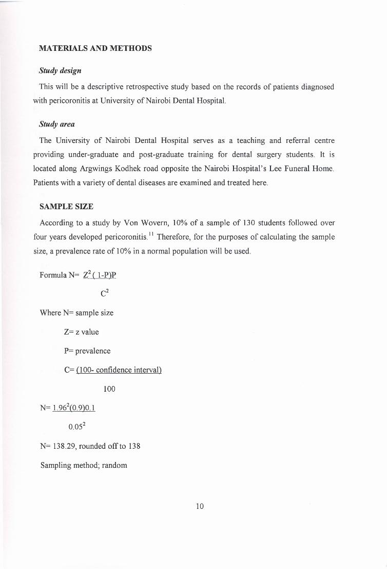
MATERIALS AND METHODS
Study design
This will be a descriptive retrospective study based on the records of patients diagnosed
with pericoronitis at University of Nairobi Dental Hospital.
Study area
The University of Nairobi Dental Hospital serves as a teaching and referral centre
providing under-graduate and post-graduate training for dental surgery students. It is
located along Argwings Kodhek road opposite the Nairobi Hospital's Lee Funeral Home.
Patients with a variety of dental diseases are examined and treated here.
SAMPLE SIZE
According to a study by Von Wovern, 10% of a sample of 130 students followed over
four years developed pericoronitis. 11 Therefore, for the purposes of calculating the sample
size, a prevalence rate of 10% in a normal population will be used.
Formula N= Z2 ( I-P)P
C2
Where N= sample size
Z= z value
P= prevalence
C> (100- confidence interval)
100
N= 1.962(0.9)0.1
N= 138.29, rounded off to 138
Sampling method; random
10

DATA COLLECTION AND ANALYSIS
Information from the patient records will be collected using a data collection form.
Data to be collected includes: age, sex, file number, number of episodes reported, age at
first and last episodes, site of recurrence and presenting complaints.
The data will then be analyzed using computer software and presented in the form of bar
graphs and pie charts.
Inclusion criteria
All cases recorded at the MOS clinic files from the period 1993-2003.
Exclusion criteria
Records with the required information is missing
Ethical considerations
1. Approval will be sought from the relevant authorities
2. All information collected will be treated confidentially and no patients' names will be
used.
3. The information gained from the study will be applied such as to benefit all patients
equally.
Problems anticipated
1. Inaccuracy of patients' records at the M.O.S. and O.D. clinics
2. Time constraints
11

PERCIEVED BENEFITS
1. The study will contribute to the body of knowledge on the patterns of recurrence of
pericoronitis.
2. The results will be used as an aid to better diagnosis and management of recurrent
pericoronitis by dentists and oral surgeons.
3. The results will be submitted in partial fulfilment of the degree of Bachelor of
Dental Surgery of the University of Nairobi.
12

BUDGETARY REQUIREMENTS
ITEM QUANTITY UNIT COST TOTAL COST
(shillings) (shillings)Stationery
0 typing paper One ream 600 600
0 biro pens 10 100ten0 writing paper
three reams 300 900
diskettes five 50 250Internet access Ten hours 1 shilling per minute 600typing Forty pages 20 800Journals and four 100 400abstractsbinding Two reports 200 400GRAND TOTAL 4050
13

DATA COLLECTION FORM
Fll-E AGE OF SEX NO. OF AGE AT AGE AT PRESENTING SITES OFNO. PATIENT EPISODES FIRST LAST COMPLAINT RECURRENCE
REPORTED EPISODE EPISODE ATRECURRENTEPISODES
1.2.3.4.5.6.7.8.9.10111213141516171819202122232425262728293031323334353637
I 14
II

REFERENCES
1. Pratt C.A, Hekmatt M, Barnard lD.W. and Zaki G. A.; Indicationsfor third molar
surgery. l R. ColI. Surg. Edinburgh, 43, April 1998, 105-108
2. Meurmann lH., Rajasuo A, Murtoman H, Savolcinen S; Respiratory tract infections
and concomitant pericoronitis of the wisdom teeth. BMJ April 1995 ;310: 834-836
3. Batanieh A B, Al Q. M.; The predisposingfactors of mandibular third molars in a
Jordanian population. Quintessence Int. March 2003;34(3) 227-231
4. Ackerman G., Cohen M. A., Altini M; The paradental cyst: a clinicopathological study
of 50 cases. Oral Surgery, Oral Medicine, Oral Pathology. Sep 1987,64(3): 308-312
5. Cawson R.A, Odell E.W; Cawson's Essentials of Oral Pathology and Oral Medicine.
seventh edition,© Churchill Livingstone, 2002
6. Ngassapa D, Hassanali J, Amwayi P. and Guthua S; Essentials of Orofacial Anatomy.
© Dar es Salaam University Press, 1996
7. Howe G. L; Minor Oral Surgery, third edition, © John Wright & Sons Ltd. 1986
8. National Inst. Health; NIH Consensus development conference for removal of third
molars. J Oral Surgery1980; 38: 235-236
9. Von Wovern N. V and Nielsen H.O; Thefate of impacted third molars after the age of
20. Int. J Oral Maxillofacial Surg. 1989; 18(5): 277-280
10. Minoru Y, Kiyofumi F, Masakini I, Takafumi H; Root resorption of mandibular second
molar teeth associated with the prescence of impacted third molars. Australian Dental
Journal 1999; 44(20): 112-116
11. Editkorial; Surgical removal of third molars. BMJ 10 September 1994;309: 620-621
12. Grant D.A; Periodontics in the tradition of Orban and Gottlieb, © the c.v. Mosby
Company 1979
15