Embed Size (px)
Citation preview

diagnostics
Review
Patients with Interstitial Lung Disease Secondary toAutoimmune Diseases: How to Recognize Them?
Domenico Sambataro 1,2,*, Gianluca Sambataro 3,* , Francesca Pignataro 4,Giovanni Zanframundo 5 , Veronica Codullo 5 , Evelina Fagone 3, Emanuele Martorana 3,Francesco Ferro 6, Martina Orlandi 7, Nicoletta Del Papa 4, Lorenzo Cavagna 5,Lorenzo Malatino 2 , Michele Colaci 2 and Carlo Vancheri 3
1 Artroreuma S.R.L., Outpatient clinic of Rheumatology associated with the National Health System Corso S.Vito 53, 95030 Catania, Italy
2 Department of Clinical and Experimental Medicine, Internal Medicine Unit, Cannizzaro Hospital, Universityof Catania, via Messina 829, 95100 Catania, Italy; [email protected] (L.M.); [email protected] (M.C.)
3 Regional Referral Centre for Rare Lung Diseases, A. O. U. “Policlinico-Vittorio Emanuele” Dept. of Clinicaland Experimental Medicine, University of Catania, via S. Sofia 68, pavillon 3 floor 1, 95123 Catania, Italy;[email protected] (E.F.); [email protected] (E.M.); [email protected] (C.V.)
4 Scleroderma clinic, Department of Rheumatology, ASST G. Pini, 20122 Milan, Italy;[email protected] (F.P.); [email protected] (N.D.P.)
5 Division of Rheumatology, Hospital IRCCS Policlinico S. Matteo Foundation of Pavia, 27100 Pavia, Italy;[email protected] (G.Z.); [email protected] (V.C.); [email protected] (L.C.)
6 Rheumatology Unit, Department of Clinical and Experimental Medicine, University of Pisa, 56126 Pisa, Italy;[email protected]
7 Department of Experimental and Clinical Medicine, Division of Rheumatology AOUC, University ofFlorence, 50139 Florence, Italy; [email protected]
* Correspondence: [email protected] (D.S.); [email protected] (G.S.);Tel.: +39-3318-009-340 (G.S.)
Received: 9 March 2020; Accepted: 7 April 2020; Published: 9 April 2020�����������������
Abstract: The diagnostic assessment of patients with Interstitial Lung Disease (ILD) can be challengingdue to the large number of possible causes. Moreover, the diagnostic approach can be limited by theseverity of the disease, which may not allow invasive exams. To overcome this issue, the referralcenters for ILD organized Multidisciplinary Teams (MDTs), including physicians and experts incomplementary discipline, to discuss the management of doubtful cases of ILD. MDT is currentlyconsidered the gold standard for ILD diagnosis, but it is not often simple to organize and, furthermore,rheumatologists are still not always included. In fact, even if rheumatologic conditions represent acommon cause of ILD, they are sometimes difficult to recognize, considering the variegated clinicalfeatures and their association with all possible radiographic patterns of ILD. The first objective of thisreview is to describe the clinical, laboratory, and instrumental tests that can drive a diagnosis towarda possible rheumatic disease. The secondary objective is to propose a set of first-line tests to performin all patients in order to recognize any possible rheumatic conditions underlying ILD.
Keywords: interstitial lung disease; idiopathic pulmonary fibrosis; interstitial pneumonia withautoimmune features; systemic sclerosis; myositis; antisynthetase syndrome; Raynaud’s phenomenon;Sjögren’s syndrome; nailfold videocapillaroscopy; multidisciplinary team
1. Introduction
The diagnostic assessment of patients with Interstitial Lung Disease (ILD) can be challenging,since this condition is associated with several possible diseases [1]. The utility of invasive exams
Diagnostics 2020, 10, 208; doi:10.3390/diagnostics10040208 www.mdpi.com/journal/diagnostics

Diagnostics 2020, 10, 208 2 of 23
should be also balanced, between the amount of histological information provided and the risk of sideeffects, mainly in elderly patients or in subjects with a severe disease [2]. Currently, the gold standardin the diagnosis and management of ILD is the Multidisciplinary Team (MDT), in which physiciansof several disciplines discuss doubtful cases to reach a confident diagnosis [3]. Lung involvement iscommon in rheumatic conditions such as vasculitis and Connective Tissue Diseases (CTDs), but theidentification of an autoimmune disease underlying ILD can be difficult for the presence of nuancedclinical pictures. Moreover, the lung can be the first manifestation of a CTD (preceding the onsetof a definite autoimmune disease by years) as the dominant or even as the sole organ involvementduring an autoimmune disease. Obviously, the correct diagnostic assessment of ILD patients hasimportant consequences in prognosis and treatment. Although the rheumatologic evaluation hasproved to be effective in reducing invasive exams, currently the rheumatologist figure is not widelypresent in MDTs [3,4]. The reasons can be numerous: lack of rheumatologists (or of rheumatologistswith lung expertise), difficulty in organizing MDTs, and the underestimation of the rheumatologist’spotential role.
The first objective of this review is to describe the main clinical and serological features, as well asthe instrumental evaluation useful for physicians to guide the diagnosis toward a possible autoimmunedisease. This review aims to summarize the main clinical and serological features suggestive for anILD secondary to rheumatic disorders. Rather than describing and collecting all clinical information ofrheumatology competence, we tried to emphasize the most important clinical signs, that should alsobe recognized by non-rheumatology specialists who study and treat patients with ILD associated withAutoimmune Disorders (ADs).
The secondary aim of this work is to expose our modus operandi for the identification of a possiblerheumatic disease in patients with ILD.
2. Clinical Signs
Clinical signs can immediately supply important elements for a diagnosis of autoimmunediseases. However, their actual recognition may require confirmation by rheumatologists withappropriate expertise.
2.1. Arthritis, Arthralgia, and Morning Stiffness
Arthritis is a clinical condition characterized by pain and stiffness of one or more joints due toan inflammatory process. The classical signs of inflammation (particularly the joint swelling) arepresent with variable degree. Therefore, arthritis is generally a simple diagnosis, even though severalconditions may mimic it and should be excluded by a rheumatologist. The patient’s history itself isfundamental during a correct diagnostic work-up. Typically, the joint pain is more intense at rest andduring the first hours of the day (h 3–6 a.m.) and may be reduced by movement. Moreover, pain isassociated with joint morning stiffness, prolonged more than 30 min up to hours. Conversely, the paindue to osteoarthritis (very common in the general population and not related to ILD) is associated withmovement, mainly at the beginning, whereas it is generally absent at rest and overnight. Moreover,in the course of osteoarthritis, joint stiffness lasts few minutes, in any case less than 30 min. Furtherclinical symptoms could be useful for differential diagnosis, such as pain and stiffness relief withwarmth (i.e., shower, paraffin, mud baths) in osteoarthritis. The presence of arthritis per se is notalways a sign of chronic inflammatory arthropathy, such as in the case of Rheumatoid Arthritis (RA)that could be linked with ILD. Other pathologic conditions may lead to acute inflammation of oneor more joints, such as infectious events, microcrystalline arthritis, or osteoarthritis itself. Hence,clinical presentation and duration are fundamental information to make diagnosis and to differentiateADs-ILD versus the mere coexistence of two separate pathologic disorders.
Considering the rheumatic conditions that can cause ILD, the most frequent disease characterizedby arthritis is RA. Nonetheless, arthritis also has a prevalence of 16% in Sjögren’s Syndrome (SjS) [5],15–20% in Systemic Sclerosis (SSc) [6], and up to 90% in Antisynthetase Syndrome (ASS) [7]. There is a

Diagnostics 2020, 10, 208 3 of 23
broad spectrum of presentation ranging from a mono- or oligoarticular form, more common in SSc, toa polyarticular form involving small joints typical of RA. The presence of arthritis justifies a deeperdiagnostic search aimed at considering a secondary form of ILD.
Isolated arthralgia is a highly non-specific symptom that may be present in all rheumatic diseasesand not necessarily limited to these. The strategy on how to investigate an arthralgia for a possibleprogression to arthritis is still under discussion. The term “Clinically Suspect Arthralgia” (CSA) hasbeen coined to identify patients with arthralgia for less than a year, which the rheumatologist suspectsmay progress toward arthritis on the basis of his personal clinical experience [8]. CSA representsabout 7% of patients with arthralgia who refer to an outpatient clinic of rheumatology and, of these,20% develop RA during the follow-up [9]. The characteristics of arthralgia at risk of RA are thefollowing: joint symptoms onset < 1 year, symptoms located in metacarpophalangeal joints, morningstiffness ≥ 60 min, most severe symptoms present in the early morning, presence of a first-degreerelative with RA, difficulty in making a fist, positive squeeze test of metacarpophalangeal joints [10].The presence of at least 4 parameters to define CSA shows 70.5% sensitivity and 93.6% specificity.
Arthralgia affecting the scapular and pelvic girdles in association with a severe morning stiffnessand elevated C-Reactive Protein (CRP) and/or Erythrocyte Sedimentation Rate (ESR) could orientatetoward a diagnosis of Polymyalgia Rheumatica (PMR). The current PMR criteria show a high sensitivity,but a low specificity in the distinction between PMR and other inflammatory, mainly autoimmune,diseases [11]. Sometimes, the distinction between PMR and late-onset RA or oligo-amyopathicmyopathy can be very difficult. Recently, cases of association between ILD and PMR have been reported,although the possible association between the two pathologies needs further investigations [12].Respiratory symptoms in the course of PMR should be investigated in relation to possible ILD,especially if asthenia, arthralgias, and morning stiffness do not show response to low-dose steroidtherapy. In these cases, the diagnosis should be re-evaluated and the possible presence of CTD shouldbe taken into consideration.
2.2. Sicca Syndrome and Glandular Swelling
Xerophthalmia and xerostomia are very common symptoms. The prevalence in the generalpopulation is 5–30% and 0.02–40%, respectively, with an increase correlated with the age of thepopulation [13,14].
Patients with CTDs frequently report these symptoms that represent the main features of SjS.CTDs are only one of the possible causes of dryness and are among the rarest, considering that theprevalence of SjS is 0.03% of the general population [15]. Sicca syndrome is also described in 25% ofpatients with RA [16] and in up to 71% of those with SSc [17], not necessarily associated with SjS.
About one-third of patients with SjS experience episodes of swelling of the salivary or lachrymalglands [18]. It has been demonstrated that parotid swelling can anticipate the onset of sicca syndromeand subsequent confirmation of the diagnosis of SjS by up to 14 years [19]. The evaluation of a glandularswelling is relevant, considering that 49% of patients with SjS-ILD did not show sicca symptoms at theonset of the disease [20]. Glandular swelling is considered a criterion of disease activity in SjS andshould be clinically evaluated. Nonetheless, ultrasounds provide useful information on the glandularstructure, helping in the disease diagnosis and stadiation [21].
In the course of SjS, parotid and submandibular salivary glands are the most common sites oflymphoma of the mucosa-associated lymphoid tissue, which typically occurs as a persistent and hardmonolateral swelling and has an important impact on prognosis [22].
2.3. Raynaud’s Phenomenon, Digital Ulcers, and Pitting Scars
Raynaud’s phenomenon (RP) owes its name to Maurice Raynaud, who described it for the firsttime in his doctoral thesis in 1862. RP is characterized by a change of the color of the fingers in3 subsequent phases: white (during ischemic phases), blue (during hypoxic phases), and red (during

Diagnostics 2020, 10, 208 4 of 23
revascularization) (Figure 1). At least the first two phases are needed to recognize the presence ofRP [23].
Diagnostics 2020, 10, x FOR PEER REVIEW 4 of 22
revascularization) (Figure 1). At least the first two phases are needed to recognize the presence of RP [23].
Figure 1. The hands in patients with Interstitial Lung Disease (ILD) secondary to rheumatic conditions. A: Raynaud’s phenomenon in ischemic phase during a provocation test; B: mechanic’s hands; C and D: Gottron’s signs; E: Gottron papules; F: sclerodactyly with clawed shape in systemic sclerosis; G: pitting scars in a patient with systemic sclerosis.
This condition affects 3–5% of the general population with a marked increase in prevalence in cold regions [24]. RP is usually generated by an external stimulus, mainly the exposure to cold, but also emotional factors and vibrations. It may be aggravated by cigarette smoking, caffeine intake, or drugs including non-selective beta-blockers and chemotherapy, among others. In response to cold, subjects with RP have a marked sympathetic-mediated vasoconstriction involving the pre-capillary sphincters, leading to the white coloring of the skin. In normal conditions, the reduction of the capillary flow determines the activation of the endothelium with dilatation of the upstream vessels which guarantees the nutritional flow downstream [25]. In RP, this mechanism of protection is less efficient. Thus, RP was subclassified into primary, where the vasospasm is not sufficient to generate a downstream damage, and secondary, where, in association with structural damage and endothelial dysfunction, damage can occur.
Up to 90% of RP cases are classified as primary [26]. Secondary RP is observed in more than 90% of cases of SSc or Mixed Connective Tissue Disease (MCTD) and in up to 2/3 of patients with ASS, and it is considered in the classification criteria of these conditions [27,28]. RP is also observed in 18–45% of patients with Systemic Lupus Erythematosus (SLE), 20% with Dermatomyositis (DM)/Polymyositis (PM), and 10–20% with RA [24]. Nailfold videocapillaroscopy (NVC) and autoantibody panel play a pivotal role in the recognition of secondary RP.
The transition from primary to secondary RP occurs in 3.2% of patients per year with a mean time of 10 years from the onset of RP [29]. This supports the importance of a tight follow-up.
The presence of cyanosis at the extremities without the ischemic phase of RP is defined as “acrocyanosis”, a clinical entity frequent in the general population that could also be considered part of the clinical picture of a CTD.
Digital Ulcers (DUs) represent the clinical manifestation of microvascular damage. It is estimated that approximately 50% of patients with SSc develop at least one DU during their clinical history. Moreover, the presence of DUs is a classification criterion of SSc and is associated with more severe subsets of the disease [27,30].
Figure 1. The hands in patients with Interstitial Lung Disease (ILD) secondary to rheumatic conditions.(A): Raynaud’s phenomenon in ischemic phase during a provocation test; (B): mechanic’s hands; (C,D):Gottron’s signs; (E): Gottron papules; (F): sclerodactyly with clawed shape in systemic sclerosis; (G):pitting scars in a patient with systemic sclerosis.
This condition affects 3–5% of the general population with a marked increase in prevalence in coldregions [24]. RP is usually generated by an external stimulus, mainly the exposure to cold, but alsoemotional factors and vibrations. It may be aggravated by cigarette smoking, caffeine intake, or drugsincluding non-selective beta-blockers and chemotherapy, among others. In response to cold, subjectswith RP have a marked sympathetic-mediated vasoconstriction involving the pre-capillary sphincters,leading to the white coloring of the skin. In normal conditions, the reduction of the capillary flowdetermines the activation of the endothelium with dilatation of the upstream vessels which guaranteesthe nutritional flow downstream [25]. In RP, this mechanism of protection is less efficient. Thus, RP wassubclassified into primary, where the vasospasm is not sufficient to generate a downstream damage,and secondary, where, in association with structural damage and endothelial dysfunction, damagecan occur.
Up to 90% of RP cases are classified as primary [26]. Secondary RP is observed in more than 90%of cases of SSc or Mixed Connective Tissue Disease (MCTD) and in up to 2/3 of patients with ASS, and itis considered in the classification criteria of these conditions [27,28]. RP is also observed in 18–45% ofpatients with Systemic Lupus Erythematosus (SLE), 20% with Dermatomyositis (DM)/Polymyositis(PM), and 10–20% with RA [24]. Nailfold videocapillaroscopy (NVC) and autoantibody panel play apivotal role in the recognition of secondary RP.
The transition from primary to secondary RP occurs in 3.2% of patients per year with a mean timeof 10 years from the onset of RP [29]. This supports the importance of a tight follow-up.
The presence of cyanosis at the extremities without the ischemic phase of RP is defined as“acrocyanosis”, a clinical entity frequent in the general population that could also be considered part ofthe clinical picture of a CTD.
Digital Ulcers (DUs) represent the clinical manifestation of microvascular damage. It is estimatedthat approximately 50% of patients with SSc develop at least one DU during their clinical history.
Diagnostics 2020, 10, 208 5 of 23
Moreover, the presence of DUs is a classification criterion of SSc and is associated with more severesubsets of the disease [27,30].
DUs can be extremely painful and have an important impact on patients’ quality of life [31].They are frequently complicated by infection and the presence of osteomyelitis is observed in 42% ofpatients with infected DUs [32]. Healing is very slow with a mean time of 76 days in pure DUs, 93 incalcinosis, and 281 in gangrene [33].
Digital Pitting Scars (DPSs) are defined as “pinhole-sized digital concave depressions withhyperkeratosis” [34]. Observed in 34–53% of cases [30], they have greater weight than DUs in theclassification criteria for SSc (3 vs. 2 points) [27]. Unlike DUs, their localization is not limited to thefingertips, but they are also present laterally, including on the radial surface of the second and thirdfinger and the ulnar surface of the first finger. The factors that seem to be determinant in the genesis ofDPSs are ischemia, cold exposure, and micro-traumatisms [34]. Usually not painful, they tend to healfaster than DUs (25 days) [33].
It is important to evaluate the possible coexistence of other signs, considering that DUs and DPSscommonly occur in the presence of RP or skin sclerosis and, therefore, in these cases, they stronglysupport the diagnosis of SSc.
2.4. Puffy Hands and Skin Sclerosis
Puffy Hands (PHs) are often considered the first sign of SSc after the onset of RP [35], but theyare also observed in Undifferentiated CTD and MCTD and considered an additional criterion in thedefinition of Very Early Diagnosis of SSc (VEDOSS) [36]. Moreover, their presence satisfies 2 of the9 points necessary for the classification of SSc [27]. This phase is characterized by the presence ofedema of the hands that may remain stable or evolve, even after a long time, toward fibrosis. A state ofpersistent hypoxia, the presence of proinflammatory and profibrotic cytokines, fibroblast activation,oxidative stress, microvascular damage, and ineffectiveness of neoangiogenic mechanisms graduallycause the appearance of fibrosis [37]. Skin fibrosis is the hallmark of SSc. In particular, the presenceof skin fibrosis of both hands extending proximal to the metacarpophalangeal joints is a sufficientclassification criterion [27]. Progressively, fibrosis can become so severe as to cause contracture of thejoints with a serious impact on the patients’ quality of life. This has a prevalence of 31% of patientswith SSc [38].
The presence of skin sclerosis on the extremities (generally hands) and face identifies the limitedcutaneous subset of Ssc, while the involvement of the trunk and the proximal limbs defines the diffuseone [39].
The degree of cutaneous involvement can be measured with the modified Rodnan skin score:the obtained value correlates with disease activity, disease severity, and mortality in the course ofSSc [40].
2.5. Gottron’s Papules and Gottron’s Sign
Described for the first time by Gottron in 1930, this cutaneous manifestation consists of violaceousnon-palpable macules (Gottron’s sign) or raised papules (Gottron’s papules) on the surface of boneprominence (Figure 1) [41]. Characteristically, the lesions occur over the metacarpophalangeal andinterphalangeal joints and more rarely in the elbows, knees, and/or feet [42]. Gottron’s papules havebeen observed in about 50% of patients with ASS [43] and in up to 87% of patients with DM and areconsidered a pathognomonic sign of this disease [44,45].
In view of the pathognomonicity of Gottron’s sign, it is interesting to note that the sign is includedamong the classification criteria for Interstitial Pneumonia with Autoimmune Features (IPAF) [46].On the contrary, in the prospective cohort of IPAF no case showed Gottron’s sign [47]. It is possible toimagine that the simultaneous presence of Gottron’s sign and ILD would lead the MDT toward thedefinition of CTD-ILD.

Diagnostics 2020, 10, 208 6 of 23
2.6. Mechanic’s Hands and Hiker’s Feet
Mechanic’s Hands (MHs) was described for the first time in 1979 as a hyperkeratotic andnon-pruritic eruption of the hands with scaling, fissuring, and hyperpigmentation. It typically shows asymmetrical involvement of the ulnar surface of the first finger and the radial surface of the others,principally the second and third finger [48]. These alterations resemble a manual laborer’s hands,but only 1 out of the 8 patients observed actually did manual work. Four of these patients had adiagnosis of MCTD, 3 of DM, and 1 of SLE, but all manifested a myositic involvement. The presenceof MHs is observed in 40% of patients with DM and in 30–70% of those with ASS [49] and is part ofboth classification criteria for ASS proposed in 2010 and 2011 [28]; however, the clinical importanceof MHs is still under study. Indeed, MHs appear to be associated with an increased risk of systemicinvolvement, especially lung, and a lower risk of malignancy in DM [50].
Skin lesions similar to those seen on the hands can also be observed in the feet. In this case,the hyperkeratosis is associated with cracking and dryness and involves the plantar surface, includingthe toes, reminiscent of the feet of long-distance walkers. The term “hiker’s feet” was chosen to identifythis cutaneous sign, observed in DM and ASS and associated in 90% of cases with MH [51].
2.7. Heliotrope Rash
Its name is due to the typical color that recalls the flower Heliotropium. It is a violaceous erythemainvolving the upper eyelid and periorbital tissue, that can also extend to the cheeks and nasolabial fold.Frequently, the rash is associated with overt edema up to reducing eyelid opening. This is considered acharacteristic sign of DM, and it has also been observed in 14% of patients with ASS [45,52].
2.8. Shawl Sign, V-Sign, and Holster Sign
They consist of a red rash, which may be either flat or raised, involving the upper back, shouldersand arms (shawl sign), the skin in the anterior area of the neck and upper chest with a “V”-shapedpattern (V-sign), or the external area of the hip (holster sign) [53]. They are considered characteristicsigns of DM [45].
2.9. Telangiectasias
These are enlarged capillaries visible on the skin surface. They occur characteristically in SSc,mainly in limited cutaneous form, so that they are included in the classification criteria [27]. In SSc,telangiectasias are commonly localized in the face, lips, hands, and inside the mouth and mightbe associated with the presence of pulmonary arterial hypertension [54]. Telangiectasias can alsobe observed in DM and ASS, commonly in periungual areas, but also in periorbital and gingivalareas [53,55]. Telangiectasias are considered in the clinical domain of the classification criteria forIPAF, even if limited to those on the palmar surface [46]. For the purpose of the definition of IPAF,it would seem useful to also extend the location of telangiectasias to the other areas commonly involvedduring CTDs.
2.10. Calcinosis
Calcinosis is defined as the accumulation of insoluble calcium salts in various tissues. Classically,calcinosis is classified into five classes: (i) metastatic calcinosis, often associated with malignancies,with abnormal serum level of phosphorous and calcium that commonly affects the wall of arteriesand internal organs; (ii) tumoral calcinosis, initially limited to a rare form of genetic disease andcharacterized by a high level of serum phosphorous and normal level of calcium, then generallyunderstood as any form of large calcification; (iii) dystrophic calcinosis, commonly observed inADs, particularly in SSc, DM, and MCTD. In this case, there is a normal serum level of calcium andphosphorous, and the areas principally involved are skin and subcutaneous tissue; (iv) idiopathiccalcification, that occurs in healthy individuals with normal serum levels of calcium and phosphorous;

Diagnostics 2020, 10, 208 7 of 23
(v) calciphylaxis, typical of patients with chronic renal failure, characterized by an alteration of theserum levels of calcium and phosphorous and involvement of the vessels that can lead to ischemia [56].
Calcinosis affects approximately 25% of patients with SSc, 20% of adults with DM, and up to70% of those having the juvenile form [57,58]. The pathogenesis of calcinosis is poorly understood.Chronic inflammation, local microtraumas, and hypoxia are thought to play an important role [59].The most common sites are the skin and subcutaneous tissue in SSc and proximal limb muscles inDM [60]. However, calcinosis can occur in other areas, including paravertebral sites, and reach the sizeof tumoral calcinosis [61]. Calcifications occur in the chronic phase of disease—in SSc, about 9 yearsafter onset and more commonly in limited cutaneous SSc [62]. Anecdotally, the calcification anticipatesthe diagnosis of SSc [61].
Another type of subcutaneous formation that can be found at the bone prominence is therheumatoid nodule. Even though not frequently reported in clinical practice, it is characteristicof long-standing RA; thus, it could represent a useful sign to address the clinician toward therheumatological diagnosis.
2.11. Muscle Weakness
Muscle weakness is a common sign of myopathies. Ten different patterns have been describedthat should guide the examiner in the diagnosis. In the course of inflammatory myopathies, muscleweakness is often associated with myalgia and principally manifests three patterns: (i) proximal“limb-girdle” weakness that symmetrically affects the proximal muscles of arms and legs, with lessinvolvement of the distal muscle. This pattern is the most frequent and common to many myopathiesand is therefore not specific; (ii) prominent neck extensor weakness, also called “dropped headsyndrome”, where amyotrophic lateral sclerosis and myasthenia gravis should also be considered inthe differential diagnosis; (iii) episodic pain, weakness, and myoglobinuria, mainly linked to intensephysical exercise in an untrained subject, but it has rarely been described as not related to physicalexercise in subjects with DM/PM [63]. Muscle weakness can also be observed during PMR and,therefore, requires subsequent examinations in order to be defined.
2.12. Dysphagia
During the course of PM, patients may be affected by dysphagia, due to the muscle involvementof pharynx and the upper third of the esophagus. This condition may become severe because of theimpairment of patients’ ability to eat and drink.
Another kind of dysphagia regards the smooth muscular wall of the lower part of esophagusin SSc patients. The visceral hypotonia and dyskinesia develop slowly during the patients’ clinicalhistory, affecting the ability to swallow and the quality of life in various degrees.
Finally, SjS patients may also complain of dysphagia because of the dryness of the mouth andpharynx exacerbated during the meal.
2.13. Fever of Unknown Origin
An unexplained fever may be present in the course of all ADs related to ILD. The sign is highlynon-specific and needs to be properly investigated. Generally, fever secondary to CTDs is < 38 ◦C,while higher temperatures should be always investigated to exclude concomitant infectious diseases.
3. Laboratory Exams
Laboratory exams can play a pivotal role in the diagnostic assessment of ILD, supporting a specificdiagnosis of ADs, above all in patients with nuanced accompanying clinical features. For convenience,we distinguish general exams from autoimmune exams.
Diagnostics 2020, 10, 208 8 of 23
4. General Laboratory Exams
A panel of general laboratory tests usually gives useful information regarding the levelof inflammation, eventual hepatic or renal dysfunction, CTD-related alterations, and possiblecomorbidities. This panel should consider a complete blood test, ESR, CRP, complement fractions C3and C4, Serum Protein Electrophoresis (SPEP), urine test, creatinine, Alanine Aminotransferase (ALT),Aspartate Aminotransferase (AST), Creatinine Phosphokinase (CPK), Lactic Dehydrogenase (LDH),myoglobin, and aldolase.
A complete blood count should be always carried out in all ILD patients. It can be useful to assessthe level of hemoglobin in order to regulate the use of drugs with a high risk of myelo-suppression.Anemia may be secondary to chronic inflammation. In this view, the increased number of WhiteBlood Cells (WBCs) and their formula can suggest inflammation (sustained by ADs or infections).The reduction of platelets or WBCs may be useful for diagnostic purposes, given that this is consideredin the diagnostic criteria for SLE [64].
Increased levels of ESR, CRP, C3, and C4 are associated with inflammation, but they are not ableto distinguish between infectious or autoimmune origin. The dosage of procalcitonin may be helpfulin the former case. Moreover, they can be burdened by several cases of false positives or negativesand their value should be carefully evaluated in the absence of a concordant clinic [65]. However,ESR and CRP are commonly high in autoimmune inflammation, especially in RA, in which they areincluded as criteria [66]. The reduction of the fractions of complement C3 and C4 can be relatedto active SLE (therefore included in the classification criteria) and with Anti-Neutrophil CytoplasmAntibody (ANCA)-Associated Vasculitis (AAV) [67,68]. SPEP is able to provide interesting information.The finding of an increased portion of α2 globulins is considered a sign of inflammation, and the studyof γ globulins in SPEP is also of some interest in ILD patients. Although hypergammaglobulinemiais not included in any classification criteria of CTDs, it has commonly been found in these patients,mainly in SLE and SjS [69]. On the contrary, hypogammaglobulinemia is a marker of common variableimmunodeficiency, a condition that can produce ILD in the lung, mainly resembling an advanced stageof sarcoidosis in High-Resolution Computed Tomography (HRCT) [70].
Urine test, creatinine, and transaminases are useful in the assessment of the kidney and liverfunctions in order to manage possible treatments, such as Disease-Modifying Anti-Rheumatic Drugs(DMARDs) or antibiotics. The urine test diagnostic value is essential for the recognition of proteinuria,hematuria, and/or chronic renal failure, potentially linked to SLE or AAV.
AST, LDH, CPK, myoglobin, and aldolase are muscular enzymes, generally increased in patientswith an active inflammatory muscular involvement in Idiopathic Inflammatory Myopathies (IIMs).PM, DM, and ASS are included in this group, being conditions associated with potentially severeILD [71,72]. During IIMs, one or more of these enzymes can have high serum levels, but they canalso be elevated in other conditions such as hepatic injury or ILD itself [73]. Therefore, it could bereasonable to consider second-line tests to support a suspected diagnosis of IIMs [74].
5. Autoantibodies
The search of autoantibodies is very useful for the diagnostic assessment of ILD patients, due tothe general high association with CTDs. They can reflect the autoimmune activity of B cells, but theirpositivity should be always considered in association with the clinical picture. Indeed, a singleautoantibody positivity without appropriate clinical findings may be not related to an establisheddisease or may be stochastically positive or anticipate the disease onset. In our opinion, we candistinguish first-line and second-line autoimmune exams.

Diagnostics 2020, 10, 208 9 of 23
6. First-Line Autoimmunity Exams
In this class, we consider Antinuclear Antibodies (ANAs), Rheumatoid Factor (RF),Anti-Citrullinated Protein Antibody (ACPA), Double-Stranded DNA (DsDNA), ANCA, and theExtractable Nuclear Antigen (ENA) profile. The latter group, generally available in commercial kits,includes anti-Ro/SSA, anti-La/SSB, anti-Ribonucleoprotein (RNP), anti-Sm, anti-topoisomerase I (Scl70),and anti-Jo1.
ANAs can be positive in all ADs, typically in SLE [64], and they are also considered in the IPAFcriteria, a research classification similar to the concept of undifferentiated CTD. The inclusion of ANAsin these criteria is reasonable, considering that they may precede the disease onset by 5 years [75].However, ANA positivity can be found in a large number of conditions not related to ILD and even innormal subjects [76,77]. Therefore, as well as for all potentially autoimmune items, they should beconsidered along with the other clinical features for every patient. In addition to the titer, the patternof ANA positivity could also be very useful [78]. Indeed, a centromeric pattern is pathognomonic forthe presence of Anticentromere Antibodies (ACAs). Of great interest is the nucleolar and cytoplasmicpositivity, because these can suggest positivity for Antisynthetase Antibodies (ATSAs) regardless ofthe seric titer and should be studied in depth, looking for the presence of Myositis-Associated orMyositis-Specific Antibodies (MAAs and MSAs).
RF and ACPA are specific antibodies for the diagnosis of RA [3]. Both these antibodies can bepositive several years before the disease onset and the combined positivity seems to have 100% risk ofdeveloping RA within 5 years [79]. However, their diagnostic value is different. RF can be positivealso in other conditions, not necessarily of autoimmune origin, and even in up to 25% of the generalpopulation [80]. Therefore, RF positivity is generally considered as low and high titers using two timesthe upper limit as the cutoff for IPAF criteria, and three times the limit for RA ones [46,66]. ACPAs aremore specific for RA than for RF, but they can be positive in about 1% of normal subjects also (who areat risk for RA in any case) [79–82]. ACPAs are also interesting for the pathogenesis of ILD: the lungcould be the first site of injury in RA [83]. Increased evidences support citrullination of lung peptidesdue to cigarette smoking and other environmental triggers, as well as possible microbial molecularmimicry. These conditions could lead to the production of ACPAs and the subsequent developmentof RA.
DsDNAs are highly specific for SLE and their presence in healthy subjects is very uncommon [64,84].Despite the utility in the diagnosis and prognosis of SLE, the utility in the management of ILD patientsseems to be limited, considering the rarity of ILD-SLE [72]. Moreover, DsDNAs were considered in theserological criteria of IPAF. In prospective studies of IPAF patients, these autoantibodies were rarelyrecognized, even at low titer, and not confirmed during follow-up, thus suggesting a possible falsepositivity [85,86].
ANCAs are a group of autoantibodies specific for AAV, but they can be positive in severalother ADs [87]. They are generally divided into proteinase-3 (PR3), specific for Granulomatosiswith Polyangiitis (GPA); Myeloperoxidase (MPO), more specific for Microscopic Polyangiitis (MPA)and Eosinophilic Granulomatosis with Polyangiitis (EGPA); and atypical ANCA [87]. Despite theabovementioned trend of specificity for AAV, ANCAs can be positive in several other conditions(infections, CTD, inflammatory bowel disease, some of which are able to justify ILD). Moreover,EGPA with PR3-ANCA positivity, as well as GPA associated with MPO-ANCA are not uncommon [88].For this reason, ANCAs are not currently included in any classification criteria, neither for AAV nor forIPAF [89]. However, in recent studies, patients with Idiopathic Pulmonary Fibrosis (IPF) and ANCApositivity developed vasculitis (MPA and GPA) [86,90]. This is in line with the current knowledgethat recognizes the Usual Interstitial Pneumonia (UIP) pattern as the most common ILD in AAV [91].For this reason, we considered it appropriate to include ANCA in the first-line exams for the diagnosisof ILD patients, mainly if with a UIP pattern or imaging resembling sarcoidosis [92].

Diagnostics 2020, 10, 208 10 of 23
As already mentioned, the ENA profile includes autoantibodies that are highly specific for severalCTDs. However, their utility in ILD patients has different values. Anticentromere Antibody (ACA,also named CENtromere Protein B, CENP-B) and anti-Scl70 are associated with SSc [93], but theformer is protective for ILD, while the latter is associated with severe forms [94]. Anti-Sm andAnti-RNP can be present in several CTDs, but are specific for SLE, while anti-U1-RNP is specificfor MCTD [95]. Anti-RNP is generally associated with mild myositis and ILD [96]. Anti-SSA andanti-SSB are associated with SjS, but the latter antibody was excluded by new classification criteriafor SjS [97]. In fact, anti-SSB alone without accompanying anti-SSA is uncommon and unrelated toSjS [98]. Anti-SSA can be directed to the subunit of 52 or 60 kD [99]. Both are specific for SjS, but theycan be found in several other CTDs, especially SSA52 kD, which is frequently associated with IIMs [96].It should be taken into consideration that anti-SSA can be positive at ANA negative for the loss ofanti-SSA60 kD during preparation, but also because SSA52 kD recognizes a cytoplasmic antigen [96].SSA52 kD is mainly associated with ILD in both IIMs and SjS [69]. The ENA profile also includesanti-Jo1, the most frequent antisynthetase antibody and the unique one included in ENA commercialkits. It recognizes histidyl-tRNA synthetase, a cytoplasmic protein; therefore, similarly to anti SSA52kD, it can be positive with ANA negative [96]. Anti-Jo1 is associated with arthritis, myositis, and ILDin IIMs and mainly ASS [100,101].
7. Second-Line Autoimmunity Exams
A number of other, generally rare, MSAs and MAAs can be associated with ILD, the largest partbelonging to the family of IIMs [96].
Among the MAAs, Pm/scl75 kD and Pm/scl100 kD (based on the protein recognized) are found inIIMs, overlap syndromes, and SSc, generally seronegative for Scl70 or ACA [102] and are associatedwith mild myositis and ILD with better outcome compared to Scl70 + SSc [103]. These autoantibodieswere not found in healthy subjects [104]. Another uncommon antibody that can be found in severalCTDs is anti-Ku. Patients with this antibody can have myositis and ILD, the latter refractory tosteroids [105]. Moreover, Anti-Mitochondrial Antibody (AMA) M2 antibody, greatly specific forprimary biliary cholangitis, can be associated with IIMs and ILD [106], but no evidence is reported inliterature regarding the clinical features of AMA M2 + ILD patients.
Among MSAs, rare ATSAs different from anti-Jo1 are included (e.g., PL7, PL12, EJ, OJ, Ks).Regarding cytoplasmic antigens, they can be positive regardless of ANA. They are generally associatedwith amyopathic or mild form or myositis, but potentially severe ILD. Increased evidences suggest apossible specific disease subset for each antibody specificity [107]. Anti-Mi2 and antibodies to smallubiquitin-like modifier activating enzyme (anti-SAE) are associated with the diagnosis of juvenile DMand skin involvement in DM, respectively, but both myositis and ILD seem to have a good prognosiswhen associated to these antibodies [108]. The most dangerous MSAs can be considered anti-MelanomaDifferentiation-Associated 5 gene (anti-MDA5), generally associated with clinically amyopathic DMand rapidly progressive ILD [109]. Several other MSAs can be useful for the diagnosis of IIMs, but theirrole in the assessment of ILD is not currently studied in depth [96]. Some of the most common areTIF1, anti-signal recognition particles, anti-SAE, antibody to 3-hydroxy-3methyl-glutaryl-Coenzyme AReductase (anti-HMGCR).
A distinct mention should be made for Antiphospholipid Antibodies (APLAs), anti-cardiolipinantibodies, and anti-β2glicoprotein I antibodies. They are specific for Antiphospholipid Syndrome(APS), alone or associated with other CTDs (mainly SLE), but they are also present in 1%–5% of healthysubjects [110]. No associations are currently reported regarding ILD and APL. However, it should beconsidered that Idiopathic Pulmonary Fibrosis (IPF) shows a pro-thrombotic status and vascular eventsare common [111]. In this disease, Lupus Anticoagulant (LAC) is reported positive in about 21% ofpatients [112], a proportion significantly higher compared to what is reported in the general population.LAC is reported also in 35% of SLE patients and is a diagnostic criterion for this condition [64,113],

Diagnostics 2020, 10, 208 11 of 23
however, it is hard to suppose an overlap with SLE in these patients given the lack of other features.A possible overlapping condition between IPF and APS is an argument of interest.
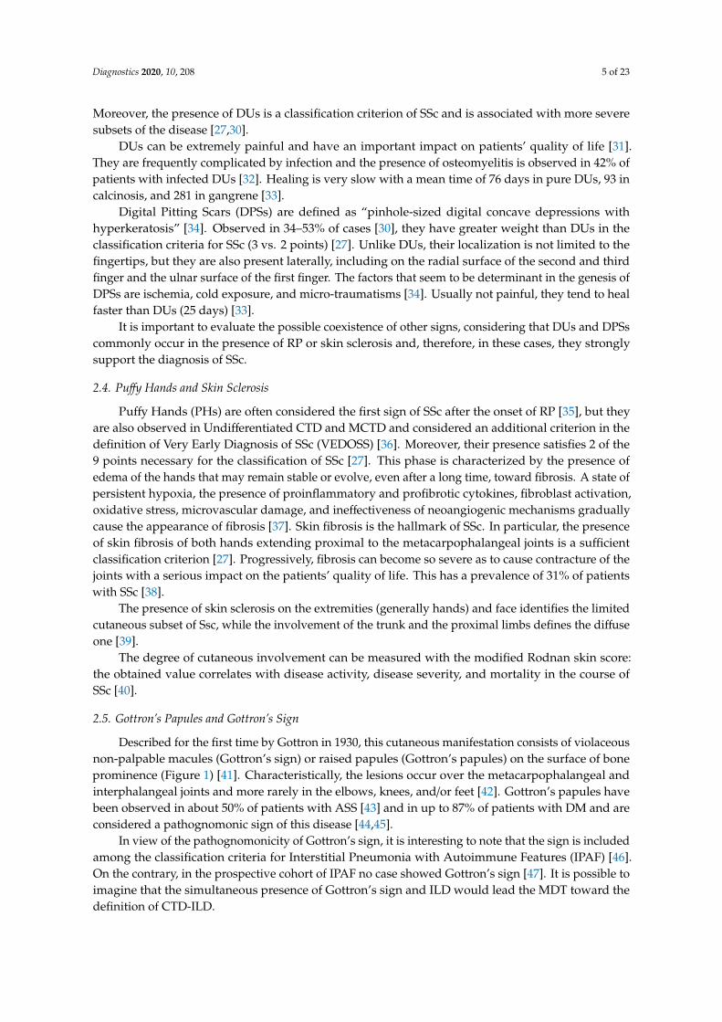
Figure 2 shows the main autoantibodies useful for the diagnosis of autoimmune ILD.
Diagnostics 2020, 10, x FOR PEER REVIEW 10 of 22
It recognizes histidyl-tRNA synthetase, a cytoplasmic protein; therefore, similarly to anti SSA52 kD, it can be positive with ANA negative [96]. Anti-Jo1 is associated with arthritis, myositis, and ILD in IIMs and mainly ASS [100,101].
7. Second-Line Autoimmunity Exams
A number of other, generally rare, MSAs and MAAs can be associated with ILD, the largest part belonging to the family of IIMs [96].
Among the MAAs, Pm/scl75 kD and Pm/scl100 kD (based on the protein recognized) are found in IIMs, overlap syndromes, and SSc, generally seronegative for Scl70 or ACA [102] and are associated with mild myositis and ILD with better outcome compared to Scl70 + SSc [103]. These autoantibodies were not found in healthy subjects [104]. Another uncommon antibody that can be found in several CTDs is anti-Ku. Patients with this antibody can have myositis and ILD, the latter refractory to steroids [105]. Moreover, Anti-Mitochondrial Antibody (AMA) M2 antibody, greatly specific for primary biliary cholangitis, can be associated with IIMs and ILD [106], but no evidence is reported in literature regarding the clinical features of AMA M2 + ILD patients.
Among MSAs, rare ATSAs different from anti-Jo1 are included (e.g., PL7, PL12, EJ, OJ, Ks). Regarding cytoplasmic antigens, they can be positive regardless of ANA. They are generally associated with amyopathic or mild form or myositis, but potentially severe ILD. Increased evidences suggest a possible specific disease subset for each antibody specificity [107]. Anti-Mi2 and antibodies to small ubiquitin-like modifier activating enzyme (anti-SAE) are associated with the diagnosis of juvenile DM and skin involvement in DM, respectively, but both myositis and ILD seem to have a good prognosis when associated to these antibodies [108]. The most dangerous MSAs can be considered anti-Melanoma Differentiation-Associated 5 gene (anti-MDA5), generally associated with clinically amyopathic DM and rapidly progressive ILD [109]. Several other MSAs can be useful for the diagnosis of IIMs, but their role in the assessment of ILD is not currently studied in depth [96]. Some of the most common are TIF1, anti-signal recognition particles, anti-SAE, antibody to 3-hydroxy-3methyl-glutaryl-Coenzyme A Reductase (anti-HMGCR).
A distinct mention should be made for Antiphospholipid Antibodies (APLAs), anti-cardiolipin antibodies, and anti-β2glicoprotein I antibodies. They are specific for Antiphospholipid Syndrome (APS), alone or associated with other CTDs (mainly SLE), but they are also present in 1%–5% of healthy subjects [110]. No associations are currently reported regarding ILD and APL. However, it should be considered that Idiopathic Pulmonary Fibrosis (IPF) shows a pro-thrombotic status and vascular events are common [111]. In this disease, Lupus Anticoagulant (LAC) is reported positive in about 21% of patients [112], a proportion significantly higher compared to what is reported in the general population. LAC is reported also in 35% of SLE patients and is a diagnostic criterion for this condition [64,113], however, it is hard to suppose an overlap with SLE in these patients given the lack of other features. A possible overlapping condition between IPF and APS is an argument of interest.
Figure 2 shows the main autoantibodies useful for the diagnosis of autoimmune ILD.
Figure 2. Spectrum of autoantibodies associated with autoimmune interstitial lung disease.Legend: AAVs: ANCA (Anti-Neutrophil Cytoplasm Antibody)-Associated Vasculitis; ACPA:Anti-Citrullinated Protein Antibody; ANA: Antinuclear Antibodies; APLA: Anti-phospholipidantibodies; ATSA: Anti-T-RNA-synthetase antibodies; IIMs: Idiopathic Inflammatory Myopathies;IPAF: Interstitial Pneumonia with Autoimmune Features; MAAs: Myositis-associated antibodies;MSAs: Myositis-specific antibodies; RA: Rheumatoid Arthritis; RF: Rheumatoid Factor SjS: Sjögren’sSyndrome; SSDs: Scleroderma Spectrum Disorders.
8. Instrumental Evaluation
Several instrumental exams are able to support a diagnosis of AD underlying an ILD, however,they are not always available in an outpatient setting. In this section, we propose a set of first- andsecond-line instrumental exams that are useful in the assessment of ILD.
9. First-Line Instrumental Exams
NVC is a useful tool to study in vivo the density and morphology of capillaries of the fingersthrough a magnification of 200 folds. NVC represents an easy, non-invasive, and non-expensivetechnique, generally used to evaluate patients with RP in order to detect a possible SSc [114]. Themost important parameters for the diagnosis are the presence of giant capillaries (capillaries witha diameter ≥50 µ) and Avascular Areas (AAs, distance between two capillary loops ≥ 500 µ) [114].NVC in SSc has proved to be useful not only in diagnosis but also in prognosis. The Number ofmicrohEMOrrages (NEMO score) is useful to assess the disease activity in SSc, whereas the meannumber of capillaries and AAs can be helpful to stratify the risk of developing complications suchas DUs [115–118]. Recently, a pathologic NVC with scleroderma pattern was also found in patientswith IIMs without RP [119], and the presence of bushy capillaries was associated with the diagnosis ofIIMs [120]. Therefore, NVC can be a useful tool to assess ILD patients in order to select patients inwhom it can be appropriate to look for MAAs and MSAs.
The diagnosis of SjS, generally suggested by the presence of sicca syndrome, can be studied bysalivary gland ultrasound or sialo-scintigraphy; according to the current criteria, impairment in theglandular function should be demonstrated [97]. In fact, the ocular staining score should be assessed byexperienced ophthalmologists, whereas Schirmer’s Test (ST) and Unstimulated Salivary Flow Rate Test(USFRT) can be performed easily in an outpatient clinic assessing ILD [121]. ST is a simple test aimedto measure lacrimal production in 5 min through a measured strip in the lower eyelid. A production of

Diagnostics 2020, 10, 208 12 of 23
<5 mm in 5 min is considered pathological [122]. USFRT can assess the production of saliva by invitingpatients to collect their saliva by passive drool in a measured tube in 5 min. The test is consideredpositive for a production < 0.1 mL/min [123].
Pulmonary Function Tests (PFTs) have a role in the follow-up rather than in the diagnosis ofADs-ILD. Forced Vital Capacity (FVC) and Diffusion Lung Capacity for Carbon Monoxide (DLCO)can describe the severity of restrictive lung damage. This is true mainly for RA, while in sclerodermaspectrum disorders and IIMs, conflicting results are reported in the literature regarding the possibilitythat PFT could appropriately assess the evolution of lung damage and the response to treatment [72].DLCO is in fact undermined by the frequent presence in these conditions of pulmonary arteryhypertension associated with ILD.
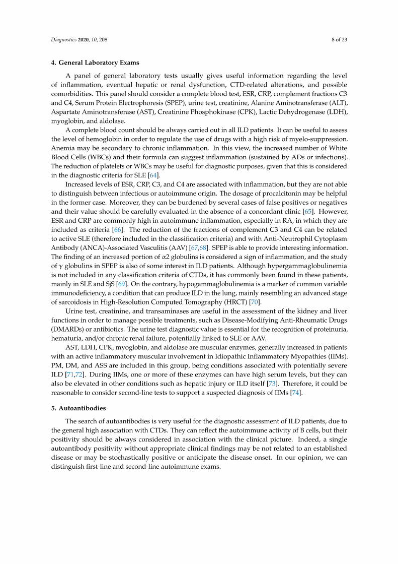
High-Resolution Computed Tomography (HRCT) should be considered a first-line exam for theassessment of ILD patients. Non-Specific Interstitial Pneumonia (NSIP) is the most frequent ILDpattern in CTDs with lung involvement, but all patterns can be observed. UIP is the most commonpattern in ILDs associated with RA and AAV, however it is also present in about 10% of ILD-SjSfrequently before the onset of sicca syndrome and in the late stage of SSc. Lymphocytic interstitialpneumonia is a rare pattern but is closely related with the diagnosis of SjS. Combined patterns arealso common, especially for NSIP-OP. The latter pattern is a common feature of IIM-ILD [124–131].Figure 3 shows an indicative proportion of the frequency of each HRCT pattern for each rheumaticdisease [47,124–131]. The proportions reported can widely vary in the studies, depending from thecriteria used for the patients’ enrollment and/or HRCT classification, as well as the stage of the diseaseat the time of the study. HRCT may also be useful as a prognostic factor in the assessment of ILDseverity and progression. Emerging evidences support the role of many quantification scores in theevaluation of prognosis and response to treatment in several CTDs [132–134].
Diagnostics 2020, 10, x FOR PEER REVIEW 12 of 22
at the time of the study. HRCT may also be useful as a prognostic factor in the assessment of ILD severity and progression. Emerging evidences support the role of many quantification scores in the evaluation of prognosis and response to treatment in several CTDs [132–134].
Figure 3. Legend: AAV: ANCA-Associated Vasculitis; LIP: Lymphocytic Interstitial Pneumonia; IPAF: Interstitial Pneumonia with Autoimmune Features; IIMs: Idiopathic Inflammatory Myopathies; NSIP: Non-Specific Interstitial Pneumonia; RA: Rheumatoid Arthritis; SLE: Systemic Lupus Erythematosus; SjS: Sjögren’s Syndrome; SSD: Scleroderma Spectrum Disorders; UIP: Usual Interstitial Pneumonia.
10. Second-Line Instrumental Exams
Several other instrumental exams can be useful in the diagnostic assessment of ILD patients with suspected AD. The most important is biopsy, performed depending on the tissue involved. Lung biopsy can be useful to assess the ILD pattern with more precision, but the acquired information should be balanced with the risk of the procedure [2]. It should be taken into account that, although small studies have reported histological differences between the UIP pattern in RA and IPF (fewer fibroblastic foci and more CD4+ cells in RA), no evidences strongly support a confident differential diagnosis between these two conditions [135]. A similar consideration should be made for sarcoidosis. Indeed, up to 35% of these patients show non-caseating necrotizing granulomas; so, for them a possible alternative diagnosis of AAV or RA should be considered [136]. Minor salivary gland biopsy can be very useful in the diagnosis of ILD underlying SjS, mainly considering the possible seronegative subset of these patients [69]. The kidney and the upper and lower respiratory tract may be involved by inflammation during AAV, but about 30% of histological exams of the respiratory tract can give a false negative result [137]. Muscle biopsy may be useful for the diagnosis of IIMs and the differential diagnosis among the forms including them [138]. However, several subsets of ILD-IIMs had small muscular involvement and, therefore, it could be useful to perform several exams (e.g., echography, electromyography, magnetic resonance imaging) in order to choose the appropriate tissue to collect for a confident diagnosis.
Finally, kidney biopsy can be considered for the diagnostic definition of ILD patients with suspected SLE and/or AAVs.
11. Conclusions
The differential diagnosis of conditions underlying ILD is a fascinating topic involving many physicians of different specialties. Currently, the gold standard for the diagnostic assessment of ILD is MDT and the presence of rheumatologists among them has proved to be useful to reduce the need for invasive exams [4,139]. The role of rheumatologists in MDT is probably more important considering the possible selection bias in the study of ADs-ILD. In fact, AD-ILD patients can refer to
Figure 3. Legend: AAV: ANCA-Associated Vasculitis; LIP: Lymphocytic Interstitial Pneumonia; IPAF:Interstitial Pneumonia with Autoimmune Features; IIMs: Idiopathic Inflammatory Myopathies; NSIP:Non-Specific Interstitial Pneumonia; RA: Rheumatoid Arthritis; SLE: Systemic Lupus Erythematosus;SjS: Sjögren’s Syndrome; SSD: Scleroderma Spectrum Disorders; UIP: Usual Interstitial Pneumonia.

Diagnostics 2020, 10, 208 13 of 23
10. Second-Line Instrumental Exams
Several other instrumental exams can be useful in the diagnostic assessment of ILD patients withsuspected AD. The most important is biopsy, performed depending on the tissue involved. Lungbiopsy can be useful to assess the ILD pattern with more precision, but the acquired informationshould be balanced with the risk of the procedure [2]. It should be taken into account that, althoughsmall studies have reported histological differences between the UIP pattern in RA and IPF (fewerfibroblastic foci and more CD4+ cells in RA), no evidences strongly support a confident differentialdiagnosis between these two conditions [135]. A similar consideration should be made for sarcoidosis.Indeed, up to 35% of these patients show non-caseating necrotizing granulomas; so, for them a possiblealternative diagnosis of AAV or RA should be considered [136]. Minor salivary gland biopsy canbe very useful in the diagnosis of ILD underlying SjS, mainly considering the possible seronegativesubset of these patients [69]. The kidney and the upper and lower respiratory tract may be involvedby inflammation during AAV, but about 30% of histological exams of the respiratory tract can give afalse negative result [137]. Muscle biopsy may be useful for the diagnosis of IIMs and the differentialdiagnosis among the forms including them [138]. However, several subsets of ILD-IIMs had smallmuscular involvement and, therefore, it could be useful to perform several exams (e.g., echography,electromyography, magnetic resonance imaging) in order to choose the appropriate tissue to collect fora confident diagnosis.
Finally, kidney biopsy can be considered for the diagnostic definition of ILD patients withsuspected SLE and/or AAVs.
11. Conclusions
The differential diagnosis of conditions underlying ILD is a fascinating topic involving manyphysicians of different specialties. Currently, the gold standard for the diagnostic assessment of ILD isMDT and the presence of rheumatologists among them has proved to be useful to reduce the need forinvasive exams [4,139]. The role of rheumatologists in MDT is probably more important consideringthe possible selection bias in the study of ADs-ILD. In fact, AD-ILD patients can refer to the specialistwho is perceived as more useful for the clinical condition. In this hypothesis, patients with mildILD can refer to a rheumatologist, while ILD patients with poor rheumatologic symptoms could bedirected to pulmonologists. Several conditions (e.g., SjS, AAVs, RA) can have ILD as the main clinicalmanifestation of the disease. Moreover, ILD can be the first manifestation of the disease, raisingquestions about a possible pathogenic role of the lung as an autoimmune source. This is one of thereasons that led to the definition of the IPAF criteria, in order to select patients with primary ILD atrisk of developing ADs or those who harbor a subclinical CTD.
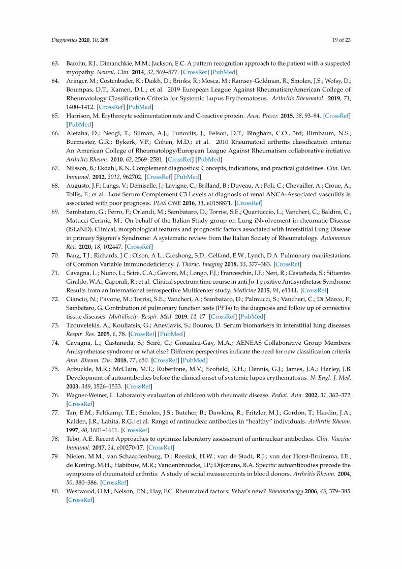
Close collaboration between rheumatologists and pulmonologists, both experienced in themanagement of ILD, is therefore useful for the assessment of these patients, but currently it is notwidely diffused and standardized. In this review, we report our approach in Figure 4. The first step isa careful clinical examination performed by both pulmonologists and rheumatologists. All patientsshould undergo to HRCT, NVC, PFTs, and a first-line serological assessment. Based on the resultsobtained, patients can perform second-line exams as reported in the figure. After the study, patientswith inconclusive diagnosis could be discussed within the MDT.
Finally, it is auspicious that the growing knowledge in the field and the interest to better definecomplex clinical pictures led different specialists to gather together their know-how into sharedrecommendations in order to manage autoimmune ILD patients as well as possible.

Diagnostics 2020, 10, 208 14 of 23
Diagnostics 2020, 10, x FOR PEER REVIEW 13 of 22
the specialist who is perceived as more useful for the clinical condition. In this hypothesis, patients with mild ILD can refer to a rheumatologist, while ILD patients with poor rheumatologic symptoms could be directed to pulmonologists. Several conditions (e.g., SjS, AAVs, RA) can have ILD as the main clinical manifestation of the disease. Moreover, ILD can be the first manifestation of the disease, raising questions about a possible pathogenic role of the lung as an autoimmune source. This is one of the reasons that led to the definition of the IPAF criteria, in order to select patients with primary ILD at risk of developing ADs or those who harbor a subclinical CTD.
Close collaboration between rheumatologists and pulmonologists, both experienced in the management of ILD, is therefore useful for the assessment of these patients, but currently it is not widely diffused and standardized. In this review, we report our approach in Figure 4. The first step is a careful clinical examination performed by both pulmonologists and rheumatologists. All patients should undergo to HRCT, NVC, PFTs, and a first-line serological assessment. Based on the results obtained, patients can perform second-line exams as reported in the figure. After the study, patients with inconclusive diagnosis could be discussed within the MDT.
Figure 4. Our methodology in the diagnostic assessment of patients with interstitial lung disease. Legend: AAV: ANCA-Associated Vasculitis; ALT: Alanine Transaminase; ACA: Anticentromere Antibody; ACPA: Anti-Citrullinated Protein Antibody; ANA: Antinuclear Antibody; ANCA: Anti-Neutrophil Cytoplasmic Antibody; AST: Aspartate Transaminase; C3 and C4: Complement Fraction 3 and 4; CPK: Creatine Phosphokinase; CRP: C-Reactive Protein; DsDNA: Double-Stranded DNA; ESR: Erythrocyte Sedimentation Rate; HRCT: High-Resolution Computed Tomography; LDH: Lactic Dehydrogenase; NVC: Nailfold Video Capillaroscopy; PET: Positron Emission Tomography; PFTs: Pulmonary Function Tests; RF: Rheumatoid Factor.
Finally, it is auspicious that the growing knowledge in the field and the interest to better define complex clinical pictures led different specialists to gather together their know-how into shared recommendations in order to manage autoimmune ILD patients as well as possible.
Abbreviations
Alanine Aminotransferase ALT ANCA-Associated Vasculitis AAV Anti-3-hydroxy-3methyl-glutaryl-Coenzyme A Reductase
Anti-HMGCR
Anti-Citrullinated Protein Antibody ACPA Anti-Neutrophil Cytoplasm Antibody ANCA
Figure 4. Our methodology in the diagnostic assessment of patients with interstitial lung disease.Legend: AAV: ANCA-Associated Vasculitis; ALT: Alanine Transaminase; ACA: AnticentromereAntibody; ACPA: Anti-Citrullinated Protein Antibody; ANA: Antinuclear Antibody; ANCA:Anti-Neutrophil Cytoplasmic Antibody; AST: Aspartate Transaminase; C3 and C4: ComplementFraction 3 and 4; CPK: Creatine Phosphokinase; CRP: C-Reactive Protein; DsDNA: Double-StrandedDNA; ESR: Erythrocyte Sedimentation Rate; HRCT: High-Resolution Computed Tomography; LDH:Lactic Dehydrogenase; NVC: Nailfold Video Capillaroscopy; PET: Positron Emission Tomography;PFTs: Pulmonary Function Tests; RF: Rheumatoid Factor.
Author Contributions: Conceptualization: D.S. and G.S.; data curation: D.S., G.S., F.P., M.O., F.F., E.M., E.F.,G.Z., and V.C.; formal analysis: D.S., G.S., N.D.P., L.C., M.C., L.M., and C.V.; funding acquisition: not applicable;investigation: all authors; methodology: all authors; resources: not applicable; software: not applicable;supervision: N.D.P., L.C., M.C., L.M., and C.V.; validation: D.S., G.S., N.D.P., L.C., M.C., L.M., and C.V.;visualization: all authors; writing—original draft: all authors; writing—review and editing: D.S., G.S., N.D.P.,L.C., M.C., L.M., and C.V. All authors have read and agreed to the published version of the manuscript.
Acknowledgments: Contribution made with funds for University Research—Research plan 2016–2018—project#1A Dept. Of Clinical and Experimental Medicine “Molecular and clinical markers—early instruments in metabolicand chronic-degenerative pathologies”.
Conflicts of Interest: C.V. is part of the F. Hoffmann-La Roche Ltd. Scientific board. He has received consulting feesand/or speaker fees from AstraZeneca, Boehringer Ingelheim, Chiesi, F. Hoffmann-La Roche Ltd., and Menarini.The authors G.S., D.S., E.F., G.Z., V.C., L.C., M.C., M.O., F.P., N.D.P., L.M. declare that they have no conflictof interest.
Abbreviations
Alanine Aminotransferase ALTANCA-Associated Vasculitis AAVAnti-3-hydroxy-3methyl-glutaryl-Coenzyme A Reductase Anti-HMGCRAnti-Citrullinated Protein Antibody ACPAAnti-Neutrophil Cytoplasm Antibody ANCAAnti-Small ubiquitin-like modifier activating enzyme Anti SAEAnti-Topoisomerase I Scl70Anticentromere Antibody ACAAnti-Mitochondrial Antibody AMAAntinuclear Antibody ANAAntiphospholipid Syndrome APSAntiphospholipid Antibody APLA

Diagnostics 2020, 10, 208 15 of 23
Antisynthetase Antibody ATSAAntisynthetase Syndrome ASSAspartate Aminotransferase ASTAutoimmune Disorders ADsAvascular Areas AAsCENtromere Protein B CENP-BClinically Suspect Arthralgia CSAConnective Tissue Diseases CTDsC-Reactive Protein CRPCreatinine Phosphokinase CPKDermatomyositis DMDiffusion Lung Capacity for Carbon Monoxide DLCODigital Pitting Scars DPSsDigital Ulcer DUDisease-Modifying Anti-Rheumatic Drugs DMARDsDouble-Stranded DNA DsDNAEosinophilic Granulomatosis with Polyangiitis EGPAErythrocyte Sedimentation Rate ESRExtractable Nuclear Antigen ENAForced Vital Capacity FVCGranulomatosis with Polyangiitis GPAHigh-Resolution Computed Tomography HRCTIdiopathic Inflammatory Myopathies IIMsIdiopathic Pulmonary Fibrosis IPFInterstitial Lung Disease ILDInterstitial Pneumonia with Autoimmune Features IPAFLactic Dehydrogenase LDHLupus Anticoagulant LACMechanic’s Hands MHMelanoma Differentiation-Associated 5 gene MDA5Microscopic Polyangiitis MPAMixed Connective Tissue Disease MCTDMultidisciplinary Team MDTMyeloperoxidase MPOMyositis-Associated Antibodies MAAsMyositis-Specific Antibodies MSAsNailfold Videocapillaroscopy NVCNonspecific Interstitial Pneumonia NSIPNumber of microhEMOrrages NEMOPolymyalgia Rheumatica PMRPolymyositis PMProteinase-3 PR3Puffy Hands PHPulmonary Function Tests PETRaynaud’s Phenomenon RPRheumatoid Arthritis RARheumatoid Factor RFRibonucleoprotein RNPSchirmer’s Test STSerum Protein Electrophoresis SPEPSjögren’s Syndrome SjSSystemic Lupus Erythematosus SLESystemic Sclerosis SScUnstimulated Salivary Flow Rate Test USFRTUsual Interstitial Pneumonia UIPVery Early Diagnosis of SSc VEDOSSWhite Blood Cell WBC

Diagnostics 2020, 10, 208 16 of 23
References
1. Antoniou, K.M.; Margaritopoulos, G.A.; Tomassetti, S.; Bonella, F.; Costabel, U.; Poletti, V. Interstitial LungDisease. Eur. Respir. Rev. 2014, 23, 40–54. [CrossRef] [PubMed]
2. Cottin, V. Lung biopsy in interstitial lung disease: Balancing the risk of surgery and diagnostic uncertainty.Eur. Respir. J. 2016, 48, 1274–1277. [CrossRef] [PubMed]
3. Furini, F.; Carnevale, A.; Casoni, G.; Guerrini, G.; Cavagna, L.; Govoni, M.; Scirè, C.A. The role of theMultidisciplinary evaluation of interstitial lung diseases: Systematic literature review of the current evidenceand future perspectives. Front. Med. 2019, 6, 246. [CrossRef] [PubMed]
4. Levi, Y.; Israeli-Shani, L.; Kuchuk, M.; Epstein Shochet, G.; Koslow, M.; Shitrit, D. Rheumatological assessmentis important for interstitial lung disease diagnosis. J. Rheumatol. 2018, 45, 1509–1514. [CrossRef]
5. Ramos-Casal, M.; Brito-Zeror, P.; Seror, R.; Bootsma, H.; Bowman, S.J.; Doner, T.; Gottemberg, J.E.; Mariette, X.;Theander, E.; On behalf of the EULAR Sjögren’s Syndrome Task Force; et al. Characterization of systemicdisease in primary Sjögren’s syndrome: EULAR-SS Task Force recommendations for articular, cutaneous,pulmonary and renal involvements. Rheumatology 2015, 54, 2230–2238. [CrossRef]
6. Lorand, V.; Czirjak, L.; Minier, T. Musculoskeletal involvement in systemic sclerosis. Presse Med. 2014, 43,e315–e328. [CrossRef]
7. Tripoli, A.; Marasco, E.; Cometi, L.; De Stefano, L.; Marcucci, E.; Furini, F.; Barsotti, S.; Cavagna, L. One yearin review 2019: Idiopathic inflammatory myopathies. Clin. Exp. Rheumatol. 2020, 38, 1–10.
8. Van Steenbergen, H.W.; van Nies, J.A.B.; Huizinga, T.W.J.; Bloem, J.L.; Reijnierse, M.; van der Helm-vanMil, A.H.M. Characterising arthralgia in the preclinical phase of rheumatoid arthritis using MRI. Ann. Rheum.Dis. 2015, 74, 1225–1232. [CrossRef]
9. Van Steenbergen, H.W.; Mangnus, L.; Reijnierse, M.; Huizinga, T.W.; van der Helm-van Mil, A.H. Clinicalfactors, anticitrullinated peptide antibodies and MRI-detected subclinical inflammation in relation toprogression from clinically suspect arthralgia to arthritis. Ann. Rheum. Dis. 2016, 75, 1824–1830. [CrossRef]
10. Van Steenbergen, H.W.; Aletaha, D.; Beaart-van de Voorde, L.J.J.; Brouwer, E.; Codreanu, C.; Combe, B.;Fonseca, J.E.; Hetland, M.L.; Humby, F.; Kvien, T.K.; et al. EULAR definition of arthralgia suspicious forprogression to rheumatoid arthritis. Ann. Rheum. Dis. 2017, 76, 491–496. [CrossRef]
11. Gonzalez-Gay, M.A.; Garcia-Porrua, C.; Salvarani, C.; Olivieri, I.; Hunder, G.G. Polymyalgia manifestation indifferent conditions mimicking polymyalgia rheumatica. Clin. Exp. Rheumatol. 2000, 18, 755–759. [PubMed]
12. Sambataro, G.; Sambataro, D.; Pignataro, F.; Torrisi, S.E.; Vancheri, A.; Pavone, M.; Palmucci, S.; Del Papa, N.;Vancheri, C. Interstitial Lung Disease in patients with Polymyalgia Rheumatica: A case series. Respir. Med.Case Rep. 2019, 26, 126–130. [CrossRef] [PubMed]
13. Phadatare, S.P.; Momin, M.; Nighojkar, P.; Askarkar, S.; Singh, K.K. A comprehensive review on dry eyedisease: Diagnosis, medical management, recent developments, and future challenges. Adv. Pharm. 2015,2015, 704946. [CrossRef]
14. Agostini, B.A.; Cericato, G.O.; da Silveira, E.R.; Giacomelli Nascimento, G.; dos Santos Costa, F.;Thomson, W.M.; Demarco, F.F. How common is dry mouth? Systematic review and meta-regressionanalysis of prevalence estimates. Braz. Dent. J. 2018, 29, 606–618. [CrossRef]
15. Sambataro, D.; Sambataro, G.; Dal Bosco, Y.; Polosa, R. Present and future of biologic drugs in primarySjögren’s syndrome. Expert Opin. Biol. Ther. 2017, 17, 63–75. [CrossRef]
16. Hamideh, F.; Prete, P.E. Ophthalmologic manifestations of rheumatic disease. Semin Arthritis Rheum. 2001,30, 217–241. [CrossRef]
17. Kobak, S.; Oksel, F.; Aksu, K.; Kabasakal, Y. The frequency of sicca symptoms and Sjögren’s syndrome inpatients with systemic sclerosis. Int. J. Rheum. Dis. 2013, 16, 88–92. [CrossRef]
18. Stefanski, A.L.; Tomiak, C.; Pleyer, U.; Dietrich, T.; Rudiger Burmester, G.; Dorner, T. The diagnosis andtreatment of Sjögren’s syndrome. Dtsch Arztebl. Int. 2017, 114, 354–361.
19. Wang, S.L.; Zou, Z.J.; Yu, S.F.; Zhu, J.R. Recurrent swelling of parotid glands and Sjögren’s syndrome. Int. J.Oral. Maxillofac. Surg. 1993, 22, 362–365. [CrossRef]
20. Gao, H.; Zou, Y.; Zhang, X.; He, J.; Zhang, J.; Sun, Y.; Li, Z. Interstitial ling disease in non-sicca onset primarySjögren’s syndrome: A large-scale case-control study. Int. J. Rheum. Dis. 2018, 21, 1423–1429. [CrossRef]

Diagnostics 2020, 10, 208 17 of 23
21. Seror, R.; Bowman, S.J.; Brito-Zeron, P.; Theander, E.; Bootsma, H.; Tzioufas, A.; Gottenberg, J.E.;Ramos-Casals, M.; Dorner, T.; Ravaud, P.; et al. EULAR Sjögren’s syndrome disease activity index(ESSDAI): A user guide. RMD Open 2015, 1, e000022. [CrossRef]
22. Alunno, A.; Leone, M.C.; Giacomelli, R.; Gerli, R.; Carubbi, F. Lymphoma and lymphomagenesis in primarySjögren’s syndrome. Front. Med. 2018, 5, 102. [CrossRef] [PubMed]
23. Maverakis, E.; Patel, F.; Kronenberg, D.; Chung, L.; Fiorentino, D.; Allanore, Y.; Guiducci, S.; Hesselstrand, R.;Hummers, L.; Duong, C.; et al. International consensus criteria for the diagnosis of Raynaud’s phenomenon.J. Autoimmun. 2014, 48–49, 60–65. [CrossRef] [PubMed]
24. Block, J.A.; Sequeira, W. Raynaud’s phenomenon. Lancet 2001, 357, 2042–2048. [CrossRef]25. Wigley, F.M.; Flavahan, N.A. Raynaud’s phenomenon. N. Eng. J. Med. 2016, 375, 556–565. [CrossRef]
[PubMed]26. Pope, J.E. Raynaud’s phenomenon (primary). BMJ Clin. Evid. 2013, 2013, 1119. [PubMed]27. Van den Hoogen, F.; Khanna, D.; Fransen, J.; Johnson, S.R.; Baron, M.; Tyndall, A.; Matucci Cerinic, M.;
Naden, R.; Riemekasten, G.; Carreira, P.; et al. Classification criteria of Systemic Sclerosis: An ACR-EULARcollaborative initiative. Arthritis Rheum. 2013, 65, 2737–2747. [CrossRef]
28. Witt, L.J.; Curran, J.J.; Strek, M.E. The diagnosis and treatment of antisynthetase syndrome. Clin. Pulm. Med.2016, 23, 218–226. [CrossRef]
29. Spencer-Green, G. Outcomes in primary Raynaud phenomenon: A meta-analysis of the frequency, rates,and predictors of transition to secondary disease. Arch. Intern. Med. 1998, 58, 595–600. [CrossRef]
30. Khimdas, S.; Harding, S.; Bonner, A.; Zummer, B.; Baron, M.; Pope, J.; Canadian Scleroderma ResearchGroup. Association with digital ulcers in a large cohort of systemic sclerosis: Results from the CanadianScleroderma Research Group registry. Arthritis Care Res. 2011, 63, 142–149. [CrossRef]
31. Mouthon, L.; Mestre-Stanislas, C.; Berezné, A.; Rannou, F.; Guilpain, P.; Revel, M.; Pagnoux, C.; Guillevin, L.;Fermanian, J.; Poideraudeau, S. Impact of digital ulcers on disability and health-related quality of life insystemic sclerosis. Ann. Rheum. Dis. 2010, 69, 214–217. [CrossRef] [PubMed]
32. Giuggioli, D.; Manfredi, A.; Colaci, M.; Lumetti, F.; Ferri, C. Osteomyelitis complicating scleroderma digitalulcers. Clin. Rheumatol. 2013, 32, 623–627. [CrossRef] [PubMed]
33. Amanzi, L.; Braschi, F.; Fiori, G.; Galluccio, F.; Miniati, I.; Guiducci, S.; Conforti, M.L.; Kaloudi, O.; Nacci, F.;Sacu, O.; et al. Digital ulcers in scleroderma: Staging, characteristics and sub-setting through observation of1614 digital ulcers. Rheumatology 2010, 49, 1374–1382. [CrossRef] [PubMed]
34. Maeda, M.; Matubara, K.; Hirano, H.; Watabe, H.; Ichiki, Y.; Mori, S. Pitting scars in progressive systemicsclerosis. Dermatology 1993, 187, 104–108. [CrossRef]
35. Maddali Bongi, S.; Del Rosso, A.; Passalacqua, M.; Miccio, S.; Matucci Cerinic, M. Manual lymph drainageimproving upper extremity edema and hand function in patients with systemic sclerosis in edematous phase.Arthritis Care Res. 2011, 63, 1134–1141. [CrossRef]
36. Matucci Cerinic, M.; Allanore, Y.; Czirjak, L.; Tyndall, A.; Muller-Ladner, U.; Denton, C.; Valentini, G.;Distler, O.; Fligelstone, K.; Tyrrel-Kennedy, A.; et al. The challenge of early systemic sclerosis of the EULARScleroderma Trial and Research group (EUSTAR) community. It is time to cut the Gordian knot and developa prevention or rescue strategy. Ann. Rheum. Dis. 2009, 68, 1377–1380. [CrossRef]
37. Gabrielli, A.; Avvedimento, E.V.; Krieg, T. Scleroderma. N. Eng. J. Med. 2009, 360, 1989–2003. [CrossRef]38. Avouac, J.; Walker, U.; Tyndall, A.; Kahan, A.; Matucci Cerinic, M.; Allanore, Y.; EUSTAR group.
Characteristics of joint involvement and relationships with systemic inflammation in systemic sclerosis:Results from the EULAR Scleroderma Trial and Research Group (EUSTAR) database. J. Rheumatol. 2010, 37,1488–1501. [CrossRef]
39. Le Roy, E.C.; Black, C.; Fleischmajer, R.; Jablonska, S.; Krieg, T.; Medsger, T.A., Jr.; Rowwell, N.; Wollheim, F.Scleroderma (systemic sclerosis): Classification, subsets and pathogenesis. J. Rheumatol. 1988, 15, 202–205.
40. Khanna, D.; Furst, D.E.; Clements, P.J.; Allanore, Y.; Baron, M.; Czirjak, L.; Distler, O.; Foeldvari, I.; Kuwana, M.;Matucci Cerinic, M.; et al. Standardization of the modified Rodnan skin score for use in clinical trials ofsystemic sclerosis. J. Scleroderma Relat. Disord. 2017, 2, 11–18. [CrossRef]
41. Gottron, H. Hautveranderungen bei Dermatomyositis. In VIII Congress Intern Dermatol et Syphilogr; Lomholt:Copenhagen, Denmark, 1931; pp. 826–830.
42. Callen, J.P. Dermatomyositis. Lancet 2000, 355, 53–57. [CrossRef]

Diagnostics 2020, 10, 208 18 of 23
43. Jensen, M.L.; Lokke, A.; Hilberg, O.; Hyldgaaed, C.; Bendstrup, E.; Tran, D. Clinical characteristics andoutcome in patients with antisynthetase syndrome associated interstitial lung disease: A retrospective cohortstudy. Eur. Clin. Respir. J. 2019, 6, 1583516. [CrossRef]
44. Balci, M.A.; Donmez, S.; Saritas, F.; Bas, V.; Pamuk, O.N. The epidemiology of dermatomyositis innorthwestern Thrace region in Turkey: Epidemiology of Dermatomyositis in Turkey. Rheumatol. Int. 2017,37, 1519–1525. [CrossRef]
45. Euwer, R.L.; Sontheimer, R.D. Dermatologic aspects of myositis. Curr. Opin. Rheumatol. 1994, 6, 583–589.[CrossRef] [PubMed]
46. Fischer, A.; Antoniou, K.M.; Brown, K.K.; Cadranel, J.; Corte, T.J.; du Bois, R.M.; Lee, J.S.; Leslie, K.O.;Lynch, D.A.; Matteson, E.L.; et al. An official European Respiratory Society / American Thoracic Societyresearch statement: Interstitial pneumonia with autoimmune features. Eur. Respir. J. 2015, 46, 976–987.[CrossRef]
47. Sambataro, G.; Sambataro, D.; Torrisi, S.E.; Vancheri, A.; Colaci, M.; Pavone, M.; Pignataro, F.; Del Papa, N.;Palmucci, S.; Vancheri, C. Clinical, serological and radiological features of a prospective cohort of InterstitialPneumonia with Autoimmune Features (IPAF) patients. Respir. Med. 2019, 150, 154–160. [CrossRef]
48. Stahl, N.I.; Klippel, J.H.; Decker, J.L. A cutaneous lesion associated with myositis. Ann. Intern. Med. 1979, 91,577–579. [CrossRef] [PubMed]
49. Sohara, E.; Saraya, T.; Sato, S.; Tsujimoto, N.; Watanabe, T.; Takata, S.; Tanaka, Y.; Ishii, H.; Takizawa, H.;Goto, H. Mechanic’s hands revisited: Is this sign still useful for diagnosis in patients with lung involvementof collagen vascular disease? BMC Res. Notes 2014, 7, 303. [CrossRef] [PubMed]
50. Gusdorf, L.; Moruzzi, C.; Goetz, J.; Lipsker, D.; Sibilia, J.; Cribier, B. Mechanics hands in patients withantisynthetase syndrome: 25 cases. Ann. Dermatol. Venereol. 2019, 146, 19–25. [CrossRef]
51. Cox, J.T.; Gullotti, G.M.; Mecoli, C.A.; Lahouti, A.H.; Albayda, J.; Paik, J.; Danoff, S.K.; Mammen, A.L.;Christopher-Stine, L. “Hiker’s feet”: A novel cutaneous finding in the inflammatory myopathies. Clin.Rheumatol. 2017, 36, 1683–1686. [CrossRef] [PubMed]
52. Noguchi, E.; Uruha, A.; Suzuki, S.; Hamanaka, K.; Ohnuki, Y.; Tsugawa, J.; Watanabe, Y.; Nakahara, J.;Shiina, T.; Suzuki, N.; et al. Skeletal muscle involvement in antisynthetase syndrome. JAMA Neurol. 2017, 74,992–999. [CrossRef] [PubMed]
53. Marvi, U.; Chung, L.; Fiorentino, D.F. Clinical presentation and evaluation of dermatomyositis. Indian J.Dermatol. 2012, 57, 375–381. [PubMed]
54. Shah, A.A.; Wigley, F.M.; Hummers, L.K. Telangiectases in scleroderma: A potential clinical marker ofpulmonary arterial hypertension. J. Rheumatol. 2010, 37, 98–104. [CrossRef] [PubMed]
55. Vuillard, C.; Pineton de Chambrun, M.; de Prost, N.; Guerin, C.; Schmidt, M.; Dargent, A.; Quenot, J.P.;Preau, S.; Ledoux, G.; Neuville, M.; et al. Clinical features and outcome of patients with acute respiratoryfailure revealing anti-synthetase or anti-MDA-5 dermato-pulmonary syndrome: A French multicenterretrospective study. Ann. Intensive Care 2018, 8, 87. [CrossRef] [PubMed]
56. Boulman, N.; Slobodin, G.; Rozenbaum, M.; Rosner, I. Calcinosis in rheumatic disease. Semin Arthritis Rheum.2005, 34, 805–812. [CrossRef] [PubMed]
57. Valenzuela, A.; Chung, L. Management of calcinosis associated with systemic sclerosis. Curr. Treat. OptionsRheum. 2016, 2, 85. [CrossRef]
58. Katsuyuki Shinjo, S.; de Souza, F.H.C. Update on the treatment of calcinosis in dermatomyositis. Rev. Bras.Rheumatol. 2013, 53, 211–214. [CrossRef]
59. Valenzuela, A.; Chung, L. Calcinosis: Pathophysiology and management. Curr. Opin. Rheumatol. 2015, 27,542–548. [CrossRef]
60. Sanyal, S.; Atwal, S.S.; Mndal, D.; Garga, U.C. Radiographic patterns of soft tissue calcinosis in juveniledermatomyositis and its clinical implications. J. Clin. Diagn. Res. 2014, 8, RD08–RD11. [CrossRef]
61. Sambataro, D.; Sambataro, G.; Zaccara, E.; Maglione, W.; Vitali, C.; Del Papa, N. Tumoral calcinosis of thespine in the course of systemic sclerosis: Report of a new case and review of the literature. Clin. Exp.Rheumatol. 2015, 33, 175–178.
62. Bartoli, F.; Fiori, G.; Braschi, F.; Amanzi, L.; Bruni, C.; Blagojevic, J.; Bellando Randone, S.; Cometi, L.; de SouzaMuller, C.; Guiducci, S.; et al. Calcinosis in systemic sclerosis: Subsets, distribution and complication.Rheumatology 2016, 55, 1610–1614. [CrossRef] [PubMed]
Diagnostics 2020, 10, 208 19 of 23
63. Barohn, R.J.; Dimanchkie, M.M.; Jackson, E.C. A pattern recognition approach to the patient with a suspectedmyopathy. Neurol. Clin. 2014, 32, 569–577. [CrossRef] [PubMed]
64. Aringer, M.; Costenbader, K.; Daikh, D.; Brinks, R.; Mosca, M.; Ramsey-Goldman, R.; Smolen, J.S.; Wofsy, D.;Boumpas, D.T.; Kamen, D.L.; et al. 2019 European League Against Rheumatism/American College ofRheumatology Classification Criteria for Systemic Lupus Erythematosus. Arthritis Rheumatol. 2019, 71,1400–1412. [CrossRef] [PubMed]
65. Harrison, M. Erythrocyte sedimentation rate and C-reactive protein. Aust. Prescr. 2015, 38, 93–94. [CrossRef][PubMed]
66. Aletaha, D.; Neogi, T.; Silman, A.J.; Funovits, J.; Felson, D.T.; Bingham, C.O., 3rd; Birnbaum, N.S.;Burmester, G.R.; Bykerk, V.P.; Cohen, M.D.; et al. 2010 Rheumatoid arthritis classification criteria:An American College of Rheumatology/European League Against Rheumatism collaborative initiative.Arthritis Rheum. 2010, 62, 2569–2581. [CrossRef] [PubMed]
67. Nilsson, B.; Ekdahl, K.N. Complement diagnostics: Concepts, indications, and practical guidelines. Clin. Dev.Immunol. 2012, 2012, 962702. [CrossRef] [PubMed]
68. Augusto, J.F.; Langs, V.; Demiselle, J.; Lavigne, C.; Brilland, B.; Duveau, A.; Poli, C.; Chevailler, A.; Croue, A.;Tollis, F.; et al. Low Serum Complement C3 Levels at diagnosis of renal ANCA-Associated vasculitis isassociated with poor prognosis. PLoS ONE 2016, 11, e0158871. [CrossRef]
69. Sambataro, G.; Ferro, F.; Orlandi, M.; Sambataro, D.; Torrisi, S.E.; Quartuccio, L.; Vancheri, C.; Baldini, C.;Matucci Cerinic, M.; On behalf of the Italian Study group on Lung iNvolvement in rheumatic Disease(ISLaND). Clinical, morphological features and prognostic factors associated with Interstitial Lung Diseasein primary Sjögren’s Syndrome: A systematic review from the Italian Society of Rheumatology. AutoimmunRev. 2020, 18, 102447. [CrossRef]
70. Bang, T.J.; Richards, J.C.; Olson, A.L.; Groshong, S.D.; Gelfand, E.W.; Lynch, D.A. Pulmonary manifestationsof Common Variable Immunodeficiency. J. Thorac. Imaging 2018, 33, 377–383. [CrossRef]
71. Cavagna, L.; Nuno, L.; Scirè, C.A.; Govoni, M.; Longo, F.J.; Franceschin, I.F.; Neri, R.; Castañeda, S.; SifuentesGiraldo, W.A.; Caporali, R.; et al. Clinical spectrum time course in anti Jo-1 positive Antisynthetase Syndrome:Results from an International retrospective Multicenter study. Medicine 2015, 94, e1144. [CrossRef]
72. Ciancio, N.; Pavone, M.; Torrisi, S.E.; Vancheri, A.; Sambataro, D.; Palmucci, S.; Vancheri, C.; Di Marco, F.;Sambataro, G. Contribution of pulmonary function tests (PFTs) to the diagnosis and follow up of connectivetissue diseases. Multidiscip. Respir. Med. 2019, 14, 17. [CrossRef] [PubMed]
73. Tzouvelekis, A.; Kouliatsis, G.; Anevlavis, S.; Bouros, D. Serum biomarkers in interstitial lung diseases.Respir. Res. 2005, 6, 78. [CrossRef] [PubMed]
74. Cavagna, L.; Castaneda, S.; Scirè, C.; Gonzalez-Gay, M.A.; AENEAS Collaborative Group Members.Antisynthetase syndrome or what else? Different perspectives indicate the need for new classification criteria.Ann. Rheum. Dis. 2018, 77, e50. [CrossRef] [PubMed]
75. Arbuckle, M.R.; McClain, M.T.; Rubertone, M.V.; Scofield, R.H.; Dennis, G.J.; James, J.A.; Harley, J.B.Development of autoantibodies before the clinical onset of systemic lupus erythematosus. N. Engl. J. Med.2003, 349, 1526–1533. [CrossRef]
76. Wagner-Weiner, L. Laboratory evaluation of children with rheumatic disease. Pediat. Ann. 2002, 31, 362–372.[CrossRef]
77. Tan, E.M.; Feltkamp, T.E.; Smolen, J.S.; Butcher, B.; Dawkins, R.; Fritzler, M.J.; Gordon, T.; Hardin, J.A.;Kalden, J.R.; Lahita, R.G.; et al. Range of antinuclear antibodies in “healthy” individuals. Arthritis Rheum.1997, 40, 1601–1611. [CrossRef]
78. Tebo, A.E. Recent Approaches to optimize laboratory assessment of antinuclear antibodies. Clin. VaccineImmunol. 2017, 24, e00270-17. [CrossRef]
79. Nielen, M.M.; van Schaardenburg, D.; Reesink, H.W.; van de Stadt, R.J.; van der Horst-Bruinsma, I.E.;de Koning, M.H.; Habibuw, M.R.; Vandenbroucke, J.P.; Dijkmans, B.A. Specific autoantibodies precede thesymptoms of rheumatoid arthritis: A study of serial measurements in blood donors. Arthritis Rheum. 2004,50, 380–386. [CrossRef]
80. Westwood, O.M.; Nelson, P.N.; Hay, F.C. Rheumatoid factors: What’s new? Rheumatology 2006, 45, 379–385.[CrossRef]

Diagnostics 2020, 10, 208 20 of 23
81. Sambataro, G.; Sambataro, D.; Torrisi, S.E.; Vancheri, A.; Pavone, M.; Rosso, R.; Schisano, M.; Crimi, C.;Pignataro, F.; Fischer, A.; et al. State of the art in interstitial pneumonia with autoimmune features:A systematic review on retrospective studies and suggestions for further advances. Eur. Respir. Rev. 2018, 27,170139. [CrossRef]
82. Hensvold, A.H.; Frisell, T.; Magnusson, P.K.; Holmdahl, R.; Askling, J.; Catrina, A.I. How well do ACPAdiscriminate and predict RA in the general population: A study based on 12 590 population-representativeSwedish twins. Ann. Rheum. Dis. 2017, 76, 119–125. [CrossRef]
83. Perry, E.; Kelly, C.; Eggleton, P.; De Soyza, A.; Hutchinson, D. The lug in ACPA-positive rheumatoid arthritis:An initiating site of injury? Rheumatology 2014, 53, 1940–1950. [CrossRef] [PubMed]
84. Azizah, M.R.; Azila, M.N.; Zulkifli, M.N.; Norita, T.Y. The prevalence of antinuclear, anti-dsDNA, anti-Smand anti-RNP antibodies in a group of healty blood donors. Asian Pac. J. Allergy Immunol. 1996, 14, 125–128.[PubMed]
85. Sebastiani, M.; Cassone, G.; De Pasquale, L.; Cerri, S.; Della Casa, G.; Vacchi, C.; Luppi, F.; Salvarani, C.;Manfredi, A. Interstitial Pneumonia with Autoimmune Features: A signle center prospective follow-upstudy. Autoimmun Rev. 2020, 19, 102451. [CrossRef] [PubMed]
86. Sambataro, G.; Vancheri, A.; Torrisi, S.E.; Colaci, M.; Pavone, M.; Libra, A.; Martorana, E.; Rosso, R.;Pignataro, F.; Del Papa, N.; et al. The morphological domain does not affect the rate of progression to definedautoimmune diseases in Interstitial Pneumonia with Autoimmune Features (IPAF) patients. Chest 2020, 157,238–242. [CrossRef]
87. Radice, A.; Sinico, R.A. Antineutrophil cytoplasmic antibodies (ANCA). Autoimmunity 2005, 38, 93–103.[CrossRef]
88. Houben, E.; Bax, W.A.; van Dam, B.; Slieker, W.A.T.; Verhave, G.; Frerichs, F.C.P.; van Eijk, I.C.; Boersma, W.G.;de Kuyper, G.T.M.; Penne, E.L. Diagnosing ANCA-associated vasculitis in ANCA positive patients. Medicine2016, 95, e5096. [CrossRef]
89. Pagnoux, C. Updates in ANCA-associated vasculitis. Eur. J. Rheumatol. 2016, 3, 122–133. [CrossRef]90. Liu, G.Y.; Ventura, I.B.; Achtar-Zadeh, N.; Elicker, B.M.; Jones, K.D.; Wolters, P.J.; Collard, H.R.;
Adegunsoye, A.; Strek, M.E.; Ley, B. Prevalence and clinical significance of Antineutrophil CytoplasmicAntibodies in North American Patients with Idiopathic Pulmonary Fibrosis. Chest 2019, 4, 715–723. [CrossRef]
91. Katsumata, Y.; Kawaguchi, Y.; Yamanaka, H. Interstitial Lung Disease with ANCA-associated vasculitis.Clin. Med. Insights Circ. Respir. Pulm. Med. 2015, 9, 51–56. [CrossRef]
92. Chiarenza, A.; Esposto Ultimo, L.; Falsaperla, D.; Travali, M.; Foti, P.V.; Torrisi, S.E.; Schisano, M.; Mauro, L.A.;Sambataro, G.; Basile, A.; et al. Chest imaging using signs, symbols, and naturalistic images: A practicalguide for radiologists and non-radiologists. Insights Imaging 2019, 10, 114. [CrossRef] [PubMed]
93. Domsic, R.T.; Medsger, T.A. Autoantibodies and their role in scleroderma clinical care. Curr. Treat. OptionsRheum. 2016, 2, 239–251. [CrossRef]
94. Iniesta Arandia, N.; Simeon-Aznar, C.P.; Guillen Del Castillo, A.; Colunga Arguelles, D.; Rubio-Rivas, M.;Trapiella Martinez, L.; Garcia Hernandez, F.J.; Saez Comet, L.; Egurbide Arberas, M.V.; Ortego-Centeno, N.;et al. Influence of antibody profile in clinical features and prognosis in a cohort of Spanish patients withSystemic sclerosis. Clin. Exp. Rheumatol. 2017, 35 (Suppl. 106), 98–105.
95. Migliorini, P.; Baldini, C.; Rocchi, V.; Bombardieri, S. Anti-Sm and Anti-RNP antibodies. Autoimmunity 2005,38, 47–54. [CrossRef] [PubMed]
96. Palterer, B.; Vitiello, G.; Carraresi, A.; Giudizi, M.G.; Cammelli, D.; Parronchi, P. Bench to bedside review ofmyositis autoantibodies. Clin. Mol. Allergy 2018, 16, 5. [CrossRef] [PubMed]
97. Shiboski, C.H.; Shiboski, S.C.; Seror, R.; Criswell, L.A.; Labetoulle, M.; Lietman, T.M.; Rasmussen, A.;Scofield, H.; Vitali, C.; Bowman, S.J.; et al. 2016 American College of Rheumatology/European LeagueAgainst Rheumatism Classification criteria or Primary Sjögren’s Syndrome: A Consensus and data-Drivenmethodology involving three international patient cohorts. Arthritis Rheumatol. 2017, 69, 35–45. [CrossRef]
98. Baer, A.N.; Mc Adams DeMarco, M.; Shiboski, S.C.; Lam, M.Y.; Challacombe, S.; Daniels, T.E.; Dong, Y.;Greenspan, J.S.; Kirkham, B.W.; Lanfranchi, H.E.; et al. The SSB-positive/SSA-negative antibody profile isnot associated with key phenotypic features of Sjögren’s syndrome. Ann. Rheum. Dis. 2015, 74, 1557–1561.[CrossRef]
99. Franceschini, F.; Cavazzana, I. Anti-Ro/SSa and La/SSB antibodies. Autoimmunity 2005, 38, 58–63. [CrossRef]

Diagnostics 2020, 10, 208 21 of 23
100. González-Gay, M.A.; Montecucco, C.; Selva-O’Callaghan, A.; Trallero-Araguas, E.; Molberg, O.; Andersson, H.;Rojas-Serrano, J.; Perez-Roman, D.I.; Bauhammer, J.; Fiehn, C.; et al. Timing of onset affects arthritispresentation pattern in antisynthetase syndrome. Clin. Exp. Rheumatol. 2018, 36, 44–49.
101. Cavagna, L.; Nuño, L.; Scirè, C.A.; Govoni, M.; Longo, F.J.; Franceschini, F.; Neri, R.; Castañeda, S.; SifuentesGiraldo, W.A.; Caporali, R.; et al. Serum Jo-1 Autoantibody and isolated arthritis in the AntisynthetaseSyndrome: Review of the literature and report of the experience of AENEAS Collaborative Group. Clin. Rev.Allergy Immunol. 2017, 52, 71–80. [CrossRef]
102. D’Aoust, J.; Hudson, M.; Tatibouet, S.; Wick, J.; Canadian Scleroderma Research Group; Mahler, M.; Baron, M.;Fritzler, M.J. Clinical and serologic correlates of anti-PM-Scl antibodies in systemic sclerosis: A multicenterstudy of 763 patients. Arthritis Rheumatol. 2014, 66, 1608–1615. [CrossRef] [PubMed]
103. Guillen-Del Castillo, A.; Pilar Simeòn-Aznar, C.; Fonollosa-Pla, V.; Alonso-Vila, S.; Reverte-Vinaixa, M.M.;Munoz, X.; Pallisa, E.; Selva-O’allaghan, A.; Fernandez-Codina, A.; Vilardell-Tarrès, M. Good outcome ofinterstitial lung disease in patients with scleroderma associated to anti-PM/Scl antibody. Semin ArthritisRheum. 2014, 44, 331–337. [CrossRef] [PubMed]
104. Muro, Y.; Hosono, Y.; Sugiura, K.; Ogawa, Y.; Mimori, T.; Akiyama, M. Anti-PM/Scl antibodies are found inJapanese patients with various systemic autoimmune conditions besides myositis and scleroderma. ArthritisRes. Ther. 2015, 17, 57. [CrossRef] [PubMed]
105. Rigolet, A.; Musset, L.; Dubourg, O.; Maisonobe, T.; Grenier, P.; Charuel, J.L.; Behin, A.; Herson, S.; Amoura, Z.;Benveniste, O. Inflammatory myopathies with anti-ku antibodies: A prognosis dependent on associatedlung disease. Medicine 2012, 91, 95–102. [CrossRef]
106. Albayda, J.; Khan, A.; Casciola-Rosen, L.; Corse, A.M.; Paik, J.J.; Cristopher-Stine, L. Inflammatory myopathyassociated with anti-mitochondrial antibodies: A distinct phenotype with cardiac involvement. Semin ArthritisRheum. 2018, 47, 552–556. [CrossRef]
107. Cavagna, L.; Trallero Araguas, E.; Meloni, F.; Cavazzana, I.; Rojas-Serrano, J.; Feist, E.; Zanframundo, G.;Morandi, V.; Meyer, A.; Pereira da Silva, J.A.; et al. Antisynthetase antibodies specificities: Impact on clinicalspectrum time course of Antisynthetase syndrome. J. Clin. Med. 2019, 8, 2013. [CrossRef]
108. Nakashima, R. Clinical significance of myositis-specific autoantibodies. Immunol. Med. 2018, 41, 103–112.[CrossRef]
109. Koga, T.; Fujikawa, K.; Horai, Y.; Okada, A.; Kawashiri, S.Y.; Iwamoto, N.; Suzuki, T.; Nakashima, Y.;Tamai, M.; Arima, K.; et al. The diagnostic utility of anti-melanoma differentiation-associated gene 5antibody testing for predicting the prognosis of Japanese patients with DM. Rheumatology 2012, 51, 1278–1284.[CrossRef]
110. Biggioggero, M.; Meroni, P.L. The geoepidemiology of the antiphospholipid antibody syndrome. Autoimmun.Rev. 2010, 9, A299–A304. [CrossRef]
111. Torrisi, S.E.; Vancheri, A.; Pavone, M.; Sambataro, G.; Palmucci, S.; Vancheri, C. Comorbidities of IPF: Howdo they impact in prognosis. Pulm. Pharmacol. Ther. 2018, 53, 6–11. [CrossRef]
112. Bargagli, E.; Madioni, C.; Bianchi, N.; Refini, R.M.; Cappelli, R.; Rottoli, P. Serum analysis of coagulationfactors in IPF and NSIP. Inflammation 2014, 37, 10–16. [CrossRef] [PubMed]
113. Woo, K.S.; Kim, K.E.; Kim, J.M.; Han, J.Y.; Chung, W.T.; Kim, K.H. Prevalence and clinical associations oflupus anticoagulant, anticardiolipin antibodies, anti beta2-glycoprotein I antibodies in patients with systemiclupus erythematosus. Korean J. Lab. Med. 2010, 30, 38–44. [CrossRef] [PubMed]
114. Cutolo, M.; Sulli, A.; Smith, V. How to perform and interpret capillaroscopy. Best Pract. Res. Clin. Rheumatol.2013, 27, 237–248. [CrossRef] [PubMed]
115. Sambataro, D.; Sambataro, G.; Zaccara, E.; Maglione, W.; Polosa, R.; Afeltra, A.M.; Vitali, C.; Del Papa, N.Nailfold videocapillaroscopy micro-haemorrhage and giant capillary counting a san accurate approach for asteady state definition of disease activity in systemic sclerosis. Arthritis Res. Ther. 2014, 16, 462. [CrossRef]
116. Andracco, R.; Irace, R.; Zaccara, E.; vettori, S.; Maglione, W.; Riccardi, A.; pignataro, F.; Ferrara, R.;Sambataro, D.; Sambataro, G.; et al. The cumulative number of micro-haemorrhages and micro-thrombosesin nailfold videocapillaroscopy is a good indicator of disease severity in systemic sclerosis: A validationstudy of the NEMO score. Arthritis Res. Ther. 2017, 19, 133. [CrossRef]
117. Sebastiani, M.; Manfredi, A.; Vukatana, G.; Moscatelli, S.; Riato, L.; Bocci, M.; Iudici, M.; Principato, A.;Mazzuca, S.; Del Medico, P.; et al. Predictive role of capillaroscopic skin ulcer risk index in systemic sclerosis:A multicentre validation study. Ann. Rheum. Dis. 2012, 71, 67–70. [CrossRef]

Diagnostics 2020, 10, 208 22 of 23
118. Pignataro, F.; Maglione, W.; Minniti, A.; Sambataro, D.; Sambataro, G.; Campanaro, F.; Valentini, G.; Vitali, C.;Del Papa, N. NEMO score in nailfold videocapillaroscopy is a good tool to assess both steady state levels andovertime changes of disease activity in patients with systemic sclerosis: A comparison with the proposedcomposite indices for this disease status entity. Arthritis Res. Ther. 2019, 21, 258. [CrossRef]
119. Sebastiani, M.; Triantafyllias, K.; Manfredi, A.; Gonzalez-Gay, M.A.; Palmou-Fontana, N.; Cassone, G.;Drott, U.; Delbruck, C.; Rojas-Serrano, J.; Bertolazzi, C.; et al. Nailfold Capillaroscopy Characteristics ofAntisynthetase Syndrome and possible clinical associations: Results of a multicenter International Study.J. Rheumatol. 2019, 46, 279–284. [CrossRef]
120. Manfredi, A.; Sebastiani, M.; Campomori, F.; Pipitone, N.; Giuggioli, D.; Colaci, M.; Praino, E.; Ferri, C.Nailfold Videocapillaroscopy alterations in Dermatomyositis and Systemic Sclerosis: Toward identificationof a specific pattern. J. Rheumatol. 2016, 43, 1575–1580. [CrossRef]
121. Whitcher, J.P.; Shiboski, C.H.; Shiboski, S.C.; Heidenreich, A.M.; Kitagawa, K.; Zhang, S.; Hamann, S.;Larkin, G.; McNamara, N.A.; Greenspan, J.S.; et al. A simplified quantitative method for assessingkeratoconjunctivitis sicca from the Sjögren’s Syndrome International Registry. Am. J. Ophtalmol. 2010, 149,405–415. [CrossRef]
122. Stevens, S. Schirmer’s test. Community Eye Health 2011, 24, 45. [PubMed]123. Navazesh, M.; Kumar, S.K.; University of Southern California School of Dentistry. Measuring salivary flow:
Challenges and opportunities. J. Am. Dent. Assoc. 2008, 139, 35S–40S. [CrossRef] [PubMed]124. Bouros, D.; Wells, A.U.; Nicholson, A.G.; Colby, T.V.; Polychronopoulos, V.; Pantelidis, P.; Haslam, P.L.;
Vassilakis, D.A.; Black, C.M.; du Bois, R.M. Histopathologic subsets of fibrosing alveolitis in patients withsystemic sclerosis and their relationship to outcome. Am. J. Respir. Crit. Care Med. 2002, 165, 1581–1586.[CrossRef] [PubMed]
125. Dong, X.; Zhou, J.; Guo, X.; Li, Y.; Xu, Y.; Fu, Q.; Lu, Y.; Zheng, Y. A retrospective analysis of distinguishingfeatures of chest HRCT and clinical manifestation in primary Sjögren’s syndrome-related interstitial lungdisease in a Chinese population. Clin. Rheumatol. 2018, 37, 2981–2988. [CrossRef]
126. Tanaka, N.; Kim, J.S.; Newell, J.D.; Brown, K.K.; Cool, C.D.; Meehan, R.; Emoto, T.; Matsumoto, T.; Lynch, D.A.Rheumatoid arthritis-related lung diseases: CT findings. Radiology 2004, 232, 81–91. [CrossRef]
127. Lega, J.C.; Cottin, V.; Fabien, N.; Thivolet-Béjui, F.; Cordier, J.F. Interstitial lung disease associated withanti-PM/Scl or anti-aminoacyl-tRNA synthetase autoantibodies: A similar condition? J. Rheumatol. 2010, 37,1000–1009. [CrossRef]
128. Kang, E.H.; Lee, E.B.; Shin, K.C.; Im, C.H.; Chung, D.H.; Han, S.K.; Song, Y.W. Interstitial lung diseasein patients with polymyositis, dermatomyositis and amyopathic dermatomyositis. Rheumatology 2005, 44,1282–1286. [CrossRef]
129. Cottin, V. Idiopathic interstitial pneumonias with connective tissue diseases features: A review. Respirology2016, 21, 245–258. [CrossRef]
130. Enomoto, N.; Egashira, R.; Tabata, K.; Hashisako, M.; Kitani, M.; Waseda, Y.; Ishizuka, T.; Watanabe, S.;Kasahara, K.; Izumi, S.; et al. Analysis of systemic lupus erythematosus-related interstitial pneumonia:A retrospective multicentre study. Sci. Rep. 2019, 9, 7355. [CrossRef]
131. Maillet, T.; Goletto, T.; Beltramo, G.; Dupuy, H.; Jouneau, S.; Borie, R.; Crestani, B.; Cottin, V.; Blockmans, D.;Lazaro, E.; et al. Usual interstitial pneumonia in ANCA-associated vasculitis: A poor prognostic factor.J. Autoimmun. 2020, 106, 102338. [CrossRef]
132. Guisado Vasco, P.; Silva, M.; Duarte, M.A.; Sambataro, G.; Bertolazzi, C.; Pavone, M.; Martin-Garrido, I.;Martin-Segarra, O.; Luque-Pinilla, J.M.; Santilli, D.; et al. Quantitative assessment of interstitial lung diseasein Sjögren’s syndrome. PLoS ONE 2019, 14, e0224772. [CrossRef] [PubMed]
133. Ariani, A.; Silva, M.; Seletti, V.; Bravi, E.; Saracco, M.; Parisi, S.; De Gennaro, F.; Idolazzi, L.; Caramaschi, P.;Benini, C.; et al. Quantitative Chest computed tomography is associated with two prediction models ofmortality in interstitial lung disease related to systemic sclerosis. Rheumatology 2017, 56, 922–927. [CrossRef][PubMed]
134. Nurmi, H.M.; Kettunen, H.P.; Suoranta, S.K.; Purokivi, M.K.; Karkkainen, M.S.; Selander, T.A.;Kaarteenaho, R.L. Several high-resolution computed tomography findings associate with survival andclinical features in rheumatoid arthritis-associated interstitial lung disease. Respir. Med. 2018, 134, 24–30.[CrossRef] [PubMed]

Diagnostics 2020, 10, 208 23 of 23
135. Kim, E.J.; Collard, H.R.; King, T.E., Jr. Rheumatoid arthritis-associated interstitial lung disease: The relevanceof histopathologic and radiographic pattern. Chest 2009, 136, 1397–1405. [CrossRef]
136. Ohshimo, S.; Guzman, J.; Costabel, U.; Bonella, F. Differential diagnosis of granulomatous lung disease:Clues and pitfalls: Number 4 in the series “pathology for the clinician” edited by Peter Dorfmuller andAlberto Cavazza. Eur. Respir. Rev. 2017, 26, 170012. [CrossRef]
137. Yates, M.; Watts, R.A.; Bajema, I.M.; Cid, M.C.; Crestani, B.; Hauser, T.; Hellmich, B.; Holle, J.U.; Laudien, M.;Little, M.A.; et al. EULAR/ERA-EDTA recommendations for the management of ANCA-associated vasculitis.Ann. Rheum. Dis. 2016, 75, 1583–1594. [CrossRef]
138. Stenzel, W.; Goebel, H.H.; Aronica, E. Review: Immune-mediated necrotizing myopathies—A heterogeneousgroup of diseases with specific myopathological features. Neuropathol. Appl. Neurobiol. 2012, 38, 632–646.[CrossRef]
139. Chaudhuri, N.; Spencer, L.; Greaves, M.; Bishop, P.; Chaturvedi, A.; Leonard, C. A review of themultidisciplinary diagnosis if Interstitial Lung Diseases: A retrospective analysis in a single UK specialistcentre. J. Clin. Med. 2016, 5, 66. [CrossRef]
© 2020 by the authors. Licensee MDPI, Basel, Switzerland. This article is an open accessarticle distributed under the terms and conditions of the Creative Commons Attribution(CC BY) license (http://creativecommons.org/licenses/by/4.0/).