Embed Size (px)
Citation preview

G. Schlag H. Redl (Eds.)
Pathophysiology of Shock, Sepsis, and Organ Failure
With 540 Figures, some in Color, and 115 Tables
Springer-Verlag Berlin Heidelberg New York London Paris Tokyo Hong Kong Barcelona Budapest

Günther Schlag, M. D. Heinz Redl, Ph. D. Ludwig-Boltzmann Institute for Experimental and Clinical Traumatology Donaueschingenstraße 13, A-1200 Vienna, Austria
ISBN 3-540-54223-X Springer-Verlag Berlin Heidelberg New York ISBN 0-387-54223-X Springer-Verlag New York Berlin Heidelberg
This work is subject to copyright. All rights are reserved, whether the whole or part of the material is concerned, specifically the rights of translation, reprinting, reuse of illustrations, recitation, broadcasting, reproduction on microfilm or in any other way, and storage in data banks. Duplication of this publication or parts thereof is permitted only under the provisions of the German Copyright Law of September 9, 1965, in its current version, and permission for use must always be obtained from Springer-Verlag. Violations are liable for prosecution under the German Copyright Law. © Springer-Verlag Berlin Heidelberg 1993 Printed in Germany The use of general descriptive names, registered names, trademarks, etc. in this publication does not imply, even in the absence of a specific statement, that such names are exempt from the relevant protective laws and regulations and therefore free for general use. Product liability: The publishers cannot guarantee the accuracy of any information about dosage and application contained in this book. In every individual case the user must check such information by consulting the relevant literature. Typesetting: K+V Fotosatz GmbH, Beerfelden 23/3145-5 4 3 2 1 0 - Printed on acid-free paper Universität»-
Bibliothek München
Table of Contents
Introduction: "Organ in Shock", "Early Organ Failure", "Late Organ Failure" ( G . SCHLAG and H. R E D L ) 1
Trauma, Shock and Development of the Organ in Shock and Early Organ Failure (SIRS) 5 Shock, Sepsis, and Multiple Organ Failure: The Result of Whole-Body Inflammation (R. J. A. G O R I S ) 7 The Role of Complement (M. H E I D E M A N and A. BENGTSSON) 25 Activation of Humoral Systems. The Role of Coagulation, Fibrinolysis, and the Plasma Kallikrein-Kinin System (A. O. A A S E N and L. Bu0) 36 Proteinases (M. J O C H U M , W. M A C H L E I D T , H. N E U H O F , and H. FRITZ) . . 46 Cellular Mechanisms of Leukocyte Adhesion (AsRAR B. M A L I K and Y A N W A N G ) 61 Eicosanoids in Trauma and Traumatic Shock (H. N E U H O F ) 79 Radical Related Cell Injury (H. R E D L , H. G A S S E R , S. HALLSTRÖM, and G . SCHLAG) 92 Humoral Mechanisms (D.B. H O Y T , W . G . JUNGER, and A . N . OZKAN) . . . I l l Monocyte and Lymphocyte Responses Following Trauma (R. S T E P H A N , A. AYALA, and L H . CHAUDRY) 131 Metabolic Response to Trauma (J. A R N O L D , D. L E I N H A R D T , and R.A. LITTLE) 145 Morphology of the Lung as a Consequence of Direct and Indirect Trauma ( G . SCHLAG, H. R E D L , and J. PRETORIUS) 161 Permeability Changes (M. SEYR and N . J . M U T Z ) 176 Cardiac Function During Hypovolemia (D.L. T R A B E R , J. M E Y E R , and L.D. TRABER) 194 Cardiodepressant Factors (S. HALLSTRÖM, B. K O I D L , U. MÜLLER, K. W E R D A N , and G . SCHLAG) . . 200 Response of the Macrocirculation ( G . GUTIERREZ and S.D. BROWN) 215 Response of the Microcirculation: Tissue Oxygenation ( G . I . J . M . BEERTHUIZEN) 230 Cardiovascular Function in Acute Burns (C. S. Cox, Jr., D. L. T R A B E R , J.B. ZWISCHENBERGER, and D . N . H E R N D O N ) 242
VIII Table of Contents Morphology of the Liver in Shock (H.P. D I N G E S , G . S C H L A G , and H. R E D L ) 2 5 7 Bacterial Translocation (M. R. M A I N O U S and E. A. D E I T C H ) 2 6 5 Bacterial Translocation During Traumatic Shock in Baboons ( G . SCHLAG, H. R E D L , J. DAVIES, C.J.J, VAN V U U R E N , and P. SMUTS) . . . 2 7 9 Bacterial Translocation in Polytrauma Patients (B.F. R U S H , Jr.) 2 9 2 Bacterial Translocation in Burns (S.T. Z E I G L E R , D.L. T R A B E R , and D. H E R N D O N ) 3 0 0 Hypoxic Damage (U . H A G L U N D ) 3 1 4 Reperfusion Injury in the Small Intestine (B.J. Z I M M E R M A N , H. A R N D T , P. K U B E S , H. K U R T E L , and D.N. GRANGER) 3 2 2 Kidney Blood Flow Changes in Shock (J.C. P A S S M O R E , J.T. F L E M I N G , A.E . J I M E N E Z , R.N. G A R R I S O N , and A.S. LÜBBE) 3 3 6 Central Nervous System Response to Trauma (E. K I R K M A N and R.A. L ITTLE) 3 5 2 Rat and Mouse Models of Hypovolemic-Traumatic Shock (LH. CHAUDRY, P. W A N G , G . S I N G H , J . G . H A U P T M A N , and A. AYALA) . . 3 7 1 Hypovolemic-Traumatic Shock Models in Baboons ( G . SCHLAG, H. R E D L , Z . K H A K P O U R , J. DAVIES, and J. P R E T O R I U S ) 3 8 4 The Development of the Sepsis and Multi-Organ-Dysfunction-Syndrome (MODS) 4 0 3 The Active Principle of Bacterial Lipopolysaccharides (Endotoxins) for Cytokine Induction (H. L O P P N O W , H.-D. F L A D , E.T. RIETSCHEL, and H. BRADE) 4 0 5 LPS Plasma Levels in Patients (A.O. A A S E N ) 4 1 7 Bacterial Exotoxins and Vascular Injury ( W . SEEGER, N. S U T T O R P , F. G R I M M I N G E R , and S. BHAKDI) 4 2 7 Complement in Sepsis (A. BENGTSSON, H. R E D L , and G . SCHLAG) 4 4 7 Activation of Humoral Systems: Coagulation, Fibrinolysis, and Plasma Kallikrein-Kinin Systems (A. O. A A S E N and L. Buo) 4 5 9 The Cytokine Network in Trauma and Sepsis I: TNF and IL-8 (H. R E D L , G . S C H L A G , S. B A H R A M I , J. DAVIES, A. W A A G E , M. C E S K A , W . A . B U U R M A N , and G . A D O L F ) 4 6 8 The Cytokine Network in Sepsis II: IL-1 and IL-6 (A. W A A G E , H. R E D L , G . S C H L A G , and U . S C H A D E ) 4 9 1 Platelet-Activating Factor in Shock, Sepsis, and Organ Failure (D. H O S F O R D , M. KOLTAI, and P. BRAQUET) 5 0 2 Endotoxin Activation of Eicosanoid Production by Macrophages (J. A. C O O K , J. G E I S E L , G . E . T E M P E L , W . C . W I S E , and P.V. H A L U S H K A ) . 5 1 8 Proteolytic Enzyme Systems (M. J O C H U M , W . M A C H L E I D T , and H. F R I T Z ) 5 3 1
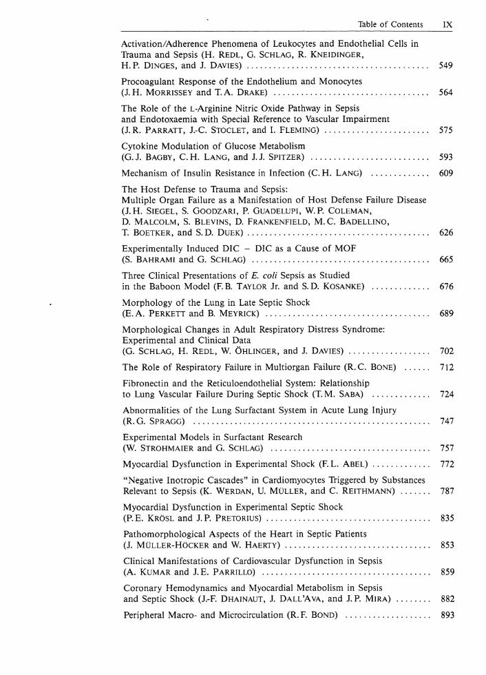
Table of Contents IX Activation/Adherence Phenomena of Leukocytes and Endothelial Cells in Trauma and Sepsis (H. R E D L , G. SCHLAG, R. K N E I D I N G E R , H.P . D I N G E S , and J. DAVIES) 5 4 9 Procoagulant Response of the Endothelium and Monocytes (J .H. M O R R I S S E Y and T.A. DRAKE) 5 6 4 The Role of the L-Arginine Nitric Oxide Pathway in Sepsis and Endotoxaemia with Special Reference to Vascular Impairment (J. R. P A R R A T T , J.-C. STOCLET, and I. FLEMING) 5 7 5 Cytokine Modulation of Glucose Metabolism (G.J. BAGBY, C .H. L A N G , and J.J. SPITZER) 5 9 3 Mechanism of Insulin Resistance in Infection (C. H. L A N G ) 6 0 9 The Host Defense to Trauma and Sepsis: Multiple Organ Failure as a Manifestation of Host Defense Failure Disease (J .H. S I E G E L , S. G O O D Z A R I , P. GUADELUPI , W . P . C O L E M A N , D. M A L C O L M , S. BLEVINS, D. FRANKENFIELD, M.C . B A D E L L I N O , T. B O E T K E R , and S.D. DUEK) 6 2 6 Experimentally Induced DIC - DIC as a Cause of M O F (S. B A H R A M I and G. SCHLAG) 6 6 5 Three Clinical Presentations of E. coli Sepsis as Studied in the Baboon Model (F. B. TAYLOR Jr. and S. D. KOSANKE) 6 7 6 Morphology of the Lung in Late Septic Shock (E. A. P E R K E T T and B. MEYRICK) 6 8 9 Morphological Changes in Adult Respiratory Distress Syndrome: Experimental and Clinical Data (G. S C H L A G , H. R E D L , W . ÖHLINGER, and J. DAVIES) 7 0 2 The Role of Respiratory Failure in Multiorgan Failure (R.C. BONE) 7 1 2 Fibronectin and the Reticuloendothelial System: Relationship to Lung Vascular Failure During Septic Shock (T. M. SABA) 7 2 4 Abnormalities of the Lung Surfactant System in Acute Lung Injury (R.G. SPRAGG) 7 4 7 Experimental Models in Surfactant Research ( W . S T R O H M A I E R and G. SCHLAG) 7 5 7 Myocardial Dysfunction in Experimental Shock (F. L. A B E L ) 7 7 2 "Negative Inotropic Cascades" in Cardiomyocytes Triggered by Substances Relevant to Sepsis ( K . W E R D A N , U . MÜLLER, and C. R E I T H M A N N ) 7 8 7 Myocardial Dysfunction in Experimental Septic Shock (P.E. KRÖSL and J.P. PRETORIUS) 8 3 5 Pathomorphological Aspects of the Heart in Septic Patients (J. MÜLLER-HÖCKER and W . HAERTY) 8 5 3 Clinical Manifestations of Cardiovascular Dysfunction in Sepsis (A. K U M A R and J. E. PARRILLO) 8 5 9 Coronary Hemodynamics and Myocardial Metabolism in Sepsis and Septic Shock (J.-F. DHAINAUT, J. D A L L ' A V A , and J. P. M I R A ) 8 8 2 Peripheral Macro- and Microcirculation (R. F. B O N D ) 8 9 3
X Table of Contents Relationship Between Oxygen Demand and Oxygen Supply in Severe Sepsis (J .-L. V I N C E N T , J . BAKKER, and P.-G. S ILANCE) 9 0 8 Hepatic Responses to Bacterial Endotoxin (LPS) (A. P. BAUTISTA,
I .V . D E A C I U C , H. J A E S C H K E , Z . SPOLARICS, and J . J . SPITZER) 9 1 5 Experimental Liver Failure (R. R. BECK) 9 3 5 Hepatic Dysfunction in Shock and Organ Failure (P.E. B A N K E Y and F.B. C E R R A ) 9 4 8 Sepsis Related Renal Morphological Alterations and the Functional Correlates (H.A. LINARES) 9 6 1 The Kidney in Sepsis (A. S. T O N N E S E N ) 9 7 3 Neurologic Abnormalities in Sepsis (M. J. BRESLOW and R. J . TRAYSTMAN) 9 9 6 The Multiple Organ or System Failure Syndrome (A.E. BAUE) 1 0 0 4 Models of Endotoxemia in Rodents (S. B A H R A M I , H. R E D L , and G. SCHLAG) 1 0 1 9 Models of Endotoxemia in Sheep (D.L. T R A B E R , L.D. T R A B E R , H. R E D L , and G. SCHLAG) 1 0 3 1 Rodent Models of Endotoxemia and Sepsis (LH. CHAUDRY, A. A Y A L A , G. S I N G H , P. W A N G , and J .G . HAUPTMAN) . 1 0 4 8 Sheep and Pigs as Animal Models of Bacteremia (D.J. D E H R I N G ) 1 0 6 0 Live Escherichia coli Sepsis Models in Baboons (G. SCHLAG, H. R E D L , J . DAVIES, C . J . J , VAN V U U R E N , and P. SMUTS) . . 1 0 7 6 Animal Models of Endotoxemia and Sepsis ( J . A. SPITZER) 1 1 0 8 Models of Sepsis: Subacute Peritonitis in Sheep and Rats ( C M . M A R T I N , A. N E A L , and W . J . SIBBALD) 1 1 1 9 Chronic Models of Endotoxemia and Sepsis: Lessons from Both a Canine Peritonitis and a Human Endotoxemia Model (A.C. D I X O N and J . E . P A R R I L L O ) 1 1 3 2 Subject Index 1 1 4 3
Proteinases M . J O C H U M 1 , W . M A C H L E I D T 2 , H . N E U H O F 3 , and H F R I T Z 1
1 Department of Clinical Chemistry and Clinical Biochemistry in the Department of Surgery, Klinikum Innenstadt, University of Munich, Nußbaumstraße 20, 8000 Munich 2, FRG 2 Institute of Physiological Chemistry, Physical Biochemistry and Cell Biology of the University of Munich, Geothestraße 33, 8000 Munich 2, FRG 3 Department of Internal Medicine, Division of Clinical Pathophysiology and Experimental Medicine, University of Giessen, Klinikstrasse 36, 6300 Giessen, FRG
C O N T E N T S
Introduction 46 PMN Elastase and Cathepsins B and L as Potent Effectors of Unspecific Proteolysis of Vital Proteins 48
PMN Elastase 48 Cathepsins B and L 48
PMN Elastase and Macrophage-Derived Cathepsin B Release in the Posttraumatic Course 49
Methods 49 Plasma Levels of PMN Elastase and Cathepsin B as Early Indicators of Forthcoming Organ Failure 50 Plasma Levels of Phagocyte Proteinases and Posttraumatic Septic Complications 52
Phagocyte Proteinases in Bronchoalveolar Lavage Fluids as Indicators of Lung Tissue Damage 53 PMN Elastase-Induced Fibrinogen Split Product as an Indicator of Unspecific Proteolysis in Vivo 55 Conclusion 55
Aspects of Future Therapeutic Approaches in Humans 56
Use of Elastase Inhibitors in the Isolated Rabbit Lung Model 56
Conclusion 58 References 58
Introduction Of the multitude of inflammatory mediators and reactions investigated hitherto and claimed as being at least contributory to the development of an inflammatory process, proteolysis-induced patho-mechanisms seem to play a major role in the primary response and ultimate outcome of the organism to inflammatory stimuli such as tissue destruction due to multiple trauma and major surgery or invasive microbes and endotoxins in sepsis. Independent of the etiology of the insult, the inflammation response is primarily directed towards inactivating and eliminating the deleterious agents and to initiate the process of repair and healing. Yet the activation of the complex interacting cellular and humoral defense mechanisms necessary for this purpose carries with it the risk of damaging healthy tissue as well, thus perpetuating the inflammatory process. In this respect, the lysosomal serine proteinase elastase and the cysteine proteinases cathepsin B and L of the primary inflammatory cells, polymor
phonuclear (PMN) granulocytes and monocytes/ macrophages, respectively, are supposed to be potent effectors of proteolytic tissue damage if they are discharged extracellularly in high amounts during activation and disintegration of the phagocytes.
Moreover, the contact between proenzymes of the humoral cascade systems and damaged vascular endothelium together with activators (e.g., tissue factor, plasminogen activator) liberated from those cells results in the formation of system-specific proteases, whose activity is largely responsible for hemostasis and closure of the wound. Yet some of these proteolytic enzymes (plasma kallikrein, thrombin, plasmin, complement esterases) also produce additional mediators of inflammation such as the vasoactive kinins, the hemostatic and edema-forming fibrin monomers and fibrin peptides, or the anaphylactic complement factors (C3a, C4a, and C5a). System-specific proteases themselves, together with a series of polypeptides formed as a result of their proteolytic activity, act as potent chemotaxins and bring about the seques-
Proteinases 47 tration and activation of inflammatory cells (particularly PMN granulocytes) in the wound area.
In addition to lysosomal proteinases cytokines, arachidonic acid metabolites and highly reactive oxygen species are also extracellularly liberated during cell stimulation. By this means, vital structural elements (basal membranes, cell receptors, fibronectin, elastin, collagens, proteoglycans, etc.) as well as humoral factors, including a wide variety of plasma proteins in close vicinity to the phagocytizing cells, may be impaired unless the lysosomal proteolytic enzymes and oxidants are inactivated by their physiological regulators, the proteinase inhibitors (e.g., a,-proteinase inhibitor, a! -antichymotrypsin, a2-macroglobulin, cysteine proteinase inhibitors) and antioxidants (superoxide dismutase, catalase, glutathione redox system, ceruloplasmin).
However, due to an overstressed phagocytic activity of P M N granulocytes and monocytes/macrophages during severe inflammation, the main antagonist of PMN elastase, the a {-proteinase inhibitor (a!PI) , is highly susceptible not only to proteolytic cleavage by cysteine proteinases (see below) and by metalloproteinases released from PMN granulocytes (Knäuper et al. 1990), fibroblasts or mononuclear cells (Winyard et al. 1991), but also to oxidative denaturation of the reactive inhibitory site in the molecule (Travis and Salvesen 1983). This enables an unrestricted local digestive activity by PMN elastase combined with fatal consequences for the hemostasis system. As shown by several authors (reviewed by Jochum 1988; Machovich and Owen 1990), PMN elastase can easily degrade and
inactivate the principal inhibitors (antithrombin III, a2-p\&smm inhibitor, plasminogen activator inhibitor 1, Cl-inactivator) of proteinases of the blood cascade systems (clotting, fibrinolysis, complement), thus allowing the maintenance of life-threatening consumption of hemostasis factors and the additional production of potent stimulators of the phagocytes as mentioned above. Moreover, the proteolytic inactivation of the blood cascade inhibitors may be greatly enhanced by their oxidative denaturation in the surroundings of phagocytizing cells (Stief and Heimburger 1988).
Just recently, the proteolytic degradation of the main human cysteine proteinase antagonists, cystatin C and kininogen, by PMN elastase has also been demonstrated (Abrahamson et al. 1991; Kleniewski and Donaldson 1988). This may further aggravate the destructive potency of monocyte/ macrophage-derived cysteine proteinases which are extracellularly discharged due to serious incidents such as multiple trauma and sepsis.
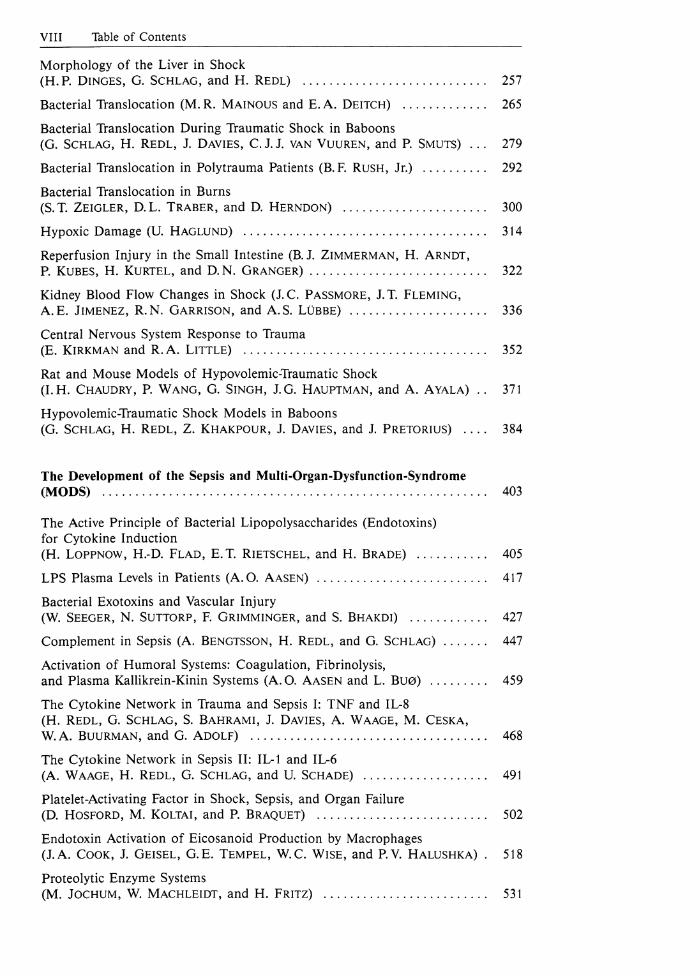
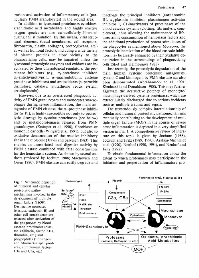
The tremendously complex interrelationship of cellular and humoral proteolytic pathomechanisms eventually contributing to the development of multiple organ failure (MOF) in the course of severe acute inflammation is depicted in a very simplified version in Fig. 1. A comprehensive review of literature on this topic is given by Jochum (1988), Jochum and Fritz (1989, 1990), Assfalg-Machleidt et al. (1990), Neuhof (1990, 1991), and Neuhof and Fritz (1992).
To obtain fundamental information about the extent to which proteinases may participate in the initiation and perpetuation of inflammatory pro-
Fig. 1. Schematic depiction of humoral and cellular proteolytic pathomechanisms involved in the development of multiple organ failure (MOF). Destructive proteases (elastase, cathepsin B) and other cell constituents are released after activation of the phagocytes by blood cascade proteinases (plasma kallikrein, factor Xlla, thrombin, etc.) and polypeptides (fibrinogen and fibronectin split products, complement factors C3a and C5a, etc.)
Plasminogen _ Plasmin Fibronectin (FN), Fibrinogen ( F )
48 M. Jochum et al. cesses, the following indications - as a modification of the Koch-Dale criteria - have to be proven in experimental and clinical studies: 1. The inflammatory potency (e.g., destruction of
vital proteins) of the respective phagocyte proteinases (PMN elastase, cathepsin B) has first to be shown in in vitro experiments.
2. The release of the lysosomal proteinases and the activation of proteolytic blood cascade enzymes has to be verified in correlation to the severity of the inflammation.
3. The consumption of proteinase inhibitors and other plasma factors susceptible to proteolytic degradation should coincide with the occurrence of proteolytic activity.
4. Specific split products of functional proteins generated by the proteolytic action of lysosomal proteinases have to be shown in correlation to the extracellular release of these enzymes.
5. The use of specific exogenous proteinase inhibitors as therapeutic tools should prevent or at least reduce to some extent severe signs of inflammation.
In the remainder of this chapter, characteristic results relating to these demands, obtained from experimental and clinical studies (the latter dealing primarily with multiple trauma), are given in more detail. Using the same specific test systems, our findings and those of other authors, which will be also briefly mentioned, demonstrate clearly the involvement of proteolytic pathomechanisms in acute inflammation such as trauma-induced organ failure. Therefore, proteinase inhibition may prove to be an indispensable therapeutic approach in the future to reduce the incident of organ failure and mortality in multiply injured patients.
PMN Elastase and Cathepsins B and L as Potent Effectors of Unspecific Proteolysis of Vital Proteins PMN Elastase
With regard to the pathological mechanisms involved in severe inflammation, the neutral protease, elastase, from the azurophilic granules of the PMN granulocytes, stands out as being significant among the lysosomal phagocyte enzymes currently recognized. Elastase is not only numerically predominant ( 3 - 5 ng/106 PMN cells) but also has practically no substrate specificity in the neutral pH range. The latter was shown by the unspecific de
gradation of a great variety of humoral and structural proteins such as clotting, fibrinolysis, and complement factors, immunoglobulins, transport proteins, and proteinase inhibitors as well as basal membrane proteins, cell receptors, fibronectin, elastin, collagens, proteoglycans, etc. Up to 1990 more than 90 papers were published describing the in vitro proteolysis of at least 45 proteins by isolated PMN release (reviews of literature given by Havemann and Gramse 1984, Jochum 1988, and Machovich and Owen 1990), and there is still a growing number of new reports on this topic. In view of the deleterious consequences due to the in-activation of the main clotting, fibrinolysis, and cysteine proteinase inhibitors, as mentioned in the introduction, the recently published abrogation of the thrombin inhibiting activity of heparin cofactor II (Pratt et al. 1990) and of the clotting factor Va and Vi l la inactivating capacity of protein S (Oates and Salem 1991) and activated protein C (Eckle et al. 1991 a) by PMN elastase is also of great interest. Moreover, with respect to trauma-induced primary lung dysfunctions, elastase-mediated endothelial cell injury (Smedly et al. 1986; Inauen et al. 1990) and proteolytic destruction of lung surfactant-associated proteins by neutrophil elastase (Pison et al. 1989) are especially worth mention. Finally, the in-activation of the neutrophil stimulating tumor necrosis factor and its receptors that can be provoked by elastase (Scuderi et al. 1991; van Kessel et al. 1991; Nortier et al. 1991; Proteu et al. 1991) opens interesting new questions concerning the auto-regulatory mechanisms of an inflammatory process. Additional modulations of the inflammatory reaction may be also brought about by new activities of the generated split products of vital proteins. Thus, elastase-produced IgG fragments inhibit Chemotaxis and oxidative burst (Eckle et al. 1991b), whereas proteolysed elastin (Senior et al. 1980) and a{?\ (Banda et al. 1988a) as well as elastase-o^PI complexes (Banda et al. 1988b) turned out to be potent chemoattractants. In addition, the inhibition of human neutrophil superoxide generation by proteolysed and complexed a r antichymotrypsin has been demonstrated just recently (Kilpatrick et al. 1991).
Cathepsins B and L
Compared to PMN elastase, the amount of the lysosomal cysteine proteinases, cathepsins B and L, in phagocytic cells is much lower (Kominami et al. 1985; Bando et al. 1986). PMN granulocytes contain only minor amounts of cysteine proteinases
Proteinases 49 (cathepsin B around 1 ng/106 cells), whereas this class of proteinases predominates in cells of the monocyte/macrophage system (cathepsin B 0.2 |ng/106, cathepsin L 0.03 ng/106 of rat resident peritoneal macrophages; no data are available for human cells so far).
Many cytosolic and extracellular proteins have been shown to be degraded by cathepsins B and L in vitro (Barrett and Kirschke 1981; Barrett et al. 1988) , including a {-proteinase inhibitor (Johnson et al. 1986), immunoglobulin G (Billing et al. 1991), elastin (Mason et al. 1986), various types of collagens (Maciewicz et al. 1990), and basement membranes (Baricos et al. 1988). In general, cathepsin B seems to be markedly less active on natural protein substrates than cathepsin L. Very recently this difference has been explained by the existence of an "occluding loop" in the cathepsin B structure, hindering the access of protein substrates to the active site cleft of the enzyme and favoring a dipeptidylcarboxypeptidase specificity (Musil et al. 1991).
The elastinolytic cysteine proteinase activity of alveolar macrophages (Chapman and Stone 1984) seems to be mainly due to cathepsin L (Reilly et al. 1989) , which is 100-fold more active on elastin than cathepsin B (Mason et al. 1986). (^-Proteinase inhibitor is effectively cleaved and inactivated by catalytical amounts of cathepsin L (Johnson et al. 1986). On the other hand, cystatin C, an inhibitor of cysteine proteinases, is rapidly degraded by PMN elastase (as already mentioned above) and converted into a fragment with drastically reduced inhibitory function (Abrahamson et al. 1991). This would explain the strong positive correlation between cathepsin B and neutrophil elastase activity and the negative correlation between elastase activity and cystatin C content observed in purulent sputum (Buttle et al. 1990).
Against synthetic substrates, cathepsin B is optimally active around pH 6.0, but many proteins are degraded more effectively at a lower pH. On the other hand, significant activity of cathepsin B can be observed even at pH 7.0-8 .0 , with a half-life of about 30 min at pH 7.4 (Machleidt et al. 1986). The enzyme seems to be stabilized by the presence of large protein substrates (Sloane 1990). Cathepsin L is rapidly inactivated at neutral pH but its half-life at pH 7.4 is still about 8 min (Machleidt et al. 1986). An acidic pH milieu favorable for cysteine proteinase activity can be expected in the microen-vironment of adherent macrophages (Silva et al. 1988). Moreover, a form of cathepsin B has been found in sputum that, unlike the enzyme isolated
from liver or spleen, is unusually stable at neutral and mildly alkaline pH (Buttle et al. 1988). Although the molecular basis for these physicochemi-cal differences has not been fully elucidated, the altered pH stability may be pathobiochemically relevant. For full activity of cysteine proteinases a reductive milieu is required which is probably provided by cysteine within the lysosome (Lloyd 1986). We have experimental evidence, however, that a significant portion of the cathepsin B activity of blood plasma and peritoneal exudate is preserved in the absence of thiols.
Cathepsin B and cathepsin L are completely different in their affinity for their endogenous protein inhibitors (cystatins, stefins, kininogens). Cathepsin L is quasi-irreversibly bound to most inhibitors (dissociation constants, K j<5pM) whereas cathepsin B forms only loose complexes with cystatin C (Ki = 0.8nM) and LMW-kininogen (K{
= 390 nM). We have shown that cathepsin B activity in blood plasma and peritonitis exudate results from active enzyme dissociating from its inactive inhibitor complexes upon dilution in the assay (Assfalg-Machleidt et al. 1988, 1990). Liberation of active cathepsin B by dissociation may occur in vivo wherever the local concentration of complexes is diminished. In contrast, due to its pH instability and its tight binding to inhibitors, cathepsin L activity should be extremely shortlived when released from the lysosome and should not be detectable in the circulation or in inflammatory secretions. The high proteolytic potential of cathepsin L (see above) could become destructive, however, when the enzyme is discharged in an acid microenviron-ment under conditions of a local proteinase-in-hibitor imbalance.
PMN Elastase and Macrophage-Derived Cathepsin B Release in the Posttraumatic Course Methods
Serially drawn citrated plasma and broncho-alveolar lavage fluid (BALF) samples were used to quantify proteinases, inhibitors, and other plasma proteins as well as their split products.
Measurements of the lysosomal serine proteinase PMN elastase were carried out with a commercially available, highly specific two-site sandwich enzyme immunoassay kit (E. Merck, Darmstadt, Germany), which detects elastase only as an
50 M. Jochum et al. inactive complex with a{Pl (detailed description by Neumann and Jochum 1984). The normal range of circulating complexed elastase in healthy people due to the physiological turnover of PMN cells amounts to 6 0 - 1 2 0 n g / m l without proteolytically active enzyme being detectable. Since, in contrast to plasma, BALF samples may contain active elastase in addition to the complex, parts of these specimens were also incubated with a surplus of a!PI in vitro and reassayed for an increase in elastase-o^PI complex as a measure of enzymatic elastase activity in vivo.
The cysteine proteinase cathepsin B activity (upper normal plasma level: 50 mU/1) was quantified using a specific fluorometric peptide substrate in combination with the cysteine proteinase inhibitor E-64 as described by Assfalg-Machleidt et al. (1988, 1990). Caseinolytic activity of the local body fluids due to active elastase and/or cathepsin B was detected using resorufin-labeled casein (Boehr-inger, Mannheim, Germany) as a substrate. The method is outlined in detail by the manufacturer (Boehringer).
The coagulation proteinases plasma kallikrein and thrombin were estimated by their cleavage activity on the chromogenic peptide substrates S-2302 (Kabi, Stockholm, Sweden) and Chromozym TH (Boehringer, Mannheim, Germany), respectively, after the turnover of the proenzymes to the active proteinases.
The inhibitory activities of antithrombin III and a } PI were measured with commercially available test systems (Coatest Antithrombin, Kabi, Stockholm, Sweden; a {-Antitrypsin Farbtest, Boehringer, Mannheim, Germany) applying chromogenic peptide substrates for thrombin (S-2238) and trypsin (BAPA), respectively. The antigen levels of the inhibitors were quantified with commercial radial immunodiffusion plates (NOR-Partigen plates, Behringwerke, Marburg, Germany). Opsonic activity, antigen levels, and split products of the opsonins IgG and complement factor C3 were determined as outlined by Billing et al. (1988, 1990, 1991) and Machleidt et al. (1992).
An elastase-specific split product of the Aa-chain of fibrinogen, called fibrino-elastase-peptide (FEP; equivalent to the Aa 1-21 peptide described by Weitz et al. 1986), was quantified with a two-step competitive immunoassay just recently developed in our laboratory by Gippner-Steppert (1991).
Clinical methods, criteria for grading the severity of the disesaes, and treatment of patients are extensively described in the original publications cited in the following sections.
Plasma Levels of PMN Elastase and Cathepsin B as Early Indicators of Forthcoming Organ Failure
In an early preliminary clinical trial involving 27 polytraumatized patients, we were able to show that severe injuries of at least two body regions (thorax, abdomen, skeletal system) induced a prompt and more or less long-lasting release of PMN elastase into the circulation (Dittmer et al. 1986). Measurements were carried out every 4 h up to the 28th h and thereafter every 6 h up to the 52nd h after trauma. The maximum increase between 5-and 30-fold of normal of PMN elastase plasma levels with 8 - 1 2 h after the traumatic event correlated well with the clinical injury severity score. However, due to logistic limitations of the study, it was not possible to estimate a predictive value of the extracellularly discharged PMN elastase for the development and outcome of trauma-induced (multiple) organ failure.
In a second approach involving 24 multiply injured patients, we were able to demonstrate that the primary activation of PMN granulocytes immediately after the polytraumatic event is followed by repetitive increases of complexed elastase in plasma in those patients who developed ARDS and additional organ failure (Jochum et al. 1989). This multiple organ insufficiency in our patients was mainly due to septic complications. In agreement with findings of Nuytinck et al. (1986) and Redl et al. (1987), elevated plasma levels of elastase correlated well with the severity of injuries and the occurrence of multiple organ failure. Moreover, they discriminated to a reasonable degree at an early stage in the clinical course between later survival or mortality.
To clarify further the potential role elastase -and in addition cathepsin B - may play in progressive posttraumatic complications, we just recently accomplished a more extended exploration. One hundred severely injured patients fulfilling previously defined entry criteria were enrolled in a prospective study (directed by Drs. Nast-Kolb and Waydhas, Department of Surgery, Klinikum Innenstadt, University of Munich) on inflammatory mediators and multiorgan failure associated with polytraumatic events (Nast-Kolb et al. 1992; Waydhas et al. 1992). Collection of blood samples and recording of clinical data were started within 30 min after arrival of the patients in the emergency department (about 1 h after the accident) and continued on a 6-h basis up to 48 h. Subsequently, the monitoring interval was extended to 24 h for a period of 14 days. Thereafter the clinical course was re-
Proteinases 51
^ <—>
.2 o tx ^ \ i 1 I u A « rr ** 11
c o
U o
_i_ _1_ 7 8
days 10 11 12 13 14
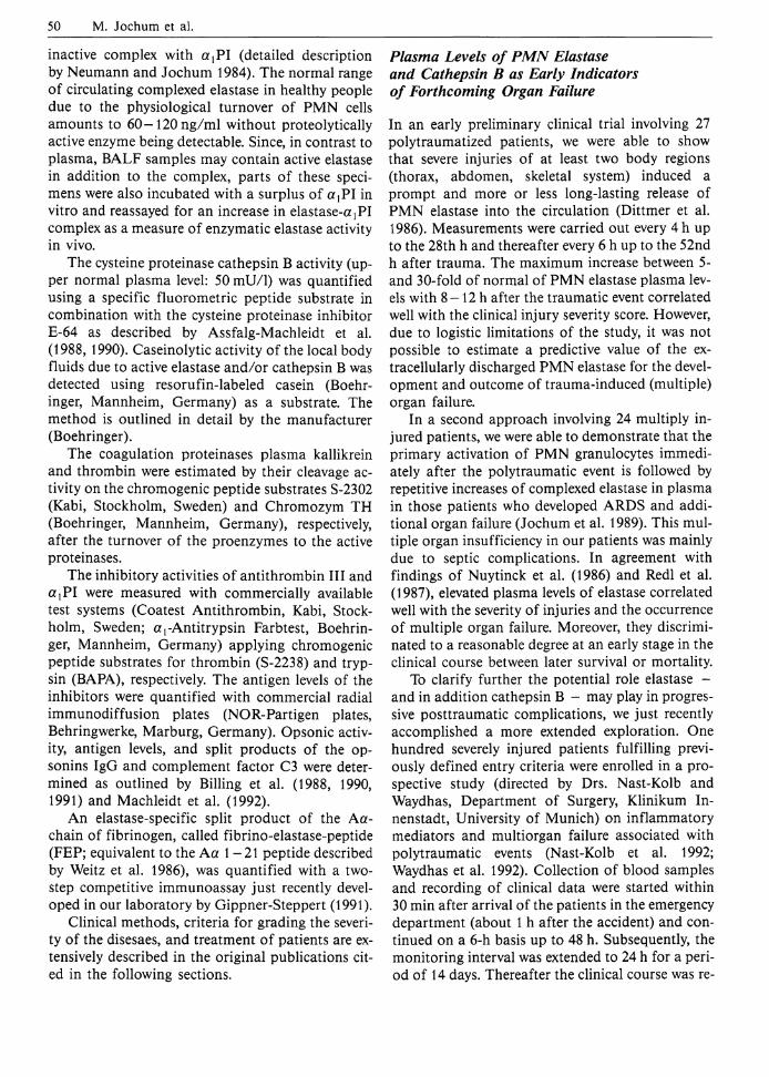
Fig. 2 a, b. Mean values (±SEM) of a PMN elastase (in complex with a,-proteinase inhibitor) and b cathepsin B in plasma of multiply injured patients assigned to three outcome groups: *—*, group I (n = 16): nonsurvivors with multiple organ failure; 0 - - - 0 group II (n = 47): survivors with reversible organ failure; +—+ group III (n = 37): survivors without organ failure. */?<0.05, **/?<0.01 group I vs. group II; # p<0.05 , ••p<0M group I vs. group III; A p<0.05 , A A p<0 .01 group II vs. group III
corded until either transfer to a general ward or death of the patient.
Retrospectively, the patients could be assigned to three different groups: 16 out of them died due to multiple organ failure 3 - 2 8 days (mean survival time: 16 days) after the traumatic incident (group I), 47 patients survived the development of organ failure (group II), and 37 patients overcame the accident without evident signs of organ dysfunctions (group III).
The extracellular release of PMN elastase and monocyte/macrophage-derived cathepsin B into plasma of the patients in the three outcome groups is depicted in Fig. 2. PMN elastase was in all groups elevated significantly above normal (upper range
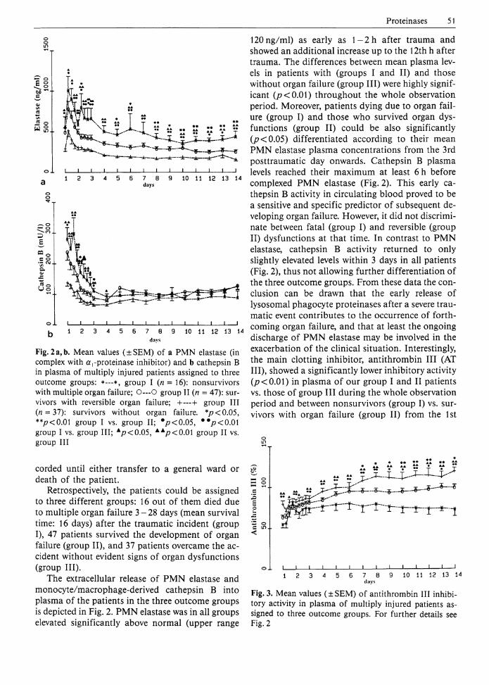
120ng/ml) as early as l - 2 h after trauma and showed an additional increase up to the 12th h after trauma. The differences between mean plasma levels in patients with (groups I and II) and those without organ failure (group III) were highly significant (/?<0.01) throughout the whole observation period. Moreover, patients dying due to organ failure (group I) and those who survived organ dysfunctions (group II) could be also significantly 0?<0.05) differentiated according to their mean PMN elastase plasma concentrations from the 3rd posttraumatic day onwards. Cathepsin B plasma levels reached their maximum at least 6 h before complexed P M N elastase (Fig. 2). This early cathepsin B activity in circulating blood proved to be a sensitive and specific predictor of subsequent developing organ failure. However, it did not discriminate between fatal (group I) and reversible (group II) dysfunctions at that time. In contrast to PMN elastase, cathepsin B activity returned to only slightly elevated levels within 3 days in all patients (Fig. 2), thus not allowing further differentiation of the three outcome groups. From these data the conclusion can be drawn that the early release of lysosomal phagocyte proteinases after a severe traumatic event contributes to the occurrence of forthcoming organ failure, and that at least the ongoing discharge of P M N elastase may be involved in the exacerbation of the clinical situation. Interestingly, the main clotting inhibitor, antithrombin III (AT III) , showed a significantly lower inhibitory activity 0?<0.01) in plasma of our group I and II patients vs. those of group III during the whole observation period and between nonsurvivors (group I) vs. survivors with organ failure (group II) from the 1st
*- o c in <
_L_ 7 8 thiVS
10 11 12 13 14
Fig. 3. Mean values (±SEM) of antithrombin III inhibitory activity in plasma of multiply injured patients assigned to three outcome groups. For further details see Fig. 2
52 M. Jochum et al. Table 1. Prognostic values of plasma levels of PMN elastase, cathepsin B, and antithrombin III on hospital admission for prediction of organ failure
Accuracy Sensitivity Specificity PPV NPV W (%) W (Vo) W
Elastase (>200ng/ml) 69 83 44 73 58 Cathepsin B (>190mU/ l ) 63 50 89 90 48 Antithrombin III (<80%) 69 79 52 74 59 PPV, Positive predictive value; NPV, negative predictive value. The discrimination values (in brackets) were evaluated in a former study (Nast-Kolb et al. 1991) Table 2. Prognostic values of plasma levels of antithrombin III and PMN elastase on hospital admission and on the 3rd posttraumatic day for prediction of death
Accuracy Sensitivity Specificity PPV NPV (<7o) (%)
Antithrombin III (<60%) 68 80 66 32 94 Elastase (>500ng/ml) 86 56 91 56 91 PPV, Positive predictive value; NPV, negative predictive value. The discrimination values (in brackets) were evaluated in a former study (Nast-Kolb et al. 1991)
posttraumatic week onwards (Fig. 3). Similar results were obtained concerning the rapid turnover of the coagulation proenzymes prekallikrein and prothombin to kallikrein and thrombin, the latter being the essential target proteinase of AT III. Thus, plasma levels of AT III, prekallikrein and prothrombin clearly below 80% of normal in the early posttraumatic phase were strikingly associated with the later appearance of severe organ dysfunctions, indicating that in addition to the release of lysosomal proteinases an overwhelming activation of the humoral proteolytic cascade systems is also conductive to the perpetuation of the posttraumatic inflammatory process. In Table 1, prognostic calculations (accuracy, sensitivity, specificity, positive and negative predictive value) are given for reliable discrimination levels of PMN elastase, cathepsin B, and antithrombin III to indicate forthcoming organ failure or uneventful recovery already on the patient's admission. The highly predictive value of early, low AT III inhibitory activities for a forthcoming exacerbation of posttraumatic organ dysfunctions eventually leading to ARDS and multiple organ failure was also emphasized by Schramm and Spannagl (1991), reporting on 57 prospectively studied trauma patients. Furthermore, in our extended study an entry level of AT III activity below 60% of normal was highly predictive for a later fatal outcome. The same held true for PMN elastase concentrations above 5-fold of normal from the 3rd posttraumatic day onwards.
The corresponding prognostic data are given in Table 2.
Plasma Levels of Phagocyte Proteinases and Posttraumatic Septic Complications
Since blunt trauma as well as infectious complications and sepsis initiate the release of phagocyte proteinases, we compared the plasma level patterns of PMN elastase and cathepsin B (besides a variety of other inflammatory mediators) in subgroups of trauma patients suffering from infection/sepsis and those who did not sustain these entities (Waydhas et al. 1992). In about 70% of patients with organ failure early dysfunctions (usually respiratory insufficiency) became obvious during the first 2 posttraumatic days. Late organ failure predominantly due to liver failure emerged after the 3rd day post trauma, whereas multiple organ failure was diagnosed in 32% of all group I and II patients between days 6 and 8. Infections came up around the 3rd day post trauma and remained fairly constant for about 1 week. Bacterial sepsis occurred slightly delayed. According to the three outcome groups infection/sepsis was verified for 81%/50% of the patients in group I, for 74%/36% in group II, and for 2 4 % / 5 % in group III, respectively. Summarizing the clinical data, it turned out that in 97% of patients with an early onset of organ failure infection started at least 2 days later than the organ insuffi-
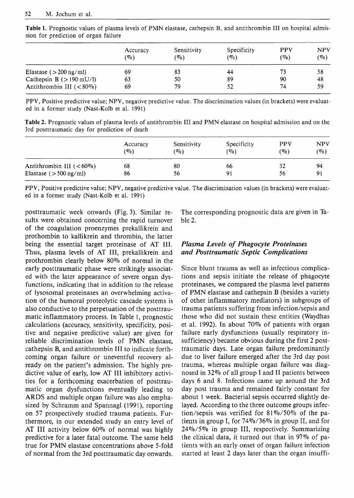
ciencies. In only half of those patients did infection or sepsis lead to subsequent deterioration of the clinical situation. In contrast, in 50% of the 18 patients with a late onset of organ failure, infection and sepsis preceded the disturbance of organ functions and seemed to have pathogenic significance. Interestingly, out of many other inflammation markers (Waydhas et al. 1992) only PMN elastase showed an obvious correlation with infection or sepsis. Since significantly higher plasma levels were measurable even before onset of these entities compared to a similar posttraumatic course without such complications (Fig. 4 shows measurements in group II patients as an example), the conclusion can be drawn that granulocytic proteinase may facilitate and maintain the occurrence of posttraumatic infections and sepsis by interfering with the immune system (e.g., by degradation of comple-
o o in
o l I I I I I I I I l I I I I I ! 1 2 3 4 5 6 7 8 9 10 11 12 13 14
days o o in
o 1 I I I I I I I I I I I i I I
k 1 2 3 4 5 6 7 8 9 10 11 12 13 14 days
Fig. 4 a, b. Mean values (±SEM) of PMN elastase (in complex with a,-proteinase inhibitor) in plasma of trauma patients with reversible organ failure (group II). a Patients with infection (*—*; n = 35) and without infection (O—O; n = 12). */?<0.05 infection vs. no infection, b Patients with sepsis (*—*; n = 17) and without sepsis (O—O; n = 30). *p<0.05; **<0.01 sepsis vs. no sepsis
Proteinases 53 ment factors and immunoglobulins). This assumption is also confirmed by recently published data on progressive organ failure in a prospectively studied group of well-defined multiply injured patients (Nerlich 1991). Although no information is given about the time of onset of the septic state, significantly higher PMN elastase plasma levels were presented for 18 septic in comparison to 17 nonseptic patients from the 24th h up to the 8th day post trauma.
Phagocyte Proteinases in Bronchoalveolar Lavage Fluids as Indicators of Lung Tissue Damage
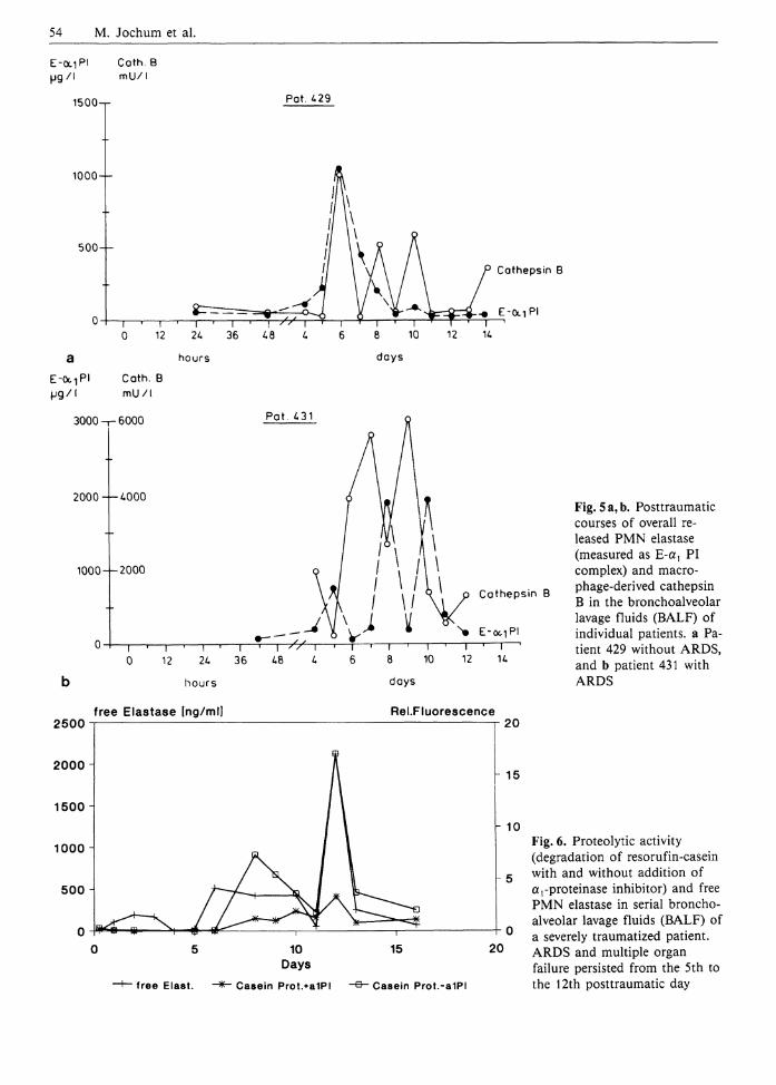
In another clinical study on multiply injured patients (a detailed study protocol is given by Sturm 1991) we were especially interested in local proteol-ysis-induced mechanisms which might be involved in the pathogenesis of the most severe lung dysfunction, the acute respiratory distress syndrome (ARDS). Daily-drawn BALF samples (method described by Obertacke et al. 1991) allowed us to confirm a significantly increased local discharge of the phagocyte proteinases elastase and cathepsin B in subjects at high risk of developing ARDS (Jochum 1991). As depicted in Fig. 5 (as an example), both proteinases showed a repeated though not congruent release in all patients, which indicates more or less permanent activation of the phagocytes throughout the 14-day posttraumatic observation period. Yet, a distinctly lower stimulation of both types of phagocytic cells became obvious in the non-ARDS subjects compared to patients with overt ARDS.
Although the a)PI antigen levels in all BALF specimens of the traumatized patients were far above the normal values of healthy volunteers, these amounts were apparently not sufficient in most cases to inhibit completely the PMN elastase released in the local epithelial milieu of the alveoli, despite an up to 40-fold molar surplus of the inhibitor over the enzyme. Therefore, the obviously deficient inhibitory capacity of a ,PI may have been caused by proteolytic (e.g., probably due to cathepsin B, L) and oxidative denaturation as well. Applying special assay systems, both ways of a {PI destruction were demonstrated by Schraufstatter et al. (1984) at least in some individual BALF samples of patients with manifest ARDS. Though other authors could not detect elastase activity against high-molecular-weight protein substrates in BALF samples of ARDS patients (Well et al. 1985;
54 M. Jochum et al. E-<x iP i C a t h . B u g / l m U / l
1 5 0 0 - r Pat . £ 2 9
1000 —
500
a E - O C T P I
H g / I 3 0 0 0 -
C a t h e p s i n B
E-Oc iP I
h o u r s
C a t h . B
m U / l
P a t . 431
2 0 0 0 — 4 0 0 0
1000
C a t h e p s i n B
b h o u r s
free Elastase [ng/ml]
d a y s
Rel.Fluorescence
Fig. 5 a, b. Posttraumatic courses of overall released PMN elastase (measured as E-a^ PI complex) and macro-phage-derived cathepsin B in the bronchoalveolar lavage fluids (BALF) of individual patients, a Patient 429 without ARDS, and b patient 431 with ARDS
2500
2000 -
1500
1000 -
500
20
- 15
10
free Elast.
10 Days
Casein Prot.*a1PI
20
• Casein Prot.-a1PI
Fig. 6. Proteolytic activity (degradation of resorufin-casein with and without addition of a pro te inase inhibitor) and free PMN elastase in serial bronchoalveolar lavage fluids (BALF) of a severely traumatized patient. ARDS and multiple organ failure persisted from the 5th to the 12th posttraumatic day
Proteinases 55 Wewers et al. 1988), we were recently able to show caseinolytic activity in a variety of BALF samples (kindly given to us by Dr. Obertacke, Department of Surgery, University of Essen) from trauma patients with severe lung dysfunction (Machleidt et al. 1993). In vitro incubation of these specimens with otj PI abolished this proteolytic activity almost completely (Fig. 6) and gave rise to an additional increase of the elastase-c^PI complex, indicating that the enzymatic activity still present in the alveolar fluid was mostly due to PMN elastase. This finding does, however, not exclude the proteolytic action of cathepsins in situ, since these enzymes have a much shorter half-life than elastase and may therefore be less active in the stored body fluids.
PMN Elastase-Induced Fibrinogen Split Product as an Indicator of Unspecific Proteolysis in Vivo
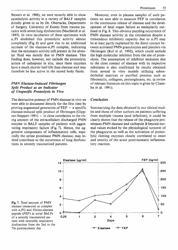
The destructive potency of PMN elastase in vivo we were able to document directly for the first time by proving augmented generation of FEP - a specific elastase-induced split product of fibrinogen (Gipp-ner-Steppert 1991) - in close correlation to the rising amount of the extracellulary discharged PMN enzyme in BALF samples of patients with aggravating respiratory failure (Fig. 7). Hence, the aggressive components of inflammatory cells, especially the serine proteinase PMN elastase, may indeed contribute to the occurrence of lung dysfunctions in severely traumatized patients.
Moreover, even in plasma samples of such patients we were able to measure FEP in correlation to the continuous release of elastase and the development of fatal organ failure as exemplarily outlined in Fig. 8. This obvious puzzling occurrence of PMN elastase activity in the circulation despite a tremendous inhibitory capacity due to a t P I may be at least partly explained by the direct contact between activated PMN granulocytes and platelets via fibrinogen (Ruf et al. 1992), which could exclude the high molecular inhibitor from the site of proteolysis. The assumption of inhibitor exclusion due to the close contact of elastase with its respective substrates is also confirmed by results obtained from several in vitro models utilizing subendothelial matrices or purified proteins such as fibronectin, collagens, proteoglycans, etc. (a review of relevant literature on this topic is given by Cham-ba et al. 1991).
Conclusion
Summarizing the data obtained in our clinical studies and those of other authors on patients suffering from multiple trauma (and infection), it could be clearly shown that the release of the phagocyte proteinases PMN elastase and cathepsin B beyond normal values evoked by the physiological turnover of the phagocytes as well as the activation of proteolytic clotting enzymes closely correlated to onset and severity of the acute posttraumatic inflammatory reaction.
Elastase lug/ml] F E P [ng/ml]
Fig. 7. Total amount of PMN elastase (measured as complex with a,PI) and fibrino-elastase peptide (FEP) in serial BALFs of a severely traumatized patient with reversible respiratory dysfunction from the 2nd to the 7th posttraumatic day

56 M. Jochum et al. Elastase [ng/ml] FEP [ng/mll
500
400
300
200
- 100
Elastase FEP
Fig. 8. Plasma levels of PMN elastase (in complex with a,-PI) and fibrino-elastase peptide (FEP) in a poly traumatized patient with fatal multiple organ failure. Resp. F, respiratory failure; DIC, disseminated intravascular coagulation; Ren. F.y renal failure; ARDS, acute respiratory distress syndrome; Hep. F, hepatic failure
A presumable inflammatory effector role proved especially true for PMN elastase, because the destructive potency of this proteinase in vitro could be convincingly demonstrated in vivo as well, at least on vital humoral proteins in relation to the propagation of organ dysfunctions in traumatized patients. In contrast, the role of released lysosomal cysteine proteinases, cathepsins B and L, as effectors of inflammation in vivo is not yet clear. Considering the relatively poor proteolytic potency of cathepsin B in vitro, it seems probable that simultaneously released cathepsin L, which is not detectable as active enzyme in the circulation, is responsible for destructive proteolysis at local sites of inflammation.
Moreover, the studies corroborated without doubt that, in patients who have incurred severe trauma and infection, the underlying cellular and humoral biochemical events are quite similar regardless of the etiology of the insult. Thus, the clinical outcome seems to be only due to the magnitude of the inflammatory response, which of course is highest if infection is superimposed on trauma.
Furthermore, it turned out that an excessive local (a{Pl) and systemic (AT III) consumption of proteinase inhibitors occurs as a main consequence of the release of lysosomal phagocyte proteinases and the activation of proteolytic blood cascade enzymes during severe inflammatory reactions. Since this entity seems to be the most critical cause which allows the proteolysis-induced (multiple) organ damage, supplementation of the body's inhibitor potential by exogenous proteinase inhibitors against elastase and thrombin - isolated from hu
man material or produced by gene technology as discussed by Fritz et al. (1992) - should be a highly promising therapeutic approach in traumatized patients.
Aspects of Future Therapeutic Approaches in Humans Use of Elastase Inhibitors in the Isolated Rabbit Lung Model
Although novel therapeutic approaches ultimately require validation in well-controlled clinical trials, it is virtually always necessary to obtain preliminary data in animals before employing new drugs (e.g., recombinant or synthetic proteinase inhibitors) in humans. Thus, progress in trauma-induced multiorgan failure research continues to depend upon studies using clinically relevant animal models which fulfil commonly accepted criteria of specific organ dysfunctions.
With respect to the interstitial edema formation associated with respiratory failure, the first organ dysfunction that becomes obvious after a severe traumatic event, the isolated perfused and ventilated rabbit lung turned out to be a reliable model to study new therapeutic approaches for the improvement of lung function (Neuhof 1990; Neuhof and Fritz 1992). In this model isolated rabbit lungs are perfused with Krebs-Henseleit starch buffer solution in a closed circuit and are ventilated by means of a Starling pump. The lungs are suspended on an electronic balance, allowing changes in lung weight indicating edema formation due to a suit-
Proteinases 57 able stimulus to be monitored continuously. The pressure in the pulmonary artery and the left atrium are recorded simultaneously to reveal changes in pulmonary vascular resistance.
To understand the role phagocyte proteinases may play in the pathogenesis of pulmonary edema, two different pathomechanisms have to be considered with regard to therapeutic consequences. The almost reversible formation of interstitial edema in the early posttraumatic stage seems to be caused mainly by functional alterations (e.g., changing of the microvascular tone, increase of capillary filtration pressure, loosening of interendothelial junctions) which are induced by bradykinin, histamine, or arachidonic acid metabolites. The irreversible vascular leakage in the later phases of the respiratory distress syndrome (ARDS), however, is due to structural injury of the vessels and alveoli, which may be primarily caused by lysosomal proteinases released from activated granulocytes and macrophages (detailed description by Neuhof 1990 and Neuhof and Fritz 1992). Since especially proteolytically active PMN elastase in the presence of inactivated a{Pl was found in the bronchoalveolar lavage fluids of patients suffering from later stages of ARDS, we for the first time used suitable elastase inhibitors to prevent otherwise irreversible vascular permeability due to elastase action in the rabbit lung model perfused with isolated human PMN granulocytes.
As long as these cells were not additionally stimulated in the lung circulation, they did not induce changes in vascular resistance or permeability during a control period of 4 - 6 h. However, upon stimulation with antigen-antibody complexes or heat-denatured immunoglobulins, which were added to the perfusion fluid, pulmonary artery pressure and lung weight increased. The rise in pulmonary vascular resistance was caused by thromboxane generated by the burst reaction of the granulocytes. Thus, the pressure increase could be blocked by a thromboxane receptor antagonist, whereas the edema formation continued. When PMN cells were stimulated in vitro and injected into the pulmonary circulation after finishing their burst reaction, there was no occurrence of either thromboxane generation or a pressure reaction. Yet edema formation still became obvious.
Using prestimulated granulocytes or PMN cells stimulated within the pulmonary circulation, the development of vascular leakage as reflected by an increase in lung weight was paralleled by a rising level of P M N elastase in the perfusion fluid. Electron microscopy of such lungs showed vacuolated granulocytes sticking to the endothelium of alveolar capil
laries. The endothelial cells were injured and partially loosened from the vessel wall below the contact area.
Similar results were obtained if purified PMN elastase was added to the perfusion fluid, reflecting the formation of interstitial edema. Again, pulmonary artery pressure remained unchanged, indicating that the edema was a true permeability edema and not caused by hemodynamic changes in filtration pressure.
The granulocyte-evoked edema formation could be largely suppressed by prophylactic infusion of the relatively specific recombinant elastase inhibitor eglin c (originally isolated from the leech Hirudo medicinalis as described by Seemüller et al. 1986), which is highly resistant to oxidative and proteolytic inactivation. Moreover, eglin c also prevented the increase in fluid passage through monolayers of cultured endothelial cells following their exposure to supernatants of activated granulocytes (Suttorp et al. 1989).
In contrast to the protective effect of eglin c, addition of isolated humans a!PI to the perfusate up to a 40-fold molar surplus over the released elastase amount failed to inhibit the pulmonary permeability increase if PMN cells were stimulated in situ. This indicates at least a significant oxidative inactivation of the inhibitor by the released reactive oxygen species.
In view of the clinical need of an oxidant-resis-tant PMN elastase inhibitor, we also tested a variant of the aprotinin molecule. Replacement of the lysine residue in the reactive center of the original plasmin/ trypsin inhibitor by valine using bioengineering techniques created a selective, nonoxidizable inhibitor against PMN elastase (Wenzel et al. 1986). Similar to the effect of eglin c, the vascular leakage evoked by in situ stimulation of granulocytes in the pulmonary circulation was remarkably reduced by this inhibitor, thus further confirming the assumption that, out of all granulocytic proteinases, elastase contributes essentially to the irreversible edema formation.
Yet despite the fact that these elastase inhibitors have shown a high efficacy in our experimental lung model, their clinical application has to be considered with caution, especially concerning a putative antigenicity of these nonhuman proteins in long-term studies. Thus, a therapeutic approach with an isolated, purified human ajPI preparation is still the only clinical choice at present. To overcome the inactivation mechanisms of this inhibitor in vivo, however, high amounts for substitution will be necessary.
58 M. Jochum et al.
Conclusion The disturbance of pulmonary vascular permeability due to multiple trauma and infection is caused by complex pathomechanisms in which a variety of mediators are involved. Of these, arachidonic acid metabolites, toxic oxygen species, and proteinases can be considered as important end products which directly affect the endothelial and epithelial barriers. Since especially lysosomal proteinases from granulocytes and probably also from macrophages seem to be mainly responsible for the pulmonary vascular leakage that occurs in the later stages of ARDS and multiple organ failure, future concepts for prophylaxis and therapy should include not only blockers and antagonists of arachidonic acid metabolites and antioxidants, but primarily proteinase inhibitors which are resistant to oxidative and proteolytic inactivation.
Concerning a putative antigenicity of nonhu-man proteins, inhibitors of human origin seem to be preferable. Such inhibitors, however, can be easily inactivated by proteolysis and oxidation, in addition to the fact that the natural sources for the isolation of proteinase inhibitors from human material are very limited. Thus, the design of highly effective inhibitory proteins on the basis of human inhibitor molecules by molecular modeling and their production by recombinant DNA technology is the most promising approach to obtain the quantities necessary for proteinase inhibition therapy in the future (Fritz et al. 1992). Acknowledgements. We are very grateful to the participating clinical colleagues mentioned in the text for their intensive collaboration. Part of the work was financially supported by the Sonderforschungsbereich 207 of the University of Munich (grants G5 to M.J. and Gl to W.M.).
References Abrahamson M, Mason RW, Hansson H, Buttle DJ,
Grubb A, Ohlsson K (1991) Human cystatin C. Role of the N-terminal segment in the inhibition of human cysteine proteinases and in its inactivation by leukocyte elastases. Biochem J 273:621-626
Assfalg-Machleidt I, Jochum M, Klaubert W, Inthorn D, Machleidt W (1988) Enzymatically active cathepsin B dissociating from its inhibitor complexes is elevated in blood plasma of patients with septic shock and some malignant tumors. Biol Chem Hoppe Seyler 369 [Suppl]:263-269
Assfalg-Machleidt I, Jochum M, Nast-Kolb D, Siebeck M, Billing A, Joka T, Rothe G, Valet G, Zauner R,
Scheuber HP, Machleidt W (1990) Cathepsin B - indicator for the release of lysosomal cysteine proteinases in severe trauma and inflammation. Biol Chem Hoppe Seyler 371 [Suppl]:211-222
Banda MJ, Rice AG, Griffin GL, Senior RM (1988 a) a,-Proteinase inhibitor is a neutrophil chemoattrac-tant after proteolytic inactivation by macrophage elastase. J Biol Chem 263:4481-4484
Banda MJ, Rice AG, Griffin GL, Senior RM (1988 b) The inhibitor complex of human alpha 1-proteinase inhibitor and human leukocyte elastase is a neutrophil chemoattractant. J Exp Med 167:1608-1617
Bando Y, Kominami E, Katunuma N (1986) Purification and tissue distribution of rat cathepsin L. J Biochem 100:35-42
Baricos WH, Zhou Y, Mason RW, Barrett AJ (1988) Human kidney cathepsins B and L. Characterization and potential role in degradation of glomerular basement membrane. Biochem J 252:301-304
Barrett AJ, Kirschke H (1981) Cathepsin B, cathepsin H, and cathepsin L. Methods Enzymol 80:535-561
Barrett AJ, Buttle DJ, Mason RW (1988) Lysosomal cysteine proteinases. In: ISI atlas of science biochemistry. Institute for Scientific Information, Philadelphia, pp 256-260
Billing A, Fröhlich D, Jochum M, Kortmann H (1988) Impaired phagocytosis in peritonitis exudate secondary to complement consumption. Surg Res Commun 3:335-345
Billing A, Fröhlich D, Jochum M, Kortmann H (1990) Deficient phagocytosis in abdominal sepsis: the influence of intraperitoneal substitution of opsonins -first results. Surg Res Commun 9:297-302
Billing A, Fröhlich D, Assfalg-Machleidt I, Machleidt W, Jochum M (1991) Proteolysis of defensive proteins in peritonitis exudate: pathobiochemic aspects and therapeutic approach. Biomed Biochim Acta 50:399-402
Buttle DJ, Bonner BC, Burnett D, Barrett AJ (1988) A catalytically active high-Mr form of human cathepsin B from sputum. Biochem J 254:693-699
Buttle DJ, Burnett D, Abrahamson M (1990) Levels of neutrophil elastase and cathepsin B activities, and cystatins in human sputum: relationship to inflammation. Scand J Clin Lab Invest 50:509-516
Chamba A, Afford SC, Stockley RA, Burnett D (1991) Extracellular proteolysis of fibronectin by neutrophils: characterization and the effects of recombinant cytokines. Am J Respir Cell Mol Biol 4:330-337
Chapman HA Jr, Stone OL (1984) Comparison of live human neutrophil and alveolar macrophage elasto-lytic activity in vitro. J Clin Invest 74:1693 — 1700
Dittmer H, Jochum M, Fritz H (1986) Freisetzung von granulozytärer Elastase und Plasmaproteinveränderungen nach traumatisch-hämorrhagischem Schock. Unfallchirurg 89:160-169
Eckle I, Seitz R, Egbring R, Kolb G, Havemann K (1991 a) Protein C degradation in vitro by neutrophil elastase. Biol Chem Hoppe Seyler 372:1007-1013
Eckle I, Kolb G, Havemann K (1991b) Inhibition of
Proteinases 59 neutrophil Chemotaxis by elastase-generated IgG fragments. Scand J Immunol 34:359-364
Fritz H, Collins J, Jochum M (1992) Proteinase inhibitor candidates for therapy of enzyme-inhibitor imbalances. In: Grassi C, Travis J, Casali L, Luisetti M (eds) Current concepts in the biochemistry of pulmonary emphysema. Springer, Berlin Heidelberg New York/ Bi and Gi, Verona Publishers, pp 101-112
Gippner-Steppert C (1991) Entwicklung eines spezifischen Testsystems für den Nachweis der Bildung eines proteloytischen Spaltproduktes des Fibrinogenes durch lysosomale PMN-Elastase sowie Untersuchungen am Miniplasminogen, einem Elastase-spe-zifischen Spaltprodukt des Plasminogens. Dissertation, Chem. Faculty of the Technical University of Munich
Havemann K, Gramse M (1984) Physiology and pathophysiology of neutral proteinases of human granulocytes. Adv Exp Med 167:1-20
Idell S, Kucich U, Fein A, Kueppers F, James HL, Walsch PN, Weinbaum G, Colman RW, Cohen AB (1985) Neutrophil elastase-releasing factors in bronchoal-veolar lavage from patients with adult respiratory distress syndrome. Am Rev Respir Dis 132:1098-1105
lnauen W, Granger DN, Meininger CJ, Sendling ME, Granger HJ, Kvietys PR (1990) Anoxia-reoxygena-tion-induced, neutrophil-mediated endothelial cell injury: role of elastase. Am J Physiol 259:H925-H931
Jochum M (1988) Lysosomale Faktoren aus polymorphkernigen Granulozyten: pathobiochemische, diagnostische und therapeutische Aspekte. Habilitationsschrift, Med. Faculty of the LM-University of Munich
Jochum M (1991) Specific proteins of inflammatory cells and a,-proteinase inhibitor in alveolar epithelial lining fluid of polytraumatized patients: do they indicate posttraumatic lung failure? In: Sturm JA (ed) Posttraumatic acute respiratory distress syndrome. Springer, Berlin Heidelberg New York, pp 193-211
Jochum M, Fritz H (1989) Pathobiochemical mechanisms in inflammation. In: Faist E, Ninnemann JL, Green DR (eds) Immune consequences of trauma shock, and sepsis. Springer, Berlin Heidelberg New York, pp 165-172
Jochum M, Fritz H (1990) Elastase and its inhibitors in intensive care medicine. Biomed Prog 3:55-59
Jochum M, Dwenger A, Joka T, Sturm JA (1989) Posttraumatic plasma levels of mediators of organ failure. Prog Clin Biol Res 308:673-681
Johnson DA, Barrett AJ, Mason RW (1986) Cathepsin L inactivates a, -proteinase inhibitor by cleavage in the reactive site region. J Biol Chem 261:14748-14751
Kilpatrick L, Johnson JL, Nickbarg EB, Wang ZM, Clifford TF, Banach M, Cooperman BS, Douglas SD, Rubin H (1991) Inhibition of human neutrophil superoxide generation by a,-antichymotrypsin. J Immunol 146:2388-2393
Kleniewski J, Donaldson V (1988) Granulocyte elastase cleaves human high molecular weight kininogen and destroys its clot-promoting activity. J Exp Med 167:1895-1907
Knäuper V, Reinke H, Tschesche H (1990) Inactivation of human plasma a,-proteinase inhibitor by human PMN leucocyte collagenase. FEBS Lett 263:355-357
Kominami E, Tsukahara T, Bando Y, Katunuma N (1985) Distribution of cathepsins B and H in rat tissues and peripheral blood cells. J Biochem 98:87-93
Lloyd JB (1986) Disulphide reduction in lysosomes. The role of cysteine. Biochem J 237:271-272
Machleidt W, Ritonja A, Popovic T, Kotnik M, Brzin J, Turk V, Assfalg-Machleidt I, Müller-Esterl W (1986) Human cathepsins B, H and L: characterization by amino acid sequences and some kinetics of inhibition by the kininogens. In: Turk V (ed) Cysteine proteinases and their inhibitors. De Gruyter, Berlin, pp 3 - 1 8
Machleidt W, Assfalg-Machleidt I, Billing A, Fröhlich D, Joka T, Nast-Kolb D (1993) The role of lysosomal cysteine proteinases as markers of macrophage activation and as non-specific mediators of inflammation. In: Faist E, Meakins J, Schildberg FW (eds) Host defense dysfunctions in trauma, shock, and sepsis. Springer, Berlin Heidelberg New York, pp 459-463
Machovich R, Owen WG (1990) The elastase-mediated pathway of fibrinolysis. Blood Coagul Fibrinolysis 1:79-90
Maciewicz RA, Wotton SF, Etherington DJ, Duance VC (1990) Susceptibility of the cartilage collagens types II, IX and XI to degradation by the cysteine proteinases, cathepsins B and L. FEBS Lett 269:189-193
Mason RW, Johnson DA, Barrett AJ, Chapman HA (1986) Elastinolytic activity of human cathepsin L. Biochem J 233:925-927
Musil D, Zucic D, Turk D, Engh RA, Mayr I, Huber R, Popovic T, Turk V, Towatari T, Katunuma N, Bode W (1991) The refined 2.15 Ä X-ray crystal structure of human liver cathepsin B: the structural basis for its specificity. EMBO J 10:2321-2330
Nast-Kolb D, Jochum M, Waydhas Ch, Schweiberer L (1991) Die klinische Wertigkeit biochemischer Faktoren beim Polytrauma. In: Rehn J, Schweiberer L, Tscherne H (eds) Hefte zur Unfallheilkunde. Springer, Berlin Heidelberg New York, pp 1-162
Nast-Kolb D, Waydhas Ch, Jochum M, Machleidt W, Duswald KH, Schweiberer L, Fritz H (1992) Biochemical mediators in monitoring multiple injured patients. Circ Shock (submitted)
Neriich ML (1991) Progressive organ failure. In: Sturm JA (ed) Posttraumatic acute respiratory distress syndrome. Springer, Berlin Heidelberg New York, pp 4 5 - 5 6
Neuhof H (1990) Role of proteinases in the pathophysiology of organ failure. In: Schlag G, Redl H, Siegel JH (eds) Shock, sepsis and organ failure. Springer, Berlin Heidelberg New York, pp 404-420
Neuhof H (1991) Actions and interactions of mediator systems and mediators in the pathogenesis of ARDS and multiorgan failure. Acta Anaesthesiol Scand 35 [Suppl 95]:7-14
Neuhof H, Fritz H (1992) Proteinases as mediators of the disturbance of pulmonary vascular permeability in sepsis, polytrauma, and ARDS. In: Rügheimer E (ed)

60 M. Jochum et al.: Proteinases New aspects on respiratory failure. Springer, Berlin Heidelberg New York, pp 6 7 - 7 6
Neumann S, Jochum M (1984) Elastase-öj-proteinase inhibitor complex. In: Bergmeyer HU, Bergmeyer J, Graßl M (eds) Methods of enzymatic analysis, 3rd edn, vol 5. Verlag Chemie, Weinheim, pp 184-195
Nortier J, Vandenabeele P, Noel E, Bosseloir Y, Goldman M, Deschodt-Lanckman (1991) Enzymatic degradation of tumor necrosis factor by activated human neutrophils: role of elastase. Life Sei 49:1879-1886
Nuytinck JKS, Goris JA, Redl H, Schlag G, van Munster PJJ (1986) Posttraumatic complications and inflammatory mediators. Arch Surg 121:886-890
Oates AM, Salem HH (1991) The binding and regulation of protein S by neutrophils. Blood Coagul Fibrinolysis 2:601-607
Obertacke U, Joka T, Reuter M, Schmit-Neuerburg KP (1991) Bronchoalveolar lavage.In: Sturm JA (ed) Adult respiratory distress syndrome. Springer, Berlin Heidelberg New York, pp 17-21
Pison U, Tarn EK, Caughey GH, Hawgood S (1989) Proteolytic inactivation of dog lung surfactant-associated proteins by neutrophil elastase. Biochem Biophys Acta 992:251-257
Pratt CW, Tobin RV, Chuch FC (1990) Interaction of heparin cofactor II with neutrophil elastase and cathepsin G. J Biol Chem 265:6092-6097
Proteu F, Brockhaus M, Wallach D, Engelmann H, Nathan CF (1991) Human neutrophil elastase releases a ligand-binding fragment from the 75-kDa tumor necrosis factor (TNF) receptor. J Biol Chem 266: 18846-18853
Redl H, Pacher R, Woloszczuk W (1987) Acute pulmonary failure. Comparison of neopterin and granulocyte elastase in septic and non-septic patients. In: Bair JA, Pfleiderer W, Wächter H (eds) Biochemical and clinical aspects of pteridines. De Gruyter, Berlin, pp 289-304
Reilly JJ Jr, Mason RW, Chen P, Joseph LJ, Sukhatme VP, Yee R, Chapman HA (1989) Synthesis and processing of cathepsin L, an elastase, by human alveolar macrophages. Biochem J 257:493-498
Ruf A, Schlenk RF, Maras A, Morgenstern E, Patscheke H (1993) Contact-induced neutrophil activation by platelets in human cell suspensions and whole blood. Blood (in press)
Schramm W, Spannagl M (1991) Differences in activation of coagulation and fibrinolysis after polytrauma with respect to the development of ARDS. In: Sturm JA (ed) Posttraumatic acute respiratory distress syndrome. Springer, Berlin Heidelberg New York, pp 7 5 - 8 7
Schraufstätter I, Revak SD, Cochrane CG (1984) Biochemical factors in pulmonary inflammatory disease. Fed Proc 43:2807-2810
Scuderi P, Nez PA, Duerr ML, Wong BJ, Valdez CM (1991) Cathepsin G and leukocyte elastase inactivate human tumor necrosis factor and lymphotoxin. Cell Immunol 135:299-313
Seemüller U, Dodt J, Fink E, Fritz H (1986) Proteinase inhibitors of the leech Hirudo medicinalis (hirudins, bdellins, eglins). In: Barrett AJ, Salvesen GS (eds) Proteinase inhibitors. Elsevier, Amsterdam, pp 337-359
Senior RM, Griffin GL, Mecham RP (1980) Chemotac-tic activity of elastin-derived peptides. J Clin Invest 66:859-862
Silva I A, Murrills RJ, Etherington DJ (1988) Microelec-trode studies on the acid microenvironment beneath adherent macrophages and osteoclasts. Exp Cell Res 175:266-276
Sloane BF (1990) Cathepsin B and cystatins: evidence for a role in cancer progression. Semin Cancer Biol 1:137-152
Smedly LA, Tonnesen MG, Sandhaus RA, Haslett C, Guthrie LA, Johnston RB Jr (1986) Neutrophil-medi-ated injury to endothelial cells. Enhancement by endotoxin and essential role of neutrophil elastase. J Clin Invest 77:1233-1243
Stief TW, Heimburger N (1988) Oxidative inactivation of purified human alpha-2-antiplasmin, antithrombin III, and Cl-inhibitor. Thromb Res 49:581-589
Sturm JA (ed) (1991) Adult respiratory distress syndrome. An aspect of multiple organ failure. Results of a prospective clinical study. Springer, Berlin Heidelberg New York
Suttorp N, Nolte A, Neuhof H (1989) Bedeutung der gra-nulozytären Elastase für die endotheliale Permeabilität. In: Suttorp N (ed) Zellbiologische Untersuchungen zur Pathogenese des akuten Atemnotsyndroms des Erwachsenen. Habilitationsschrift, University of Giessen
Travis J, Salvesen GS (1983) Human plasma proteinase inhibitors. Annu Rev Biochem 52:655-709
Van Kessel KPM, van Strijp JAG, Verhoef J (1991) Inactivation of recombinant human tumor necrosis fac-tor-o? by proteolytic enzymes released from stimulated human neutrophils. J Immunol 147:3862-3868
Waydhas C, Nast-Kolb D, Jochum M, Trupka A, Lenk S, Fritz H, Duswald K-H, Schweiberer L (1992) Inflammatory mediators, infection, sepsis, and multiorgan failure after severe trauma. Arch Surg 127:460-467
Weitz JI, Landmann SL, Crowley KA, Birken S, Morgan FJ (1986) Development of an assay for in vivo human neutrophil elastase activity. J Clin Invest 78:155-162
Wewers MD, Herzyk DJ, Gadek JE (1988) Alveolar fluid neutrophil elastase activity in the adult respiratory distress syndrome is complexed to alpha-2-macro-globulin. J Clin Invest 82:1260-1264
Wenzel HR, Beckmann J, Mehlich A, Schnabel E, Tschesche H (1986) Semisynthetic conversion of the bovine trypsin inhibitor (Kunitz) into an efficient leu-kocyte-elastase inhibitor by specific valine for lysine substitution in the reactive site. In: Voetter W, Bayer E, Ovchinnikov YA, Ivanov VT (eds) Chemistry of peptides and proteins, vol 3. De Gruyter, Berlin, pp 105-117
Winyard PG, Zhang Z, Chidwick K, Blake DR, Carell RW, Murphy G (1991) Proteolytic inactivation of human a!-antitrypsin by human stromelysin. FEBS Lett 279:91-94


![SOFA: Sequential [Sepsis-Related] Organ Failure Assessment](https://img.dokumen.tips/doc/110x75/58e983651a28aba6498b5711/sofa-sequential-sepsis-related-organ-failure-assessment.jpg)