Embed Size (px)
Citation preview
patients: the experience of the Italian Bone Marrow TransplantGroup. Ann N Y Acad Sci. 2005;1054:186-95.
20. LeBoulch Ph and the LentiGlobin clinical trial study group. (Abs)Ninth Cooley’s Anemia Symposium, NY Acad Sci Oct 21-24, 2009.
21. Sadelain M, Boulad F, Lisowki L, Moi P, Riviere I. Stem cell engineer-ing for the treatment of severe hemoglobinopathies.Curr Mol Med.2008;8(7):690-7.
22. Thein SL, Menzel S. Discovering the genetics underlying foetalhaemoglobin production in adults. Br J Haematol. 2009;145(4):455-67.
23. Cao A, Rosatelli MC, Monni G, Galanello R. Screening for tha-lassemia: a model of success. Obstet Gynecol Clin North Am.2002;29(2):305-28.
24. Goossens V, Harton G, Moutou C, Traeger-Synodinos J, Van Rij M,Harper JC. ESHRE PGD Consortium data collection IX: cycles from
January to December 2006 with pregnancy follow-up to October2007. Hum Reprod. 2009;24(8):1786-810.
25. Fiorentino F, Biricik A, Nuccitelli A, De Palma R, Kahraman S, SertyelS, et al. Rapid protocol for pre-conception genetic diagnosis of singlegene mutations by first polar body analysis: a possible solution forthe Italian patients. Prenat Diagn. 2008;28(1):62-4.
26. Kuliev A, Rechitsky S, Verlinsky O, Tur-Kaspa I, Kalakoutis G,Angastiniotis M, et al. Preimplantation diagnosis and HLA typing forhaemoglobin disorders. Reprod Biomed Online. 2005;11(3):362-70.
27. Forni GL, Puntoni M, Boeri E, Terenzani L, Balocco M. The influenceof treatment in specialized centers on survival of patients with tha-lassemia major. Am J Hematol. 2009;84(5):317-8.
28. TAG Thalassemia Action Group: http://www.thalsite.com/tag.shtml29. Cooley’s Anemia Foundation: http://www.thalassemia.org/30. Thalassemia International Federation: http://www.thalassaemia.org.cy
Editorials and Perspectives
348 haematologica | 2010; 95(3)
Pathophysiology and treatment of the myelodysplastic syndrome with isolated 5q deletionMartin Jädersten
Center for Experimental Hematology, Department of Medicine, Karolinska Institutet, Karolinska University Hospital, Stockholm, SwedenE-mail: [email protected]. doi:10.3324/haematol.2009.019141
(Related Original Articles on pages 398 and 406)
During the past few years important breakthroughshave been made in the understanding of the 5q-syndrome. Scientists have struggled for decades to
unravel the pathophysiology, and several candidate geneslocated on 5q have been proposed. Now, gene dosage anddeletion of micro-RNA due to haploinsufficiency are rec-ognized as key mediators of the disease. Moreover, theclinical management of patients with isolated 5q deletionhas changed radically since lenalidomide was shown tohave remarkable activity in this particular subgroup ofmyelodysplastic syndrome (MDS), although safety con-cerns have been raised recently. These issues will be dis-cussed in detail.
Clinically, the 5q- syndrome is characterized by amacrocytic anemia, normal or slightly elevated plateletcounts, hypolobated megakaryocytes, and a long-termrisk of evolution into acute myeloid leukemia (AML) ofaround 10%.1 The World Health Organization classifica-tion of 2008 recognizes MDS associated with isolateddel(5q) as a distinct entity, defined as MDS with less then5% bone marrow blasts in addition to del(5q) as the solekaryotypic abnormality.2
Novel insights into the pathogenesis of myelodysplasticsyndrome with isolated del(5q)Haploinsufficiency of the ribosomal gene RPS14 impairs erythropoiesis
MDS with isolated del(5q) is a true stem cell disease,with more than 90% of the hematopoietic stem cellsbeing part of the clone.3 The del(5q) abnormality hasbeen demonstrated in B cells as well as in natural killer(NK) cells.3-5 The bone marrow is generally normo- orhypercellular.
It has been an enigma how an expanded clonal progen-itor pool could result in anemia, but impaired erythroiddevelopment is strongly implied. The commonly deletedregion (CDR) at 5q has been shown to contain around 40
genes expressed in hematopoietic progenitor cells.6 Noneof these has been found to be mutated, and understandingthe role of haploinsufficiency of one or more genes with-in this region has proven to be a great challenge.
In 2008 Ebert et al. performed a series of experimentsknocking-down each of the 40 implicated genes locatedwithin the CDR at 5q32-33. They elegantly demonstratedthat decreased expression of RPS14 results in poor ery-throid development and increased erythroid apoptosis.Moreover, forced expression of RPS14 in cells from MDSpatients with del(5q) reduced the erythroid impairment.7
RPS14 is a component of the ribosomal subunit 40S.Intriguingly, germline mutations of other ribosomal pro-tein genes, in particular RPS19, have been shown to causeDiamond-Blackfan anemia, which is also characterized bya macrocytic anemia. Furthermore, animal models havedemonstrated that defects in ribosomal genes result inineffective erythropoiesis and an increased risk of cancer.8
RPS14 deficiency is a plausible explanation of the anemiaof 5q- syndrome. However, it does not explain the elevat-ed platelet counts or the stem cell expansion.
Loss of micro-RNA leads to thrombocytosis and contributes to clonal expansion
Starczynowski, Karsan and others recently screened formicro-RNA close to the CDR at 5q and identified miR-145and miR-146a as being significantly expressed in CD34+
bone marrow cells and significantly less expressed indel(5q) marrow cells compared to normal bone marrowcells. Knock-down of these two micro-RNA in mice result-ed in a phenotype mimicking key features of the 5q- syn-drome: hypolobated megakaryocytes and peripheralthrombocytosis. Moreover, the innate immune responsepathway was shown to be modulated by predicted targetsfor miR-145 and miR-146a. Two genes in the Toll-likereceptor signaling pathway were verified as true targets:TIRAP (miR-145) and TRAF6 (miR-146a). TIRAP interacts
with TRAF6 and subsequently results in activation ofnuclear factor-κ B. Next it was elegantly demonstratedthat a proportion of mice transplanted with TRAF6-over-expressing marrow developed marrow failure or AMLover time.9
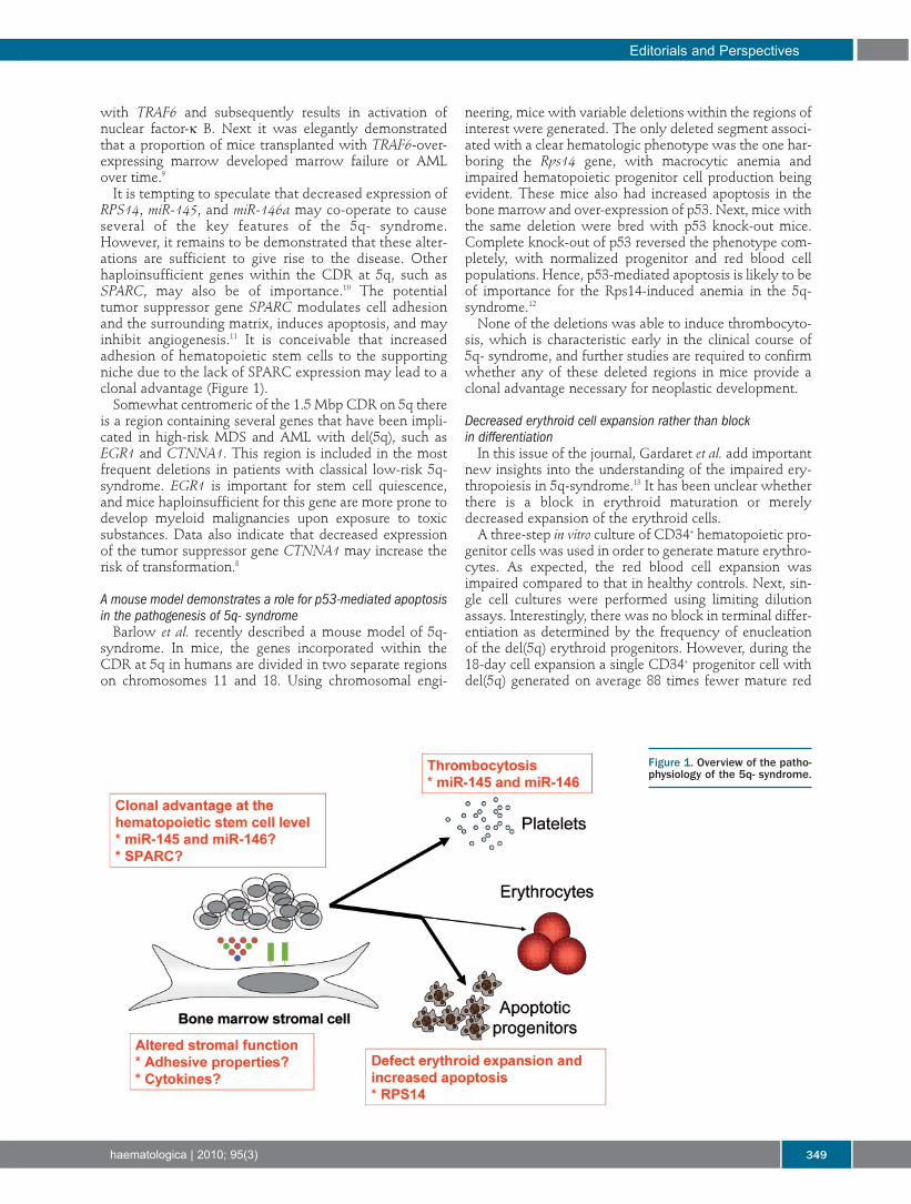
It is tempting to speculate that decreased expression ofRPS14, miR-145, and miR-146a may co-operate to causeseveral of the key features of the 5q- syndrome.However, it remains to be demonstrated that these alter-ations are sufficient to give rise to the disease. Otherhaploinsufficient genes within the CDR at 5q, such asSPARC, may also be of importance.10 The potentialtumor suppressor gene SPARC modulates cell adhesionand the surrounding matrix, induces apoptosis, and mayinhibit angiogenesis.11 It is conceivable that increasedadhesion of hematopoietic stem cells to the supportingniche due to the lack of SPARC expression may lead to aclonal advantage (Figure 1).
Somewhat centromeric of the 1.5 Mbp CDR on 5q thereis a region containing several genes that have been impli-cated in high-risk MDS and AML with del(5q), such asEGR1 and CTNNA1. This region is included in the mostfrequent deletions in patients with classical low-risk 5q-syndrome. EGR1 is important for stem cell quiescence,and mice haploinsufficient for this gene are more prone todevelop myeloid malignancies upon exposure to toxicsubstances. Data also indicate that decreased expressionof the tumor suppressor gene CTNNA1 may increase therisk of transformation.8
A mouse model demonstrates a role for p53-mediated apoptosisin the pathogenesis of 5q- syndrome
Barlow et al. recently described a mouse model of 5q-syndrome. In mice, the genes incorporated within theCDR at 5q in humans are divided in two separate regionson chromosomes 11 and 18. Using chromosomal engi-
neering, mice with variable deletions within the regions ofinterest were generated. The only deleted segment associ-ated with a clear hematologic phenotype was the one har-boring the Rps14 gene, with macrocytic anemia andimpaired hematopoietic progenitor cell production beingevident. These mice also had increased apoptosis in thebone marrow and over-expression of p53. Next, mice withthe same deletion were bred with p53 knock-out mice.Complete knock-out of p53 reversed the phenotype com-pletely, with normalized progenitor and red blood cellpopulations. Hence, p53-mediated apoptosis is likely to beof importance for the Rps14-induced anemia in the 5q-syndrome.12
None of the deletions was able to induce thrombocyto-sis, which is characteristic early in the clinical course of5q- syndrome, and further studies are required to confirmwhether any of these deleted regions in mice provide aclonal advantage necessary for neoplastic development.
Decreased erythroid cell expansion rather than block in differentiation
In this issue of the journal, Gardaret et al. add importantnew insights into the understanding of the impaired ery-thropoiesis in 5q-syndrome.13 It has been unclear whetherthere is a block in erythroid maturation or merelydecreased expansion of the erythroid cells.
A three-step in vitro culture of CD34+ hematopoietic pro-genitor cells was used in order to generate mature erythro-cytes. As expected, the red blood cell expansion wasimpaired compared to that in healthy controls. Next, sin-gle cell cultures were performed using limiting dilutionassays. Interestingly, there was no block in terminal differ-entiation as determined by the frequency of enucleationof the del(5q) erythroid progenitors. However, during the18-day cell expansion a single CD34+ progenitor cell withdel(5q) generated on average 88 times fewer mature red
Editorials and Perspectives
haematologica | 2010; 95(3) 349
Figure 1. Overview of the patho-physiology of the 5q- syndrome.

blood cells compared to the number produced by a normalCD34+ progenitor cell. The deficit of RPS14 was highlypronounced in the del(5q) cultured cells, in particular dur-ing the first 11 days of culture, and this correlated with alower proliferation rate.13 These results suggest that thereis impaired erythroid cell expansion in 5q- syndromerather than a deficiency in differentiation. It is attractive tohypothesize that this reduced proliferative ability is aresult of the RPS14 deficiency.
Alterations of the bone marrow stroma in 5q- syndromeIn another study published in this issue of the journal,
Ximeri et al. provide evidence that the bone marrow stro-ma in patients with 5q- syndrome is altered. Irradiatedbone marrow stromal layers from patients with del(5q)were shown to have an impaired ability to support thegrowth of CD34+ progenitor cells from healthy donors.However, after achieving a complete response to lenalido-mide, new stromal cells from the same patients were cul-tured. The progenitor supporting capacity was thenincreased and did not differ from that of stromal layersfrom healthy controls. Bone marrow cytokines were alsoanalyzed, and it was found that stromal cell-derived factor1 (SDF-1) and intracellular adhesion molecule 1 (ICAM-1),in particular, were up-regulated by lenalidomide.14 SDF-1binds to the receptor CXCR4 on hematopoietic stem cellsand is crucial for homing to the stem cell niche. This indi-cates that the stroma is significantly altered in 5q- syn-drome and that lenalidomide directly or indirectly,through reduction of the malignant clone, may reversethis defect.
The role of lenalidomide in the management of myelodysplastic syndrome with isolated del(5q)Standard of care
Most international guidelines propose erythropoieticgrowth factors as the first-line therapy of anemia in MDSwith isolated del(5q), provided that the patients have areasonable probability of response according to a predic-tive model based on transfusion requirements and serumerythropoietin level.15-18 Allogeneic stem cell transplanta-tion is not recommended up front; it may, however, beconsidered in selected patients as signs of disease progres-sion are manifested.
Lenalidomide has unparalleled efficacyIn studies pioneered by List et al., the immunomodula-
tory drug lenalidomide has shown remarkable efficacy inMDS with del(5q).19,20 The first large phase 2 trial (MDS-003) enrolled 148 transfusion-dependent MDS patientswith del(5q) and low or intermediate-1 risk diseaseaccording to the International Prognostic Scoring System.Of these patients, 67% achieved transfusion independen-cy and 45% entered complete cytogenetic remission.19
Important side effects included severe neutropenia andthrombocytopenia, which occurred in around half of thepatients and were managed with dose interruptions andsupportive granulocyte colony-stimulating factor. Themedian response duration was around 2 years.20 Long-term responders exist, although del(5q) cells may still bedetected in the marrow with sensitive methods in thesepatients. Hence, there is no evidence of cure.
Controversial role of lenalidomide in the management of the disease
Lenalidomide was approved by the US Food and DrugAdministration already in 2005. However, the EuropeanMedicines Agency (EMEA) requested extended studies.Despite its unquestionable efficacy, the EMEA laterrefrained from approval of lenalidomide in May 2008 dueto safety concerns, in particular the observed rate ofleukemic evolution.
In a long-term follow-up of the MDS-003 trial it wasclearly demonstrated that cytogenetic response wasinversely associated with AML evolution. Partial and com-plete cytogenetic responders had a 15% risk of their dis-ease transforming into AML, while evolution intoleukemia occurred in 67% of non-cytogenetic respon-ders.20 In untreated patients with classical 5q- syndrome,the long-term risk of AML has previously been estimatedto be no more than 10%.1 However, the patients includedin the lenalidomide study were not treated at the time ofdiagnosis, all were transfused, and some had additionalkaryotypic abnormalities or a slight increase of blasts,making a direct comparison inappropriate.
Göhring et al. recently published a comprehensiveanalysis of the outcome of the 42 German patientsenrolled in the MDS-003 trial.21 Of these 42 patients, 19patients fulfilled the criteria for MDS associated with iso-lated del(5q). Leukemic transformation occurred in 15 of42 patients (36%) overall. Evolution to AML occurred inseven of the 19 patients (37%) with MDS with isolateddel(5q) and a normal blast count. Furthermore, karyotypicevolution was observed in 17 of 42 patients, and 11 ofthese had complex karyotypes.21 This high degree of dis-ease progression was very unexpected based on the his-torical data referred to above. On the other hand, datafrom cohorts of patients suitable for comparisons have notbeen published. It cannot be excluded that this markedpropensity for leukemic transformation actually reflectsthe natural course of the disease. The issue of potentialnegative effects of lenalidomide on long-term outcome isnow under intense investigation.
Recent data suggest that there may be prognostic hetero-geneity within classical low-risk del(5q) MDS. We recentlydescribed a patient with a small clone of p53 mutated cellsin the marrow already at time of diagnosis. This subclonegradually expanded during treatment with lenalidomide,despite a complete erythroid and partial cytogeneticresponse. Eventually the clonal expansion resulted inleukemic transformation.22 The frequency of patients withp53 mutation is currently being assessed with next genera-tion sequencing in a larger cohort of patients.
Mechanisms of action of lenalidomideLenalidomide has a broad mode of action, including
inhibition of tumor necrosis factor-α and interleukin-6,induction of caspase-mediated apoptosis, cell adhesion,angiogenesis and stimulation of T cells and NK cells viainduction of interleukin-2 and interferon-γ.23
Wei et al. showed that lenalidomide potently inhibitstwo phosphatases that regulate the cell cycle, Cdc25C andPP2Acα. The genes for both these phosphatases are locat-ed at 5q, and are deleted in the majority of patients with5q- syndrome.24 Several studies have shown that del(5q)
Editorials and Perspectives
350 haematologica | 2010; 95(3)

cells are significantly more sensitive to lenalidomide com-pared to non-(del5q) cells.10,24,25 It is, therefore, conceivablethat the haploinsufficiency for the genes for Cdc25C andPP2Acα may induce this increased sensitivity.
As discussed above, the SPARC gene - located withinthe CDR at 5q and thus haploinsufficient - may be ofimportance for the clonal dominance in MDS. Intriguingly,lenalidomide restored SPARC expression to normal levelsin del(5q) progenitors.10 This may further explain whydel(5q) cells are particularly sensitive to this drug.
Immune effects such as stimulation of NK cells may alsobe of importance for the remarkable clinical effects oflenalidomide.23 As described above, lenalidomide poten-tially affects the stromal cells in patients with 5q- syn-drome, although it cannot be ruled out that elimination ofthe del(5q) clone itself may alter the stroma so that itregains its normal functions.14
Conclusions and future perspectivesThe recent identification of RPS14, miR-145, and miR-
146a has delineated key aspects of the pathogenesis ofMDS with isolated del(5q), and further understanding ofthe affected cellular pathways may lead to potential newtargets for treatment. The emerging drug lenalidomide ishighly efficient, although recent data have raised safetyconcerns regarding the risk of disease progression. Largecohort studies or, preferably, prospective randomized tri-als are necessary to clarify this issue. Until sufficient dataexist, lenalidomide should be used with great caution inMDS with del(5q).
Martin Jädersten, MD PhD is a Specialist in Hematology andInternal Medicine at the Hematology Center, KarolinskaUniversity Hospital, Stockholm, Sweden and a post-doctorateresearcher at the Center for Experimental Hematology, KarolinskaInstitutet, Stockholm, Sweden.
Conflicts of interest: Participation in Celgene advisory boardmeetings. Funding: Research funding include: Fellowship2009/06 awarded by the European Hematology Association, TheMDS Foundation Young Investigator’s Grant 2006, The RobertLundberg Foundation, KI/SLL post-doctorate grant 2009.
References
1. Giagounidis AA, Germing U, Haase S, Hildebrandt B, SchlegelbergerB, Schoch C, et al. Clinical, morphological, cytogenetic, and prognos-tic features of patients with myelodysplastic syndromes and del(5q)including band q31. Leukemia. 2004;18(1):113-9.
2. Swerdlow SH, Campo E, Harris NL, Jaffe ES, Pileri SA, Stein H, et al.WHO Classification of Tumours of Haematopoietic and LymphoidTissues. Lyon: IARC Press, 2008.
3. Nilsson L, Astrand-Grundstrom I, Arvidsson I, Jacobsson B,Hellstrom-Lindberg E, Hast R, et al. Isolation and characterization ofhematopoietic progenitor/stem cells in 5q-deleted myelodysplasticsyndromes: evidence for involvement at the hematopoietic stem celllevel. Blood. 2000;96(6):2012-21.
4. Jaju RJ, Jones M, Boultwood J, Kelly S, Mason DY, Wainscoat JS, et al.Combined immunophenotyping and FISH identifies the involvement
of B-cells in 5q- syndrome. Genes Chromosomes Cancer. 2000;29(3):276-80.
5. Kiladjian JJ, Bourgeois E, Lobe I, Braun T, Visentin G, Bourhis JH, etal. Cytolytic function and survival of natural killer cells are severelyaltered in myelodysplastic syndromes. Leukemia. 2006;20(3):463-70.
6. Boultwood J, Fidler C, Strickson AJ, Watkins F, Gama S, Kearney L, etal. Narrowing and genomic annotation of the commonly deletedregion of the 5q- syndrome. Blood. 2002;99(12):4638-41.
7. Ebert BL, Pretz J, Bosco J, Chang CY, Tamayo P, Galili N, et al.Identification of RPS14 as a 5q- syndrome gene by RNA interferencescreen. Nature. 2008;451(7176):335-9.
8. Ebert BL. Deletion 5q in myelodysplastic syndrome: a paradigm forthe study of hemizygous deletions in cancer. Leukemia. 2009;23(7):1252-6.
9. Starczynowski DT, Kuchenbauer F, Argiropoulos B, Sung S, Morin R,Muranyi A, et al. Identification of miR-145 and miR-146a as media-tors of the 5q- syndrome phenotype. Nat Med. 2010;16(1):49-58.
10. Pellagatti A, Jadersten M, Forsblom AM, Cattan H, Christensson B,Emanuelsson EK, et al. Lenalidomide inhibits the malignant clone andup-regulates the SPARC gene mapping to the commonly deletedregion in 5q- syndrome patients. Proc Natl Acad Sci USA. 2007;104(27):11406-11.
11. Framson PE, Sage EH. SPARC and tumor growth: where the seedmeets the soil? J Cell Biochem. 2004;92(4):679-90.
12. Barlow JL, Drynan LF, Hewett DR, Holmes LR, Lorenzo-Abalde S,Lane AL, et al. A p53-dependent mechanism underlies macrocyticanemia in a mouse model of human 5q- syndrome. Nat Med. 2010;16(1):59-66.
13. Garderet L, Kobari L, Mazurier C, De Witte C, Giarratana M-C, PérotC, et al. Unimpaired terminal erythroid differentiation and preservedenucleation capacity in myelodysplastic 5q(del) clones: a single cellstudy. Haematologica. 2010;95(3):398-405.
14. Ximeri M, Galanopoulos A, Klaus M, Parcharidou A, Giannikou K,Psyllaki M, et al. on behalf of the Hellenic MDS Study Group. Effectof lenalidomide therapy on hematopoiesis of patients withmyelodysplastic syndrome associated with chromosome 5q deletion.Haematologica. 2010;95(3):406-14.
15. Alessandrino EP, Amadori S, Barosi G, Cazzola M, Grossi A, LiberatoLN, et al. Evidence- and consensus-based practice guidelines for thetherapy of primary myelodysplastic syndromes. A statement from theItalian Society of Hematology. Haematologica. 2002;87(12):1286-306.
16. Bowen D, Culligan D, Jowitt S, Kelsey S, Mufti G, Oscier D, et al.Guidelines for the diagnosis and therapy of adult myelodysplasticsyndromes. Br J Haematol. 2003;120(2):187-200.
17. NCCN Clinical Practice Guidelines in Oncology / MyelodysplasticSyndromes. 2008 [Available from: www.nccn.org]
18. The Nordic MDS Group Care Program. 2008 [Available from:www.nmds.org/ez4/index.php?/nmds/Nordic-Care-Programme]
19. List A, Dewald G, Bennett J, Giagounidis A, Raza A, Feldman E, et al.Lenalidomide in the myelodysplastic syndrome with chromosome5q deletion. N Engl J Med. 2006;355(14):1456-65.
20. Melchert M, List A. Targeted therapies in myelodysplastic syndrome.Semin Hematol. 2008;45(1):31-8.
21. Gohring G, Giagounidis A, Busche G, Kreipe HH, Zimmermann M,Hellstrom-Lindberg E, et al. Patients with del(5q) MDS who fail toachieve sustained erythroid or cytogenetic remission after treatmentwith lenalidomide have an increased risk for clonal evolution andAML progression. Ann Hematol. 2009 Oct 24. [Epub ahead of print]
22. Jadersten M, Saft L, Pellagatti A, Gohring G, Wainscoat JS, BoultwoodJ, et al. Clonal heterogeneity in the 5q- syndrome: p53 expressing pro-genitors prevail during lenalidomide treatment and expand at diseaseprogression. Haematologica. 2009;94(12):1762-6.
23. Bartlett JB, Dredge K, Dalgleish AG. The evolution of thalidomideand its IMiD derivatives as anticancer agents. Nat Rev Cancer.2004;4(4):314-22.
24. Wei S, Chen X, Rocha K, Epling-Burnette PK, Djeu JY, Liu Q, et al. Acritical role for phosphatase haplodeficiency in the selective suppres-sion of deletion 5q MDS by lenalidomide. Proc Natl Acad Sci USA.2009;106(31):12974-9.
25. Tehranchi R, Fadeel B, Schmidt-Mende J, Forsblom AM, EmanuelssonE, Jadersten M, et al. Antiapoptotic role of growth factors in themyelodysplastic syndromes: concordance between in vitro and invivo observations. Clin Cancer Res. 2005;11(17):6291-9.
Editorials and Perspectives
haematologica | 2010; 95(3) 351