Embed Size (px)
Citation preview

Pathophysiological regulation of white adipocyte exocytosis of different
adiponectin molecular forms
Saliha Musovic
Department of Physiology/Metabolic physiology
Institute of Neuroscience and Physiology
Sahlgrenska Academy, University of Gothenburg
Gothenburg 2019

Cover illustration by Marina Kalds Said: Depiction of the different adiponectin molecular forms in the circulation.
Pathophysiological regulation of white adipocyte exocytosis of different adiponectin molecular forms © Saliha Musovic 2019 [email protected] ISBN 978-91-7833-308-0 (PRINT) ISBN 978-91-7833-309-7 (PDF) Printed in Gothenburg, Sweden 2019 Printed by BrandFactory

To my brother and parents


Pathophysiological regulation of white adipocyte exocytosis of different adiponectin molecular forms
Saliha Musovic Department of Physiology/Metabolic physiology,
Institute of Neuroscience and Physiology Sahlgrenska Academy, University of Gothenburg
Gothenburg, Sweden
ABSTRACT In this thesis we have identified mechanisms involved in the exocytosis of different adiponectin molecular forms in health and in metabolic disease. We have also studied similarities and differences in depot-specific adipocyte adiponectin release. In paper I we show that the physiological regulation of subcutaneous white adipocyte adiponectin exocytosis involves β3 adrenergic receptors (β3ARs) and Exchange Protein directly Activated by cAMP, isoform 1,
(Epac1) signalling. Furthermore, we show that adiponectin secretion is disturbed in obesity/type 2 diabetes induced catecholamine resistance due to reduced abundance of the key proteins β3ARs and Epac1. This condition of catecholamine resistance is further associated with a ~50% reduction of circulating high-molecular weight (HMW) adiponectin. In paper II we show that β3AR-activation rapidly triggers the release of HMW adiponectin-containing vesicle whereas insulin induces release of smaller molecular forms, with delayed time-kinetics. We moreover demonstrate that both catecholamine-triggered exocytosis of HMW adiponectin and the insulin-induced secretion of smaller adiponectin forms is entirely diminished in adipocytes from obese/type 2 diabetic mice. The equivalent regulation of secretion of different adiponectin molecular forms by catecholamines and insulin was confirmed in human adipocytes, thus defining a novel role of β3ARs in human adipocyte function. In paper III we propose that adiponectin exocytosis is regulated by sympathetic nerve endings, co-releasing noradrenaline and ATP within the adipose tissue. Secretion measurements confirmed that noradrenaline (elevates cAMP), like adrenaline in paper I, triggers adiponectin exocytosis. Extracellular ATP was shown to augment the exocytotic process, largely due to its elevation of intracellular Ca2+. We also show that defect purinergic signalling together with reduced white adipose tissue noradrenaline content likely aggravates the catecholamine resistance observed in paper I and II. Finally we describe regulation of mouse visceral adipocyte adiponectin secretion in paper IV. As demonstrated in subcutaneous adipocytes (paper I-III), visceral adipocyte adiponectin secretion is also stimulated by activation of β3AR and Epac1. In obese/diabetic conditions,
visceral adipocytes are likewise unresponsive to stimulation with catecholamines, but the underlying molecular defect does not involve reduced levels of neither β3AR nor Epac1, thus
differing from observations in subcutaneous adipocytes. In conclusion, our results suggest that secretory defects in obesity/type 2 diabetes, attributed to catecholamine resistance, underlie the reduced levels of HMW adiponectin in metabolic disease.
Keywords: White adipocytes, adiponectin exocytosis/secretion, health and metabolic disease
ISBN 978-91-7833-308-0
SAMMANFATTNING PÅ SVENSKA
Övervikt och fetma är ett alltmera alarmerande hälsoproblem, som i förlängningen kan
leda till utveckling av typ 2-diabetes och hjärt-kärlsjukdomar. I dagsläget dör fler av
överviktsrelaterade sjukdomar än av undervikt. Under metabolt hälsosamma
omständigheter lagras fett i kroppens stora energiförråd – den vita fettväven. Den vita
fettväven delas ytterligare in i två grupper baserat på anatomisk placering; subkutant
fett (under huden) och visceralt fett (i bukhålan). På senare år har forskningen visat att
vit fettväv även kan frisätta biologiskt aktiva molekyler direkt till blodet, som
möjliggör kommunikation med kroppens många andra organ.
Adiponektin som frisätts från vita fettceller (adipocyter) är ett så kallat anti-diabetisk
hormon, som minskar risken för att utveckla typ 2-diabetes genom att bland annat öka
glukosupptaget från blodet och därmed förbättra insulinkänsligheten. Vid fetma eller
typ 2-diabetes sjunker nivåerna av adiponektin i blodet. Framställning av exogent
adiponektin har visat sig komplicerat då hormonet kan frisättas till blodet i olika
komplexa molekylära former. Senare tids forskning indikerar att dessa olika
molekylära former kan ha varierande fysiologiska effekter. Den mest komplexa
strukturen, hög-molekylärt adiponektin har visats ha mest fördelaktiga egenskaper
gällande den metabola hälsan. Trots flera studier om effekter av adiponektin är
kunskapen om vad som reglerar frisättningen av hormonet relativt okänd. Vår grupp
har tidigare visat att signalmolekylen cAMP, via aktivering av proteinet Epac, triggar
snabb frisättning av adiponektin från subkutana vita adipocyter.
I denna avhandling visar vi för första gången att frisättning av hög-molekylärt
adiponektin från subkutana adipocyter, stimuleras av adrenerg β3-signalering och
Epac1. Vidare, visar vi även hur denna stimulering sannolikt sker till följd av påverkan
av närliggande sympatiska nerver som frisätter noradrenalin (aktiverar β3-receptorn)
och ATP, som vi spekulerar potentierar adiponektin-frisättningen. Tidigare utförda
studier pekar på att bukspottskörtelhormonet insulin även kan öka mängderna frisatt
adiponektin. I motsats till insulin, som cirkulerar högt i blodet efter målintag, så är
blodnivåer av noradrenalin istället ökade vid fasta.

Ur ett fysiologiskt perspektiv är sannolikheten liten att såväl både cirkulerande insulin
och noradrenalin skulle påverka samma hormon. Våra resultat demonstrerar att insulin
endast ökade frisättningen av de mindre molekylära formerna utan någon påverkan på
frisatt högmolekylärt adiponektin.
Obesa/diabetiska möss hade oförändrade cirkulerande nivåer av adiponektin med
avseende på alla molekylära former men kraftigt sänkta nivåer av hög-molekylärt
adiponektin. Subkutana fettceller, isolerade från obesa/diabetiska möss, kunde inte
svara på adrenerg-stimulering till följd av en nedreglering av β3-receptorn och Epac1
men även minskat innehåll av noradrenalin i fettväven. Studier på viscerala fettceller
visar att frisättningen av adiponektin, likt fynd från subkutana fettceller, också kan
stimuleras av adrenerg stimulering. Dessutom såg vi att viscerala fettceller från
obesa/diabetiska möss frisatte mindre adiponektin till följd av adrenerg stimulering
jämfört med metabolt friska möss. Den defekta frisättningen orsakades dock inte av
minskat genuttryck av varken β3-receptorn eller Epac1.
Sammantaget, indikerar våra resultat att adiponektinfrisättningen regleras via liknande
mekanismer i subkutana och viscerala fettceller. Den uppvisade defekta frisättningen
av adiponektin vid fetma/diabetes tros dock ha skilda bakomliggande molekylära
orsaker. Våra fynd bidrar till en ökad kunskap om de cellulära mekanismerna som
reglerar frisättningen av olika former av adiponektin vid hälsa och metabol sjukdom.
Vi föreslår att defekt adrenerg stimulerad frisättning av högmolekylärt adiponektin
ligger till grund för de uppvisade sänka nivåerna vid metabol sjukdom så som fetma
eller typ 2-diabetes.

LIST OF PAPERS
This thesis is based on the following studies, referred to in the text by their Roman numerals.
I. Komai AM*, Musovic S*, Peris EF, Alrifaiy A, El Hachmane MF, Johansson M, Asterholm IW, Olofsson CS. White adipocyte adiponectin exocytosis is stimulated via β3-Adrenergic signaling and activation of Epac1: catecholamine resistance in obesity and type 2 diabetes. Diabetes. 2016; 65(11):3301-3313
* The authors contributed equally and their names appear in alphabetical order.
II. Musovic S, Komai AM, Banke EN, Noor UA, Asterholm IW, Olofsson CS.
Epinephrine and insulin stimulates white adipocyte secretion of diverse adiponectin forms: evidence for blunted exocytosis of high-molecular weight adiponectin in diabesity-induced catecholamine resistance. Manuscript
III. Musovic S, Komai AM, Micallef P, Wu Y, Asterholm IW, Olofsson CS.
Sympathetic innervation and purinergic signaling in regulation of white adipocyte adiponectin secretion. Manuscript
IV. Musovic S, Olofsson CS. Adrenergic stimulation of adiponectin
secretion in visceral mouse adipocytes: blunted release in high-fat diet induced obesity. Submitted
Publications not included in this thesis
Komai AM, Brannmark C, Musovic S, Olofsson CS. PKA-independent cAMP stimulation of white adipocyte exocytosis and adipokine secretion: Modulations by Ca2+ and ATP. J Physiol. 2014; 592(23):5169-5186
Brannmark C, Lovfors W, Komai AM, Axelsson T, El Hachmane MF,
Musovic S, Paul A, Nyman E, Olofsson CS. Mathematical modeling of white adipocyte exocytosis predicts adiponectin secretion and quantifies the rates of vesicle exo- and endocytosis. J Biol Chem. 2017;292(49):20032-20043

TABLE OF CONTENT
INTRODUCTION ............................................................................................. 1
WHITE ADIPOSE TISSUE .................................................................................. 1 Cellular composition of WAT ................................................................... 2 Lipid metabolism ...................................................................................... 2 Sympathetic innervation of WAT ............................................................. 4 Endocrine functions of WAT .................................................................... 4
ADIPONECTIN ................................................................................................ 5 Circulating molecular forms of adiponectin ............................................. 6 Receptor mediated physiological outcomes ............................................ 7 HMW adiponectin as predictor of metabolic disease .............................. 8
SECRETORY PATHWAYS OF HORMONE SECRETION .......................................... 9 Regulated exocytosis............................................................................... 9 Epac-dependent cAMP signalling .......................................................... 10 Regulation of adiponectin secretion ...................................................... 11
AIMS .............................................................................................................. 13
MATERIAL AND METHODS ........................................................................ 15
Cell culture ............................................................................................. 15 Isolation of primary white adipocytes ..................................................... 16 Measurements of white adipocyte secretion ......................................... 17 Ratiometric calcium imaging .................................................................. 17 Gene expression analysis ..................................................................... 18 siRNA transfection ................................................................................. 19 Data analysis ......................................................................................... 19
RESULTS AND DISCUSSION ...................................................................... 21
PAPER I ....................................................................................................... 21 Gene expression of adrenergic receptor subtypes and Epac isoforms . 21 Adrenergic stimulation of adiponectin in white adipocytes occur via activation of β3ARs and Epac1 ............................................................. 21 The role of Ca2+ in adrenergically stimulated adiponectin secretion ..... 23 Blunted adiponectin secretion in adipocytes isolated from high-fat diet fed mice ................................................................................................. 24
SUMMARY OF FINDINGS IN PAPER I ................................................................ 26 PAPER II ...................................................................................................... 27
Insulin and adrenaline/CL-stimulated adiponectin secretion involves different signalling pathways and show dissimilar time-kinetics ............ 27 The role of Ca2+and cAMP in insulin-induced adiponectin release ....... 28

Both adrenaline- and insulin-stimulated adiponectin secretion is blunted in adipocytes isolated from obese/diabetic mice ................................... 29 Adrenaline triggers exocytosis of high-molecular weight adiponectin whereas insulin induces release of smaller adiponectin forms ............. 30 The stimulated secretion of HMW adiponectin is abrogated in adipocytes from obese/ diabetic mice ...................................................................... 30 HMW adiponectin secretion is triggered by catecholamines also in human adipocytes .................................................................................. 31
SUMMARY OF FINDINGS IN PAPER II .............................................................. 32 PAPER III ..................................................................................................... 34
Extracellularly applied noradrenaline stimulates white adipocyte adiponectin exocytosis/secretion ........................................................... 34 The role of extracellular ATP in the regulation of adiponectin release .. 34 The significance of extracellular ATP .................................................... 35 Effects of noradrenaline and ATP on adiponectin release in primary adipocytes isolated from lean and obese/diabetic mice ........................ 36 Effects of noradrenaline and ATP on secretion of HMW adiponectin ... 37
SUMMARY OF FINDINGS PAPER III ................................................................. 38 PAPER IV .................................................................................................... 39
Visceral adipocyte adiponectin release is stimulated via β3ARs and
Epac1 ..................................................................................................... 39 Role of exocytotic proteins in blunted adiponectin release from visceral HFD-adipocytes ..................................................................................... 42
SUMMARY OF FINDINGS IN PAPER IV ............................................................. 42
FINAL DISCUSSION ..................................................................................... 43
Catecholamine resistance ..................................................................... 43 Adrenergic signalling stimulates release of HMW adiponectin.............. 44 Role of sympathetic innervation in regulation of adiponectin release ... 45 Adipose-depot specific adiponectin release in health and metabolic disease .................................................................................................. 46
PATHOPHYSIOLOGICAL RELEVANCE .............................................................. 48
ACKNOWLEDGMENTS ............................................................................... 51
REFERENCES .............................................................................................. 52

ABBREVATIONS
[Ca2+]i Intracellular levels of Ca2+
AC Adenylyl cyclase
AdipoR1 Adiponectin receptor 1
AdipoR2 Adiponectin receptor 2
ADR Adrenaline
AR Adrenergic receptor
ATP Adenosine triphosphate
cAMP Cyclic adenosine monophosphate
cDNA Complementary DNA
Chow adipocytes Primary adipocytes isolated from chow-fed mice
CL CL316,243
CSF Cerebral spinal fluid
DEXA Dexamethasone
Epac Exchange protein directly activated by cAMP
ER Endoplasmic reticulum
FBS Fetal bovine serum
FFA Free fatty acids
FSK Forskolin
GDP Guanosine diphosphate
GEF Guanine nucleotide exchange factor
GLUT4 Glucose transporter 4
GTP Guanosine triphosphate
GWAT Gonadal white adipose tissue
HFD High-fat diet
HFD adipocytes Primary adipocytes isolated from HFD-fed mice

HMW High-molecular weight
IBMX 3-isobutyl-1-methylxanthine
IWAT Inguinal white adipose tissue
LMW Low-molecular weight
MMW Middle-molecular weight
NA Noradrenaline
NBCS Newborn calf serum
PDE3B Phosphodiesterase 3B
PI3K Phosphoinositide 3-kinase
PKA Protein kinase A
PKB Protein kinase B
PPAR Peroxisome proliferator activated receptor
SAT Subcutaneous adipose tissue
siRNA Small interfering RNA
SNARE Soluble N-ethylmaleimide-sensitive factor attachment protein receptor
SNS Sympathetic nervous system
SVF Stromal vascular fraction
T2D Type 2 diabetes
TH Tyrosine hydroxylase
TZD Thiazolidinedione
VAT Visceral adipose tissue
VAMP Vesicle-associated membrane protein
WAT White adipose tissue
WHO World Health Organisation

1
INTRODUCTION
World Health Organisation (WHO) reported in 2016 that over 1.9 billion adults were
overweight whereof 650 million were classified as obese. Global prevalence of
overweight and obesity has nearly tripled since the seventies. Formerly considered a
western world health issue the occurrence of overweight and obesity has reached
epidemic proportions with increased numbers now in low- and middle income
countries. Overweight and obesity is characterised by an excessive expansion of the
white adipose tissue (WAT), an organ traditionally considered a passive energy
reservoir. However, discoveries from the last two decades have established WAT as
an important metabolic organ with essential functions in whole body physiology,
conveyed by the secretion of bioactive molecules termed adipokines (Kershaw & Flier,
2004). The growing epidemic caused by obesity and obesity-associated comorbidities
such as type 2 diabetes (T2D) calls for an increased understanding of the role of
adipose tissue in regard to metabolic health.
White adipose tissue Adipose tissue is divided into white and brown adipose tissue that differ both in terms
of morphology and functionality. In humans, WAT is further divided into regional
depots; subcutaneous adipose tissue (SAT) is situated underneath the skin and visceral
adipose tissue (VAT) is found surrounding the internal organs in the abdominal cavity
(Trujillo & Scherer, 2006). The distinction between subcutaneous and visceral depots
is attributed to inherent differences in cellular composition, metabolic properties and
microenvironment. Distribution of WAT is highly individual and depends on factors
such as age, nutrition, gender and genetics (Wajchenberg et al., 2002). For example,
men are more inclined to store fat in visceral compartments while premenopausal
women have a greater tendency to accumulate fat in the SAT depots (Karastergiou et
al., 2012). In rodents, WAT is also a multi-depot organ as in humans, nonetheless there
are some differences. For instance, rodents have a visceral fat pad located in the
perigonadal region called gonadal white adipose tissue (GWAT), which is absent in
humans (Chusyd et al., 2016).

2
Cellular composition of WAT
Primary white adipocytes contain one large lipid droplet, accounting for ~95% of total
cell volume. Due to their high lipid-storage capacity, white adipocytes are
heterogeneous in size and can vary from 25-200 μM (Meyer et al., 2013).
Adipose tissue can enlarge either as a results of bigger adipocyte size due to increased
lipid storage (hypertrophy) or higher differentiation rate of preadipocytes to mature
adipocytes (hyperplasia; Rutkowski et al., 2015). WAT consists of not only adipocytes
but also of other cell types which account for a significant proportion of total cell
number (Fig. 1). Collectively, these nonadipocytes are called the stromal vascular
fraction (SVF) and include preadipocytes, endothelial cells, pericytes, monocytes,
macrophages and other cell types; all important for the maintenance of adipose tissue
homeostasis. Pericytes and endothelial cells provide the surrounding cells and
vasculature important growth and developmental factors while immune cells are
important for clearance of apoptotic/necrotic cells (Trujillo & Scherer, 2006).
Pathological conditions can have a strong impact on the cellular composition of the
SVF. For example, obesity is associated with local inflammation due to increased
number of immune cells (Kanneganti & Dixit, 2012).
Lipid metabolism Lipid mobilisation and utilisation are two fundamental processes in the white
adipocyte. During times of energy deficiency (fasted state), lipids stored as
triglycerides are degraded and released as free fatty acids (FFAs) in the circulation to
be taken up and utilised as energy by non-adipose organs. This breakdown of lipids is
termed lipolysis and several studies highlight WAT adrenergic receptors
(ARs; Lafontan & Berlan, 1993) as the primary physiological regulators of stimulated
lipolysis (Duncan et al., 2007; Hellstrom et al., 1997).
Fig. 1: Illustration of the heterogeneous cellular composition of WAT surrounded by vasculature

3
Activation of ARs results in various intracellular responses depending on receptor
subtype. βAR activation elevates cytosolic cyclic adenosine monophosphate (cAMP)
levels through activation of adenylyl cyclases (ACs), which catalyse the synthesis of
cAMP from adenosine triphosphate (ATP). In contrast, activation of α2AR decreases
cAMP through inhibition of ACs while α1AR elevate cytoplasmic calcium levels
through activation of phospholipase C (Lafontan & Berlan, 1993). Lipolysis is
stimulated through elevations of cAMP and further downstream activation of protein
kinase A (PKA; fig 2: left) which phosphorylates hormone-sensitive lipase, the main
enzyme that hydrolyses triglycerides to FFAs (Anthonsen et al., 1998).
Furthermore, decreased βAR-stimulated lipolysis has also been linked to obesity
(Lonnqvist et al., 1992).
Insulin is secreted by pancreatic β-cells upon elevation of blood glucose (fed state).
Actions of white adipocyte insulin-signalling decreases circulating FFAs, both via
stimulation of FFAs uptake and inhibition of lipolysis (Stahl et al., 2002).
In summary, activation of membrane bound insulin receptors induces an intracellular
signalling cascade (Fig. 2: right) involving phosphorylation of both
phosphoinositide 3-kinases (PI3Ks) and Akt/Protein kinase B (PKB). Additionally
Akt/PKB activates phosphodiesterase 3B (PDE3B) which breaks down cAMP
(Degerman et al., 2011). PKB-signalling is also involved in the insulin-stimulated
glucose uptake, increasing the presence of glucose transporter 4 (GLUT4) in the
adipocyte membrane (Furtado et al., 2002). Obesity is linked with an elevated basal
(unstimulated) lipolysis caused by impaired insulin sensitivity.
Fig. 2: Receptor-mediated signalling pathways involved in white adipocyte lipid metabolism. To the left, PKA-dependent βAR signalling pathway. To the right PKB-dependent insulin receptor action.

4
Sympathetic innervation of WAT
The sympathetic nervous system (SNS) impact adipose tissue physiology through
actions on for example lipolysis. Evidence propose that the primary involvement of
SNS in lipid metabolism is mediated via direct innervations of sympathetic
postganglionic nerves (Bartness et al., 2010; Youngstrom & Bartness, 1995), co-
releasing noradrenaline (NA) together with ATP (Burnstock, 2007). NA has higher
affinity for the β3AR than adrenaline (ADR; Tate et al., 1991), which is the
predominant receptor subtype in rodents. The abundance of β3AR in human WAT is
lower (Berkowitz et al., 1995) and the role of this receptor subtype in human adipocyte
physiology is not yet completely understood, although an involvement in lipolysis has
been reported (Baskin et al., 2018). Effect of extracellular ATP are mediated via
purinergic signalling. Numerous purinergic receptors are expressed in WAT and their
roles in adipogenesis and lipid metabolism have been described (Tozzi & Novak,
2017). The possible role of parasympathetic nervous innervation has been studied less.
However the anatomical closeness of the vagus nerve to the visceral fat depot indicates
a possible link. Also, the autonomic nervous system is known to have impact on
peripheral tissues via both branches of innervation (Bartness et al., 2014).
Endocrine functions of WAT In the late 80s, WAT started getting recognised as an endocrine organ releasing several
adipokines, and it was reported that the release of some adipokines were disturbed in
metabolic disease. Since then, many additional adipokines have been identified.
Adipsin (complement factor D) was the first adipokine described (Cook et al., 1987).
It plays an important role in the immune system (Xu et al., 2001) and also has several
positive metabolic effects such as improvement of pancreatic β-cell function (Lo et al.,
2014). The discovery of the white adipocyte hormones leptin (Zhang et al., 1994) and
adiponectin (Scherer et al., 1995) further established the importance of adipose tissue
endocrine function. Leptin receptor action regulates appetite in the brain by signalling
for satiety and increasing energy expenditure (Kelesidis et al., 2010). Circulating levels
of leptin correlates positively to body weight and adipocyte size (Lonnqvist et al.,
1997). Furthermore, serum levels of leptin are elevated in obese individuals.

5
Adiponectin Adiponectin is a 30 kDa, 244-247 amino acid long polypeptide. Full-length
(monomeric) adiponectin consists of four distinct regions; an N-terminal signalling
sequence, a non-conserved variable region, a collagenous domain and a C-terminal
globular domain (Fig. 3; Scherer et al., 1995). Human adiponectin is encoded by the
ADIPOQ gene and expression is amplified during white adipocyte differentiation.
Moreover, adiponectin gene levels is regulated by several cellular transcriptional
factors such as peroxisome proliferator activated receptor γ (PPARγ; Maeda et al.,
2001). Even though adiponectin gene expression has been detected in other cell types
(Krause et al., 2008; Uribe et al., 2008), the possible contribution of non-adipocytes to
circulating adiponectin levels seem unlikely (Wang et al., 2013).
Circulating levels of adiponectin are exceedingly high (~0.01% of total plasma
proteins) compared to other traditional endocrine hormones such as insulin or leptin
(Arita et al., 1999). Serum adiponectin levels display a strong sexual dimorphism and
women have higher levels than men (Combs et al., 2003). As mentioned earlier, pre-
menopausal females accumulate more adipose tissue in the subcutaneous regions.
Some studies show that subcutaneous adipocytes secrete more adiponectin compared
to visceral, thus contributing more to circulating serum levels of the adipokine (Fisher
et al., 2002; Meyer et al., 2013). The male sex hormone testosterone has been reported
to reduced adiponectin synthesis as well as interfere with cellular post-translational
modifications (Xu et al., 2005) and might contribute to the observed gender difference
in regards to circulating adiponectin levels.
Fig. 3: The four amino acid sequence regions that full-length adiponectin (monomeric) polypeptide consists of.

6
Circulating molecular forms of adiponectin
Adiponectin undergoes several post-translational modifications in the endoplasmic
reticulum (ER) prior to release from the white adipocyte (Wang et al., 2007). Full-
length adiponectin is present in the circulation as different molecular structures, linked
together with covalent disulphide bonds (Fig. 4). The different forms are in literature
classified as following; trimeric low-molecular weight (LMW), hexameric middle-
molecular weight (MMW) and the most complex structure which is oligomeric high-
molecular weight (HMW) adiponectin (Pajvani et al., 2003; Scherer et al., 1995).
Moreover, different physiological clearance of the diverse forms has been reported
(Halberg et al., 2009).
Interconversion does not occur after release into the blood stream and is thus only
regulated on cell level. Disulphide bond formation occurs in the ER though oxidation.
Earlier studies have shown the involvement of ER-chaperones ERp44 and Ero1-α in
the regulation of higher adiponectin complex formation (Qiang et al., 2007; Wang et
al., 2007). In addition, a third ER-resident protein, DsbA-L, has been identified to have
a key role in the regulation of adiponectin multimerisation, due to its high intrinsic
characteristics to form disulphide bonds. This was confirmed in studies on transgenic
mice, overexpressing DsbA-L in the adipose tissue. These animals had enhanced
adiponectin multimerisation, compared to wild-type littermates, as revealed by
increased circulating levels and adipocyte content of HMW adiponectin (Liu et al.,
2012).
Fig. 4: Different circulating adiponectin molecular forms.

7
Receptor mediated physiological outcomes
Adiponectin mediates its endocrine effects through two identified receptors;
Adiponectin Receptor 1 (AdipoR1) and Adiponectin Receptor 2 (AdipoR2). The two
receptors display high amino acid sequence homology and are seven-transmembrane
receptors, similar to traditional G-coupled receptors, however with a cytoplasmic N-
terminal and an extracellular C-terminal. AdipoR1 is ubiquitously expressed and
speculated to act via AMPK signalling while AdipoR2 is mainly present in the liver
and proposed to regulate expression of PPARα. Reports suggest that AdipoR1 displays
high affinity to the globular region of adiponectin while full-length adiponectin is
required to activate AdipoR2 (Yamauchi et al., 2003). Earlier studies in skeletal
muscle, reveal that adiponectin increases glucose uptake and β-oxidation, thus
improving muscle insulin sensitivity (Kuoppamaa et al., 2008; Yamauchi et al., 2002).
In liver, adiponectin suppresses glucose production by downregulating rate-limiting
enzymes involved in gluconeogenesis. Further actions of adiponectin in liver include
stimulation of glycolysis, β-oxidation and suppression of lipid accumulation. (Combs
et al., 2001; Liu et al., 2012). Via these overall favourable outcomes on glucose and
lipid metabolism, adiponectin improves systemic insulin sensitivity.
The wide distribution of adiponectin receptors in several other tissues contributes to
the extensive physiological effects of adiponectin. Basic science studies have shown
adiponectin to have cardioprotective properties, by increasing angiogenesis, providing
protection from foam cell formation and development of atherosclerosis (Shimada et
al., 2004; Wang & Scherer, 2008). Quite contradictory, recent studies suggest
adiponectin as a risk factor for cardiovascular health issues (Dadson et al., 2015,
Holland et al., 2011). In addition to the peripheral effects on insulin sensitivity,
adiponectin also display central effects. In the brain, adiponectin has been reported to
increase energy output with no effect on appetite. Initially, it was speculated that
adiponectin does not cross the blood-brain barrier (Spranger et al., 2006), however the
trimeric LMW adiponectin form has been detected in human cerebrospinal fluid (CSF).
Peripheral administration of adiponectin in serum results in a parallel increase in CSF,
but at 1000-fold lower concentration (Kusminski et al., 2007).

8
The T-cadherin receptor has been proposed as a third adiponectin-binding receptor.
This receptor type is highly expressed in endothelial and smooth muscle cells with
significant roles in cell growth and vasculature development. Yet the T-cadherin
receptor shows no interactions to neither globular nor trimeric adiponectin (LMW) but
has instead been proposed to specifically bind to the higher complex structures (Hug
et al., 2004), signifying the importance of higher order structures for the full biological
function of adiponectin.
HMW adiponectin as predictor of metabolic disease Obesity can result in disturbed WAT functions resulting in dysfunctional plasma levels
of several known adipokines. Other significant characteristics of obese WAT are
higher degree of inflammation and local deprivations of oxygen supply. Obesity-
caused WAT expansion is often linked to insulin resistance. The relationship between
adiponectin and metabolic disease has been studied in several studies. Adiponectin
depleted mice have decreased insulin sensitivity (Nawrocki et al., 2006), whereas
transgenic mice, overexpressing full-length adiponectin are more metabolically
healthy and less inclined to be affected by high-fat diet (HFD) feeding (Otabe et al.,
2007). Moreover, the cross-breeding of mice displaying the obese genetic phenotype
ob/ob with transgenic adiponectin mice improved circulating glucose and triglyceride
levels, thus improving overall metabolic health (Kim et al., 2007). Nevertheless, other
studies suggest that the insulin-sensitising properties of adiponectin can be attributed
to the explicit effects of HMW adiponectin. Circulating HMW adiponectin levels are
decreased in T2D individuals compared to healthy controls (Hara et al., 2006; Tabara
et al., 2008; Zhu et al., 2010). Pharmacological treatment with tiazolidinediones
(TZDs; PPAR-γ agonists) in T2D patients, increased circulating HMW adiponectin
together with improved insulin sensitivity (Pajvani et al., 2004). In regards to
adiponectin, observed cellular actions of TZDs on the white adipocyte include
increased gene expression, secretion and multimerisation (Iwaki et al., 2003; Phillips
et al., 2009; Yu et al., 2002;).

9
Secretory pathways of hormone secretion Intercellular communication is essential for mammalian multicellular systems, such as
the white adipose tissue. There are two ways in which the cells can interact with other
cells, either through direct cell-cell interactions or via secreted products. Due to the
large size and charge of proteins they cannot be transported over the lipid bilayer via
diffusion mechanisms and therefore need to relay on other transporting systems such
as exocytosis. In eukaryotic cells, there are two types of exocytosis; constitutive and
regulated. Constitutive exocytosis operates via unstimulated vesicular transport to the
plasma membrane whereas regulated exocytosis is reliant on signal transduction
pathways induced by extracellular stimulation.
Regulated exocytosis Polypeptides, synthesised and modified in the ER pass the Golgi complex to the trans-
Golgi where initial formation of immature vesicles occur. In endocrine cells that
release hormones, mature secretory vesicles can be coarsely categorised in functional
pools. Vesicles belonging to the readily releasable pool are situated close to the plasma
membrane and are rapidly released upon triggering of the right stimulatory signal
(Burgoyne & Morgan, 2003). Observations with electron microscopy show that this
vesicle pool is rather small and quickly exhausted. Electrophysiological studies and
live cell imaging of vesicle exocytosis in hormone releasing cells further reveal that
stimulated exocytosis is a rapid cellular event, with fusion of single vesicles occurring
within milliseconds (Huang et al., 2007; Kasai et al., 2012).
In most neuronal and endocrine cells, fusion of readily releasable vesicles with the
plasma membrane is triggered by rises in intracellular Ca2+ ([Ca2+]i) due to influx
through membrane-bound calcium channels and/or release from [Ca2+]i stores
(Burgoyne & Morgan, 2003). Whole-cell patch-clamp recordings of exocytotic events
carried out in adrenal chromaffin cells verified the presence of a limited readily
releasable pool of catecholamine-containing vesicles that upon Ca2+ elevations
released their cargo to the circulation for systemic effects on target tissues (Heinemann
et al., 1994). Another typical example is the pancreatic β-cell, where insulin-containing
vesicles are released in a Ca2+-dependent manner when blood glucose levels rises

10
(Barg et al., 2002; Olofsson et al., 2009). When the readily releasable pool is depleted,
immature vesicles from the reserve pool of vesicles need to undergo Ca2+, cAMP
and/or ATP dependent modifications and/or physical translocation in order to gain
release competence (Burgoyne & Morgan, 2003). The replenishment of the readily
releasable vesicle pool is necessary in order to maintain hormone release over longer
time periods.
Soluble-N-ethylmaleimede-sensitive factor attachment protein receptor (SNAREs) are
a group of membrane-associated proteins that are involved in different stages of the
exocytotic process (Kasai et al., 2012). SNAREs are located both on the secretory
vesicle itself and on the target cell membrane. Upon signal transduction, these SNARE
proteins will together form a tightly bound four helix complex, which is thought to be
involved in providing the energy necessary for fusion. The specific cellular events
leading up to complete fusion is not fully understood but suggested to involve direct
involvement of SNAREs (Han et al., 2017).
Epac-dependent cAMP signalling The intracellular signalling molecule cAMP, plays important roles in most cell types
affecting differentiation, cell growth, apoptosis, secretion and many other cellular
processes. Elevations of intracellular cAMP can trigger two identified signalling
pathways involving actions on either PKA or Exchange protein directly activated by
cAMP (Epac; Holz et al., 2006). While the role of PKA-signalling has been firmly
established in the regulation of stimulated lipolysis, the significance of Epac-signalling
in white adipocyte physiology is less investigated. Epacs are a novel group of cAMP-
binding proteins that exists in two isoforms; Epac1 and Epac2. The two isoforms share
extensive sequence homology but differ in tissue distribution. Epac1 is expressed in
most cell types whereas Epac2 expression is restricted to neuroendocrine cell types
(Schmidt et al., 2013). cAMP plays a role in modulating the exocytotic pathway in
several endocrine cell types. For instance, in pancreatic β-cells, PKA and Epac2
cAMP-signalling is essential for the regulation of insulin granule dynamics (Shibasaki
et al., 2007). A similar augmenting effect of cAMP on exocytosis has been defined in
adrenal chromaffin cells (Novara et al., 2004).

11
Further downstream, Epac can activate several small GTPases belonging to the Ras
superfamily, recognised to be involved in vesicle trafficking and exocytosis. Epac is a
guanine nucleotide exchange factor (GEF) which upon stimulation by an upstream
signal catalyses the exchange of a guanosine diphosphate (GDP) to guanosine
triphosphate (GTP) thus activating the small GTPases (Simanshu et al., 2017).
Regulation of adiponectin secretion The possible role of regulated adiponectin secretion was first proposed by Scherer and
colleagues in 1995. In their first article about adiponectin they suggested that insulin
stimulates adiponectin release, presenting evidence from data in cultured 3T3-L1
adipocytes (Scherer et al., 1995). Since, several studies have shown various outcomes
of insulin-treatment on adiponectin in regards to gene expression, synthesis and
release. Long-term exposure (>4 h) to insulin in both cultured and isolated rodent
adipocytes induced adiponectin release (Blumer et al., 2008; Cong et al., 2007; Lim et
al., 2015; Pereira & Draznin, 2005) and the effect was shown to act via PI3K- and
PDE3B-dependent pathways (Cong et al., 2007). Interestingly, shorter time exposure
to insulin (30-120 min) has also been shown to induce adiponectin release in a PI3K-
dependent manner, without contribution of increased gene expression or protein
synthesis. Subcellular fractionation post insulin-exposure revealed that the highest
content of adiponectin was localised close to the plasma membrane fraction (Bogan &
Lodish, 1999; Lim et al., 2015), suggesting that insulin affects the release-process of
already synthesised and modified adiponectin.
The effect of catecholamines on adiponectin release has been investigated in a few
studies. In human VAT explants, an inhibitory outcome was observed on adiponectin
gene expression as a result of long-term (>8 h) exposure with βAR- or cAMP-agonists
(Delporte et al., 2002) and similar results have been observed in mouse adipose tissue
(Delporte et al., 2002; Fasshauer et al., 2001). In isolated GWAT rat adipocytes,
treatment with the βAR agonist isoprenaline for 4-24 h inhibited adiponectin secretion
while stimulating lipolysis (Cong et al., 2007). The studies described above have thus
investigated long-term/chronic effects of catecholamines on adiponectin release.

12
In 2014, our group was first to define that white adipocyte exocytosis (measured as
increase in membrane capacitance) and adiponectin secretion in both cultured and
human primary adipocytes is triggered by elevations of cAMP via activation of Epac,
in a PKA-independent manner (Fig. 5). The recruitment of new releasable vesicles
from the reserve pool was shown to occur in a Ca2+-dependent manner. Thus, in Ca2+-
free conditions, the readily releasable pool will be depleted and exocytosis can not
sustain for longer time-periods, even in the presence of cAMP. Furthermore we have
shown that a combination of Ca2+ and ATP potentiates adiponectin exocytosis via
direct effects on the release process itself (Komai et al., 2014). These findings propose
an acute stimulatory effect of catecholamines on adiponectin secretion (through the
activation of βARs) and suggests adrenergic signalling as a physiological regulator of
adiponectin release.
Fig 5: Model of white adipocyte exocytosis. Elevations of cAMP trigger the release of readily releasable vesicles whereas intracellular rises of Ca2+ and ATP augment the exocytotic event.

13
AIMS
The overall aim of this thesis was to define the (patho-) physiological regulation of
adiponectin exocytosis/secretion as well as the depot-specific adipocyte adiponectin
release.
The specific aims of the four papers included in this thesis were to:
I. Study the effects of catecholamines on subcutaneous white adipocyte adiponectin exocytosis/secretion and how adrenergically stimulated adiponectin release is affected by diet-induced diabetes/obesity.
II. Explore the similarities and differences between adiponectin secretion stimulated by catecholamines and insulin in subcutaneous white adipocytes.
III. Investigate the role of sympathetic innervation and purinergic signalling in the regulation of adiponectin exocytosis/secretion in subcutaneous white adipocytes.
IV. Define the regulation of visceral white adipocyte adiponectin secretion in health and in metabolic disease.

14

15
MATERIAL AND METHODS
The reader is referred to paper I-IV for a more detailed description of material and
methods used in this thesis.
Cell culture 3T3-L1 adipocytes are a well-known in vitro white adipocyte model of murine cell
origin and was first isolated and cloned in the seventies (Greenberger & Aaronson,
1974). The cell line is extensively used in studies of white adipocyte development,
lipid metabolism and endocrine function (Fasshauer et al., 2001; Scherer et al., 1995).
3T3-L1 preadipocytes have a fibroblast like morphology and can within ten days be
differentiated into mature lipid-storing adipocytes (Fig. 6).
3T3-L1 preadipocytes were cultured in flasks and kept at 37°C and 5% CO2 together
with high-glucose DMEM containing 10% newborn calf serum (NBCS) and 1%
penicillin-streptomycin (P/S). When cells reached ~70% confluency, they were
trypsinised (3 min, 37°C ) and seeded on either plastic- or glass-bottom dishes or 12-
well plates. Cells were grown to higher confluency prior to differentiation (~90%).
Differentiation (D1) was initiated by addition of insulin (850 nM), dexamethasone
(1 μM) and 3-isobutyl-1-methylxanthine (IBMX; 0.5 mM) in high glucose DMEM
with 10% fetal bovine serum (FBS) and 1% penicillin-streptomycin (P/S).
The exposure to this mixture, at early stages of cell differentiation, upregulates
Fig. 6: Differentiation of 3T3-L1 adipocytes from fibroblast-like cells (day 0) to mature lipid-storing adipocytes (day 8-9).

16
essential adipocyte genes; as result both glucose uptake and triglyceride synthesis
increases. After 48 hours the medium was replaced with fresh medium (DMEM;
10% FBS, 1% P/S) containing only insulin (D2). At this point, the 3T3-L1 adipocytes
start to accumulate several lipid droplets. Thereafter, medium was changed every
second day up until day 8-9 of differentiation, when experiments were carried out.
Isolation of primary white adipocytes Human subcutaneous white adipocytes were isolated from adipose tissue biopsies.
Mouse inguinal and gonadal white adipocytes were isolated according to established
protocol (Ruan et al., 2003; Fig. 7) from 8-16 week old C57BL/6J mice, either fed
chow or HFD (60% kcal from fat) for 8 weeks Adipose tissue was collected and
minced into small pieces and thereafter digested in collagenase type II (1 mg/mL in
KRHG, 3% BSA) for 45-60 min at 37 °C. Following the incubation with collagenase,
the adipose tissue suspension was poured through a 100 μM nylon mesh into a tube.
Adipocytes floating on top were washed with buffer (KRHG, 1% BSA). Adipocytes
were allowed to float to the surface and buffer solution beneath the adipocyte layer,
containing SVF cells, was removed with a syringe. Isolated adipocytes were moved
to a separate tube and allowed to float in order to facilitate removal of more excessive
buffer by using a smaller syringe. Human adipocytes were incubated overnight and
secretion measurements proceeded the following day. Mouse adipocytes were directly
used for adipokine secretion experiments or frozen at -80°C for further analysis. All
animal and human studies were approved by the regional ethical review board.
Fig. 7: Illustration depicting the procedure for isolation of primary adipocytes

17
Measurements of white adipocyte secretion
Isolated adipocytes (10-15% v/v) or 3T3-L1 adipocytes, cultured on 12 well plates,
were incubated in 5 mM glucose extracellular solution containing test substances for
30 min at 32˚C under gentle shaking conditions. Primary cell incubation was
terminated by centrifugation of cell suspension in diisononyl phthalate oil followed by
snap freezing in dry ice. This allows for separation of adipocytes from media,
containing the secreted product. Tubes were cut through at two levels, separating cells
from media and removing the oil layer in the middle. Following incubations with
3T3-L1 adipocytes, the medium was collected and centrifuged in order to remove
detached cells. Supernatant was collected following centrifugation and adipocytes
were lysed in lysis buffer containing protease inhibitors. All samples were stored in
-80 ˚C. Secreted adiponectin was measured with specific ELISA and normalised to
total protein content obtained with the Bradford assay.
Capacitance measurements Exocytosis was measured in 3T3-L1 adipocytes with membrane capacitance
measurements using the patch-clamp method (Neher & Marty, 1982). Membrane
capacitance can be calculated from equation Cm = A x Ɛ/d where A is the membrane
surface area. The thickness of the plasma membrane (d) and the specific membrane
capacitance Ɛ are constant. Exocytosis results in an increase of the membrane area and
is thus proportional to the capacitance. Hence, membrane capacitance (Cm) is
proportional to the plasma membrane area (A). The rate of vesicle fusion with plasma
membrane (exocytosis) was measured as capacitance over time.
Ratiometric calcium imaging ([Ca2+]i) levels were measured with dual-wavelength ratiometric imaging in 3T3-L1
adipocytes. The cells were loaded with the fluorescent Ca2+ dye fura-2-AM together
with mild detergent Pluronic for 45 minutes prior to measurement. Fura-2-AM is a
highly membrane permeable ester that is hydrolysed by cellular esterases once inside
the cell. Fura-2 can be excited at two wavelengths depending on whether it is bound to
Ca2+ or not. The excitation maximum for unbound fura-2 is 380 nm while it is 340 nm
for Ca2+-bound fura-2. The emitted light is collected at 510 nm.

18
The ratio (Fura-2bound/Fura-2unbound) allows for calculations of [Ca2+]i concentrations
over time. Ratiometric imaging reduces the impact variations in fura-loading as well
as declined fluorescence signal due to bleaching between experiments. [Ca2+]i was
calculated using the equation 5 of Grynkiewicz et al 1985 and a Kd of 224 nM.
(Grynkiewicz et al., 1985).
Gene expression analysis Quantitative real-time RT-PCR can be used to measure expression of genes of interest.
The method requires complementary DNA (cDNA) as a starting material, which can
be made from mRNA with the enzyme reverse transcriptase. Assessment of RNA
purity, following isolation, is validated through spectrophotometrical absorbance
measurements at 260 and 280 nm. Amplifications of cDNA sequences of interest can
be detected with fluorescent labelling dyes. In this thesis, the fluorescent dye SYBR
green was used which binds to newly synthesised double-stranded DNA (see Fig. 8 for
more details). The amount of synthesised cDNA is directly proportional to
fluorescence and can be detected over time.
Fig. 8: Each PCR-cycle include three steps that are repeated 40-45 times. The reaction is initialised with an increase in temperature, which results in the separation of double-stranded DNA into two single-stranded DNA (denaturation). In the next step, temperature is reduced allowing for primers to bind to denatured target DNA (annealing). In the last step, a new complementary DNA-strand is synthesised by DNA polymerase (elongation).

19
siRNA transfection
Intracellular incorporation of small interfering RNA (siRNA) can be used to
silence/suppress the expression of a specific gene in order to study its function.
In this thesis we used siRNA to silence the expression of β3AR and Epac1 in cultured
adipocytes. 3T3-L1 adipocytes were transfected with Silencer Select siRNA
(80 nmol/L) at day 6 of differentiation using transfection reagent Lipofectamine 2000.
Medium was changed eight hours post transfection. Further studies of gene expression
and secretion were carried on 60 hours after transfection with siRNA. Knockdown of
protein of interest was confirmed with gene expression analysis. Adiponectin release
after 30 minutes at 32°C was measured in order to study the functionality of the
knockdown. In these experiments scramble siRNA was used as a negative control.
Data analysis Results included in this thesis are presented as mean values ± SEM, expressed either
in fold change over basal (unstimulated conditions) or in absolute concentrations.
Statistical significance was calculated with OriginPro (OriginLab Corporation, USA).
Student’s t-test (unpaired or paired as appropriate) was used to determine significance
between two experimental groups. Analysis of variance (ANOVA) was used to
determine statistical significance between two or more independent groups and/or
conditions.

20

21
RESULTS AND DISCUSSION
Paper I In paper I of this thesis we investigated the effects of ADR and the highly selective
β3-agonist CL 316,243 (CL) on white adipocyte adiponectin exocytosis using a
combination of electrophysiological and biochemical measurements. Effects on
adiponectin release were studied in both cultured 3T3-L1 adipocytes and in primary
subcutaneous adipocytes isolated from lean or obese/diabetic mice. The current study
builds on our previously published work, showing that cAMP elevations trigger white
adipocyte release of adiponectin-containing vesicles via Epac-dependent signalling
pathways (Komai et al., 2014).
Gene expression of adrenergic receptor subtypes and Epac isoforms In order to confirm the presence of ARs in the adipocytes used in this study (Lafontan
& Berlan, 1993), we measured gene expression of all five ARs in undifferentiated and
differentiated 3T3-L1 cells. In differentiated adipocytes, β3AR expression was highly
abundant while significantly lower expression was obtained for the other receptor
subtypes. We measured mRNA levels of the cAMP-binding proteins Epac isoforms 1
(Rapgef3) and 2 (Rapgef4). Epac1 was expressed in differentiated 3T3-L1 adipocytes
while Epac2 could not be detected. Interestingly mRNA levels of Epac1 were 60%
higher in undifferentiated cells. This is in agreement with studies that show that cAMP-
stimulated adipogenesis involves both PKA- and Epac-dependent signalling pathways
(Jia et al., 2012; Petersen et al., 2008). Equivalent gene expression ratios of ARs and
Epac isoforms were obtained in primary mouse adipocytes isolated from inguinal
white adipose tissue (IWAT).
Adrenergic stimulation of adiponectin in white adipocytes occur via activation of β3ARs and Epac1
White adipocyte exocytosis was measured as increase in membrane capacitance with
the patch-clamp method. In differentiated 3T3-L1 adipocytes, infused with a non-
stimulatory pipette solution (lacking cAMP), extracellularly applied ADR (5 μM) and

22
β3-agonist CL (1 μM) triggered a similar magnitude of exocytosis.
To study the role of Epac in adrenergically stimulated adipocyte exocytosis, adipocytes
were pretreated with the membrane-permeable competitive Epac inhibitor ESI-09
(10 μM) for 30 min prior to addition of ADR. Pretreatment with ESI-09 completely
abolished the exocytosis triggered by ADR (Fig. 9: left). Previously published results
from our group demonstrate that the cAMP-produced membrane capacitance increase
in 3T3-L1 adipocytes largely represent the release of adiponectin-containing vesicles
(Komai et al., 2014; El Hachmane et al., 2015). In agreement with the capacitance
data, incubation of 3T3-L1 adipocytes with ADR or CL for 30 min stimulated
adiponectin secretion (~1.8-fold with either secretagogue). ADR can bind to several
ARs with different affinity (Lafontan & Berlan, 1993). Although the similar effects on
adiponectin release by ADR and CL suggest that adrenergically stimulated adiponectin
release is chiefly mediated via β3ARs. We further validated the role of Epac1 in
isolated IWAT adipocytes. In agreement with capacitance measurements in 3T3-L1
cells, inhibition of Epac in IWAT adipocytes abolished adiponectin secretion
stimulated by ADR or CL (Fig. 9: right).
Fig. 9: ADR and β3AR-agonist CL trigger white adipocyte adiponectin exocytosis via Epac-dependent mechanisms. Left: Example capacitance traces of 3T3-L1 adipocytes infused with a non-stimulatory pipette solution (lacking cAMP) with extracellular addition of CL or ADR as indicated by the arrows. Light blue trace represents cells pre-treated for 30 min with Epac antagonist ESI-09 prior to addition of extracellular ADR. Right: Adrenergic/β3AR-stimulation of adiponectin secretion in IWAT adipocytes during 30 min as well as effects of pretreatment with ESI-09 prior to addition of ADR or CL. Data are mean values ± SEM of 9 experiments. *P<0.05 vs control.

23
Adiponectin release stimulated by CL remained unchanged in adipocytes pretreated
with the protein synthesis inhibitor cycloheximide (10 μM). Also, short-term
incubation of cultured adipocytes with CL was without effect on gene expression level
of adiponectin. These results support our hypothesis that short-term adrenergic
stimulation triggers the release of prestored adiponectin-containing vesicles with little
contribution to increased adiponectin synthesis or expression.
Measurements of cAMP content in cells exposed to ADR or CL for 30 min revealed
that ADR elevated cAMP ~7.5-fold, while levels in CL-treated cells were only 3-fold
increased. This finding is not surprising since ADR activates all cAMP increasing
βARs. The observation that the 3-fold elevation of cAMP via β3AR signalling
stimulates adiponectin exocytosis/secretion of an equal magnitude as ADR (which
increases cAMP much more and via all βARs), further proposes little or no
contribution of β1- and β2ARs in adrenergically triggered adiponectin release.
Together our findings suggest that activation of β3ARs and Epac1 triggers release of
pre-existing adiponectin-containing vesicles with no observed contribution to neither
adiponectin gene expression nor protein synthesis.
The role of Ca2+ in adrenergically stimulated adiponectin secretion Next we studied the role of [Ca2+]i on adrenergically stimulated adiponectin secretion
as ADR is known to elevate concentrations of the ion via actions on α1ARs.
ADR -stimulated adiponectin release remained intact upon Ca2+ chelation with
BAPTA (50 μM; 30 min pretreatment), in both cultured and in primary mouse IWAT
adipocytes. Ratiometric Ca2+ measurements demonstrated a heterogeneous response
of ADR on Ca2+ elevation. The effects on Ca2+ were overall small and thus in
agreement with obtained low gene expression of α1AR. Our results are similar to
previously reported heterogeneous Ca2+ elevations in human adipocytes exposed to
NA, an effect reported to depend on an uneven intercellular distribution of AR
subtypes (Seydoux et al., 1996). We conclude that the Ca2+ elevation resulting from
α1AR activation is of little importance for adrenergically stimulated adiponectin
release.

24
Blunted adiponectin secretion in adipocytes isolated from high-fat diet fed mice To study the association between obesity/T2D and catecholamine-stimulated
adiponectin release, IWAT adipocytes were isolated from mice fed chow
(chow adipocytes) or HFD (HFD adipocytes) for 8 weeks. HFD-fed mice had elevated
serum glucose and insulin levels, representing a diabetic condition. Additionally the
HFD-fed mice had increased bodyweight (46.3 ±0.8 g. compared with 31.8 ± 0.6 g,
for chow-fed mice). There was no change in circulating adiponectin levels in HFD-fed
mice, in contrast to previous reports in obese/diabetic subject (Panidis et al., 2003;
Saltevo et al., 2008). However, we observed a significant reduction in levels of
circulating HMW adiponectin (~50%), corresponding to what has been reported in
obese/type 2 diabetic individuals (Hara et al., 2006; Tabara et al., 2008; Zhu et al.,
2010).
A combination of the cAMP-elevating agents forskolin (FSK; 10 μM) and IBMX (200
μM; FSK/IBMX), CL or ADR increased the release of adiponectin ~2-fold in IWAT
adipocytes isolated from chow-fed mice. In adipocytes from HFD-fed mice, the
stimulatory effect of FSK/IBMX was significantly reduced (~30%) compared to chow
adipocytes (Fig. 10: left). Strikingly, adiponectin secretion triggered by ADR or CL
was essentially abolished in adipocytes from obese/diabetic mice coupled with a
2-fold increase of basal release (unstimulated conditions; Fig. 10: right).
Fig. 10: Adrenergically/cAMP-stimulated adiponectin secretion from lean and obese/diabetic mice. Stimulated adiponectin secretion (left) and basal release (right) in IWAT adipocytes isolated from chow- or HFD-fed mice during 30 min of incubation. Data are from 8 chow-fed and 8 HFD-fed animals. Data are presented as mean values ± SEM. *P<0.05; **P<0.01; ***P<0.001.

25
We evaluated whether reduced adiponectin content could explain the blunted secretion
in HFD adipocytes. Several studies have proposed that the obese/diabetic state is
associated with reduced adipocyte adiponectin expression, both at the gene and protein
level (Hu et al., 1996). IWAT adipocyte adiponectin content was unaltered in HFD-
fed mice. Moreover, the fraction secreted upon extracellular stimulation with ADR or
CL was much smaller in HFD adipocytes. All things considered, our data suggests that
the adrenergically regulated adiponectin release is impaired in IWAT adipocytes from
HFD-fed mice.
Gene expression analysis of key players involved in adrenergically stimulated
adiponectin secretion supported the existence of a secretory defect. Gene expression
data showed that IWAT adipocytes from HFD-fed mice, displayed reduced mRNA
levels of β3AR (~5-fold) and Epac1 (40 %). The reduced abundance of β3AR and
Epac1 in adipocytes from HFD-fed mice was also confirmed at the protein level.
Measurements showed that Epac1 was decreased by ~30%, similar in magnitude to the
reduced effect of FSK/IBMX-stimulated adiponectin secretion observed in HFD
adipocytes. The protein level of β3AR was 35% lower in IWAT adipocytes from HFD-
fed mice.
Fig. 11: Blunted adrenaline-stimulated adiponectin secretion upon β3AR and Epac1 siRNA knockdown. A: Relative mRNA expression in 3T3-L1 adipocytes transfected with β3AR (Adrb3) or Epac1 (Rapgef3) siRNA for 60 hours. The siRNA gene expression for each gene of interest is normalised against its expression in concurrently scramble transfected cells (negative control). B-C: Adrenaline-stimulated adiponectin secretion (30 min) in β3AR or Epac1 silenced 3T3-adipocytes as well as scramble transfected cells. Data in A-C are from five separate experiments. **P<0.01; ***P<0.001.

26
To further verify the significance of β3AR and Epac1 in catecholamine-stimulated
adiponectin secretion, we carried out siRNA knockdown in 3T3-L1 adipocytes (Fig.
11A). In adipocytes with silenced β3AR expression (~60% knockdown), ADR was not
able to induce adiponectin release (Fig. 11B). Likewise, silencing of Epac1 (~50%)
resulted in an abrogated ADR effect on adiponectin secretion (Fig. 11C). Adipocytes
transfected with scramble siRNA (negative control) remained responsive.
We postulate that the reduced levels of β3AR together with lower abundance of Epac1
might provide an explanation for the blunted CL/ADR-stimulated release of
adiponectin.
Summary of findings in paper I We here move our knowledge forward and show that the physiological regulation of
adiponectin exocytosis in subcutaneous adipocytes involves catecholaminergic
signalling, mainly via β3AR and further downstream activation of Epac1 (Fig. 12: left).
We also provide evidence for a defect adrenergically stimulated adiponectin release in
subcutaneous adipocytes isolated from diet-induced obese/diabetic mice due to
reduced abundance of key proteins β3AR and Epac1, in a condition we refer to as
“catecholamine resistance” (Fig. 12: right).
Fig. 12: Graphical conclusion based on our findings in paper I. See text for further details.

27
Paper II
Several studies report that insulin induces adiponectin release from both cultured and
primary mouse adipocytes. However, in paper I, we show an alternative pathway of
catecholamine-stimulated adiponectin secretion. The cAMP/catecholamine-triggered
adiponectin exocytosis is difficult to reconcile with a stimulatory effect of insulin,
which decreases intracellular cAMP levels via activation of PDE3B (Degerman et al.,
2011). In paper II, we investigate adiponectin exocytosis/secretion in response to
insulin and ADR, using cultured 3T3-L1 adipocytes and primary subcutaneous
adipocytes isolated from mice and humans.
Insulin and adrenaline/CL-stimulated adiponectin secretion involves different signalling pathways and show dissimilar time-kinetics 3T3-L1 adipocytes were exposed to the β3AR agonist CL (1 μM) or insulin (200 nM)
for 30 min. In agreement with findings in paper I, CL stimulated adiponectin secretion
(~1.7-fold). Insulin-stimulated adiponectin release was of similar magnitude.
The combination of CL and insulin resulted in released adiponectin in amounts
comparable to stimulation with each substance separately. Reports of insulin-
stimulated adiponectin release suggest the involvement of PI3K (Blumer et al., 2008;
Cong et al., 2007; Lim et al., 2015; Pereira & Draznin, 2005). To investigate this, we
measured adiponectin release from 3T3-L1 adipocytes pretreated with PI3K-inhibitor
Wortmannin (100 nM) for 30 min prior to incubation with insulin or ADR.
Inhibition of PI3K action abolished the insulin-induced adiponectin release whereas
ADR-stimulated adiponectin release remained intact. Furthermore, inhibition of Epac
had no effect on insulin-stimulated adiponectin release while, in agreement with
paper I, ADR-triggered adiponectin secretion was diminished.
To further study the similarities and differences in more detail, we performed time-
course experiments of adiponectin release (Fig. 13). CL stimulated adiponectin release
at early time points (measurable amounts collected over 15 min in static incubations).
However, the insulin effect was first observed at 30 minutes. The observed effect of
insulin-stimulation on adiponectin release is similar to results from a previous study
(Lim et al., 2015), demonstrating that insulin was without effect on adiponectin release

28
at early time points with a more prominent effect observed at later time points
(60-120 min). Our data propose that insulin acts via mechanisms acting downstream
of the exocytotic machinery whereas catecholamines trigger release at the level of the
exocytotic machinery via a direct actions on readily releasable adiponectin vesicles.
The role of Ca2+and cAMP in insulin-induced adiponectin release
Insulin-induced adiponectin release was unaffected by Ca2+-depletion, and ratiometric
Ca2+-imaging further demonstrated that changes in intracellular Ca2+ do not underlie
insulin-stimulated adiponectin secretion. In agreement with findings in paper I, ADR
stimulated short-term (30 min incubation) adiponectin release in IWAT adipocytes
isolated from mice. Consistent with previous reports (Cong et al., 2007), insulin (20 or
200 nM) also induced adiponectin release in isolated mouse IWAT adipocytes.
Measurements of cAMP levels in cells exposed to insulin for 30 min confirmed that
cAMP was elevated and thus not involved in the stimulatory mechanisms.
The observation that adrenergic stimulation and insulin exposure both stimulate the
release of adiponectin may appear contradictory due to their opposing physiological
roles.
Fig. 13: Insulin-stimulated adiponectin release has slower time-kinetics than β3AR-triggered adiponectin secretion Time-course series in 3T3-L1 adipocytes representing the effects of insulin and CL on adiponectin release at 15, 30 and 60 minutes. Results are from 5 experiments and are expressed as mean ± SEM *P<0.05; **P<0.01; ***P<0.001 vs control (unstimulated conditions).

29
Both adrenaline- and insulin-stimulated adiponectin secretion is blunted in adipocytes isolated from obese/diabetic mice In paper I of this thesis, we demonstrate that adrenergically stimulated adiponectin
exocytosis is blunted in IWAT adipocytes isolated from mice with diet-induced
obesity/T2D, due to reduced abundance of β3ARs and Epac1 (Komai et al., 2016).
To study the effect of obesity/T2D on insulin-stimulated adiponectin release, we
exposed IWAT adipocytes isolated from chow- or HFD-fed mice (8 weeks) to insulin
(20 nM) or ADR (5 μM). Consistent with previous experiments in this study, insulin
and ADR increased the amount of released adiponectin in chow adipocytes.
In adipocytes isolated from HFD-fed mice, neither ADR nor insulin could stimulate
adiponectin release (Fig. 14: left). The latter is likely due to insulin resistance in the
HFD-fed mice, displayed by elevated glucose and insulin levels as well as reduced
insulin receptor expression. Similar to findings in paper I, basal (unstimulated)
adiponectin release tended to be elevated although this did not reach statistical
significance (P=0.067). Measurements of adipocyte adiponectin content (Fig. 14:
right) showed that the blunted secretion was not due to a reduction of the hormone
content.
Fig. 14: Insulin- and ADR-stimulated adiponectin secretion is blunted in obese/diabetic mice. Adiponectin secretion (left) and adiponectin content (right) in IWAT adipocytes isolated from chow- or HFD-fed mice (8 weeks). Results are from 5-6 chow- and 5-6 HFD-fed mice. **P<0.01; ***P<0.001 vs control.
30
There was a tendency to a small increase of adiponectin content in chow adipocytes
exposed to insulin (P=0.075 vs control), indicating that extracellular insulin
upregulates adiponectin synthesis. This slightly enhanced content observed in chow
adipocyte yielded a significant difference when compared to insulin-treated HFD
adipocytes. Adiponectin content was neither altered in cells stimulated with ADR nor
in control cells, in adipocytes from chow- or HFD-fed mice.
Adrenaline triggers exocytosis of high-molecular weight adiponectin whereas insulin induces release of smaller adiponectin forms The physiological meaning of adiponectin release stimulated via both a catabolic
(adrenergic) and an anabolic (insulin) signalling pathway is puzzling. However
adiponectin is a complex hormone existing in diverse circulating molecular form,
reported to have different effects on target tissues (Yamauchi et al., 2002).
We hypothesised that adiponectin secretion induced by adrenergic or insulin
stimulation reflects release of different adiponectin forms. To investigate this, we
measured HMW levels in medium samples from mouse IWAT adipocytes treated with
ADR or insulin for 30 min (Fig. 15). Interestingly, release of HMW adiponectin was
unaffected by insulin treatment but potently triggered by ADR.
The stimulated secretion of HMW adiponectin is abrogated in adipocytes from obese/ diabetic mice
Next we measured released HMW adiponectin from HFD adipocytes (Fig. 15).
Notable, ADR-stimulated HMW adiponectin secretion was entirely obliterated in HFD
adipocytes whereas the basal release of HMW adiponectin was unaffected. To confirm
that the blunted HMW adiponectin release was not due to HFD-induced diminished
adiponectin synthesis, we measured levels of the molecular form in homogenates from
chow and HFD adipocytes. The content of HMW adiponectin was unaltered in
adipocytes from fat mice. Thus, the obesity/T2D-associated blunted release is likely
the result of a secretory defect and not due to reduced intracellular levels.

31
In conclusion, we propose that the blunted insulin-induced release of smaller forms is
due to insulin resistance. We further speculate that the reduced stimulated release of
smaller forms is compensated for by elevated basal (unstimulated) secretion.
This would provide an explanation for the observed unaltered serum total adiponectin
levels in obese/diabetic mice. However, the adipocytes isolated from HFD-fed mice
seem unable to compensate for the diminished stimulated secretion of HMW
adiponectin, as shown by the reduced levels of serum HMW adiponectin in
obese/diabetic mice.
HMW adiponectin secretion is triggered by catecholamines also in human adipocytes
To validate our findings using mouse adipocytes for human physiology, we isolated
subcutaneous abdominal adipocytes from healthy individuals undergoing elective
surgery. In agreement with experiments in cultured and subcutaneous mouse
adipocytes; insulin and ADR both stimulated adiponectin secretion in human
adipocytes during 30 min incubations. Interestingly, CL readily stimulated adiponectin
secretion in human adipocytes (Fig. 16: left). The physiological role of β3ARs in
human adipocyte function has, as mentioned, been widely debated although β3ARs
expression has been observed in human adipocytes (Berkowitz et al., 1995; Krief et
al., 1993; Revelli et al., 1993) Our secretion measurements with CL propose a new
role for β3ARs in regulation of human adipocyte metabolism. Pretreatment of human
adipocytes with Epac inhibitor ESI-09 confirmed the involvement of Epac in
adrenergically stimulated adiponectin release, which is in agreement with earlier
Fig. 15: ADR triggers release of HMW adiponectin whereas insulin induces release of smaller adiponectin forms only. HMW adiponectin release upon stimulation with insulin or ADR (in same samples as in fig.14). Data are from adipocytes isolated from 5-6 chow- and 5-6 HFD-fed mice. ***P<0.001 vs control. ###P<0.001 vs ADR; chow.

32
observations in mouse adipocytes. Next we measured released HMW adiponectin in
our human medium samples exposed to insulin, ADR or CL for 30 min. In line with
our experiments in mouse adipocytes- both ADR and CL triggered the release of
HMW adiponectin while insulin was not able to induce secretion of this molecular
form (Fig. 16: right).
Summary of findings in Paper II In this study we show that insulin- and catecholamine stimulation have entirely diverse
roles in the regulation of adiponectin release (Fig. 17: top). Catecholamine-triggered
exocytosis involving β3ARs and Epac1 signalling results in rapid release of HMW
adiponectin containing vesicles whereas insulin induces the release of smaller forms
via PI3K-dependent mechanisms at later time-points. Additionally, we show that in
obese/diabetic conditions, short-term stimulated secretion of both total and HMW
adiponectin was blunted with a tendency to an upregulated basal release of smaller
adiponectin forms (confirmed in paper; Fig. 17:bottom). Our findings further suggest
that the inability of metabolically deprived adipocytes to respond to catecholamine
stimulation (catecholamine resistance) causes the reduction of circulatory HMW
adiponectin observed in metabolic disease.
Fig. 16: β3AR-activation stimulates release of HMW adiponectin in human subcutaneous primary adipocytes Effect of stimulatory agents insulin, ADR and CL on total adiponectin release (left) and HMW adiponectin release in the same samples (right). Results are from 8 patients. *P<0.05; ***P<0.001 vs control.

33
Fig. 17: Graphical conclusion based on our findings in paper II. See text for further
details.

34
Paper III The finding in paper I that adiponectin exocytosis is triggered by catecholamines
proposes the involvement of the SNS in regulation of adiponectin secretion and
suggests a possible regulatory role for simultaneously released ATP. In paper III of
this thesis we investigate the involvement of sympathetic innervation in the regulation
of white adipocyte adiponectin release.
Extracellularly applied noradrenaline stimulates white adipocyte adiponectin exocytosis/secretion Short-term incubation of mouse IWAT adipocytes with NA (100 nM) for 30 min
stimulated adiponectin release of a similar magnitude as demonstrated in paper I with
ADR or the β3-agonist CL (Komai et al., 2016). Further, NA-stimulated adiponectin
release was unaffected in Ca2+-depleted cells. Exocytotic recordings of vesicle fusion
revealed that NA, as expected, triggered adipocyte exocytosis via Epac-dependent
mechanisms (Fig. 18). Our data propose that NA triggers adiponectin
exocytosis/secretion through β3AR-activation, elevating cAMP levels and finally
resulting in a further downstream activation of Epac1.
The role of extracellular ATP in the regulation of adiponectin release Extracellular ATP can bind to ionotropic P2X or metabotropic P2Y receptors and
activate purinergic signalling, leading to diverse biological effects depending on
receptor subtype (Burnstock, 2007). In order to study the effects of external ATP, 3T3-
L1 adipocytes were incubated with ATP for 30 minutes. Measurements of released
adiponectin revealed that ATP stimulated adiponectin secretion ~2-fold over basal.
We have recently demonstrated that extracellular application of ATP increases
Fig. 18: Effects of NA on white adipocyte exocytosis. Example capacitance traces of 3T3-L1 cells infused with a non-stimulatory pipette solution (lacking cAMP) with extracellular addition of NA as indicated by the arrow. Light grey trace represents cells pre-treated for 30 min with Epac antagonist ESI-09 prior to addition of extracellular NA.

35
3T3-L1 adipocyte Ca2+ levels (confirmed in the current study) via activation of
P2Y2Rs (Gq-coupled and increases intracellular Ca2+; El Hachmane et al., 2018).
The ability of external ATP to elevate adipocyte Ca2+ was also confirmed in the current
study. ATP-stimulated adiponectin secretion was less potent in BAPTA-pretreated
cells during 30 min incubations and was blunted upon stimulation during 60 min
(in agreement with that Ca2+ has an important role for adiponectin vesicle
replenishment and for secretion over longer periods).
The significance of extracellular ATP ATP is a complex molecule involved in many cellular processes. In the cell, ATP
provides energy to several biochemical reactions as well as activates various kinases,
important for the signal transduction pathway. In addition to its intracellular functions,
ATP can also be released extracellularly from cells. ATP has been suggested to be
secreted via different mechanisms, such as cellular diffusion, channel proteins or
vesicle fusion. Release of ATP from sympathetic nerve terminals has already been
mentioned (Burnstock, 2007), and co-release via vesicle fusion in other cell types has
been previously shown in chromaffin cells (Rojas et al., 1985) and pancreatic β-cells
(Detimary et al., 1996). It is feasible to suggest that ATP is also released from the
white adipocyte, perhaps in adipokine-containing vesicles, but this has never been
shown. Regardless of origin (sympathetic nerve endings or the adipocyte itself),
ATP release occurs in close proximately to adipocytes and thus may act locally in high
concentration. Due to the biochemical properties of ATP, it is a very instable molecule
that is rapidly hydrolysed. Although difficult to measure, reports demonstrate a range
of local ATP concentration ranging from 10-100 μM (thus a concentration similar to
the one used in this study; Detimary et al., 1996). In white adipocytes, purinergic
signalling has been suggested to regulate adipogenesis via P2Y2. The possible role of
P2Y2 in lipid metabolism has been debated; some report that extracellular ATP
stimulates lipolysis while other show no lipolytic effect of ATP. The receptor subtype
P2Y1 has been proposed to be involved in leptin secretion (Laplante et al., 2010),
however our own gene expression data show very low levels.
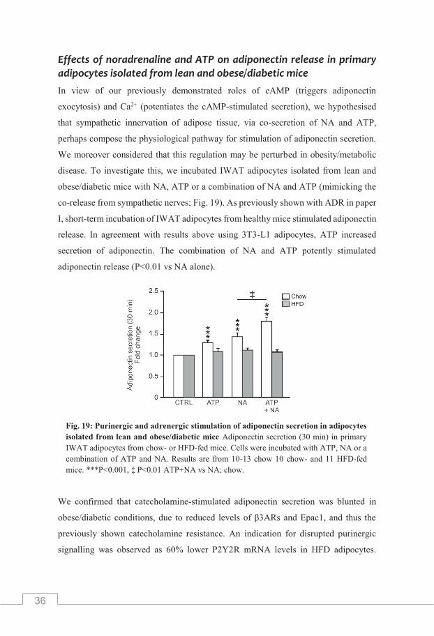
36
Effects of noradrenaline and ATP on adiponectin release in primary adipocytes isolated from lean and obese/diabetic mice In view of our previously demonstrated roles of cAMP (triggers adiponectin
exocytosis) and Ca2+ (potentiates the cAMP-stimulated secretion), we hypothesised
that sympathetic innervation of adipose tissue, via co-secretion of NA and ATP,
perhaps compose the physiological pathway for stimulation of adiponectin secretion.
We moreover considered that this regulation may be perturbed in obesity/metabolic
disease. To investigate this, we incubated IWAT adipocytes isolated from lean and
obese/diabetic mice with NA, ATP or a combination of NA and ATP (mimicking the
co-release from sympathetic nerves; Fig. 19). As previously shown with ADR in paper
I, short-term incubation of IWAT adipocytes from healthy mice stimulated adiponectin
release. In agreement with results above using 3T3-L1 adipocytes, ATP increased
secretion of adiponectin. The combination of NA and ATP potently stimulated
adiponectin release (P<0.01 vs NA alone).
We confirmed that catecholamine-stimulated adiponectin secretion was blunted in
obese/diabetic conditions, due to reduced levels of β3ARs and Epac1, and thus the
previously shown catecholamine resistance. An indication for disrupted purinergic
signalling was observed as 60% lower P2Y2R mRNA levels in HFD adipocytes.
Fig. 19: Purinergic and adrenergic stimulation of adiponectin secretion in adipocytes isolated from lean and obese/diabetic mice Adiponectin secretion (30 min) in primary IWAT adipocytes from chow- or HFD-fed mice. Cells were incubated with ATP, NA or a combination of ATP and NA. Results are from 10-13 chow 10 chow- and 11 HFD-fed mice. ***P<0.001, ‡ P<0.01 ATP+NA vs NA; chow.

37
Furthermore, measured adiponectin cell content was only marginally decreased in
HFD compared to chow adipocytes. However, analysis of the released fraction of the
total adiponectin revealed that HFD adipocytes released a smaller proportion,
indicating defect secretory capacity. Our results suggest that ATP augments the NA-
stimulated adiponectin release in a Ca2+-dependent manner. To validate the
involvement of P2Y2Rs (elevates Ca2+) in the current study, we performed secretion
measurements with the P2Y2R antagonist AR-C 118925XX (AR-C, 1 μM).
In agreement with previous findings, the combination of NA and ATP potently
stimulated adiponectin release. However, the secretory response was significantly
reduced in adipocytes treated with the antagonist.
Obesity-induced reduction of sympathetic sensitivity in white adipose tissue has been
reported (Lonnqvist et al., 1992; Reynisdottir et al., 1994). To examine the SNS
innervation in adipose tissue, we performed immunohistochemical staining of tyrosine
hydroxylase (TH), the rate-limiting enzyme for catecholamine synthesis and a
commonly used marker for sympathetic nerves. Additionally we measured adipose
tissue NA-levels. Indeed, quantification of TH and NA levels revealed reduced SNS
activity in IWAT from HFD-fed mice compared to chow. Our findings propose that
diabetes/obesity is associated with decreased SNS innervation of white adipose tissue,
resulting in blunted catecholamine-stimulated release of adiponectin and that defect
adipocyte purinergic signalling further reduces the adiponectin secretion.
Effects of noradrenaline and ATP on secretion of HMW adiponectin In order to study the effect of purinergic signalling in adiponectin release in more
detail, we measured released HMW adiponectin from IWAT adipocytes exposed to
NA or ATP for 30 minutes (Fig. 20). Preliminary results show that NA, in agreement
with findings with ADR in paper II, stimulated secretion of this molecular form.
ATP on the other hand only induced release of smaller adiponectin forms.
This demonstrates that ATP, in accordance with the finding that the nucleotide is
without effect on cytoplasmic cAMP, has no effect on release of HMW adiponectin.

38
Summary of findings Paper III In paper III of this thesis, we show that, as expected, NA stimulates adiponectin release
of a similar magnitude as that obtained with ADR. Further we show that external ATP
induces adiponectin release via Ca2+-dependent pathways. Interestingly, ATP
augments the NA-triggered adiponectin secretion and this augmentation is diminished
in adipocytes isolated from obese/diabetic mice. The blunted ATP potentiation is
paralleled by a downregulation of purinergic P2Y2R gene expression and decreased
adipose tissue NA levels (Fig. 21). Our studies propose that adiponectin exocytosis is
physiologically regulated by NA and ATP, co-released from sympathetic nerves
innervating the white adipose tissue. The combination of the disruptions of purinergic
signalling and decreased NA levels within the adipose tissue and the documented
catecholamine resistance shown in this thesis, likely jointly contributes to the
obesity/T2D-induced reduced adiponectin release and to the lower adiponectin levels
observed in metabolic disorders.
Fig. 21: Graphical conclusion based on our findings in paper III. See text for further details.
Fig. 20: Effects of NA and purinergic signalling on released HMW adiponectin. Secreted HMW adiponectin in IWAT adipocytes incubated with NA or ATP for 30 min. Results are presented in fold increase. Data are from 4 chow mice. *P<0.05.

39
Paper IV
In paper IV we studied if the pathophysiological regulation of adiponectin release in
visceral adipocytes was similar to that demonstrated in subcutaneous adipocytes
(Paper I-III). Visceral adipocytes have been shown to be more sensitive to
catecholamine stimulation due to high expression of ARs (Arner et al., 1990; Bolinder
et al., 1983). This suggest a role of adrenergic signalling in stimulation of visceral
adipocyte adiponectin release.
Visceral adipocyte adiponectin release is stimulated via β3ARs and Epac1
Incubation of visceral GWAT mouse adipocytes with FSK (10 μM) together with
IBMX (200 μM), ADR (5 μM) or β3-agonist CL (1 μM) stimulated short-term
adiponectin secretion. Measurements of released product revealed that a combination
of FSK/IBMX (elevates cAMP) increased adiponectin secretion >2-fold over basal
whereas the effect of ADR or CL was slightly lower. To study the role of Epac in
visceral adiponectin release, GWAT adipocytes were pretreated with the Epac
antagonist ESI-09 (10 μM for 30 minutes) prior to incubation with ADR (Fig. 22).
Adiponectin release caused by ADR was abolished in cells preincubated with ESI-09.
We propose that visceral adipocyte short-term adiponectin release is stimulated by the
same mechanism as shown in subcutaneous adipocytes (Paper I), involving activation
of β3ARs and Epac1.
Fig. 22: Adrenergic stimulation of adiponectin secretion in GWAT adipocytes is Epac-dependent. Adrenaline-stimulated adiponectin release in isolated primary GWAT adipocytes pretreated with Epac-antagonist ESI-09 for 30 min (10 μM). Data is expressed in fold change and from 7 mice. ***P<0.001.

40
Measurement of total adiponectin content in cells treated with either stimulatory agent,
revealed no difference in cellular levels of the hormone. This confirmed that the
stimulatory effect on adiponectin release was due to effects on the secretory
machinery, and not as a result of increased adiponectin synthesis. Further we showed
that only a small fraction (~1.5 %) of total adiponectin content was release upon
stimulation for 30 min.
Reduced stimulated adiponectin secretion in visceral adipocytes isolated from obese/diabetic mice We next investigated how diet-induced obesity/diabetes affected adrenergically
stimulated adiponectin release using adipocytes isolated from mice fed HFD (Fig. 23).
In GWAT adipocytes isolated from chow-fed mice, ADR or CL could stimulate
adiponectin release. Similar to finding in subcutaneous adipocytes, GWAT adipocytes
isolated from diet induced obese/diabetic mice had reduced levels of secreted
adiponektin upon stimulation with ADR or CL. Unstimulated adiponectin release
(basal) from GWAT was unaffected by HFD-feeding. Total adiponectin content
analysis, showed a small reduction (~20%) of the adipokine in HFD adipocytes
compared to chow controls.
In Paper I we demonstrated that in IWAT adipocytes, HFD results in a complete
abolishment of adrenergically stimulated adiponectin release largely due to reduced
levels of β3ARs and Epac 1. To investigate if similar molecular defects could explain
the reduced stimulated adiponectin release in GWAT adipocytes from obese/diabetic
mice, we performed gene expression analysis of ARs and Epac isoforms.
Fig. 23: Adrenergic stimulation of adiponectin secretion in GWAT cells from lean and obese/diabetic mice. Adrenergically stimulated adiponectin secretion from GWAT adipocytes isolated from chow- and HFD-fed mice. Data are from 10 chow- and 11 HFD-fed mice. **P<0.01; *** P<0.001.

41
Similar to findings in IWAT adipocytes, β3AR was highly expressed in chow GWAT
adipocytes (Fig. 24: left). The β1 and β2 adrenergic receptors were expressed at ~100-
fold lower levels than β3AR (not shown). Also α1DAR was detected in GWAT
adipocytes. Contrary to findings using IWAT adipocytes, there was no change in
mRNA levels of β3AR in HFD adipocytes. Comparable to findings in IWAT
adipocytes, Epac1 was expressed in GWAT adipocytes with no presence of the other
isoform (Fig. 24: right). Interestingly gene expression of Epac1 was doubled in HFD
adipocytes compared to adipocytes isolated from chow-fed mice.
We speculated that perhaps the upregulation of Epac1 was due to lower levels of
intracellular cAMP in the obese/diabetic adipocytes. We measured intracellular cAMP
levels in chow and HFD GWAT adipocytes treated with the stimulatory agents.
Rises of intracellular cAMP triggered by ADR or CL were similar in cells isolated
from chow- and HFD-fed mice. Unaltered levels of cAMP in HFD adipocytes are in
agreement with the detected maintained mRNA level of β3ARs. This is supported by
that in IWAT adipocytes isolated from HFD-fed mice, the cAMP increase in response
to CL was ~60% lower. Our results propose that diet-induced blunted adiponectin
release in visceral (GWAT) mouse adipocytes are caused by other secretory defects
than those described in subcutaneous (IWAT) mouse adipocytes.
Fig. 24: Relative gene expression of β3AR and Epac isoforms in GWAT adipocytes from lean and obese/diabetic mice. Gene expression of ARs (left) and Epac isoforms (right). Data are from 5 chow- and 5 HFD-fed mice. ***P<0.001.

42
Role of exocytotic proteins in blunted adiponectin release from visceral HFD-adipocytes We hypothesised that the reduced stimulated adiponectin release in visceral HFD
adipocytes is perhaps due to defects of the exocytotic process itself. Hormone-
containing vesicle fusion is typically mediated by interaction of SNARE proteins.
Guided by previous publications (Kawanishi et al., 2000; Olson et al., 1997; St-Denis
et al., 1999) and adipocyte micro array data, we decided to measure the expression of
a selection of proteins known to be involved in regulated exocytosis. The SNARE
SNAP-23 is present in adipocytes and has important roles in the translocation of
GLUT4 to the plasma membrane (St-Denis et al., 1999). SNAREs need to interact with
other SNAREs such as syntaxins and vesicle-associated membrane proteins (VAMPs)
in order to complete vesicle fusion (Han et al., 2017). Gene expression analysis
revealed a small reduction of SNAP-23 in HFD adipocytes (P=0.06) whereas levels of
VAMP2 and VAMP4 were more reduced (P<0.05). Also, the small GTPase Rab3a
was expressed at a slightly lower level (P<0.05) whereas Rap1 (isoforms a and b)
remained unaltered. Based on our gene expression data comparing normal and
obese/diabetic conditions, we propose that the defected stimulated adiponectin release
may be due to defects in expression of proteins important for the exocytotic process.
However, several exocytotic proteins are present in white adipocytes and their roles
are incompletely understood.
Summary of findings in paper IV In paper IV, we demonstrate that short-term adiponectin secretion in GWAT
adipocytes is stimulated via β3AR and activation of Epac1 as shown in subcutaneous
adipocytes (Paper I-III). Moreover, we show that stimulated adiponectin release in
GWAT adipocytes isolated from obese/diabetic mice is reduced. It seems that the
underlying mechanism for the disturbed adiponectin release clearly differs between
depots since the gene expression level of β3AR and Epac1 remain unaltered.

43
FINAL DISCUSSION
The metabolic health improving effects of adiponectin is widely recognised and
suggest adiponectin replacement therapy as promising pharmacological treatment of
obesity-related diseases. Eukaryotic cells are a successful (although expensive) model
system for synthesis of all molecular forms of adiponectin. Nonetheless, development
of exogenous adiponectin has experienced many difficulties. First, adiponectin cannot
be administrated orally due to its polypeptide structure. Secondly, intravenous
administration would be challenging to maintain since adiponectin has a short
biological half-life (Halberg et al., 2009). Thus, the prospect to instead increase levels
of endogenous adiponectin is attractive. Studies demonstrate that pharmacological
elevation of circulating adiponectin improves metabolic status (Pajvani et al., 2004)
and can be a promising therapeutic strategy for treatment of metabolic diseases.
Still little is known about the regulation of adiponectin secretion. In order to pave the
way for future treatments aimed at elevating secretion of the body’s own adiponectin,
we must increase our knowledge about the cellular and molecular regulation of the
adiponectin exocytosis process.
Catecholamine resistance As a natural continuation of the findings in (Komai et al., 2014), we show in paper I
of this thesis that adiponectin exocytosis is stimulated via catecholamine-activation of
β3ARs and Epac1 in subcutaneous adipocytes. We further demonstrate that
adrenergically stimulated adiponectin secretion is blunted in obesity/diabetes, in a state
we refer to as catecholamine resistance. Professor Arner with colleagues was first to
describe catecholamine resistance in subcutaneous adipocytes isolated from obese
individuals (Lonnqvist et al., 1992). The observed disturbed lipolytic activity was
attributed to decreased β2AR abundance (~70%) as well as defected downstream
signalling (reduced PKA levels). Similar to those findings, we have now demonstrated
that the inability of subcutaneous adipocytes isolated from obese/diabetic mice to
respond to catecholamine-stimulation with elevated adiponectin release is caused by
lowered density of β3ARs and Epac1.

44
Due to its wide distribution in the body, SAT has been proposed to be more likely to
affect circulating levels of secretory products. Likewise, studies show that
subcutaneous adipocytes secrete more adiponectin compared to visceral fat cells, thus
contributing more to circulating levels of the adipokine (Fisher et al., 2002; Meyer et
al., 2013). In paper I, we observed that catecholamine resistance in the subcutaneous
adipocytes is associated with a reduction of circulating HMW adiponectin (~50%).
Interestingly total adiponectin levels were unaltered in obese/diabetic mice compared
to healthy control mice. We speculated that the increased basal adiponectin release
observed in obese/diabetic adipocytes acts as a compensatory mechanism for reduced
stimulated secretory response, hence maintaining high circulating adiponectin levels.
Adrenergic signalling stimulates release of HMW adiponectin Results from paper I contributed to our hypothesis in paper II that adrenergic signalling
is explicitly involved in the regulation of HMW adiponectin secretion. Short-term
secretion measurements revealed that both ADR and β3-agonist CL triggered the
release of HMW adiponectin. Our findings from cultured and primary mouse
subcutaneous adipocytes were confirmed in isolated primary human subcutaneous
adipocytes. Although the expression of β3AR was dramatically lower in human
adipocytes compared to observations in mouse adipocytes, specific β3AR-activation
resulted in increased release of HMW adiponectin. The coeval finding that insulin also
induces adiponectin secretion lead us to further investigate the similarities and
differences between adiponectin release stimulated via the two signalling pathways.
Our studies demonstrated that insulin acts via different signalling mechanisms and
induces adiponectin release with slower time-kinetics.
The observation that adiponectin secretion was induced by two opposing signalling
molecules may appear controversial from a physiological perspective. In the body,
levels of circulating insulin are high as glucose levels rise following a meal (fed state).
Endocrine functions of insulin include increased glucose uptake in peripheral tissues.
On a cellular level, insulin maintains expression of several transcriptional factors,
essential for lipid and glucose metabolism (Furtado et al., 2002; Stahl et al., 2002).
A small increase of adiponectin content was observed in adipocytes exposed to insulin

45
for 30 minutes, proposing that insulin increases synthesis of adiponectin.
However, results from Professor Han and colleagues demonstrated that inhibition of
protein synthesis did not affect insulin-stimulated adiponectin secretion (Lim et al.,
2015) and the effect of insulin on adiponectin expression need to be further
investigated.
Under the opposite physiological condition, serum insulin drops and circulating
catecholamines increases (fasted state). In addition, adipocyte lipolysis is increased in
the fasted state to satisfy the higher FFA demand of peripheral tissues for energy
consumptions. Considering the complexity of circulating adiponectin molecular
structures, it is feasible that different mechanisms can regulate release of diverse
adiponectin forms. We considered the possibility that insulin stimulates the release of
smaller adiponectin forms. Measurements of secreted product confirmed that short-
term insulin exposure did not stimulate HMW adiponectin secretion but only the
release of smaller adiponectin forms. We found that in adipocytes from obese/diabetic
mice, the stimulated release of both HMW adiponectin and simpler forms were
disturbed due to catecholamine resistance coupled with insulin resistance.
The maintained total adiponectin levels appear to instead be achieved by the diabesity-
associated increase of unregulated release of smaller adiponectin forms.
Role of sympathetic innervation in regulation of adiponectin release
Due to the demonstrated rich innervation of white adipose tissue by the sympathetic
nervous system (Youngstrom & Bartness, 1995), we in paper III determine the role of
NA and ATP that are co-released from sympathetic nerves, in the regulation of
adiponectin exocytosis. We speculated that β3ARs are more likely to be activated by
high NA concentrations released from the close vicinity of the sympathetic nerve
terminal than by catecholamines released from the adrenal medulla into the circulation.
This suggestion was supported by quantification of catecholamine content in rat
IWAT, revealing 80-fold higher levels of NA compared to ADR (Vargovic et al.,
2011), thus implying NA as the main catecholamine regulating adiponectin exocytosis.
Our findings in paper III indeed propose that adiponectin secretion is physiologically
triggered by innervation of sympathetic nerves, NA elevates cAMP levels to trigger

46
adiponectin exocytosis and ATP acts to increase Ca2+ via P2Y2R, thus potentiating the
cAMP-stimulated secretion. This regulation is in agreement with the mechanistic
molecular control shown for cAMP and Ca2+ in our previous publication (Komai et al.,
2014).
The regulation of HMW adiponectin secretion by sympathetic innervation offers a very
precise and rapid means of controlling adiponectin secretion, perhaps in an adipose
depot specific manner. Our preliminary data indicate that only catecholamines/cAMP
trigger the release of HMW adiponectin whereas ATP, like insulin, triggers release of
smaller forms of the adipokine. However, we also demonstrate that the stimulatory
effect of ATP is largely dependent on Ca2+ while insulin-stimulated adiponectin release
occurs without alterations of intracellular Ca2+. In addition, we have previously shown
that Ca2+ can not trigger adipocyte exocytosis in the absence of cytosolic cAMP
(Komai et al., 2014). It seems unlikely that ATP affects the same adiponectin pool as
insulin, as we show that insulin-stimulated adiponectin release is Ca2+-independent.
The results thus indicate that ATP, via Ca2+ elevations (and likely by initiation of
additional signalling cascades), act on a third adiponectin pool. Clearly, the
involvement of diverse signalling pathways in secretion of different adiponectin forms
is perplex and further studies are required in order to resolve the details. .
Adipose-depot specific adiponectin release in health and metabolic disease
In the final work of this thesis (paper IV), we show that catecholamines trigger
adiponectin release also in visceral rodent (gonadal) adipocytes. Similar to results in
subcutaneous adipocytes, regulation of adiponectin secretion involves β3AR and
Epac1 signalling. Interestingly, our results demonstrate a blunted adiponectin release
in gonadal adipocytes from obese/diabetic mice. However, the underlying mechanism
for the disturbed stimulated release in visceral HFD adipocytes differ from the
catecholamine resistance defined in subcutaneous adipocytes as shown by maintained
gene expression of β3ARs and Epac1 as well as largely sustained cAMP levels; the
defects appear to lie further down the exocytotic pathway, perhaps involving proteins
essential for the adiponectin exocytosis process. It is worthy of note that local

47
altercations of cAMP concentrations, in regions near the plasma membrane where
adiponectin exocytosis occurs, may exist. Compartmentalisation of cAMP signalling
molecules such as ARs, ACs and PDEs has been reported. Furthermore, formation of
cellular cAMP gradients have been observed (Houslay & Adams, 2003) as well as a
restricted distribution of Epac1 (Schmidt et al., 2013). It is thus possible that the
signalling involved in regulation of adiponectin exocytosis/secretion is localised to
specific cellular micro-domains containing the key regulatory molecular players
(β3AR and Epac1) and that the local signalling at these sites is disturbed.
Interestingly, in cardiomyocytes, β2ARs have been suggested to be located in
cholesterol rich structures called caveolae. These lipid rich structures are enriched in
white adipocytes and are proposed to be responsible for heterogeneous distribution of
signalling proteins in different micro-domains. Unpublished findings from our group
suggest that caveolae are important for adiponectin secretion (Brännmark et al., in
preparation).
The data in this thesis propose that similar mechanisms regulate adiponectin secretion
in subcutaneous and visceral adiponectin but that different release disturbances arise
in association to obesity and diabetes. Subcutaneous and visceral adipose tissues have
different origin (Chau et al., 2014) and also differ in metabolic functions (Wajchenberg
et al., 2002). Adipocytes in visceral fat are more metabolically active as demonstrated
by increased lipolytic activity and thus higher expression of βARs together with lower
insulin-sensitivity (Arner et al., 1990; Bolinder et al., 1983). Furthermore, the size of
visceral fat mass correlates positively to insulin resistance (Kloting et al., 2010).
Individuals with enlarged VAT are at a higher risk for developing obesity-related
diseases. The anatomical position of VAT, with direct portal drainage to the liver
(Rytka et al., 2011), enables hormones such as adiponectin released from VAT to have
a direct effect on glucose metabolism though paracrine effects. Considering the above,
the similarities with regard to adiponectin release mechanisms between subcutaneous
and visceral adipocytes may actually appear more surprising than the differences.
48
Pathophysiological relevance The importance of regulated secretion of a hormone as abundant in the circulation as
adiponectin may be questioned. Also, even if adrenergic stimulation specifically
regulates release of HMW adiponectin, this form also exists at rather constantly high
serum levels (30-50% of the total adiponectin; Aso et al., 2006; Bluher et al., 2007;
Hamilton et al., 2011). Other endocrine hormones typically circulate at low levels and
are rapidly and transiently increased upon physiological demand (such as insulin that
systemically increases in response to food intake). We propose that the answer lies
within the distinct characteristics of adipose tissue in combination with where the
released adiponectin is to exert its effects.
White adipose tissue differs from other hormone releasing organs in that it is located
in diverse regional depots and contribute to as much as a fifth of the whole body mass.
The wide distribution of white adipose tissue expectedly maintains the incessantly high
systemic levels of adiponectin. It is worth speculating that the regulated secretion of
adiponectin, in particular the HMW form, is perhaps more important for local
autocrine/paracrine actions and not so much for endocrine effects. Thus, the regulated
exocytotic pathway of HMW adiponectin may exist in order to locally increase the
hormone to very high levels, acting at nearby cells, tissues and organs. The close
anatomical location of visceral adipose tissue to several metabolically active organs
such as pancreas and liver suggests an important role for visceral adiponectin release.
An alternative/complementing explanation is that the regulated secretion of HMW
adiponectin does not necessarily exist with the aim to induce acute effects on the target
tissues but instead to preserve the high circulating levels. Our data in paper I support
this hypothesis, as we demonstrate that serum levels of HMW adiponectin are
decreased by ~50% in obese and catecholamine resistant mice. Our data defining the
regulation of adiponectin secretion (HMW and smaller forms) is so far largely limited
to results from subcutaneous adipocytes. Other adipose tissue depots, such as VAT
that has been shown to differ from SAT need to be more thoroughly investigated with
respect to the regulated adiponectin release.

49
The finding presented here, showing that secretion of HMW and smaller forms of
adiponectin are stimulated under opposite metabolic conditions propose that the
regulation of adiponectin is a very complex process. More detailed future research
defining the release of specific adiponectin forms and its molecular and cellular
regulation is required to fully explain the role of adiponectin in metabolic disease.
However, we are of the opinion that our studies jointly propose that adiponectin
secretory defects underlie the reduced circulating HMW adiponectin levels reported in
metabolic disease.

50

51
ACKNOWLEDGMENTS First, my sincerest thanks to all of my colleagues, past and present, at the unit of Metabolic
Physiology for all their help and support during my PhD.
To my main supervisor, Charlotta Olofsson. Thank you for giving me the opportunity to join
your research group, first as a master student and then as a PhD student. For guiding me on the
road to become an independent researcher.
Thanks to my co-supervisors, Ingrid W Asterholm and Professor Patrik Rorsman. To all the co-
authors that have contributed to the experimental work included in this thesis. A special warm
thanks to all the great students that I have supervised over the years.
Ali, you have been my primary source for scientific discussions and an instrumental part of this
thesis work. Thank you for your compassion and endless support. You are truly a great academic
role model!
My all-time favourite duo, Birgit and Ann-Marie (BLAMA). Thank you for all the positive
encouragement and reinforcement in times of hopelessness. But also for the daily laughs and
anecdotes
Thanks to my office-buddies Ahmed, Belen and Marina for providing a relaxed and enjoyable
atmosphere.
To my oldest and dearest friend Karin, for listening when I needed to ventilate and always
providing good advice.
My parents Nazif and Mejra, for always being there for me no matter the circumstances. You are
the definition of loving parents.
To my brother and best friend Edin, for always believing in me and assuring me of my
capabilities all throughout life. You are my greatest supporter!
Financial support for studies included in this thesis were supported by the Swedish Diabetes
Foundation, the Knut and Alice Wallenberg Foundation (P. Rorsman) and the Swedish Medical
Research Council.

52
REFERENCES
Anthonsen, M. W., Ronnstrand, L., Wernstedt, C., Degerman, E., & Holm, C. (1998). Identification of novel phosphorylation sites in hormone-sensitive lipase that are phosphorylated in response to isoproterenol and govern activation properties in vitro. J Biol Chem, 273(1), 215-221
Arita, Y., Kihara, S., Ouchi, N., Takahashi, M., Maeda, K., Miyagawa, J., Hotta, K., Shimomura, I., Nakamura, T., Miyaoka, K., Kuriyama, H., Nishida, M., Yamashita, S., Okubo, K., Matsubara, K., Muraguchi, M., Ohmoto, Y., Funahashi, T., & Matsuzawa, Y. (1999). Paradoxical decrease of an adipose-specific protein, adiponectin, in obesity. Biochem Biophys Res Commun, 257(1), 79-83
Arner, P., Hellstrom, L., Wahrenberg, H., & Bronnegard, M. (1990). Beta-adrenoceptor expression in human fat cells from different regions. J Clin Invest, 86(5), 1595-1600
Aso, Y., Yamamoto, R., Wakabayashi, S., Uchida, T., Takayanagi, K., Takebayashi, K., Okuno, T., Inoue, T., Node, K., Tobe, T., Inukai, T., & Nakano, Y. (2006). Comparison of serum high-molecular weight (HMW) adiponectin with total adiponectin concentrations in type 2 diabetic patients with coronary artery disease using a novel enzyme-linked immunosorbent assay to detect HMW adiponectin. Diabetes, 55(7), 1954-1960
Barg, S., Eliasson, L., Renstrom, E., & Rorsman, P. (2002). A subset of 50 secretory granules in close contact with L-type Ca2+ channels accounts for first-phase insulin secretion in mouse beta-cells. Diabetes, 51 Suppl 1, S74-82
Bartness, T. J., Liu, Y., Shrestha, Y. B., & Ryu, V. (2014). Neural innervation of white adipose tissue and the control of lipolysis. Front Neuroendocrinol, 35(4), 473-493
Bartness, T. J., Shrestha, Y. B., Vaughan, C. H., Schwartz, G. J., & Song, C. K. (2010). Sensory and sympathetic nervous system control of white adipose tissue lipolysis. Mol Cell Endocrinol, 318(1-2), 34-43
Baskin, A. S., Linderman, J. D., Brychta, R. J., McGehee, S., Anflick-Chames, E., Cero, C., Johnson, J. W., O'Mara, A. E., Fletcher, L. A., Leitner, B. P., Duckworth, C. J., Huang, S., Cai, H., Garraffo, H. M., Millo, C. M., Dieckmann, W., Tolstikov, V., Chen, E. Y., Gao, F., Narain, N. R., Kiebish, M. A., Walter, P. J., Herscovitch, P., Chen, K. Y., & Cypess, A. M. (2018). Regulation of Human Adipose Tissue Activation, Gallbladder Size, and Bile Acid Metabolism by a beta3-Adrenergic Receptor Agonist. Diabetes, 67(10), 2113-2125
Berkowitz, D. E., Nardone, N. A., Smiley, R. M., Price, D. T., Kreutter, D. K., Fremeau, R. T., & Schwinn, D. A. (1995). Distribution of beta 3-adrenoceptor mRNA in human tissues. Eur J Pharmacol, 289(2), 223-228
Bluher, M., Brennan, A. M., Kelesidis, T., Kratzsch, J., Fasshauer, M., Kralisch, S., Williams, C. J., & Mantzoros, C. S. (2007). Total and high-molecular weight adiponectin in relation to metabolic variables at baseline and in response to an exercise treatment program: comparative evaluation of three assays. Diabetes Care, 30(2), 280-285
Blumer, R. M., van Roomen, C. P., Meijer, A. J., Houben-Weerts, J. H., Sauerwein, H. P., & Dubbelhuis, P. F. (2008). Regulation of adiponectin secretion by insulin and amino acids in 3T3-L1 adipocytes. Metabolism, 57(12), 1655-1662
Bogan, J. S., & Lodish, H. F. (1999). Two compartments for insulin-stimulated exocytosis in 3T3-L1 adipocytes defined by endogenous ACRP30 and GLUT4. J Cell Biol, 146(3), 609-620
Bolinder, J., Kager, L., Ostman, J., & Arner, P. (1983). Differences at the receptor and postreceptor levels between human omental and subcutaneous adipose tissue in the action of insulin on lipolysis. Diabetes, 32(2), 117-123
Burgoyne, R. D., & Morgan, A. (2003). Secretory granule exocytosis. Physiol Rev, 83(2), 581-632 Burnstock, G. (2007). Physiology and pathophysiology of purinergic neurotransmission. Physiol Rev, 87(2),
659-797 Chau, Y. Y., Bandiera, R., Serrels, A., Martinez-Estrada, O. M., Qing, W., Lee, M., Slight, J., Thornburn,
A., Berry, R., McHaffie, S., Stimson, R. H., Walker, B. R., Chapuli, R. M., Schedl, A., & Hastie, N. (2014). Visceral and subcutaneous fat have different origins and evidence supports a mesothelial source. Nat Cell Biol, 16(4), 367-375
Chusyd, D. E., Wang, D., Huffman, D. M., & Nagy, T. R. (2016). Relationships between Rodent White Adipose Fat Pads and Human White Adipose Fat Depots. Front Nutr, 3, 10
Combs, T. P., Berg, A. H., Obici, S., Scherer, P. E., & Rossetti, L. (2001). Endogenous glucose production is inhibited by the adipose-derived protein Acrp30. J Clin Invest, 108(12), 1875-1881
53
Combs, T. P., Berg, A. H., Rajala, M. W., Klebanov, S., Iyengar, P., Jimenez-Chillaron, J. C., Patti, M. E., Klein, S. L., Weinstein, R. S., & Scherer, P. E. (2003). Sexual differentiation, pregnancy, calorie restriction, and aging affect the adipocyte-specific secretory protein adiponectin. Diabetes, 52(2), 268-276
Cong, L., Chen, K., Li, J., Gao, P., Li, Q., Mi, S., Wu, X., & Zhao, A. Z. (2007). Regulation of adiponectin and leptin secretion and expression by insulin through a PI3K-PDE3B dependent mechanism in rat primary adipocytes. Biochem J, 403(3), 519-525
Cook, K. S., Min, H. Y., Johnson, D., Chaplinsky, R. J., Flier, J. S., Hunt, C. R., & Spiegelman, B. M. (1987). Adipsin: a circulating serine protease homolog secreted by adipose tissue and sciatic nerve. Science, 237(4813), 402-405
Dadson, K., S. Turdi, S. Hashemi, J. Zhao, N. Polidovitch, S. Beca, P. H. Backx, J. C. McDermott, and G. Sweeney. 2015. 'Adiponectin is required for cardiac MEF2 activation during pressure overload induced hypertrophy', J Mol Cell Cardiol, 86: 102-9.
Degerman, E., Ahmad, F., Chung, Y. W., Guirguis, E., Omar, B., Stenson, L., & Manganiello, V. (2011). From PDE3B to the regulation of energy homeostasis. Curr Opin Pharmacol, 11(6), 676-682
Delporte, M. L., Funahashi, T., Takahashi, M., Matsuzawa, Y., & Brichard, S. M. (2002). Pre- and post-translational negative effect of beta-adrenoceptor agonists on adiponectin secretion: in vitro and in vivo studies. Biochem J, 367(Pt 3), 677-685
Detimary, P., Jonas, J. C., & Henquin, J. C. (1996). Stable and diffusible pools of nucleotides in pancreatic islet cells. Endocrinology, 137(11), 4671-4676
Duncan, R. E., Ahmadian, M., Jaworski, K., Sarkadi-Nagy, E., & Sul, H. S. (2007). Regulation of lipolysis in adipocytes. Annu Rev Nutr, 27, 79-101
El Hachmane, M. F., Ermund, A., Brannmark, C., & Olofsson, C. S. (2018). Extracellular ATP activates store-operated Ca(2+) entry in white adipocytes: functional evidence for STIM1 and ORAI1. Biochem J, 475(3), 691-704
El Hachmane, M. F., Komai, A. M., & Olofsson, C. S. (2015). Cooling reduces cAMP-stimulated exocytosis and adiponectin secretion at a Ca2+-dependent step in 3T3-L1 adipocytes. PLoS One, 10(3), e0119530
Fasshauer, M., Klein, J., Neumann, S., Eszlinger, M., & Paschke, R. (2001a). Adiponectin gene expression is inhibited by beta-adrenergic stimulation via protein kinase A in 3T3-L1 adipocytes. FEBS Lett, 507(2), 142-146
Fasshauer, M., Klein, J., Neumann, S., Eszlinger, M., & Paschke, R. (2001b). Tumor necrosis factor alpha is a negative regulator of resistin gene expression and secretion in 3T3-L1 adipocytes. Biochem Biophys Res Commun, 288(4), 1027-1031
Fisher, F. M., McTernan, P. G., Valsamakis, G., Chetty, R., Harte, A. L., Anwar, A. J., Starcynski, J., Crocker, J., Barnett, A. H., McTernan, C. L., & Kumar, S. (2002). Differences in adiponectin protein expression: effect of fat depots and type 2 diabetic status. Horm Metab Res, 34(11-12), 650-654
Furtado, L. M., Somwar, R., Sweeney, G., Niu, W., & Klip, A. (2002). Activation of the glucose transporter GLUT4 by insulin. Biochem Cell Biol, 80(5), 569-578
Greenberger, J. S., & Aaronson, S. A. (1974). Morphologic revertants of murine sarcoma virus transformed nonproducer BALB-3T3: selective techniques for isolation and biologic properties in vitro and in vivo. Virology, 57(2), 339-346
Grynkiewicz, G., Poenie, M., & Tsien, R. Y. (1985). A new generation of Ca2+ indicators with greatly improved fluorescence properties. J Biol Chem, 260(6), 3440-3450
Halberg, N., Schraw, T. D., Wang, Z. V., Kim, J. Y., Yi, J., Hamilton, M. P., Luby-Phelps, K., & Scherer, P. E. (2009). Systemic fate of the adipocyte-derived factor adiponectin. Diabetes, 58(9), 1961-1970
Hamilton, M. P., Gore, M. O., Ayers, C. R., Xinyu, W., McGuire, D. K., & Scherer, P. E. (2011). Adiponectin and cardiovascular risk profile in patients with type 2 diabetes mellitus: parameters associated with adiponectin complex distribution. Diab Vasc Dis Res, 8(3), 190-194
Han, J., Pluhackova, K., & Bockmann, R. A. (2017). The Multifaceted Role of SNARE Proteins in Membrane Fusion. Front Physiol, 8, 5
Hara, K., Horikoshi, M., Yamauchi, T., Yago, H., Miyazaki, O., Ebinuma, H., Imai, Y., Nagai, R., & Kadowaki, T. (2006). Measurement of the high-molecular weight form of adiponectin in plasma is useful for the prediction of insulin resistance and metabolic syndrome. Diabetes Care, 29(6), 1357-1362
Heinemann, C., Chow, R. H., Neher, E., & Zucker, R. S. (1994). Kinetics of the secretory response in bovine chromaffin cells following flash photolysis of caged Ca2+. Biophys J, 67(6), 2546-2557

54
Hellstrom, L., Rossner, S., Hagstrom-Toft, E., & Reynisdottir, S. (1997). Lipolytic catecholamine resistance linked to alpha 2-adrenoceptor sensitivity--a metabolic predictor of weight loss in obese subjects. Int J Obes Relat Metab Disord, 21(4), 314-320
Holland, W. L., R. A. Miller, Z. V. Wang, K. Sun, B. M. Barth, H. H. Bui, K. E. Davis, B. T. Bikman, N. Halberg, J. M. Rutkowski, M. R. Wade, V. M. Tenorio, M. S. Kuo, J. T. Brozinick, B. B. Zhang, M. J. Birnbaum, S. A. Summers, and P. E. Scherer. 2011. 'Receptor-mediated activation of ceramidase activity initiates the pleiotropic actions of adiponectin', Nat Med, 17: 55-63.
Holz, G. G., Kang, G., Harbeck, M., Roe, M. W., & Chepurny, O. G. (2006). Cell physiology of cAMP sensor Epac. J Physiol, 577(Pt 1), 5-15
Houslay, M. D., & Adams, D. R. (2003). PDE4 cAMP phosphodiesterases: modular enzymes that orchestrate signalling cross-talk, desensitization and compartmentalization. Biochemical Journal, 370(1), 1-18
Hu, E., Liang, P., & Spiegelman, B. M. (1996). AdipoQ is a novel adipose-specific gene dysregulated in obesity. J Biol Chem, 271(18), 10697-10703
Huang, S., Lifshitz, L. M., Jones, C., Bellve, K. D., Standley, C., Fonseca, S., Corvera, S., Fogarty, K. E., & Czech, M. P. (2007). Insulin stimulates membrane fusion and GLUT4 accumulation in clathrin coats on adipocyte plasma membranes. Mol Cell Biol, 27(9), 3456-3469
Hug, C., Wang, J., Ahmad, N. S., Bogan, J. S., Tsao, T. S., & Lodish, H. F. (2004). T-cadherin is a receptor for hexameric and high-molecular-weight forms of Acrp30/adiponectin. Proc Natl Acad Sci U S A, 101(28), 10308-10313
Iwaki, M., Matsuda, M., Maeda, N., Funahashi, T., Matsuzawa, Y., Makishima, M., & Shimomura, I. (2003). Induction of adiponectin, a fat-derived antidiabetic and antiatherogenic factor, by nuclear receptors. Diabetes, 52(7), 1655-1663
Jia, B., Madsen, L., Petersen, R. K., Techer, N., Kopperud, R., Ma, T., Doskeland, S. O., Ailhaud, G., Wang, J., Amri, E. Z., & Kristiansen, K. (2012). Activation of protein kinase A and exchange protein directly activated by cAMP promotes adipocyte differentiation of human mesenchymal stem cells. PLoS One, 7(3), e34114
Kanneganti, T. D., & Dixit, V. D. (2012). Immunological complications of obesity. Nat Immunol, 13(8), 707-712
Karastergiou, K., Smith, S. R., Greenberg, A. S., & Fried, S. K. (2012). Sex differences in human adipose tissues - the biology of pear shape. Biol Sex Differ, 3(1), 13
Kasai, H., Takahashi, N., & Tokumaru, H. (2012). Distinct initial SNARE configurations underlying the diversity of exocytosis. Physiol Rev, 92(4), 1915-1964
Kawanishi, M., Tamori, Y., Okazawa, H., Araki, S., Shinoda, H., & Kasuga, M. (2000). Role of SNAP23 in insulin-induced translocation of GLUT4 in 3T3-L1 adipocytes. Mediation of complex formation between syntaxin4 and VAMP2. J Biol Chem, 275(11), 8240-8247
Kelesidis, T., Kelesidis, I., Chou, S., & Mantzoros, C. S. (2010). Narrative review: the role of leptin in human physiology: emerging clinical applications. Ann Intern Med, 152(2), 93-100
Kershaw, E. E., & Flier, J. S. (2004). Adipose tissue as an endocrine organ. J Clin Endocrinol Metab, 89(6), 2548-2556
Kim, J. Y., van de Wall, E., Laplante, M., Azzara, A., Trujillo, M. E., Hofmann, S. M., Schraw, T., Durand, J. L., Li, H., Li, G., Jelicks, L. A., Mehler, M. F., Hui, D. Y., Deshaies, Y., Shulman, G. I., Schwartz, G. J., & Scherer, P. E. (2007). Obesity-associated improvements in metabolic profile through expansion of adipose tissue. J Clin Invest, 117(9), 2621-2637
Kloting, N., Fasshauer, M., Dietrich, A., Kovacs, P., Schon, M. R., Kern, M., Stumvoll, M., & Bluher, M. (2010). Insulin-sensitive obesity. Am J Physiol Endocrinol Metab, 299(3), E506-515
Komai, A. M., Brannmark, C., Musovic, S., & Olofsson, C. S. (2014). PKA-independent cAMP stimulation of white adipocyte exocytosis and adipokine secretion: modulations by Ca2+ and ATP. J Physiol, 592(23), 5169-5186
Komai, A. M., Musovic, S., Peris, E., Alrifaiy, A., El Hachmane, M. F., Johansson, M., Wernstedt Asterholm, I., & Olofsson, C. S. (2016). White Adipocyte Adiponectin Exocytosis Is Stimulated via beta3-Adrenergic Signaling and Activation of Epac1: Catecholamine Resistance in Obesity and Type 2 Diabetes. Diabetes, 65(11), 3301-3313
Krause, M. P., Liu, Y., Vu, V., Chan, L., Xu, A., Riddell, M. C., Sweeney, G., & Hawke, T. J. (2008). Adiponectin is expressed by skeletal muscle fibers and influences muscle phenotype and function. Am J Physiol Cell Physiol, 295(1), C203-212
Krief, S., Lonnqvist, F., Raimbault, S., Baude, B., Van Spronsen, A., Arner, P., Strosberg, A. D., Ricquier, D., & Emorine, L. J. (1993). Tissue distribution of beta 3-adrenergic receptor mRNA in man. J Clin Invest, 91(1), 344-349

55
Kuoppamaa, H., Skrobuk, P., Sihvo, M., Hiukka, A., Chibalin, A. V., Zierath, J. R., & Koistinen, H. A. (2008). Globular adiponectin stimulates glucose transport in type 2 diabetic muscle. Diabetes Metab Res Rev, 24(7), 554-562
Kusminski, C. M., McTernan, P. G., Schraw, T., Kos, K., O'Hare, J. P., Ahima, R., Kumar, S., & Scherer, P. E. (2007). Adiponectin complexes in human cerebrospinal fluid: distinct complex distribution from serum. Diabetologia, 50(3), 634-642
Lafontan, M., & Berlan, M. (1993). Fat cell adrenergic receptors and the control of white and brown fat cell function. J Lipid Res, 34(7), 1057-1091
Laplante, M. A., Monassier, L., Freund, M., Bousquet, P., & Gachet, C. (2010). The purinergic P2Y1 receptor supports leptin secretion in adipose tissue. Endocrinology, 151(5), 2060-2070
Lim, C. Y., Hong, W., & Han, W. (2015). Adiponectin is released via a unique regulated exocytosis pathway from a pre-formed vesicle pool on insulin stimulation. Biochem J, 471(3), 381-389
Liu, M., Xiang, R., Wilk, S. A., Zhang, N., Sloane, L. B., Azarnoush, K., Zhou, L., Chen, H., Xiang, G., Walter, C. A., Austad, S. N., Musi, N., DeFronzo, R. A., Asmis, R., Scherer, P. E., Dong, L. Q., & Liu, F. (2012). Fat-specific DsbA-L overexpression promotes adiponectin multimerization and protects mice from diet-induced obesity and insulin resistance. Diabetes, 61(11), 2776-2786
Liu, Q., Yuan, B., Lo, K. A., Patterson, H. C., Sun, Y., & Lodish, H. F. (2012). Adiponectin regulates expression of hepatic genes critical for glucose and lipid metabolism. Proc Natl Acad Sci U S A, 109(36), 14568-14573
Lo, J. C., Ljubicic, S., Leibiger, B., Kern, M., Leibiger, I. B., Moede, T., Kelly, M. E., Chatterjee Bhowmick, D., Murano, I., Cohen, P., Banks, A. S., Khandekar, M. J., Dietrich, A., Flier, J. S., Cinti, S., Bluher, M., Danial, N. N., Berggren, P. O., & Spiegelman, B. M. (2014). Adipsin is an adipokine that improves beta cell function in diabetes. Cell, 158(1), 41-53
Lonnqvist, F., Wahrenberg, H., Hellstrom, L., Reynisdottir, S., & Arner, P. (1992). Lipolytic catecholamine resistance due to decreased beta 2-adrenoceptor expression in fat cells. J Clin Invest, 90(6), 2175-2186
Lonnqvist, F., Wennlund, A., & Arner, P. (1997). Relationship between circulating leptin and peripheral fat distribution in obese subjects. Int J Obes Relat Metab Disord, 21(4), 255-260
Maeda, N., Takahashi, M., Funahashi, T., Kihara, S., Nishizawa, H., Kishida, K., Nagaretani, H., Matsuda, M., Komuro, R., Ouchi, N., Kuriyama, H., Hotta, K., Nakamura, T., Shimomura, I., & Matsuzawa, Y. (2001). PPARgamma ligands increase expression and plasma concentrations of adiponectin, an adipose-derived protein. Diabetes, 50(9), 2094-2099
Meyer, L. K., Ciaraldi, T. P., Henry, R. R., Wittgrove, A. C., & Phillips, S. A. (2013). Adipose tissue depot and cell size dependency of adiponectin synthesis and secretion in human obesity. Adipocyte, 2(4), 217-226
Nawrocki, A. R., Rajala, M. W., Tomas, E., Pajvani, U. B., Saha, A. K., Trumbauer, M. E., Pang, Z., Chen, A. S., Ruderman, N. B., Chen, H., Rossetti, L., & Scherer, P. E. (2006). Mice lacking adiponectin show decreased hepatic insulin sensitivity and reduced responsiveness to peroxisome proliferator-activated receptor gamma agonists. J Biol Chem, 281(5), 2654-2660
Neher, E., & Marty, A. (1982). Discrete changes of cell membrane capacitance observed under conditions of enhanced secretion in bovine adrenal chromaffin cells. Proc Natl Acad Sci U S A, 79(21), 6712-6716
Novara, M., Baldelli, P., Cavallari, D., Carabelli, V., Giancippoli, A., & Carbone, E. (2004). Exposure to cAMP and beta-adrenergic stimulation recruits Ca(V)3 T-type channels in rat chromaffin cells through Epac cAMP-receptor proteins. J Physiol, 558(Pt 2), 433-449
Olofsson, C. S., Hakansson, J., Salehi, A., Bengtsson, M., Galvanovskis, J., Partridge, C., SorhedeWinzell, M., Xian, X., Eliasson, L., Lundquist, I., Semb, H., & Rorsman, P. (2009). Impaired insulin exocytosis in neural cell adhesion molecule-/- mice due to defective reorganization of the submembrane F-actin network. Endocrinology, 150(7), 3067-3075
Olson, A. L., Knight, J. B., & Pessin, J. E. (1997). Syntaxin 4, VAMP2, and/or VAMP3/cellubrevin are functional target membrane and vesicle SNAP receptors for insulin-stimulated GLUT4 translocation in adipocytes. Mol Cell Biol, 17(5), 2425-2435
Otabe, S., Yuan, X., Fukutani, T., Wada, N., Hashinaga, T., Nakayama, H., Hirota, N., Kojima, M., & Yamada, K. (2007). Overexpression of human adiponectin in transgenic mice results in suppression of fat accumulation and prevention of premature death by high-calorie diet. Am J Physiol Endocrinol Metab, 293(1), E210-218
Pajvani, U. B., Du, X., Combs, T. P., Berg, A. H., Rajala, M. W., Schulthess, T., Engel, J., Brownlee, M., & Scherer, P. E. (2003). Structure-function studies of the adipocyte-secreted hormone Acrp30/adiponectin. Implications fpr metabolic regulation and bioactivity. J Biol Chem, 278(11), 9073-9085

56
Pajvani, U. B., Hawkins, M., Combs, T. P., Rajala, M. W., Doebber, T., Berger, J. P., Wagner, J. A., Wu, M., Knopps, A., Xiang, A. H., Utzschneider, K. M., Kahn, S. E., Olefsky, J. M., Buchanan, T. A., & Scherer, P. E. (2004). Complex distribution, not absolute amount of adiponectin, correlates with thiazolidinedione-mediated improvement in insulin sensitivity. J Biol Chem, 279(13), 12152-12162
Panidis, D., Kourtis, A., Farmakiotis, D., Mouslech, T., Rousso, D., & Koliakos, G. (2003). Serum adiponectin levels in women with polycystic ovary syndrome. Hum Reprod, 18(9), 1790-1796
Pereira, R. I., & Draznin, B. (2005). Inhibition of the phosphatidylinositol 3'-kinase signaling pathway leads to decreased insulin-stimulated adiponectin secretion from 3T3-L1 adipocytes. Metabolism, 54(12), 1636-1643
Petersen, R. K., Madsen, L., Pedersen, L. M., Hallenborg, P., Hagland, H., Viste, K., Doskeland, S. O., & Kristiansen, K. (2008). Cyclic AMP (cAMP)-mediated stimulation of adipocyte differentiation requires the synergistic action of Epac- and cAMP-dependent protein kinase-dependent processes. Mol Cell Biol, 28(11), 3804-3816
Phillips, S. A., Kung, J., Ciaraldi, T. P., Choe, C., Christiansen, L., Mudaliar, S., & Henry, R. R. (2009). Selective regulation of cellular and secreted multimeric adiponectin by antidiabetic therapies in humans. Am J Physiol Endocrinol Metab, 297(3), E767-773
Qiang, L., Wang, H., & Farmer, S. R. (2007). Adiponectin secretion is regulated by SIRT1 and the endoplasmic reticulum oxidoreductase Ero1-L alpha. Mol Cell Biol, 27(13), 4698-4707
Revelli, J. P., Muzzin, P., Paoloni, A., Moinat, M., & Giacobino, J. P. (1993). Expression of the beta 3-adrenergic receptor in human white adipose tissue. J Mol Endocrinol, 10(2), 193-197
Reynisdottir, S., Wahrenberg, H., Carlstrom, K., Rossner, S., & Arner, P. (1994). Catecholamine resistance in fat cells of women with upper-body obesity due to decreased expression of beta 2-adrenoceptors. Diabetologia, 37(4), 428-435
Rojas, E., Pollard, H. B., & Heldman, E. (1985). Real-time measurements of acetylcholine-induced release of ATP from bovine medullary chromaffin cells. FEBS Lett, 185(2), 323-327
Ruan, H., Zarnowski, M. J., Cushman, S. W., & Lodish, H. F. (2003). Standard isolation of primary adipose cells from mouse epididymal fat pads induces inflammatory mediators and down-regulates adipocyte genes. J Biol Chem, 278(48), 47585-47593
Rutkowski, J. M., Stern, J. H., & Scherer, P. E. (2015). The cell biology of fat expansion. J Cell Biol, 208(5), 501-512
Rytka, J. M., Wueest, S., Schoenle, E. J., & Konrad, D. (2011). The portal theory supported by venous drainage-selective fat transplantation. Diabetes, 60(1), 56-63
Saltevo, J., Laakso, M., Jokelainen, J., Keinanen-Kiukaanniemi, S., Kumpusalo, E., & Vanhala, M. (2008). Levels of adiponectin, C-reactive protein and interleukin-1 receptor antagonist are associated with insulin sensitivity: a population-based study. Diabetes Metab Res Rev, 24(5), 378-383
Scherer, P. E., Williams, S., Fogliano, M., Baldini, G., & Lodish, H. F. (1995). A novel serum protein similar to C1q, produced exclusively in adipocytes. J Biol Chem, 270(45), 26746-26749
Schmidt, M., Dekker, F. J., & Maarsingh, H. (2013). Exchange protein directly activated by cAMP (epac): a multidomain cAMP mediator in the regulation of diverse biological functions. Pharmacol Rev, 65(2), 670-709
Seydoux, J., Muzzin, P., Moinat, M., Pralong, W., Girardier, L., & Giacobino, J. P. (1996). Adrenoceptor heterogeneity in human white adipocytes differentiated in culture as assessed by cytosolic free calcium measurements. Cell Signal, 8(2), 117-122
Shibasaki, T., Takahashi, H., Miki, T., Sunaga, Y., Matsumura, K., Yamanaka, M., Zhang, C., Tamamoto, A., Satoh, T., Miyazaki, J., & Seino, S. (2007). Essential role of Epac2/Rap1 signaling in regulation of insulin granule dynamics by cAMP. Proc Natl Acad Sci U S A, 104(49), 19333-19338
Shimada, K., Miyazaki, T., & Daida, H. (2004). Adiponectin and atherosclerotic disease. Clin Chim Acta, 344(1-2), 1-12
Simanshu, D. K., Nissley, D. V., & McCormick, F. (2017). RAS Proteins and Their Regulators in Human Disease. Cell, 170(1), 17-33
Spranger, J., Kroke, A., Mohlig, M., Bergmann, M. M., Ristow, M., Boeing, H., & Pfeiffer, A. F. (2003). Adiponectin and protection against type 2 diabetes mellitus. Lancet, 361(9353), 226-228
Spranger, J., Verma, S., Gohring, I., Bobbert, T., Seifert, J., Sindler, A. L., Pfeiffer, A., Hileman, S. M., Tschop, M., & Banks, W. A. (2006). Adiponectin does not cross the blood-brain barrier but modifies cytokine expression of brain endothelial cells. Diabetes, 55(1), 141-147
St-Denis, J. F., Cabaniols, J. P., Cushman, S. W., & Roche, P. A. (1999). SNAP-23 participates in SNARE complex assembly in rat adipose cells. Biochem J, 338 ( Pt 3), 709-715

57
Stahl, A., Evans, J. G., Pattel, S., Hirsch, D., & Lodish, H. F. (2002). Insulin causes fatty acid transport protein translocation and enhanced fatty acid uptake in adipocytes. Dev Cell, 2(4), 477-488
Tabara, Y., Osawa, H., Kawamoto, R., Tachibana-Iimori, R., Yamamoto, M., Nakura, J., Miki, T., Makino, H., & Kohara, K. (2008). Reduced high-molecular-weight adiponectin and elevated high-sensitivity C-reactive protein are synergistic risk factors for metabolic syndrome in a large-scale middle-aged to elderly population: the Shimanami Health Promoting Program Study. J Clin Endocrinol Metab, 93(3), 715-722
Tate, K. M., Briend-Sutren, M. M., Emorine, L. J., Delavier-Klutchko, C., Marullo, S., & Strosberg, A. D. (1991). Expression of three human beta-adrenergic-receptor subtypes in transfected Chinese hamster ovary cells. Eur J Biochem, 196(2), 357-361
Tozzi, M., & Novak, I. (2017). Purinergic Receptors in Adipose Tissue As Potential Targets in Metabolic Disorders. Front Pharmacol, 8, 878
Trujillo, M. E., & Scherer, P. E. (2006). Adipose tissue-derived factors: impact on health and disease. Endocr Rev, 27(7), 762-778
Uribe, M., Zamora-Valdes, D., Moreno-Portillo, M., Bermejo-Martinez, L., Pichardo-Bahena, R., Baptista-Gonzalez, H. A., Ponciano-Rodriguez, G., Uribe, M. H., Medina-Santillan, R., & Mendez-Sanchez, N. (2008). Hepatic expression of ghrelin and adiponectin and their receptors in patients with nonalcoholic fatty liver disease. Ann Hepatol, 7(1), 67-71
Wajchenberg, B. L., Giannella-Neto, D., da Silva, M. E., & Santos, R. F. (2002). Depot-specific hormonal characteristics of subcutaneous and visceral adipose tissue and their relation to the metabolic syndrome. Horm Metab Res, 34(11-12), 616-621
Wang, F., Mullican, S. E., DiSpirito, J. R., Peed, L. C., & Lazar, M. A. (2013). Lipoatrophy and severe metabolic disturbance in mice with fat-specific deletion of PPARgamma. Proc Natl Acad Sci U S A, 110(46), 18656-18661
Wang, Z. V., & Scherer, P. E. (2008). Adiponectin, cardiovascular function, and hypertension. Hypertension, 51(1), 8-14
Wang, Z. V., Schraw, T. D., Kim, J. Y., Khan, T., Rajala, M. W., Follenzi, A., & Scherer, P. E. (2007). Secretion of the adipocyte-specific secretory protein adiponectin critically depends on thiol-mediated protein retention. Mol Cell Biol, 27(10), 3716-3731
Vargovic, P., Ukropec, J., Laukova, M., Cleary, S., Manz, B., Pacak, K., & Kvetnansky, R. (2011). Adipocytes as a new source of catecholamine production. FEBS Lett, 585(14), 2279-2284
Xu, A., Chan, K. W., Hoo, R. L., Wang, Y., Tan, K. C., Zhang, J., Chen, B., Lam, M. C., Tse, C., Cooper, G. J., & Lam, K. S. (2005). Testosterone selectively reduces the high molecular weight form of adiponectin by inhibiting its secretion from adipocytes. J Biol Chem, 280(18), 18073-18080
Xu, Y., Ma, M., Ippolito, G. C., Schroeder, H. W., Jr., Carroll, M. C., & Volanakis, J. E. (2001). Complement activation in factor D-deficient mice. Proc Natl Acad Sci U S A, 98(25), 14577-14582
Yamauchi, T., Kamon, J., Ito, Y., Tsuchida, A., Yokomizo, T., Kita, S., Sugiyama, T., Miyagishi, M., Hara, K., Tsunoda, M., Murakami, K., Ohteki, T., Uchida, S., Takekawa, S., Waki, H., Tsuno, N. H., Shibata, Y., Terauchi, Y., Froguel, P., Tobe, K., Koyasu, S., Taira, K., Kitamura, T., Shimizu, T., Nagai, R., & Kadowaki, T. (2003). Cloning of adiponectin receptors that mediate antidiabetic metabolic effects. Nature, 423(6941), 762-769
Yamauchi, T., Kamon, J., Minokoshi, Y., Ito, Y., Waki, H., Uchida, S., Yamashita, S., Noda, M., Kita, S., Ueki, K., Eto, K., Akanuma, Y., Froguel, P., Foufelle, F., Ferre, P., Carling, D., Kimura, S., Nagai, R., Kahn, B. B., & Kadowaki, T. (2002). Adiponectin stimulates glucose utilization and fatty-acid oxidation by activating AMP-activated protein kinase. Nat Med, 8(11), 1288-1295
Youngstrom, T. G., & Bartness, T. J. (1995). Catecholaminergic innervation of white adipose tissue in Siberian hamsters. Am J Physiol, 268(3 Pt 2), R744-751
Yu, J. G., Javorschi, S., Hevener, A. L., Kruszynska, Y. T., Norman, R. A., Sinha, M., & Olefsky, J. M. (2002). The effect of thiazolidinediones on plasma adiponectin levels in normal, obese, and type 2 diabetic subjects. Diabetes, 51(10), 2968-2974
Zhang, Y., Proenca, R., Maffei, M., Barone, M., Leopold, L., & Friedman, J. M. (1994). Positional cloning of the mouse obese gene and its human homologue. Nature, 372(6505), 425-432
Zhu, N., Pankow, J. S., Ballantyne, C. M., Couper, D., Hoogeveen, R. C., Pereira, M., Duncan, B. B., & Schmidt, M. I. (2010). High-molecular-weight adiponectin and the risk of type 2 diabetes in the ARIC study. J Clin Endocrinol Metab, 95(11), 5097-5104

58