Embed Size (px)
Citation preview

309Vet. Res. 35 (2004) 309–324© INRA, EDP Sciences, 2004DOI: 10.1051/vetres:2004012
Original article
Pathophysiological changes occurring during Escherichia coli endotoxin and Pasteurella multocida
challenge in piglets: relationship with cough and temperature and predicitive value
for intensity of lesions
David J. HALLOYa, Sandrine BOUHETb, Isabelle P. OSWALDb, Michèle GORET-NICAISEc, Marylène KOBISCHd, Jacques MAINILe,
Pascal G. GUSTINa*
a Department of Functional Sciences, Unit of Pharmacology, Pharmacotherapy and Toxicology, Faculty of Veterinary Medicine, University of Liège, Boulevard de Colonster B-41 4000 Liège, Belgium
b INRA, Unit of Pharmacology and Toxicology, BP 3, 31931 Toulouse Cedex, Francec Human Anatomy Research Unit, Faculty of Medicine, Catholic University of Louvain (UCL),
Brussels, Belgiumd AFSSA, Laboratory of avian and swine research, Unit of mycoplasmology and Bacteriology,
BP 53, 22440 Ploufragan, Francee Department of Infectious and Parasitic Disease, Unit of Bacteriology, Faculty of Veterinary Medicine,
University of Liège, Boulevard de Colonster B-43 4000 Liège, Belgium
(Received 2 May 2003; accepted 30 January 2004)
Abstract – The aims of this study were (1) to correlate cough and body temperature (BT) with theseverity of bronchopneumonia in pigs, (2) to determine whether these clinical signs can be used toearly diagnose bronchopneumonia and (3) to assess the predictive values of cough and BT regardinglung lesions. Bronchopneumonia was induced by administering E. coli endotoxin (LPS) combinedwith Pasteurella multocida type A (PmA) in the trachea of 13 piglets. Saline-instilled negativecontrols (n = 8), PmA inoculated (n = 6) and LPS instilled (n = 5) groups were also constituted.Cough and BT were recorded daily while the bronchopneumonia severity was assessed usingbronchoalveolar lavage fluid (BALF) cytology, cytokines and measurement of lung lesion volume.Changes in expiratory breathing pattern were also measured (Penh). The combination of LPS andPmA induced a subacute bronchopneumonia characterised by macrophage, neutrophil, andlymphocyte infiltration, changes in Penh and an increase in the mRNA level of IFN-γ while IL8,IL-18 and TNF-α mRNA levels remained unchanged. The daily body weight gain of infectedanimals was significantly reduced. Cough and BT changes were proportional to the intensity of thelung inflammatory process, functional respiratory changes and to the extent of macroscopic lesions.When comparing the individual values of cough and BT to thresholds defined for both parameters,an early diagnosis of pneumonia was possible. Considering the pooled data of each group, it waspossible to define thresholds allowing an early segregation between the groups of diseased andhealthy piglets. The daily values of cough and BT were predictive for the volume of lung lesionsrecorded at the end of the trial. In conclusion, cough and BT appear as potential indicators for theintensity and the evolution of the respiratory disease. They also seem to be good predictors for themagnitude of lung lesions and weight gain recorded at the study endpoint.
lipopolysaccharides / bronchopneumonia / breathing pattern / whole body barometricplethysmography / cytokines
* Corresponding author: [email protected]

310 D.J. Halloy et al.
1. INTRODUCTION
In pigs, Pasteurella multocida type A(PmA) is the most frequent secondary path-ogen, which can generate a respiratory dis-order called swine pneumonic pasteurello-sis [31]. This disease represents the mostcommon final stage of pig respiratory myc-oplasmosis which causes important eco-nomic losses in swine herds [9, 22]. Only afew moderate clinical signs like coughingand hyperthermia are expressed during sub-acute to chronic respiratory infections withPmA explaining why infected animals areoften only detected in slaughterhouses withextended lung lesions [25]. These clinicalsigns can also be detected with a variableintensity during other respiratory diseasesinduced by Mycoplasma hyopneumoniae andBordetella bronchiseptica possibly com-bined with PmA and also in response to sev-eral viral infections often complicated withbacteria [1, 5, 9, 23, 26]. In order to preventthe development of these pneumonic lunglesions and the associated weight gain lossesby appropriate curative treatments, an earlydetection of these diseases is needed.
The quantitative assessment of someclinical signs could be very useful to deter-mine the respiratory status in swine herds,to detect diseased animals and also to recordthe response of infected animals to treat-ments. Recently, cough detection measure-ment using an on-line cough recogniser sys-tem has been described in pigs [24, 28]. Ifsuch techniques become commonly availa-ble in the future, the cough and body tem-perature could be used to improve the man-agement of respiratory diseases in swine.The relationship between the cough, bodytemperature and lung lesions is poorly doc-umented. The relationship between coughand lung damage has been investigated inpigs with enzootic pneumonia by scoringthe cough and assessing the extent of lunglesions at the slaughterhouse [25] but nosignificant correlation has been obtained. Itwas suggested that a daily monitoring ratherthan a weekly recording procedure would
have strengthened this study. Hyperthermiahas also been monitored during swine res-piratory infections involving Pasteurellamultocida but the relationship between thebody temperature with lung lesions was notinvestigated [6, 9, 15]. Finally, the relation-ships between the cough, body temperatureand other pathological parameters, scarcelymeasured in clinical conditions, as the mag-nitude of the pulmonary inflammatory proc-ess and the pulmonary functional changes,have never been investigated so far.
In this study, E. coli endotoxin, whichis known to be a common airborne pollutantin swine housings [12] has been intratrache-ally administered to predispose the animalsto a Pasteurella multocida type A infection.Cough frequency and body temperaturewere monitored during the course of thisexperimental subacute bronchopneumoniawith the aim to correlate these clinicalevents with cytokine production, broncho-alveolar lavage fluid composition, respira-tory function, weight gain and macroscopiclung lesions. The relationship between theseclinical signs and the severity of the diseasewas especially investigated to determinewhether an early diagnosis of pneumoniawas possible using these parameters. More-over, the relationships between the cough,body temperature and the major clinicalend-points recorded 14 days after the inoc-ulation were also assessed to check the pre-dictive values of the cough and body tem-perature regarding the final issue of thedisease.
2. MATERIALS AND METHODS
2.1. Animals
A total of 32 conventional piglets (9.05 ±2.82 kg) free from Mycoplasma hyopneu-moniae infection according to polymerasechain reaction (PCR) and the bacteriologi-cal culture of bronchoalveolar lavage fluidand lung tissue samples were used in this
Cough and body temperature as a predictor of bronchopneumonia 311
study. The pigs originated from the Univer-sity of Liège, Belgium, swine herd. The ani-mal experimental protocol was approved bythe local animal care committee.
2.2. Experimental design
The study was conducted to evaluate therelationship between the cough, body tem-perature, pulmonary inflammatory process,functional changes and lung lesions duringthe course of a pneumonic pasteurellosismodel combining an intra-tracheal E. coliendotoxin administration followed by asubsequent intra-tracheal inoculation withPasteurella multocida. Pigs were randomlyallocated into four groups, which weretreated according to the following sched-ule: group 1: negative controls correspond-ing to two successive inoculations withE. coli endotoxin free saline and sterilegrowth medium for Pasteurella multocida(n = 8); group 2: inoculation with E. coliendotoxin free saline plus Pasteurella mul-tocida (n = 6); group 3: inoculation withE. coli endotoxin enriched saline plus ster-ile growth medium for Pasteurella multoc-ida (n = 5); group 4: combined inoculationswith E. coli endotoxin plus Pasteurella mul-tocida (n = 13). This experimental designwas selected to investigate the individualrole of endotoxins and PmA in our model.
One day before inoculation with Pas-teurella multocida, pulmonary functionwas investigated using whole body baro-metric plethysmography and pigs were thenanaesthetised. A bronchoalveolar lavage(BAL) was performed as described belowand saline or E. coli enriched saline wasintra-tracheally instilled. A second anaes-thesia was performed at day 0 to inoculatethe animals with sterile growth medium orgrowth medium with Pasteurella multoc-ida. Then, pulmonary function measure-ments were repeated at days 1, 6, 8 and14 post-inoculation (dpi). The animals wereanaesthetised again at 14 dpi to performa last BAL, which was followed by eutha-nasia. Cough and body temperature wererecorded each day from –2 to 14 dpi.
2.3. Experimental procedure
2.3.1. Anaesthesia
The piglets were anaesthetised withxylazine 2 mg/kg IM (Rompun®, Bayer,Brussels, Belgium), ketamin 10 mg/kg IM(Imalgene®1000, Mérial, Brussels, Bel-gium), and thiopental 10 mg/kg IV (Pen-tothal®, Abott, Louvain-La-Neuve, Bel-gium).
2.3.2. Administration of Escherichia coli endotoxins and challenge with Pasteurella multocida
A 0.2 mg/mL endotoxin solution wasprepared by diluting purified endotoxins(E. coli lipopolysaccharides, serotypeO127:B8, Sigma Chemical CO, St Louis,USA) in sterile saline (9‰) and it wasinstilled in the trachea by bronchoscopy(70 cm × 6.0 mm × 2 mm, ETM GMBH,Germany) in anaesthetised animals at a rateof 1 mL/kg of the solution so that each ani-mal received 100 µg/kg of endotoxins.
A field isolate of non-toxigenic Pas-teurella multocida type A (PmA) from a pigwith bronchopneumonia was grown over-night in a tryptose sterile broth (tryptose 10 g,glucose 0.5 g, NaCl 2.5 g, yeast extract1.25 g, 500 mL water, pH = 7.4) with 10%sterile horse serum at 37 °C. The pigletswere anaesthetised and 5 mL of the brothculture (> 2 × 10⁹ Colony Forming Unit/mL)was intra-tracheally injected through a nee-dle (No. 21G) introduced in the middle extra-thoracic segment of the trachea.
2.3.3. Bronchoalveolar lavage (BAL) and lung cell isolation
Bronchoalveolar lavages (BAL) wereperformed in anaesthetised animals bybronchoscopy. Twenty milliliters of sterilephosphate-buffered saline were injected inthe right principal bronchi and at least 5 mLof BAL fluid (BALF) were recovered. One-hundred microliters of BALF were mixed

312 D.J. Halloy et al.
with 900 µL Turk solution. Fifty microlitersof this solution were placed on a Thoma cell(Superior, Germany) for a total cell count.Cytospin (Cytospin 2, Shandon (1400 tours/min, 6 min)) slides were stained with Hema-color® (Merck, Diagnostica, Darmstadt,Germany) and cytology was performed bylight microscopy (at least 100 cells werecounted).
2.3.4. Clinical observations: cough, temperature and daily weight gain
Every day, the cough frequency wasindividually counted between 8.30 am and9.00 am by one trained person able to simul-taneously observe a maximum of 10 ani-mals clearly identified by a number on theirback. The rectal temperature was measuredbetween 9.30 am and 10.00 am with a dig-ital thermometer. The cumulative coughcount (CCC) was calculated daily by add-ing the daily cough counts from the begin-ning of the experiment (–2 dpi) with the aimto obtain a clinical parameter integratingthe evolution of the pathological process.All the animals were weighed at –2 and14 dpi in order to calculate the daily weightgain of each animal.
2.3.5. Whole body barometric plethysmography: the Penh measurement
A whole body barometric plethysmo-graph (WBBP) adapted to the size of pigletswas built to assess the pulmonary functionas previously described by Halloy et al.[16]. Briefly, the pig was introduced in themain chamber of the plethysmograph and adifferential pressure signal (pressure trans-ducer, Emka Technologies, Paris, France)was measured between this WBBP box anda reference box maintained at atmosphericpressure. The pressure waveform signal gen-erated by the respiratory cycles was ampli-fied (AC264, Emka Technologies, Paris,France) and analysed by the IOX software(Emka Technologies, Paris, France) before
the Penh was calculated on a breath-by-breath basis using the following formula[18]:
Penh = (PEP/PIP) × Pause
where Pause = ((Te-RT)/RT)); Te = expi-ration time; RT = relaxation time i.e. thetime corresponding to pressure decay to36% of the total box pressure during expi-ration; PEP = peak of expiratory pressure;PIP = peak of inspiratory pressure.
Penh is an index validated in mice andcurrently used to assess airway responsive-ness in laboratory species [7, 11, 17, 19]. Inpigs, Penh has been demonstrated to be ascreening index allowing to quantify respi-ratory pattern disorders and is currently usedto investigate airway reactivity to pharma-cological agents in freely moving pigs butalso to predict the amplitude of the pulmo-nary functional changes occurring duringpulmonary diseases especially those of air-way resistance [16].
To minimise the influence of stress dueto handling and the new environment, thepiglets were previously trained by placingthem for 15 min during three consecutivedays into the WBBP. Each measurementwas performed during 10 min when the ani-mal was breathing regularly and quietly.
2.3.6. Pathology and histology
Euthanasia was performed by an IVoverdose of barbiturate at 14 dpi. The extentof macroscopic lesions was assessed byestimating the lung volume showing abnor-malities such as congestion and red or greyhepatisation. The volume of macroscopicpneumonic lesions was determined by adapt-ing a previously described method [15].The lung volume (VL) was determined bywater displacement before the lungs weretransversely cut into 2 cm slices. A 0.2 cm ×0.2 cm grid was applied to the flat sideof the cranial transverse section to assessthe proportion of injured lung tissue. Thecounts of squares corresponding to injured

Cough and body temperature as a predictor of bronchopneumonia 313
tissue and covering the total lung sectionwere measured for each section and added.The relative proportion of injured paren-chyma (Vp) was then calculated by dividingtotal lesion grid points by the total gridpoints counted. An approximate volume ofpneumonic lung was determined by multi-plying VL by Vp.
After examining the macroscopic lunglesions, specimens for light microscopy weresampled and fixed in 4% formaldehyde.The fixed specimens were embedded inparaffin, sectioned and stained with hema-toxylin and eosin.
2.3.7. RNA extraction, RT-PCR detection of cytokine mRNA and quantification of PCR products
Pneumonic lung samples collected aftereuthanasia in the right cardiac lobe weremaintained in Trizol (Life Technologies,Eragny, France) at –80 °C before they werehomogenised using a Cat homogeniser.Total RNA was extracted as recommendedby the manufacturer. The RNA was resus-pended in 50 to 200 µL of ultrapure watercontaining 0.02% (wt/vol) diethyl pyrocar-bonate (Sigma, St. Quentin Fallavier, France)and 1 mM EDTA. Total RNA was quanti-fied using a spectrophotometer at an opticaldensity of 260 nm (OD260) and the puritywas assessed by determining the OD260/OD280 ratio. All of the samples had anOD260/OD280 ratio above 1.8.
An RT-PCR procedure was performedas previously described [13] with minormodifications. Briefly, 1.5 µg of RNA werereverse transcribed using murine moloneyleukaemia virus reverse transcriptase (PointMutant; Promega, Charbonnières, France)and suspended to a final volume of 100 µL.Then, 5 µL of cDNA were amplified forcyclophilin and tested for cytokines using2.5 U of DNA Taq Polymerase enzyme(Invitrogene, Cergy Pontoise, France). Theprimer sequences, annealing temperaturesand the number of PCR cycles for IFN-γ,TNF-α, IL-18 and the house-keeping gene
cyclophilin have already been described [10,13, 14]. For IL-8, the primers used were 5’-TCTCTGGCAACCCTATGTC-3’ (sense)and 5’-AGGACCAGAGCCAGGAAGA-3’(antisense), the annealing temperatures was55 °C and 30 PCR cycles were realized.Amplified DNA were analysed followingelectrophoresis on 1% TBE (Tris-Borate-EDTA) agarose gels, which were stainedwith ethidium bromide. The level of eachPCR product was quantified by densitom-etry using a Quantity One programme (Bio-Rad, Hercules, USA). To compare the rel-ative cytokine mRNA expression levelsamong samples, the values were presentedas the ratio of the band intensity of the cyclo-philin-specific RT-PCR product over thatof the corresponding constitutively expressed“house-keeping” gene, cyclophilin.
2.3.8. Pasteurella multocida isolation and identification
At necropsy, samples of pneumonic lungswere collected from caudal and craniallobes and homogenised within a sterilemixer (Servall, Norwalk, USA) in 4 mL ofsterile saline. One-hundred microliters ofthe homogenised solution were placed onColumbia agar with 5% sheep blood (BD,Erembodegem, Belgium), Muller-Hintonchocolate agar (BD, Erembodegem, Bel-gium) and Gassner agar (Oxoïd, Drongen,Belgium). After overnight growth at 37 °Cin aerobic conditions, the colonies wereexamined using standard laboratory proce-dures and identified using the API 20 NE kit(Bio-Mérieux, Marcy-l’Étoile, France).
2.4. Data analysis and statistics
The normal and variance homogeneityhypotheses were tested by using Kol-mogorov-Smirnov and Levene tests, respec-tively. When both hypotheses were signifi-cantly (p < 0.05) confirmed, data within andbetween groups were subjected to one-wayor two-way analysis of variance (ANOVA).When the F test was significant (p < 0.05),further differences between means were
314 D.J. Halloy et al.
determined by the least square difference(LSD) Fisher procedure. The Pearson linearcorrelation coefficient was calculated toestablish statistical relationships betweenparameters. When non-normal data distri-bution was obtained, unpaired data weresubjected to Kruskal-Wallis analysis of var-iance and when the H was significant (p <0.05), further differences between the rawsums were established using Mann-Whit-ney U test. The non-parametric paired datawere analysed with Friedman analysis ofvariance before they were compared withthe Wilcoxon test. The Spearman linearcorrelation coefficient was used to calculatestatistical relationships between non-para-metric data and other parameters. All datawere expressed as means ± standard errorson the mean (SEM) while the p value defin-ing significance was set at 0.05.
To determine whether CCC and bodytemperature measured during the course ofpneumonia could be used to predict themagnitude of lung lesions, the severity ofthe pulmonary inflammation or the impactof the respiratory disease on weight gain at
the end of the experimental protocol, theindividual daily data of CCC and body tem-perature obtained from 0 up to 14 dpi werecorrelated with end-point values of theparameters measured at 14 dpi.
3. RESULTS
3.1. Changes induced in inoculated piglets
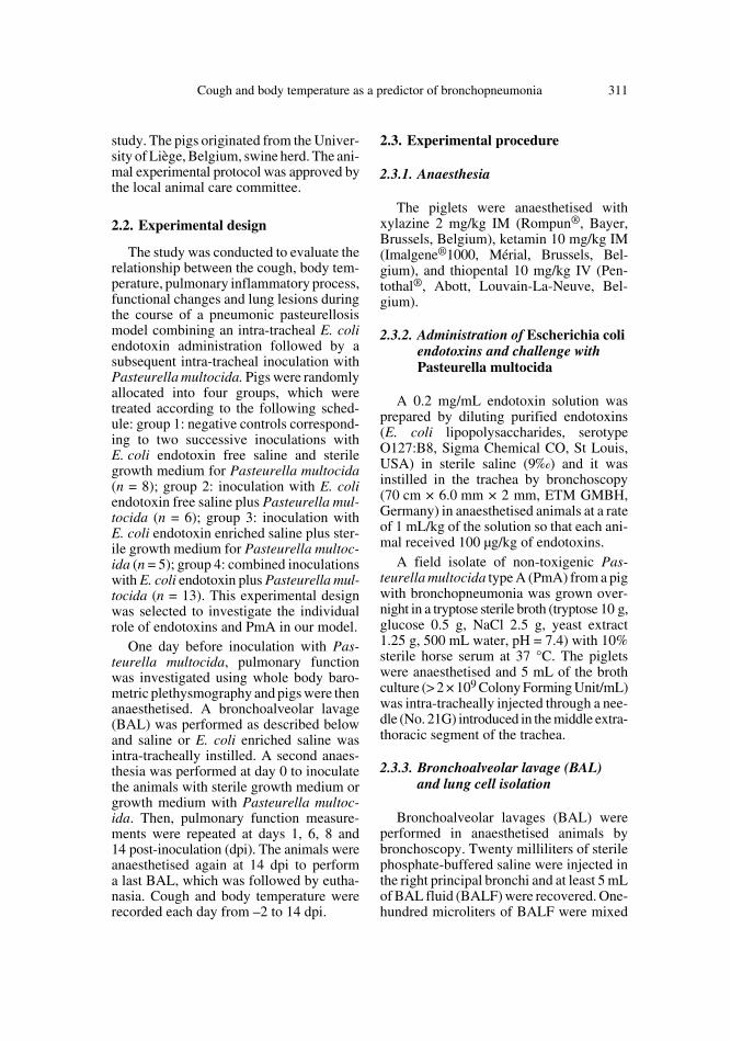
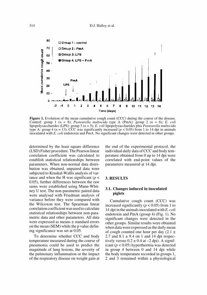
Cumulative cough count (CCC) wasincreased significantly (p < 0.05) from 1 to14 dpi in the animals inoculated with E. coliendotoxin and PmA (group 4) (Fig. 1). Nosignificant changes were detected in theother groups. Similar results were obtainedwhen data were expressed as the daily meanof cough counted one hour per day (2.1 ±2.7 and 8.1 ± 9.4 on 1 and 14 dpi respec-tively versus 0.2 ± 0.4 at –2 dpi). A signif-icant (p < 0.05) hyperthermia was detectedin group 4 between 0 and 14 dpi whilethe body temperature recorded in groups 1,2 and 3 remained within a physiological
Figure 1. Evolution of the mean cumulative cough count (CCC) during the course of the disease.Control: group 1 (n = 8); Pasteurella multocida type A (PmA): group 2 (n = 6); E. colilipopolysaccharides (LPS): group 3 (n = 5); E. coli lipopolysaccharides plus Pasteurella multocidatype A: group 4 (n = 13). CCC was significantly increased (p < 0.05) from 1 to 14 dpi in animalsinoculated with E. coli endotoxin and PmA. No significant changes were detected in other groups.
Cough and body temperature as a predictor of bronchopneumonia 315
range (Fig. 2). Except coughing and hyper-thermia observed in group 4, no other clin-ical signs appeared in any group.
Piglets receiving LPS alone (group 3)(0.24 ± 0.02 kg) or in combination withPmA (group 4) (0.19 ± 0.07 kg) presenteda similar but significant (p < 0.05) reductionin daily weight gains compared to those ofcontrol (0.45 ± 0.12 kg) and PmA inocu-lated groups (0.31 ± 0.15 kg).
The whole body barometric plethysmog-raphy measurements revealed a significantPenh increase from 8 to 14 dpi in group 4as presented in Figure 3. No significantchanges were observed in other groups.
Cell counts measured at –1 and 14 dpi arepresented in Figure 4. Significant increasesin total lung cells, macrophages, neutrophilsand lymphocytes were measured in group 4while no significant changes occurred in
other groups except in group 1 exhibiting aslight but significant increase in total lungcells, and macrophages.
Fourteen days post-inoculation, thecytokine/cyclophilin mRNA absorbanceratio of IFNγ was significantly (p < 0.05)increased in group 4 compared to the con-trol group (Fig. 5). In group 3 inoculatedwith LPS alone, a slight but significant (p <0.05) increase was also observed. No sig-nificant changes in IL-8, IL-18 and TNFαlevels were observed in any groups.
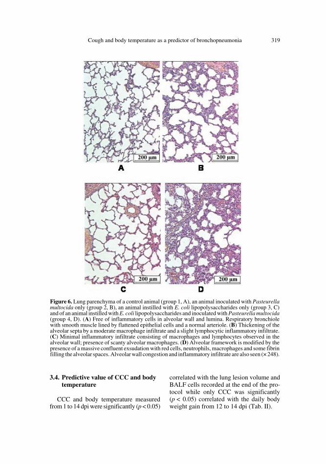
Low volume macroscopic lung lesionspreferentially located in caudal lung lobeswere detected in group 1 (controls) (34.4 ±23.3 cm3), group 2 (11.4 ± 6.2 cm3) andgroup 3 (7.0 ± 3.6 cm3). In group 1, the alve-olar walls likely consisted of flattened epi-thelial cells in close contact with capillariesand of a delicate connective tissue frame-work. It also contained scanty macrophages.
Figure 2. Evolution of the mean rectal temperature in the four groups during the course of the dis-ease. Control: group 1 (n = 8); Pasteurella multocida type A (PmA): group 2 (n = 6); E. coli lipopol-ysaccharides (LPS): group 3 (n = 5); E. coli lipopolysaccharides plus Pasteurella multocida type A:group 4 (n = 13). A significant (p < 0.05) hyperthermia was detected in group 4 between 0 and14 dpi while the body temperature recorded in groups 1, 2 and 3 remained within a physiologicalrange.

316 D.J. Halloy et al.
The alveolar spaces were well expandedand, as the bronchiolar lumina, were free ofinflammatory cells (Fig. 6A). Sometimes,the alveolar walls were enlarged by thepresence of a slight lymphocytic and a mod-erate macrophage infiltrate. However, noinflammatory infiltrate was found in thebronchi. In group 2, the alveolar frameworkwas preserved but the walls were enlargedby the presence of a slight lymphocytic anda moderate macrophage infiltrate. A subtlelymphocytic infiltrate was also seen in thebronchiolar and bronchial walls (Fig. 6B).In group 3, there was a minimal enlarge-ment of the alveolar septa due to theincrease of macrophages and lymphocytenumber. Scanty macrophages were free inthe alveoli. There was no inflammation ofthe bronchial wall (Fig. 6C). In group 4where LPS and PmA were combined, sig-nificantly more extended lung damage
(119.7 ± 39.2 cm3) was measured. Theselesions were located in cranial lobes as wellas in caudal lobes. The lung parenchymawas characterised by an accumulation ofintra-alveolar fluid, extravased red cellsand some fibrin together with polymorphsand macrophages (Fig. 6D). A moderatebronchial inflammatory infiltrate and apleuritis characterised by a lympho-plas-mocytic infiltrate and a florid reactive mes-othelial proliferation were observed. Thelesions of pleurisy were recorded in 4 of the13 animals of group 4.
No Pasteurella multocida or other swinerespiratory tract pathogens were isolatedfrom the control, LPS and PmA groups. Ingroup 4, Pasteurella multocida was iso-lated from one pig (cranial lung lobe) whileno other swine respiratory tract pathogenwas founded.
Figure 3. Evolution of the mean Penh value recorded in the different groups (unit: –). Control:group 1 (n = 8); Pasteurella multocida type A (PmA): group 2 (n = 6); E. coli lipopolysaccharides(LPS): group 3 (n = 5); E. coli lipopolysaccharides plus Pasteurella multocida type A: group 4 (n =13). Letters differing between groups indicate a significant difference (p < 0.05). When at least oneletter is common between the two groups, the difference is not significant. Within a group, thevalues were compared to those obtained on –2 dpi. Comparisons between groups were performedby testing the values measured the same day.

Cough and body temperature as a predictor of bronchopneumonia 317
3.2. Relationship between pathophysiological changes and clinical signs
Significant linear correlation coefficientswere obtained when individual values of thevolume of lung lesions, Penh, number ofBALF macrophages and neutrophils werecorrelated with the corresponding individ-ual values of CCC measured simultaneouslyduring the protocol. Penh, BALF macro-phage, neutrophil and lymphocyte numberswere also significantly (p < 0.01) correlatedwith body temperature (Tab. I). Individualvalues of Penh were correlated with the BALFneutrophil number (r = 0.64; p > 0.0001)and the volume of lung lesions (r = 0.35;p < 0.05) was measured simultaneously.
3.3. Use of cough and body temperature for the detection of diseased animals
When selecting a threshold for CCC offive coughs per animal, it appeared thatamong the animals from group 4 (n = 13),CCC exceeded this threshold at least oneday during the course of the disease in12 piglets (92.3%), all of them exhibitingsevere lung lesions at the end of the protocol(lung lesions volume: 119.6 ± 42.4 cm3).The mean detection time correspondingto the moment when the threshold wasexceeded was 4.5 ± 3.1 dpi. In this group,only one pig with extended lung lesions(120 cm3) showed CCC values remainingunder the selected threshold. In animals
Figure 4. Inflammatory lung cells counted in bronchoalveolar lavage fluids. Control: group 1 (n =8); Pasteurella multocida type A (PmA): group 2 (n = 6); E. coli lipopolysaccharides (LPS): group 3(n = 5); E. coli lipopolysaccharides plus Pasteurella multocida type A: group 4 (n = 13). Lettersdiffering between groups indicate a significant difference (p < 0.05). When at least one letter iscommon between two groups, the difference is not significant. Within a group, the values werecompared to those obtained on –2 dpi. Comparisons between groups were performed by testing thevalues measured the same day.

318 D.J. Halloy et al.
from other groups (1, 2, 3) (n = 19), onlythree (16%) developed coughs while theirmean lung lesion volume was very low(11.7 ± 5.1 cm3). The others (84%) exhib-ited low values of CCC and lung lesion vol-umes (21.5 ± 16.5 cm3). When a thresholdfor body temperature was considered(> 40.0 °C), it appeared that all animalsfrom group 4 (100%) developed hyperther-mia at least one day during the protocol(mean detection time: 2.6 ± 2.3 dpi), all ofthem having severe lung lesions (119.7 ±39.2 cm3). However, in the other groups,11 animals exceeded the threshold (57%)while their lung lesion volume was not sig-nificantly increased (21.5 ± 15.7 cm3). Whenboth criteria were combined (CCC > 5 andbody temperature > 40.0 °C), only one pig
from groups 1, 2 and 3 (5%) was consideredas a diseased animal while its lung lesionvolume was very low (4.1 cm3). In group 4(n = 13), 11 animals were detected on thebasis of this combined criteria.
To check whether the total cumulativecough counted daily in a group could beused to differentiate groups composed ofdiseased or healthy animals, a threshold offive coughs per animal multiplied by thenumber of animals included in the groupwas selected. The thresholds of CCC calcu-lated for group 1 (n = 8), 2 (n = 6), 3 (n = 5)and 4 (n = 13) were 40, 30, 25 and 75,respectively. These thresholds were neverreached for groups 1, 2 and 3 while pneu-monia was diagnosed at 2 dpi in group 4.
Figure 5. Cytokine production in lung tissue sampled at 14 dpi in: control: group 1 (n = 6);Pasteurella multocida type A (PmA): group 2 (n = 5); E. coli lipopolysaccharides (LPS): group 3(n = 5); E. coli lipopolysaccharides plus Pasteurella multocida type A: group 4 (n = 13). Total RNAwas isolated and assayed for expression of (A) IFN-γ, (B) IL-8, (C) IL-18, (D) TNF-α and thecyclophilin housekeeping genes by RT-PCR. Quantification of these cytokine mRNA is shown.Letters differing between groups indicate a significant difference (p < 0.05). When at least one letteris common between two groups, the difference is not significant.
Cough and body temperature as a predictor of bronchopneumonia 319
3.4. Predictive value of CCC and body temperature
CCC and body temperature measuredfrom 1 to 14 dpi were significantly (p < 0.05)
correlated with the lung lesion volume andBALF cells recorded at the end of the pro-tocol while only CCC was significantly(p < 0.05) correlated with the daily bodyweight gain from 12 to 14 dpi (Tab. II).
Figure 6. Lung parenchyma of a control animal (group 1, A), an animal inoculated with Pasteurellamultocida only (group 2, B), an animal instilled with E. coli lipopolysaccharides only (group 3, C)and of an animal instilled with E. coli lipopolysaccharides and inoculated with Pasteurella multocida(group 4, D). (A) Free of inflammatory cells in alveolar wall and lumina. Respiratory bronchiolewith smooth muscle lined by flattened epithelial cells and a normal arteriole. (B) Thickening of thealveolar septa by a moderate macrophage infiltrate and a slight lymphocytic inflammatory infiltrate.(C) Minimal inflammatory infiltrate consisting of macrophages and lymphocytes observed in thealveolar wall; presence of scanty alveolar macrophages. (D) Alveolar framework is modified by thepresence of a massive confluent exsudation with red cells, neutrophils, macrophages and some fibrinfilling the alveolar spaces. Alveolar wall congestion and inflammatory infiltrate are also seen (× 248).

320 D.J. Halloy et al.
4. DISCUSSION
In this model of subacute bronchopneu-monia, daily weight gain reduction andonly a few clinical signs such as cough andhyperthermia were detected. A pulmonaryinflammatory process involving macro-phages, neutrophils, lymphocytes and IFNγwas responsible for the lung lesions observedat the end of this study. Functional disordersconsisting of the alteration of the expiratoryairflow pattern also occurred. While coughand body temperature were moderatelyexpressed, their magnitude was propor-tional to the intensity of the lung inflamma-tory process, the pulmonary functionalchanges and the extent of gross lesions.Moreover, cough and body temperaturemeasured during the course of the diseasewere also related to the severity of theinflammation and lung injuries recorded atthe end of the pathological process. Onlycough was related to the reduction of thedaily weight gain.
Airway clearance of non-attached organ-isms is very effective in pigs so that theepithelium integrity needs to be impairedto successfully induce pneumonic pas-teurellosis. Mycoplasma hyopneumoniae,Aujeszky disease virus, porcine reproduc-tive and respiratory syndrome virus withBordetella bronchiseptica and crude Actin-obacillus pleuropneumoniae cytotoxin arevery effective agents to promote PmA-induced pneumonia [5, 8, 9, 22]. In ourstudy, E. coli endotoxins, which have alreadybeen shown to inhibit mucociliary clear-ance in rats and to induce a transient acutelung inflammation in pigs which remains inthe lung for a maximum of 48 h, were intrat-racheally injected to promote PmA infec-tion [20, 27]. The lung injuries observed inthe present study, especially subacute exu-dative bronchopneumonia involving migra-tion of neutrophils in alveoli and bronchi,marked alveolar macrophage prolifera-tion, and peribronchiolar and alveolar lym-phocytic infiltration were in full agreementwith studies devoted to Pasteurella multo-
cida infection [9, 15]. However, PmA wasscarcely isolated from lung lesions suggest-ing that a prolonged colonisation of the res-piratory tract was not necessary to stronglyworsen the pulmonary inflammatory proc-ess initiated by E. coli endotoxins. The factthat PmA was cleared from the lungs duringthe experiment might explain the baselinelevels of IL-8, IL-18 and TNF-α (Fig. 5)observed at 14 dpi which illustrated that theinflammatory process was decreasing inintensity. Indeed, inflammatory cytokineshave been shown to be expressed duringacute Pasteurella multocida infection [3].However in the latter study, the animalswere infected with a higher dose of bacteria(5 mL of 4.8 × 10⁹ CFU mL–1 versus 5 mLof 2 × 10⁹ CFU mL–1 in the present study)and were killed at an earlier time point ofthe respiratory infection (5 versus 14 dpi).A very transient production of cytokineswithin the first eight hours post infectionhas also been described during Actinobacil-lus pleuropneumoniae infection [2]. ThemRNA synthesis of IFN-γ detected sug-gests that lymphocytes observed within thelung tissue and the BALF participate in theregulation of both innate and acquired anti-microbial immunity [4, 30].
The Penh measured by whole bodyplethysmography is a functional parameterwhich has previously shown changes in theexpiratory airflow pattern during choliner-gic or endotoxin challenges [16]. In thisexperiment, the increase of Penh recordedon 8 and 14 dpi in group 4, was likely dueto airway obstruction and/or to a decreasein the lung elastic recoil induced by the pul-monary inflammatory process as reportedin anaesthetised piglets with other infec-tious pulmonary diseases [21]. To the bestof our knowledge, it is the first time thatsuch pulmonary functional alterations arereported in non-sedated, freely movingpiglets with bronchopneumonia. Since theraise in Penh was proportional to the inten-sity of the lung inflammatory reaction andto the extent of lung lesions, measurementof this functional parameter might be an

Cough and body temperature as a predictor of bronchopneumonia 321
interesting tool for a non-invasive follow-up of a lung disease accompanied by air-flow limitation.
Pigs from group 4 exhibited moderatecough and hyperthermia. These clinicalfindings are in agreement with previous
data and field observations collected in pigswith PmA-induced pneumonia [9]. With-out any accurate quantitative recording pro-cedure, these symptoms are not so easy todetect explaining why an early diagnosis ofsuch pulmonary diseases remains difficult
Table I. Relationship between individual values of cumulative cough count (CCC) or bodytemperature (BT) and the corresponding individual values of bronchoalveolar lavage fluid cellnumbers, Penh and volume of lung damage measured simultaneously. Regression equations withsignificant linear correlation coefficients are only indicated in this figure. The correspondingdetermination coefficients are also presented.
X Y r p ax + b R2
BT (°C) Macrophages (106/mL BALF) 0.41 0.01 0.96 x – 37.09 0.16
Neutrophils (106/mL BALF ) 0.45 0.001 0.40 x –15.73 0.20
Lymphocytes (106/mL BALF ) 0.38 0.01 0.17 x – 6.51 0.14
Penh 0.27 0.001 0.26 x – 9.29 0.07
CCC Macrophages (106/mL BALF ) 0.42 0.01 0.02 x + 0.98 0.17
Neutrophils (106/mL BALF ) 0.38 0.01 0.004 x + 0.20 0.14
Volume of lung lesions (cm3) 0.57 0.001 1.12 x + 40.71 0.32
Penh 0.35 0.0001 0.01 x + 0.77 0.12
Table II. Linear correlation coefficients (r) between clinical signs and bronchoalveolar lavage fluidcells, volume of lung lesions and daily weight gain. Values of clinical signs were day by day correlatedwith the clinical end-points recorded at d14. Days corresponding to significant (p < 0.05) r values areindicated in this table with the range of daily r values obtained during the indicated period of time.
Cumulative cough count Body temperature
Total lung cells days p.i. 3 to 14 1 to 14
r range 0.35 to 0.58 0.37 to 0.64
Macrophages days p.i. 1 to 14 1 to 14
r range 0.41 to 0.53 0.43 to 0.63
Neutrophils days p.i. 4 to 14 1 to 14
r range 0.38 to 0.50 0.51 to 0.69
Lymphocytes days p.i. 6 to 14 1 to 14
r range 0.42 to 0.48 0.48 to 0.68
Volume of lung lesions days p.i. 1 to 14 1 to 14
r range 0.31 to 0.59 0.34 to 0.55
Daily weight gain days p.i. 12 to 14 –
r range –0.39 to –0.40 –
322 D.J. Halloy et al.
in field conditions. Since other clinical signsare often missing or cannot be detected, theuse of quantitative measurement of cough-ing and body temperature might improvethe diagnosis of respiratory disorders inpigs. The relationship between coughingand lung damage has been investigated inpigs with enzootic pneumonia by scoringthe cough on a weekly basis and measuringlung lesions at slaughterhouses but no sig-nificant correlation was obtained probablydue to a non appropriate assessment ofcough and lung lesions [25]. Cough wascounted by mixing pigs in the pen for 30 sand observing the response (cough or nocough). Moreover, the extent of lung lesionswas semi-quantitatively determined by scor-ing them as a percentage of the damagedlung surface. As concluded by the authorsof this trial, a daily monitoring of cough anda better record of the lung lesion volumecould improve the predictive value of thissymptom regarding the intensity of the pul-monary disease. Hyperthermia has also beenmonitored during swine respiratory infec-tions involving Pasteurella multocida butthe relationship between the body temper-ature and lung lesions was not investigated[6, 9, 15].
The significant linear correlation coeffi-cients obtained in our study between cough,body temperature and the correspondingvalues of BALF cells, lung lesions and Penh(Tab. I) suggest that these clinical signscould be used to predict the severity andevolution of the inflammatory process andlung lesions during the course of an infec-tious respiratory disease. To assess thepotential use of CCC and body temperatureto early diagnose a respiratory disease in apig, thresholds for both parameters havebeen defined. Hence, all piglets reachingfive coughs or 40 °C at least one time duringthe protocol were considered as diseasedanimals and the accuracy of the diagnosiswas confirmed by the presence of lunglesions on 14 dpi. Applying this methodwith CCC as the criteria, 92.3% of the ani-mals with lung lesions were detected early
while only 16% of the pigs with no signif-icant lesions compared to controls wereerroneously declared as diseased (false pos-itive response). Considering body temper-ature as the discriminative criteria, the cor-responding percentages were 100 and 57%respectively. A combination of both criteriaallowed a marked reduction of the numberof false positive cases (85% of positiveresponses versus 5% of false positives).When a threshold for CCC was definedfor a group, the respiratory disease wasdetected at 2 dpi in group 4 while the others(groups 1, 2, 3) never overcame the thresh-old in our experimental conditions. Thus, ifcough recognition systems are available infield conditions in the future, this might bea real advance to determine the respiratorystatus of animals, to early and non-inva-sively diagnose respiratory diseases inherds but also to perform the follow-up ofbronchopneumonia [28, 29]. However, aprior validation of these systems is neededto confirm our observations in a large swinepopulation which contains animals withseveral common respiratory diseases. Datafrom Table II also show that BT and CCCcould be used to predict early-on the extentof major end-points regarding the intensityof respiratory disease in piglets.
We conclude that the E. coli lipopolysac-charide-induced lung inflammatory processmight be worsened by Pasteurella multoc-ida while it does not allow lung colonisa-tion. The resulting inflammatory processis characterised by an inflammatory cellinflux into the airways which limits the air-flow and by extended lesions of subacutebronchopneumonia explaining the weightgain reduction. Cough and body tempera-ture, while moderately exhibited, could beused as indicators to assess the intensity andthe evolution of the lung inflammatoryreaction and functional changes during thecourse of the disease. They are potentialpredictors for the magnitude of lung lesionsand weight gain reduction assessed at theend of the process.

Cough and body temperature as a predictor of bronchopneumonia 323
ACKNOWLEDGEMENTS
We thank N. Kirschvink for her interest andadvice during the preparation of the manuscript.The technical assistance of F. Delvaux, D. Beerensand S. Martinez is gratefully appreciated. Thiswork was supported by the Ministry of Agricul-ture (DGVI), Brussels, Belgium and by specialfunds for research (Ulg).
REFERENCES
[1] Amass S.F., Clark L.K., van Alstine W.G.,Bowersock T.L., Murphy D.A., Knox K.E.,Albregts S.R., Interaction of Mycoplasmahyopneumoniae and Pasteurella multocidainfections in swine, J. Am. Vet. Med. Assoc.204 (1994) 102–107.
[2] Baarsch M.J., Scamurra R.W., Burger K.,Foss D.L., Maheswaran S.K., Murtaugh M.P.,Inflammatory cytokine expression in swineexperimentally infected with Actinobacilluspleuropneumoniae, Infect. Immun. 63 (1995)3587–3594.
[3] Berndt A., Heller M., Kosmehl H., CytokinemRNA expression in experimental porcinepneumonia, Dtsch. Tierarztl. Wochenschr.109 (2002) 205–209.
[4] Boehm U., Klamp T., Groot M., Howard J.C.,Cellular responses to interferon-gamma, Annu.Rev. Immunol. 15 (1997) 749–795.
[5] Brockmeier S.L., Palmer M.V., Bolin S.R.,Rimler R.B., Effects of intranasal inoculationwith Bordetella bronchiseptica, porcine repro-ductive and respiratory syndrome virus, or acombination of both organisms on subsequentinfection with Pasteurella multocida in pigs,Am. J. Vet. Res. 62 (2001) 521–525.
[6] Carvalho L.F., Segales J., Pijoan C., Effect ofporcine reproductive and respiratory syn-drome virus on subsequent Pasteurella mul-tocida challenge in pigs, Vet. Microbiol. 55(1997) 241–246.
[7] Chong B.T., Agrawal D.K., Romero F.A.,Townley R.G., Measurement of bronchocon-striction using whole-body plethysmograph:comparison of freely moving versus restrainedguinea pigs, J. Pharmacol. Toxicol. Methods39 (1998) 163–168.
[8] Chung W.B., Backstrom L.R., Collins M.T.,Experimental model of swine pneumonic pas-teurellosis using crude Actinobacillus pleu-ropneumoniae cytotoxin and Pasteurella mul-tocida given endobronchially, Can. J. Vet.Res. 58 (1994) 25–30.
[9] Ciprian A., Pijoan C., Cruz T., Camacho J.,Tortora J., Colmenares G., Lopez-Revilla R.,de la Garza M., Mycoplasma hyopneumoniaeincreases the susceptibility of pigs to experi-mental Pasteurella multocida pneumonia,Can. J. Vet. Res. 52 (1988) 434–438.
[10] Darwich L., Pié S., Rovira A., Segalés J.,Domingo M., Oswald I.P., Mateu E., CytokinemRNA expression profiles in lymphoid tis-sues of pigs naturally affected by postweaningmultisystemic wasting syndrome, J. Gen.Virol 84 (2003) 2117–2125.
[11] Djuric V.J., Cox G., Overstreet D.H., Smith L.,Dragomir A., Steiner M., Genetically transmit-ted cholinergic hyperresponsiveness predis-poses to experimental asthma, Brain Behav.Immun. 12 (1998) 272–284.
[12] Donham K.J., Association of environmentalair contaminants with disease and productiv-ity in swine, Am. J. Vet. Res. 52 (1991) 1723–1730.
[13] Dozois C.M., Oswald E., Gautier N., SerthelonJ.P., Fairbrother J.M., Oswald I.P., A reversetranscription-polymerase chain reaction methodto analyze porcine cytokine gene expression,Vet. Immunol. Immunopathol. 58 (1997) 287–300.
[14] Fournout S., Dozois C.M., Yerle M., PintonP., Fairbrother J.M., Oswald E., Oswald I.P.,Cloning, chromosomal location, and tissueexpression of the gene for pig interleukin-18,Immunogenetics 51 (2000) 358–365.
[15] Hall W.F., Bane D.P., Kilroy C.R., Essex-Sorlie D.L., A model for the induction of Pas-teurella multocida type-A pneumonia in pigs,Can. J. Vet. Res. 54 (1990) 238–243.
[16] Halloy D., Kirschvinck N., Vincke G., HamoirJ., Delvaux F., Gustin P., Whole body baro-metric plethysmography: a screening methodto investigate airway reactivity and acute lunginjuries in freely moving pigs, Vet. J. (inpress).
[17] Hamelmann E., Schwarze J., Takeda K.,Oshiba A., Larsen G.L., Irvin C.G., GelfandE.W., Noninvasive measurement of airwayresponsiveness in allergic mice using baro-metric plethysmography, Am. J. Respir. Crit.Care Med. 156 (1997) 766–775.
[18] Hamelmann E., Schwarze J., Takeda K.,Oshiba A., Larsen G.L., Irvin C.G., GelfandE.W., Noninvasive measurement of airwayresponsiveness in allergic mice using baro-metric plethysmography [see comments],Am. J. Respir. Crit. Care Med. 156 (1997)766–775.

324 D.J. Halloy et al.
To access this journal online: www.edpsciences.org
[19] Hoffman A.M., Dhupa N., Cimetti L., Airwayreactivity measured by barometric whole-body plethysmography in healthy cats, Am. J.Vet. Res. 60 (1999) 1487–1492.
[20] Hosoe H., Kaise T., Ohmori K., Erdosteineenhances mucociliary clearance in rats withand without airway inflammation, J. Pharma-col. Toxicol. Methods 40 (1998) 165–171.
[21] Intraraksa Y., Engen R.L., Switzer W.P., Pul-monary and hematologic changes in swinewith Mycoplasma hyopneumoniae pneumo-nia, Am. J. Vet. Res. 45 (1984) 474–477.
[22] Kobisch M., Friis N.F., Swine mycoplas-moses, Rev. Sci. Tech. 15 (1996) 1569–1605.
[23] Kobisch M., Blanchard B., Le Potier M.F.,Mycoplasma hyopneumoniae infection in pigs:duration of the disease and resistance to rein-fection, Vet. Res. 24 (1993) 67–77.
[24] Loughmiller J.A., Spire M.F., Dritz S.S.,Fenwick B.W., Hosni M.H., Hogge S.B.,Relationship between mean body surface tem-perature measured by use of infrared thermog-raphy and ambient temperature in clinicallynormal pigs and pigs inoculated with Actino-bacillus pleuropneumoniae, Am. J. Vet. Res.62 (2001) 676–681.
[25] Morris C.R., Gardner I.A., Hietala S.K.,Carpenter T.E., Enzootic pneumonia: com-
parison of cough and lung lesions as predic-tors of weight gain in swine, Can. J. Vet. Res.59 (1995) 197–204.
[26] Thacker E.L., Thacker B.J., Janke B.H., Inter-action between Mycoplasma hyopneumoniaeand swine influenza virus, J. Clin. Microbiol.39 (2001) 2525–2530.
[27] Urbain B., Prouvost J.F., Beerens D., AnsayM., Gustin P., Acute effects of endotoxininhalation on the respiratory tract in pigs:interaction with ammonia, Inhal. Toxicol. 8(1996) 947–968.
[28] Van Hirtum A., Berckmans D., Assessing thesound of cough towards vocality, Med. Eng.Phys. 24 (2002) 535–540.
[29] Van Hirtum A., Berckmans D., Automatedrecognition of spontaneous versus voluntarycough, Med. Eng. Phys. 24 (2002) 541–545.
[30] Varma T.K., Lin C.Y., Toliver-Kinsky T.E.,Sherwood E.R., Endotoxin-induced gammainterferon production: contributing cell typesand key regulatory factors, Clin. Diagn. Lab.Immunol. 9 (2002) 530–543.
[31] Von Altrock A., Occurrence of bacterialinfectious agents in pathologically/anatomi-cally altered lungs of pigs and compilation ofresistance spectra, Berl. Muench. Tieraerztl.111 (1998) 164–172.


![Daily Dispatch (Richmond, [Va.]) 1863-02-28 [p ]mMßJa_k af" wBBP VflßMftßlA gef fißßbVßßßaflti gaat \u25a0WB-l-l*HaJ *llf](https://img.dokumen.tips/doc/110x75/5e7d0666275ac74ae50f2a55/daily-dispatch-richmond-va-1863-02-28-p-mmjak-af-wbbp-vflmftla.jpg)