Embed Size (px)
Citation preview

Pathology Course Chemical Pathology
Mike Davies Tom Marjot
Kindly sponsored by:

Outline Normal Values Fluid Balance Sodium and Potassium Acid Base Metabolic Bone Disease Pituitary Adrenal
Liver Func@on Tests Porphyria Hypoglycaemia Nutri@on Gout Cardiac Markers Plasma Proteins

Not covered
• Thyroid • Forensic Toxicology • Enzymology • Drug monitoring • Metabolic disorders + screening

Learning Objec@ves
• Understand normal ranges for Chemical Pathology
• Understand most important concepts in the module
• Prac@ce EMQs in prepara@on for exam • Provide a framework for you to revise • Scare you in to revising

Normal Values
• Don’t depend on normal values being in exam • Don’t need to learn ALL normal ranges • Be ready for the first few ques@ons in the exam will have numbers
• Get a feel for what is normal, mildly and grossly abnormal
• But which values…TELL US!

Normal Values
• Acid Base • Electrolytes • Bone metabolism • Liver Func@ons • Thyroid • Diabetes

Liver Func@on Tests
Page 8
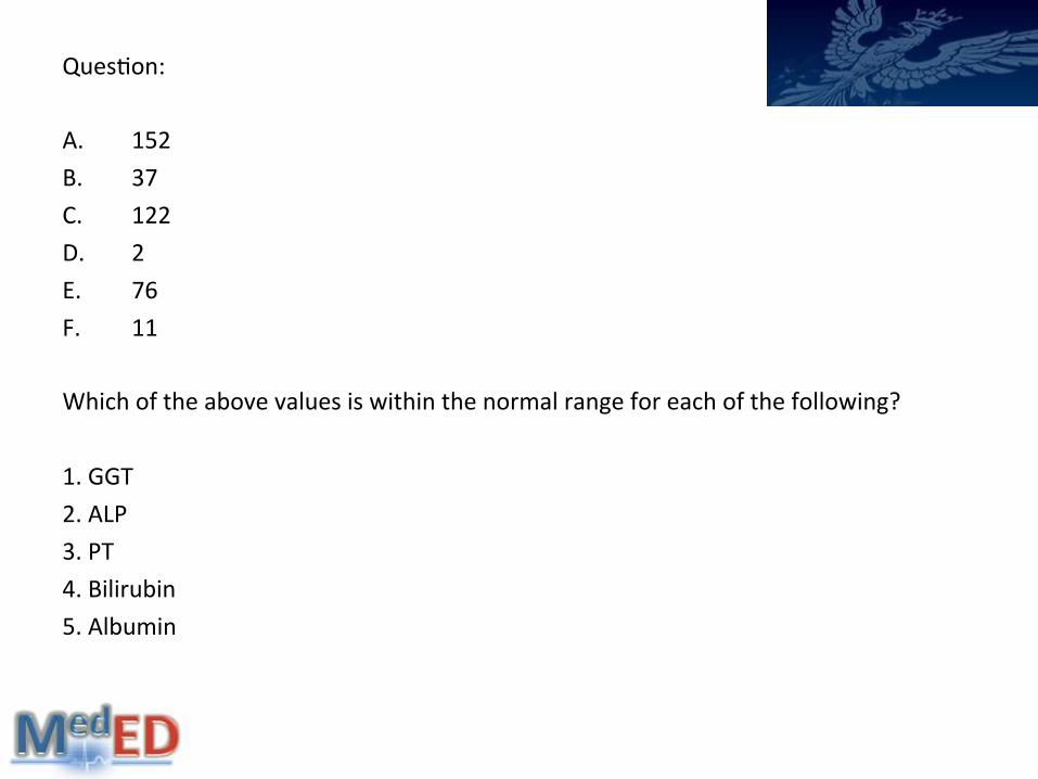
Ques@on: A. 152 B. 37 C. 122 D. 2 E. 76 F. 11
Which of the above values is within the normal range for each of the following? 1. GGT 2. ALP 3. PT 4. Bilirubin 5. Albumin
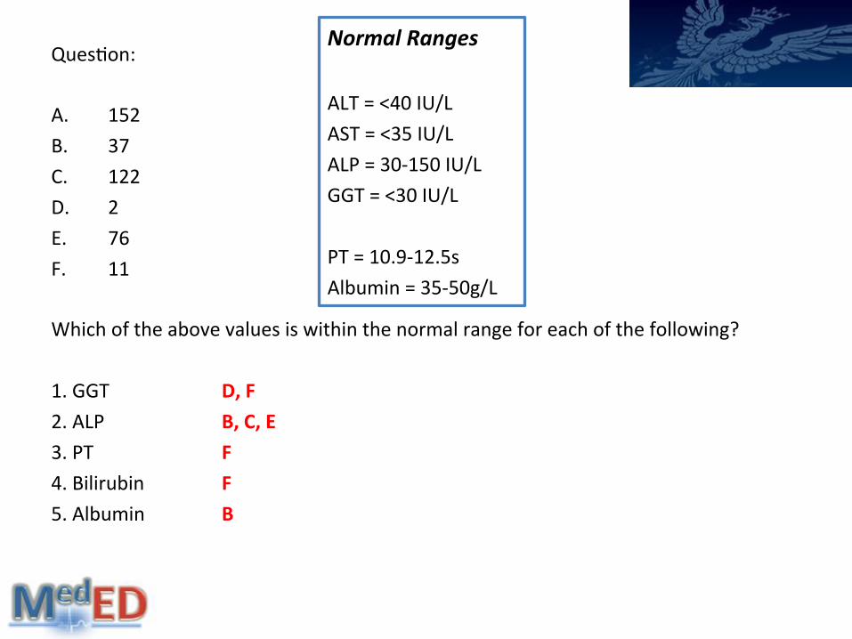
Ques@on: A. 152 B. 37 C. 122 D. 2 E. 76 F. 11
Which of the above values is within the normal range for each of the following? 1. GGT D, F 2. ALP B, C, E 3. PT F 4. Bilirubin F 5. Albumin B
Normal Ranges ALT = <40 IU/L AST = <35 IU/L ALP = 30-‐150 IU/L GGT = <30 IU/L PT = 10.9-‐12.5s Albumin = 35-‐50g/L

LFTs Normal Ranges ALT = <40 IU/L AST = <35 IU/L ALP = 30-‐150 IU/L GGT = <30 IU/L PT = 10.9-‐12.5s Albumin = 35-‐50g/L
?

LFTs LFTs
Aminotransferases (AST/ ALT)
<40iu/L
Raised when hepatocytes die
Alcoholic liver disease: AST:ALT = 2:1
Viral liver disease: AST:ALT = <1:1
Alkaline Phosphatase (ALP)
30 – 150 iu/L
Raised with cholestasis (either intrahepa@c or extrahepa@c) and bone disease, ↑++ in pregnancy
Gamma GT (GGT)
<30 iu/L
Usually elevated in chronic alcohol use
Also bile duct disease and metastases. Used to confirm hepa@c source of ↑ALP

LFTs • Acute liver failure gives ↑INR
• Chronic liver failure gives ↓albumin
• ↑ALP but no ↑GGT: suggests non-‐hepa@c cause
• Alcoholic liver disease: ↑AST>ALT (+ ↑GGT)
• Viral liver disease: ↑ALT>AST
• Transaminases >1,000 = Acute Hepa@@s (Ischameic/ Viral/ Toxic)

Ques@on A: Which of the following coagula@on factors does not require vitamin K for the gamma carboxyla@on needed for the conversion of prothrombin to thrombin? B: Which coagula@on factor is not primarily synthesised by the liver? 1. II 2. V 3. VII 4. VIII 5. X

Ques@on A: Which of the following coagula@on factors does not require vitamin K for the gamma carboxyla@on needed for the conversion of prothrombin to thrombin? B: Which coagula@on factor is not primarily synthesised by the liver? 1. II 2. V 3. VII 4. VIII 5. X

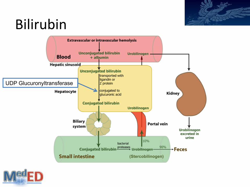
Bilirubin 5-‐17 μmol/l From the lysis of RBCs Bilirubin: End product of haem degrada@on Van den Burgh = direct reac@on measures conjugated bilirubin. Add methanol, completes reac@on giving total. Difference is the unconjugated bilirubin (indirect). Unconjugated: Increased produc@on Conjugated: Parenchymal liver disease, Obstruc@on
Bilirubin
UDP Glucuronyltransferase
(Stercobilinogen)

Gilberts
Autosomal Recessive 50% carrier, Prevalence = 5% Unconjugated hyperbilirubinaemia (>85% total frac@on). Self limi@ng, benign. Due to decreased glucuronosyltransferase Symptoms appear during stress, fas@ng.

Ques@on • Mul@ple spider naevi, Dupuytren’s contracture, Palmar erythema and Gynaecomas@a are signs of what?
• A. Jaundice • B. Hepa@@s • C. Chronic stable liver disease • D. Portal hypertension. • E. Liver failure • F. Obstruc@on of the bile ducts.

Ques@on • Mul@ple spider naevi, Dupuytren’s contracture, Palmar erythema and Gynaecomas@a are signs of what?
• A. Jaundice • B. Hepa@@s • C. Chronic stable liver disease • D. Portal hypertension. • E. Liver failure • F. Obstruc@on of the bile ducts.
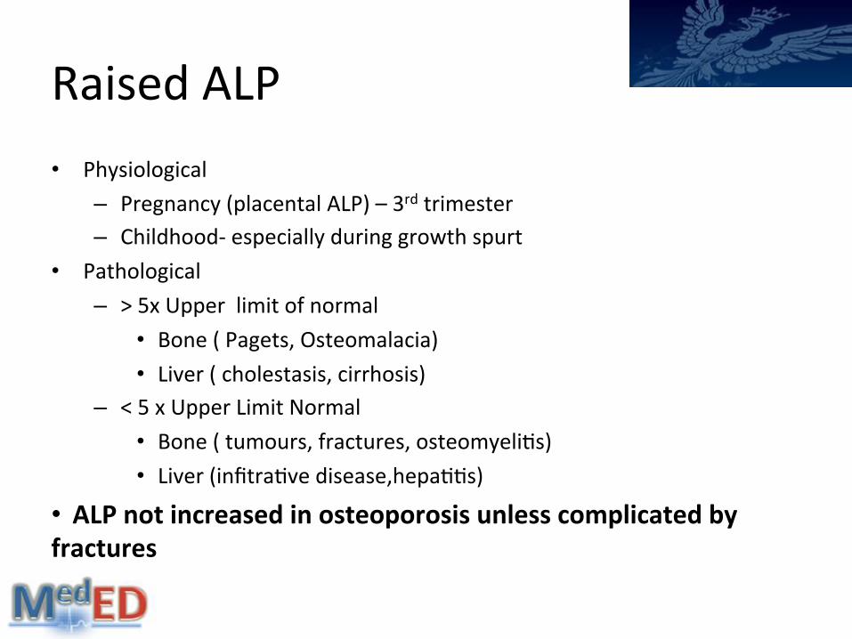
Raised ALP • Physiological
– Pregnancy (placental ALP) – 3rd trimester – Childhood-‐ especially during growth spurt
• Pathological – > 5x Upper limit of normal
• Bone ( Pagets, Osteomalacia) • Liver ( cholestasis, cirrhosis)
– < 5 x Upper Limit Normal • Bone ( tumours, fractures, osteomyeli@s) • Liver (infitra@ve disease,hepa@@s)
• ALP not increased in osteoporosis unless complicated by fractures
LFTs
Review Paper
EvaluaIon of abnormal liver funcIon tests JK Limdi, GM Hyde Postgrad Med J 2003
BSG Guidelines: Management of Abnormal LFTs
in Asymptoma@c Pa@ents
Porphyria Page 9

EMQ A: Uroporphyrinogen Decarboxylase B: Hydroxymethylbilane synthase C: Hereditary Coproporphyria D: Protoporphyrinogen Oxidase
E: Plumboporphyria F: Acute Intermiuent Porphyria G: Aminolevulinic Acid Synthase H: Congen@al Erythropoie@c Porphyria
1. Is reduced in Acute Intermiuent Porphyria?
2. Is the rate limi@ng step in heme produc@on in the liver?
3. Which form of porphyria most likely to present with cutaneous symptoms?
4. Has never been reported in the UK?
B
G
H
E

Porphyrias •7 inherited disorders caused by deficiency in enzymes along pathway •Involved in haem biosynthesis •Classified in to neuropsychiatric, cutaneous and mixed •Symptoms primarily neurological or photosensi@vity
Two main types: 1.Acute intermiuent porphyria (AIP) 2.Porphyria Cutanea Tarda
Neuropyschiatric: Acute Intermi7ent Porphyria, Plumboporphyria Cutaneous: Congen@al Erythropoie@c Porphyria, Porphyria Cutanea Tarda,
Erythropoie@c Protoporphyria, Mixed: Hereditary Coproporphyria, Variegate Porphyria

Pathway
Accumula@on of porphyrin and precursors
Aminolevulinic Acid Synthase
Neuropsychiatric

AIP • Autosomal dominant • Hydroxymethylbilane synthase deficiency • 5-‐aminolevulinic acid is neurotoxic! • Any paIent with (not photosensi@vity):
–Abdo pain, seizures, psych disturbances, peripheral neuropathy
• ↑ALA + ↑PBG in urine (“Port wine urine”) • Precipita@ng factors –alcohol, steroids, infec@on, pregnancy,
smoking, substance misuse
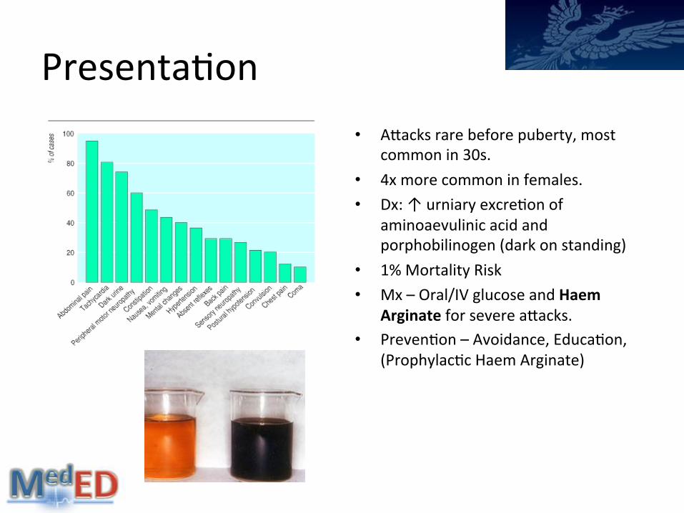
Presenta@on • Auacks rare before puberty, most
common in 30s. • 4x more common in females. • Dx: ↑ urniary excre@on of
aminoaevulinic acid and porphobilinogen (dark on standing)
• 1% Mortality Risk • Mx – Oral/IV glucose and Haem
Arginate for severe auacks. • Preven@on – Avoidance, Educa@on,
(Prophylac@c Haem Arginate)

Porphyria Cutanea Tarda Accumula@on of porphyrinogens in the skin Oxida@on to porphyrins + light = Symptoms •Inherited/ acquired •Uroporphrinogen decarboxylase deficiency •Any pa@ent with: •Vesicles on sun exposed sites (especially ayer excess EtOH), burning itching sensa@on, (subepidermal bullae) •Dx -‐↑ urinary uroporphrins + ↑ferri@n •Mx – avoidance, skin care, phlebotomy (remove iron)
Porphyria
• Clinical Review Paper
Diagnosis and management of porphyria H. Thadani, A. Deacon, T. Peters
2000

EMQ
A: Osteoarthri@s B: Ank Spon C: Gout D: Pseudogout
E: Reiters Syndrome F: Rheumatoid Arthri@s G: SLE H: Psoria@c Arthri@s
1. A 70-year-old male with stage 3 renal failure presents with severe pain in the right knee. The exquisite pain woke him up from his sleep. On examination the joint is swollen red and painful and tender.

EMQ
A: Osteoarthri@s B: Ank Spon C: Gout D: Pseudogout
E: Reiters Syndrome F: Rheumatoid Arthri@s G: SLE H: Psoria@c Arthri@s
1. A 70-year-old male with stage 3 renal failure presents with severe pain in the right knee. The exquisite pain woke him up from his sleep. On examination the joint is swollen, red and painful and tender.

EMQ
A: Osteoarthri@s B: Ank Spon C: Gout D: Pseudogout
E: Reiters Syndrome F: Rheumatoid Arthri@s G: SLE H: Psoria@c Arthri@s
2. A 45 year old male smoker presents to his GP with painfully swollen hands, sta@ng they are worse in the morning. The gentleman has also no@ced a swelling on his right elbow and more recently a numb/@ngling sensa@on in his right hand.

EMQ
A: Osteoarthri@s B: Ank Spon C: Gout D: Pseudogout
E: Reiters Syndrome F: Rheumatoid ArthriIs G: SLE H: Psoria@c Arthri@s
2. A 45 year old male smoker presents to his GP with painfully swollen hands, sta@ng they are worse in the morning. The gentleman has also no@ced a swelling on his right elbow and more recently a numb/@ngling sensa@on in his right hand.

EMQ
A: Osteoarthri@s B: Ank Spon C: Gout D: Pseudogout
E: Reiters Syndrome F: Rheumatoid Arthri@s G: SLE H: Psoria@c Arthri@s
3. A 75 year old gentlemen with chronic joint problems presents to the GP with non-‐painful pedunculated lumps behind his ears that his wife recently no@ced. When drained the lumps contain a chalky material

Gout

Gout Disorder of purine metabolism characterized by acute, recurrent auacks of synovi@s. Crystal deposi@on disease (mono sodium urate) Can be acute (Podagra if big toe) or chronic (Tophaceous) M:F = 20:1.Stable prevalence of around 1% Normal urate level = Men 0.12 – 0.42mmol/l
Women 0.12 – 0.36 mmol/l
Exquisitely painful, red, hot and swollen joint 1st MTP joint first site in 50%, involved in 90% overall

Management Acute: NSAIDS and Colchicine (an@-‐IL1 for resistance), Cor@costeroids Long term: Allopurinol, Lifestyle modifica@on, diet, treat co-‐mordibi@es ComplicaIons: Tophi, soy @ssue damage, degenera@ve arthri@s, renal disease.

Gout: True or False • Colchicine lowers urate level?
• Allopurinol should be used acutely?
• Allopurinol lowers urate levels by inhibi@ng xanthine oxidase?
• The MTP is the first joint to be affected in 90% of cases?
• Allopurinol reduces plasma levels of 6-‐mercaptopurine?
• Pseudogout crystals exhibit nega@ve birefringence under polarised light?

Gout: True or False • Colchicine lowers urate level? FALSE – prevent phagocyte transit
• Allopurinol should be used acutely? FALSE – long term
• Allopurinol lowers urate levels by inhibi@ng xanthine oxidase? TRUE
• The MTP is the first joint to be affected in 90% of cases? FALSE – 50%
• Allopurinol increases plasma levels of 6-‐mercaptopurine? TRUE
• Pseudogout crystals exhibit nega@ve birefringence under polarised light? FLASE – posi@ve birefringence

Gout Gout Pseudogout
Crystals Monosodium Urate
Pyrophosphate
Shape Needle like Rhomboid
Birefringence Strongly Nega@ve Weakly Posi@ve
Joint MTP Knee, Hip, Wrist
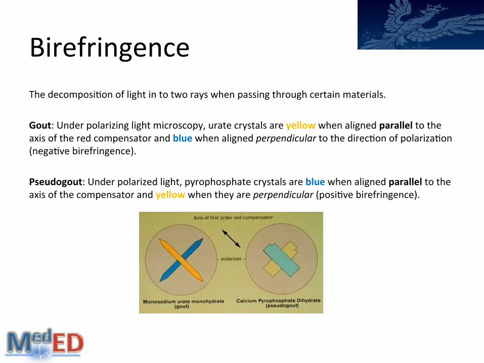
Birefringence The decomposi@on of light in to two rays when passing through certain materials. Gout: Under polarizing light microscopy, urate crystals are yellow when aligned parallel to the axis of the red compensator and blue when aligned perpendicular to the direc@on of polariza@on (nega@ve birefringence). Pseudogout: Under polarized light, pyrophosphate crystals are blue when aligned parallel to the axis of the compensator and yellow when they are perpendicular (posi@ve birefringence).

Gout
Review Paper Up-‐to-‐date management of gout
KM Jordan Curr Opin Rheumatology 2012
Gout: An Update AT Eggebeen
Am Fam Physician 2007

Hypoglycaemia Page 21

Symptoms
Neuroglycopaenic Somnolence Confusion Incoordina@on Seizures, coma NB: may be none
Adrenergic Tremors Palpita@ons Swea@ng Hunger

Cause
• DiabeIcs • Drugs: excessive • Inadequate CHO intake / missed meal
• Impaired awareness • Excessive alcohol • Strenuous Exercise • Co-‐exis@ng autoimmune condi@ons
• Non-‐DiabeIcs • Cri@cally unwell • Organ failure • Hyperinsulinism • Post gastric-‐bypass • Drugs • Extreme weight loss • Fac@@ous
C-‐pep@de
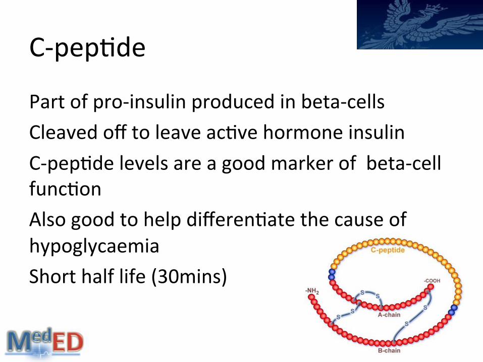
Part of pro-‐insulin produced in beta-‐cells Cleaved off to leave ac@ve hormone insulin C-‐pep@de levels are a good marker of beta-‐cell func@on Also good to help differen@ate the cause of hypoglycaemia Short half life (30mins)

Hypoglycaemia

Extras Non-‐islet cell tumour hypoglycaemia (everything low) Other tumours which cause a paraneoplas@c syndrome Secre@on of ‘big IGF-‐2’ Big IGF2 binds to IGF-‐1 receptor and insulin receptor Mesenchymal tumours ( mesothelioma /fibroblastoma) Epithelial tumours (carcinoma) Autoimmune insulin syndrome (low C-‐pepIde) An@bodies to insulin receptors Bind and s@mulate insulin release
Hypoglycaemia
Review Paper
Hypoglycaemic Disorders F.J Serivce NEJM 1995

Nutri@on


Nutri@on Site of producIon FuncIon Obesity
Grehlin Stomach, Pancreas Hypothalamus
Appe@te S@mulant ↑
NPY Hypothalamus Appe@te S@mulant ↑
LepIn White Adipose Tissue Inhibits Appe@te ↓
PYY Ileum and Colon Inhibits Appe@te ↓
AdiponecIn Adipose Tissue Inhibits Appe@te ↓

Nutri@on A: Thiamine B: Zinc C: Cobalamin D: Niacin
E: Riboflavin F: Pyridoxine G: Fluoride H: Copper
1. Is given with TB medication to prevent peripheral neuropathy?
2. The X-linked recessive Menkes Disease causes an abnormal transport and accumulation of this?
3. A deficiency classically causes a triad of diarrhoea, dementia and dermatitis?
4. A deficiency can be tested for using Schillings Test?
5. Deficiency predisposes to the development of tooth decay?
F
H
D
C
G

Cardiac Markers
Page 16

Cardiac Enzymes • CreaIne Kinase • Most widely used marker of muscle damage • Three forms -‐ dimers containing the M (muscle) and B (brain) subunits
– CK-‐MM-‐ skeletal muscles – CK-‐MB (1 & 2) – cardiac muscles – CK-‐ BB – brain – ac@vity minimal even in severe brain damage
• CK-‐MM accounts for almost en@re normal plasma ac@vity
• Causes of AbnormaliIes: – Myopthy e.g. Duchenne muscular dystrophy (>10xULN) – Myocardial Infarc@on (>10xULN) – Severe exercise (5xULN) – Physiological – Afro-‐Caribbean (<5xULN)

Cardiac Markers
Troponins Rise 4-‐6 hours post MI
Peak at 12 -‐24 hours post MI Remain elevated for 3 -‐10 days
A : Myoglobin B : Cardiac Troponin C : CK-‐MB D : Cardiac Troponin u
Plasma Proteins
Page 17

EMQ A: Alpha 1 an@-‐trypsin B: CRP C: Caeruloplasmin D: Transferrin E: Ig A
F: Ig M G: Ig G H: Total Iron Binding Capacity (TIBC) I: AFP J: CA 19.9
1. A deficiency is associated with a movement disorder and liver disease 2. Is an independent marker for cardiovascular disease 3. Is decreased in a disorder of excess iron deposition 4. A deficiency predisposes individuals to basal emphysema
C B H A

Plasma Proteins Class Protein Function Serum
Conc Albumin Oncotic Pressure 40
Source of Amino acids Ligand Binding
Acute Phase Protein
CRP ↑6-8 hrs after tissue damage (peaks after 24-28hrs
<10
Stays elevated if there's continuous stimulus α1-globulin α1-antitrypsin Major antagonist of serine proteases 2.9
Positive Acute Phase Reactant α2-globulin Haptoglobulins 2
Caeruloplasmin Copper containing protein with oxidase activity 0.35 Deficiency (i.e. Wilson's) causes body to retain copper
β-globulin Transferrin Plasma iron transport 3 Negative acute phase protein Used in assessment of Fe def/overload
LDL 1 Complements 1
-globulins IgG 14 IgA 2.5 IgM 1.5 IgD 0.03 IgE trace
Tumour Markers
PSA Prostate Ca AFP Hepatic Ca CA19-9 Pancreatic Masses CA125 Ovarian Ca/Pelvic Masses CEA Colorectal Ca βhCG Gestational Trophoblastic Disease
CRP <10mg/L Acute phase protein. Binds to damaged/dying cells to ac@vate complement system Synthesised in liver Increases 6-‐8 hrs ayer @ssue damage (trauma, infec@on, inflamma@on), & peaks ayer 24-‐48 hrs. Stays elevated if there is con@nuing s@mulus. Marker for CVD
Alpha-‐1 An@-‐trypsin Plasma protein produced by the liver. Most abundant serpin (serine protease inhibitor) Major antagonist of serine proteases released at site of @ssue injury. Main physiological role to inhibit neutrophil elastase Normal allele is M, variants are S and Z Deficiency predisposes to emphysema in adults, cirrhosis in children Smoking is a major contributor to disease progression Treatment is with purified AAT therapy.
Alpha-‐1 An@-‐trypsin
Review Paper
Alpha1-‐AnItrypsin deficiency: best clinical pracIce
NA Kalsheker J Clin Pathol 2009

Albumin Main protein of Plasma Important to the maintenance of plasma colloid onco@c pressure Deficiency results in oedema. Non-‐specific carrier protein for molecules such as fauy acids, calcium, unconjugated bilirubin, thyroxine and urate Almost always low Increase seen in severe dehydra@on Behaves as a nega@ve acute phase protein, reduced levels primarily due to increased capillary permeability. Renal and gut losses also common.

Transudate vs Exudate
Transudate Volume/ Pressure Overload Due to failure • Conges@ve heart failure • Liver cirrhosis • Hypoalbuminaemia • Peritoneal dialysis
Exudate
Inflamma@on • Malignancy • Pulmonary embolism • Parapneumonic effusions • Miscellaneous
pancrea@@s,RA, SLE TB. ie, Chronic inflamma@on

Adrenals Page 13

Phaeochromocytoma -‐Adrenal medulla tumour = ↑ Adrenaline Can occur at any age, assoc with pregnancy. •Symptoms/ Signs: (daily, weekly, monnthly) Pressure: Episodic HTN Pain: headaches Palpata@ons and Tremor Pallor Perspira@on
•Treatment: –αblockade –phenoxybenzamine –βblockade – atenolol, propanolol –Surgery: 4-‐6wks later.
• B1 – Inotropic (force) and chronotropic (rate) effect
• B2 – Bronchodilata@on and some vasodilata@on, relaxa@on of intes@nal smooth muscle
• A1 – Vasoconstric@on of smooth muscle • A2 – Vasoconstric@on of vascular
smooth muscle.
Rule of 10s •10% Bilateral •10% Malignant •10% Outside adrenal •10% Associated with familial syndromes

Phaeochromocytoma
Review Paper
Phaeochromocytoma: A Brief Review L.G. Tolstoi
Hosp Pharm. 2001

Addisonian Crisis Background Known Addison’s disease or long term steroid use, (TB pa@ents) PrecipitaIng Factor Infec@on, trauma, surgery Result Shocked: tachy, hypotensive, confused, peripherally shut down Mx Immediate: Hydrocor@sone sodium succinate 100mg IV stat Resus, Bloods, monitor BM, ? Cultures ConInued: Fluids, oral steroids, ? Glucose, address cause.

Addison’s Disease
Review Paper Adrenal Insufficiency and Addison’s Disease
R Munver R, IAVolfson Curr Urol Reports 2006

Adrenal EMQs A Cushing’s syndrome E Iatrogenic Cushing’s Syndrome B Cushing’s disease F Conn’s Syndrome C Addison’s disease G Phaeochromocytoma D Hypothyroidism H None of the Above
1. A 27-‐year-‐old man with known ulcera@ve coli@s presents to his GP with
concerns over his mood and recent weight gain.

Adrenal EMQs A Cushing’s syndrome E Iatrogenic Cushing’s Syndrome B Cushing’s disease F Conn’s Syndrome C Addison’s disease G Phaeochromocytoma D Hypothyroidism H None of the Above
2. A 45 year old lady with a chronic cough, night sweats and weight loss
presents to A+E confused, peripherally shut down. Bloods: BM =2.4, K+ =6.1, Na+ = 128.

Adrenal EMQs A Cushing’s syndrome E Iatrogenic Cushing’s Syndrome B Cushing’s disease F Conn’s Syndrome C Addison’s disease G Phaeochromocytoma D Hypothyroidism H None of the Above
3. A 65 year-‐old heavy smoker presents with haemoptysis and
dyspnoea. O/E he marked bruising, truncal obesity and bloods reveal a hypokalaemia.

Adrenal EMQs A Cushing’s syndrome E Iatrogenic Cushing’s Syndrome B Cushing’s disease F Conn’s Syndrome C Addison’s disease G Phaeochromocytoma D Hypothyroidism H None of the Above
4. Elevated cor@sol and ACTH levels which suppress on a high, but not a
low, dexamethasone suppression test.

Adrenal EMQs A Cushing’s syndrome E Iatrogenic Cushing’s Syndrome B Cushing’s disease F Conn’s Syndrome C Addison’s disease G Phaeochromocytoma D Hypothyroidism H None of the Above
4. Elevated cor@sol and ACTH levels which suppress on a high, but not a
low, dexamethasone suppression test.

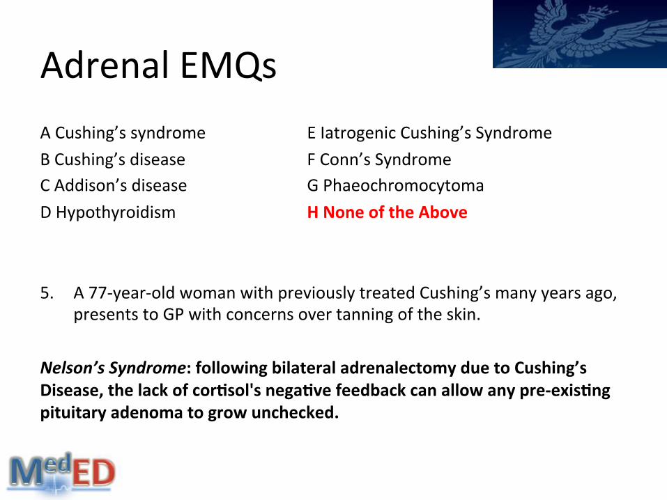
Adrenal EMQs A Cushing’s syndrome E Iatrogenic Cushing’s Syndrome B Cushing’s disease F Conn’s Syndrome C Addison’s disease G Phaeochromocytoma D Hypothyroidism H None of the Above 5. A 77-‐year-‐old woman with previously treated Cushing’s many years
ago, presents to GP with concerns over tanning of the skin.
Adrenal EMQs A Cushing’s syndrome E Iatrogenic Cushing’s Syndrome B Cushing’s disease F Conn’s Syndrome C Addison’s disease G Phaeochromocytoma D Hypothyroidism H None of the Above 5. A 77-‐year-‐old woman with previously treated Cushing’s many years ago,
presents to GP with concerns over tanning of the skin.
Nelson’s Syndrome: following bilateral adrenalectomy due to Cushing’s Disease, the lack of corIsol's negaIve feedback can allow any pre-‐exisIng pituitary adenoma to grow unchecked.

Finished!
Thank you