Embed Size (px)
Citation preview

A PATHOLOGICAL STUDY OF FIVE CASES OFPYELONEPHRITIS IN THE NEWBORN
BY
K. A. PORTER and H. McC. GILESFrom the Departments of Pathology and Paediatrics, St. Mary's Hospital, London
(RECEIVED FOR PUBUCATO?N MARCH 27, 1956)
It is increasingly recognized that many problemsconcerning the aetiology, recognition and naturalhistory of urinary tract infections are still unresolved,and this is particularly true of such infections ininfancy. There is, for example, no general agree-ment about their incidence in the first months oflife: Stansfeld (1954) has pointed out that pyelone-phritis occurs more frequently in the first year thanat any other age, and most frequently of all in thefirst month, but this distribution pattem has notbeen universally recognized. The divergence ofopinion may derive from a failure to considerpyelonephritis as a possible cause of such symptomsas vomiting, anorexia, constipation, diarrhoea andpoor weight gain, or from uncertainty in assessingthe significance of those minor abnormalities in theurine which may be the only diagnostic feature ofchronic pyelonephritis. The situation is furtherconfused by disagreement over the interpretationof various lesions which may be found in the kidneysof small infants dying of renal disease so that evennecropsy may not resolve clinical uncertainty.Among the few existing reports of the morbid
anatomical findings in cases of neonatal pyelone-phritis are those of Chown (1927) who could findonly nine cases in the earlier literature. To thesehe added detailed reports of 30 cases of 'pyelitis' ininfancy. Most of his cases showed the changes ofacute suppurative pyelonephritis but four withchronic pyuria showed in addition 'inflammatorylesions in various stages of healing'. Craig (1935)gave the post-mortem findings in six cases of acutepyelonephritis and Hunt (1936) described one casewith unilateral acute suppurative pyelonephritisfatal 13 days after birth. More recently Claireauxand Pearson (1955) have recorded chronic pyelone-phritis in a newborn infant.The histological picture of acute pyelonephritis
in early infancy is indistinguishable from that foundat any other age; in the chronic stage, however,
differences may appear which have hitherto beeninsufficiently recognized and it is the purpose of thispaper to discuss them.
MateialFive cases of pyelonephritis in infants under
1 month of age are described. All of these weremales and this is in keeping with the general tendencyof neonatal pyelonephritis to be more frequent inboys. Four of the infants were born in the samehospital within a few months of each other. (Twoother cases occurring in the same period haveapparently recovered.) The fifth case was a brotherof one of the other four. One infant died duringthe acute phase of his illness, two died later inuraemia and two succumbed to intercurrent infec-tion.
Case 1. R.E., a male negro infant, was the secondof twins born on March 25, 1954, after a normalpregnancy. He was a- breech presentation, but wasturned and extracted without difficulty. It is not knownwhether the twins were identical or fraternal. Heprogressed normally until April 12 when he was noticedto be jaundiced. Haemoglobin was 8-3 g. 00 and totalserum bilirubin 7-4 mg. %o; the direct Coombs test wasnegative and no sickling was demonstrable. On April 16he became lethargic, anorexic and febrile, and was giventerramycin. The white blood count was 35,000/c.rnm.and blood culture yielded a growth of E. coli: the urinecontained pus and also yielded E. coli. The organismsobtained from the blood and urine appeared identicalexcept that the former was sensitive and the latterresistant to terramycin. In spite of vigorous treatmentwith penicillin and streptomycin he died on April 18,aged 31 weeks.GENERAL NECROPSY FfNDiNGs. The tissues were de-
hydrated and bile-stained.The myocardium was pale and flabby and the ductus
arteriosus and foramen ovale were widely patent. Thelungs showed a few areas of collapse at the right base.The stomach and small gut were distended and
303
copyright. on M
arch 23, 2020 by guest. Protected by
http://adc.bmj.com
/A
rch Dis C
hild: first published as 10.1136/adc.31.158.303 on 1 August 1956. D
ownloaded from

ARCHIVES OF DISEASE IN CHILDHOODcontained white curdy material. The liver was deeplyjaundiced, firm and without obvious pattern. Therewas a congenital stenosis of the common bile duct.The left kidney (weight 25 g.) was considerably larger
than the right (weight 17 5 g.) and showed extensiveacute pyelonephritis. Inflammation was much lessmarked in the right kidney. No other abnormality wasseen in the genito-urinary tract.On section of the brain a large haemorrhage was seen
in the region of the right caudate nucleus which com-municated with blood in the ventricles.MICROSCOPY OF THE KmNEys. The renal capsule was
normal. Scattered throughout both the cortex andmedulla were multiple abscesses in which bacteria couldbe demonstrated. In these areas some of the glomerulihad been destroyed: others were extensively infiltratedby polymorphs or showed periglomerulitis with fibrin.Many of the tubules were dilated and contained pus.Polymorphs and some lymphocytes had infiltrated theinterstitial connective tissue. The blood vessels werenormal. The renal pelvis showed acute inflammationwith areas of necrosis and loss of lining epithelium.
Microdissection showed a number of the nephrons tobe of premature type in that they were shorter thannormal and lacked convolutions.
Case 2. C.T. was a male infant born at term onMarch 4, 1950, by spontaneous, normal delivery. Hehad a weak cry with stridorous breathing, and laryngo-scopy was thought to show a fine web across the vocalcords. Progress was slow with increasing reluctance tofeed: on May 18 he was febrile and the urine containedpus and was found to be infected with E. coli andStr. faecalis. The infection did not clear in spite oftreatment and he became increasingly acidotic andazotaemic. He died on July 20, 1950, aged 20 weeks.GENERAL NECROPSY FINDINGs. The body was slightly
wasted with pale skin but no oedema. No congenitalabnormality was found on external examination.The heart weighed 40 g. and showed left ventricular
hypertrophy. In the larynx, situated immnediately abovethe vocal cords which did not seem to be well developed,there was a very thin, triangular, transparent membrane.All lobes of both lungs showed moderate congestion andoedema and patchy areas of collapse. The intestines,liver, pancreas, spleen, pituitary, thyroid and adrenalswere normal.Each kidney weighed 20 g. and the capsules stripped
easily leaving a smooth pale surface. On section pallorwas pronounced, the cortico-medullary junctions blurredand the pelves injected. The renal vessels and theremainder of the urogenital system were normal.The brain and bones were normal.MICROSCOPY OF KmNEys. The capsule of the kidneys
showed no change. The glomeruli were reduced toabout three-quarters of their normal number. Of thosesurviving, most were normal but some showed markedpericapsular fibrosis. A few showed capsular crescentformation: the majority of the crescents had undergonefibrosis (Fig. 1) but some remained cellular. Adhesionsbetween glomerular tuft and capsule were also to be seen
FIG. 1.-Case 2. Two glomeruli showing capsular crescents whichhave become fibrous. Note the dense interstitial infiltration with
chronic inflammatory cells. (Haematoxylin and cosin x 150).
in a few. Occasional glomeruli were partly hyalinized,others had been converted into hyaline spheres. Thedamaged glomeruli tended to be grouped together intorough cortical wedges. There were no striking changesin the glomerular capillary basement membranes.'Alterative' glomerulitis was seen in some glomeruli inunscarred areas.
In the areas of glomerular damage groups of proximaltubules were dilated and lined by flattened atrophicepithelium and contained pink 'colloid' casts. Occasionaltubules were cystic. Some collecting tubules showedsimilar changes.The interstitial tissue, particularly in the cortical
wedges of glomerular and tubular damage, was heavilyinfiltrated by lymphocytes and plasma cells with veryoccasional polymorphonuclear leucocytes. There wasalso some fibrosis.The arcuate and interlobular arteries showed mild
productive endarteritis and the arterioles mild hvper-plastic arteriolosclerosis.The pelvis of the kidney appeared normal.Nephron dissection (Fig. 2) showed that the proximal
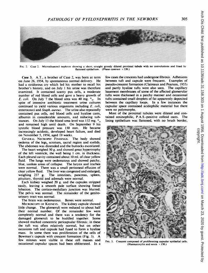
tubule was considerably shorter than normal and almostcompletely devoid of convolutions: in addition theepithelium was flattened and the lumen grossly dilated(Darmady and Stranack, 1955).
304
copyright. on M
arch 23, 2020 by guest. Protected by
http://adc.bmj.com
/A
rch Dis C
hild: first published as 10.1136/adc.31.158.303 on 1 August 1956. D
ownloaded from
PATHOLOG Y OF P YELONEPHRITIS IN THE NEWBORN
FiG. 2. Case 2. Microdissected nephron showing a short, straight grossly dilated proximal tubule with no convolutions and lined by-flattened epithelium. (Phase contrast x 220.)
Case 3. A.T., a brother of Case 2, was born at termon June 26, 1954, by spontaneous normal delivery. Hehad a stridorous cry which led his mother to recall hisbrother's history, and on July I his urine was thereforeexamined. It contained scanty pus cells, a moderatenumber of red blood cells and gave a heavy growth ofE. coli. On July 7 the blood urea was 40 mg. 00. Inspite of intensive antibiotic treatment urine culturescontinued to yield various organisms including E. coli,enterococci and Staph. aureus. The urine also repeatedlycontained pus cells, red blood cells and hyaline casts,albumin in considerable amounts, and reducing sub-stances. On July 13 the blood urea level was 132 mg. 00and remained high until death. On September 9 hissystolic blood pressure was 150 mm. He becameincreasingly acidotic, developed heart failure, and diedon November 5, 1954, aged 19 weeks.GENERAL NECROPSY FINDrNGs. The body showed
oedema of the legs, scrotum, sacral region and eyelids.The abdomen was distended and the buttocks excoriated.The heart weighed 50 g. and showed great hypertrophy
of the left ventricle, the wall being 1 cm. in thickness.Each pleural cavity contained about 10 ml. of clear yellowfluid. The lungs were oedematous and showed patchy,blue, sunken areas of collapse. The larynx and tracheawere normal. There was a small peritoneal effusion ofclear yellow fluid. The liver was congested and enlarged,weighing 237 g. The intestines, pancreas, spleen,pituitary, thyroid and adrenals were normal.Each kidney weighed 20 g. and the capsules stripped
easily, leaving a smooth pale surface showing foetallobation. The cortico-medullary junction was blurred.The pelvis was normal. The remainder of the genito-urinary tract was normal.The brain was oedematous. Bones were normal.MICROSCOPY OF KIDNEYS. The kidney capsule showed
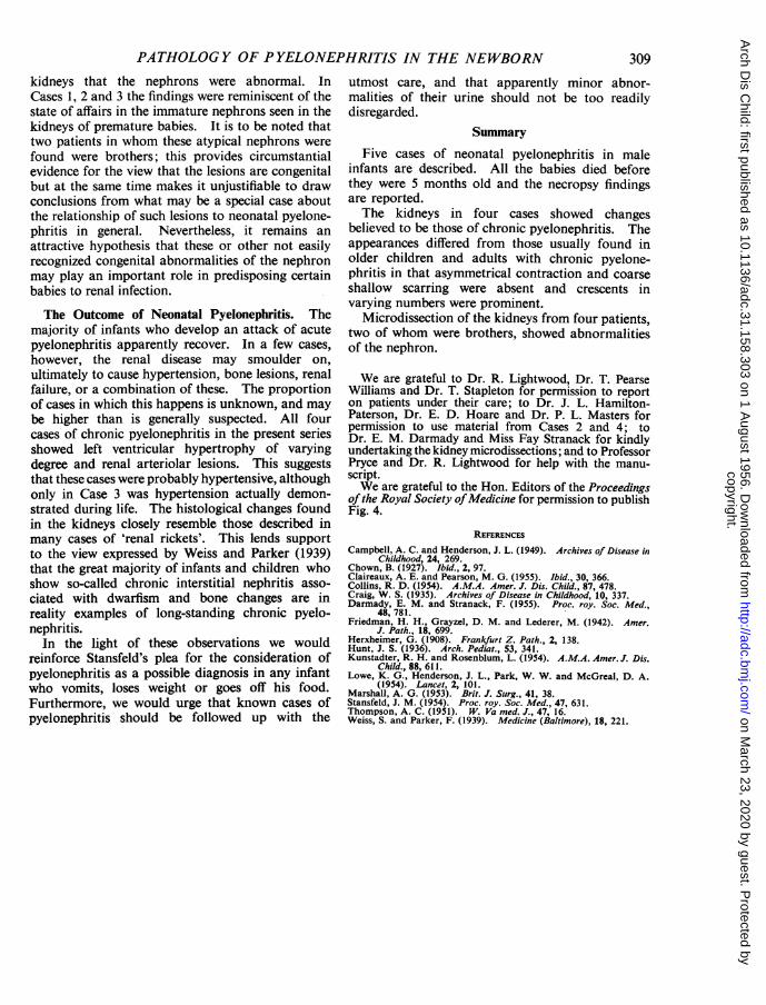
little change. The glomeruli were reduced to about halftheir normal number. Of the remainder few werecompletely normal and there was a tendency for thedamaged glomeruli to be huddled together. Someshowed marked concentric pericapsular fibrosis; in thesethe tuft was often relatively normal, but on otheroccasions tuft and capsule had fused to form a hyalinemass. In some there was proliferation of the cells ofBowman's capsule with crescent formation (Fig. 3). Afew mitoses were visible in these cell masses andoccasional capsular spaces had been obliterated. In a
few cases the crescents had undergone fibrosis. Adhesionsbetween tuft and capsule were frequent. Examples ofpseudo-crescent formation (Claireaux and Pearson, 1955)and partly hyaline tufts were also seen. The capillarybasement membranes of some of the affected glomerulartufts were thickened in a patchy manner and occasionaltufts contained small droplets of fat apparently depositedbetween the capillary loops. In a few instances thecapsular space contained acidophilic material but therewere no polymorphs.Most of the proximal tubules were dilated and con-
tained eosinophilic, P.A.S.-positive colloid casts. Thelining epithelium was flattened, with no brush border,
jIg7 &-%-FK;. 3. Crescent composed of proliferating capsular epithelial cells.
(Haematoxylin and eosin x 150.)
305
copyright. on M
arch 23, 2020 by guest. Protected by
http://adc.bmj.com
/A
rch Dis C
hild: first published as 10.1136/adc.31.158.303 on 1 August 1956. D
ownloaded from

ARCHIVES OF DISEASE IN CHILDHOOD
FJG. 4. Case 3. Numnrous dilated tubuls lined by flattenedqehelum and containig 'colloidf casts. The artery shows pro-
ductive endarteritis. (Haematoxylin and cosin x 160).
and lacked alkaline phosphatase. Many of the loops,distal tubules and colecting tubules contained pink casts,some of which were calified. A few contained pus.
Large areas of interstitial tissue, particularly in thecortex, were densely infiltrated by plasma cells, lympho-cytes, occasional eosinophils and histiocytes. In themedulla the chronic inflammatory infiltrate was muchlighter. Interstitial fibrosis was also marked.The arcuate and interlobular arteries showed marked
productive endarteritis (Fig. 4) whilst many arteriolesshowed hyperplastic arteriolosclerosis and occasionalfatty-hyaline change.The epithelium of the renal pelvis was normal.Nephron dissection showed the tubules to be
essentially the same as those in the patient's brother(Case 2).No organisms, inclusion bodies, cystine crystals or
extramedullary haemopoiesis were found in this kidney.Hyperplastic arteriolosclerosis was also found in the
arterioles of the gut wall.
Case 4. S.R. was a boy born at term on August 25,1954, by spontaneous normal delivery. On September 5he began vomiting and on September 9 the urine con-tained many pus cells and yielded a mixed growth ofmicrococci and proteus. On the same day blood cultureyielded a growth of E. coli and four days later an appar-ently identical coliform organism was cultured from theurie. On September 15 the blood urea level was
100 mg. % and the systolic blood pressure was 80 mm.He was given a prolonged course of sulphonamide andchkamphenicol, during which his general conditionimproved and his urine became free of pus cells butcontinued to yield a scanty mixed growth of enterococci,micrococci, proteus and coliforms. Blood urea fell toabout 50 mg. %0 but did not return to normal. Treat-ment was stopped on September 28. He remainedclinically well, but on October 21 the urine again con-taied pus cells and grew E. coli and Proteus, and thehaemoglobin was 8 - 9 g. %. From October 25 toNovember 11 he was given sulphonamide, penicillin andstreptomycin, in spite of which the urine continued toshow a small excess of pus cells and a mixed growth oforganisms. The blood urea level remained at 50 to60 mg. 0. His general progress was satisfactory untilJanuary 12, 1955, when he developed a slight cough: onJanuary 15 his mother found him dead in his cot. Atdeath he was aged 21 weeks.GENERAL NECROPSY FINDuIcs. The body was well
nourished, showing cyanosis of the lips and finger nailsand bile-stained fluid coming from the mouth.The heart weighed 39 g. and showed left ventricular
hypertrophy, moderate dilatation of the right side, and afew petechiae beneath the epicardium. There was frothymucopus in the trachea, main bronchi and many of thesmaller bronchi, particularly in the left lower lobe, withconsiderable injection of the lining mucosa. The lungsshowed extensive, patchy, lobular collapse, most markedin the lower lobes, and generalized oedema. Micro-scopically there was a purulent bronchioitis. The hilarlymph nodes were enlarged and haemorrhagic. The liverwas enlaged and congested. The gastro-intestinal tract,pancreas, spleen, adrenals, thyroid, thymus and pituitarywere normal.The kidneys (weight 31 g. and 27 g.) were congested.
The capsules stripped readily, exposing surfaces with afine irregular granularity. No abnormality was seen onsection. The pelves, renal vessels and the remainder ofthe urogenital system were normal.The brain and bones were normal.MICROSCOPY OF THE KmINEfys. The renal capsule was
normal. The glomeruli were slightly reduced in number.Most of the surviving glomeruli were normal, but in aroughly wedge-shaped area; they were closer togetherthan usual and showed various changes, the commonestof which was marked concentric pericapsular fibrosis,often with relatively normal tufts. However, in somethis had led to complete obliteration of the tufts so thatonly a hyaline sphere remained. Some showed adhesionsbetween tuft and capsule. Occasional crescents wereseen (Fig. 5) and a few primitive glomeruli.
In the patches where the glomeruli were affected someof the proximal tubules appeared atrophic with flattenedepithelium. However, colloid casts were few, and dilata-tion was not a feature. The distal parts of the tubulesseemed normal. Some primitive tubules were also seen.
Fibrosis and chronic inflammatory infiltration of theinterstitium with lymphocytes, plasma cells andoccasional eosinophils were features of the scarred areas.The arcuate and interlobular arteries showed slight
306l
copyright. on M
arch 23, 2020 by guest. Protected by
http://adc.bmj.com
/A
rch Dis C
hild: first published as 10.1136/adc.31.158.303 on 1 August 1956. D
ownloaded from

PATHOLOGY OF PYELONEPHRITIS IN THE NEWBORN
t
~tFIG. 5. Case 4. Glomerulus with a well defined cellular capsular
crescent. (Haematoxylin and eosin '. 350.)
productive endarteritis and the arterioles mild hyper-plastic arteriolosclerosis.The epithelium of the renal pelvis was normal.Microdissection showed little evidence of persisting
foetal nephrons. However, there were a number oflong loops of Henle.
Arteries and arterioles in other organs were normal.
Case 5. R.F. was a male infant born at term onMay 23, 1954, by spontaneous normal delivery. Hemade normal progress until June 5 when he refused hisfeeds and was noticed to be jaundiced. The liver wasenlarged and the tip of the spleen and left kidney werepalpable. On June 6 the urine contained pus cells andyielded a growth of E. coli. On June 9, the blood urealevel was 160 mg. 00. Between June 7 and July 6 hewas treated with sulphonamide, penicillin, streptomycin,terramycin, chloramphenicol and erythromycin invarious combinations. From mid-June the jaundicefaded: whether it had been due to infection or to neonatalhepatitis was uncertain. His general condition improvedslowly and irregularly and although the urine becamefree of pus cells it continued to yield a growth of proteus,coliforms and micrococci. By July 10 he was thoughtto be clinically well, and on July 28 he was dischargedfrom hospital. He did not return for follow-up but wasbelieved by his parents to be progressing satisfactorily
until September 3 when he developed a severe attack ofvomiting and diarrhoea. He was admitted to hospitalbut died on September 4, aged 144 weeks.GENERAL NECROPSY FINDINGs. The body was severely
dehydrated. The heart weighed 40 g. and showed leftventricular hypertrophy. There was bilateral basalpulmonary congestion. The tongue was flabby andfurred and there was hyperaemia of the mucous me-branes of the stomach and small intestine suggestive ofan acute gastro-enteritis. The liver and spleen were alittle enlarged. The brain and bones were normal.The kidneys (weight 27 g. and 24 g.) appeared normal
and their capsules stripped easily exposing smoothsubcapsular surfaces. No abnormalities were seen inthe remainder of the urogenital system.MICROSCOPY OF KIDNEYs. The renal capsule was
normal. The glomeruli were reduced to about three-quarters of their normal number. Many showedpericapsular fibrosis with relatively normal tufts, someshowed adhesions between tuft and capsule, others wellmarked capsular crescents. Some glomeruli had becomecompletely hyalinized. There damaged glomeruli tendedto be huddled together in rough cortical wedges.Unaffected glomeruli had undergone compensatoryhypertrophy.Many of the proximal tubules were dilated and some
contained 'colloid' casts. Some of their lining epithelialcells were atrophic, others showed hyaline dropletdegeneration. Distal and collecting tubules appearednormal. There were occasional casts in the collectingtubules, some showing calcification with tubular syncytialcell formation around them.
There was increased medullary interstitial and peri-vascular fibrous tissue, with occasional foci of calcifica-tion in cortex and mnedulla and a scattered infiltrationby lymphocytes, plasma cells and occasional eosinophilsand neutrophils.The arcuate and interlobular arteries showed mild
productive endarteritis, whilst the walls of occasionalarterioles showed hyperplastic arteriolosclerosis.The renal pelvis was lightly infiltrated by chronic
inflammatory cells and subepithelial fibrosis wasprominent.These kidneys were not microdissected.Arteries and arterioles in other organs were unaffected.
DiscussionMorphologial Differences from Pyelonephritis in
Adults. Case 1 died after an acute illness ofrelatively brief duration. The kidneys showed thetypical histological picture of acute pyelonephritis,differing in no way from that seen in the olderchild or adult. In the remaining cases, there was amore prolonged illness, dunrng which nitrogenretention and persistent pus cells and organisms inthe urine led to the clinical diagnosis of chronicpyelonephritis. The histology of the kidneys in allfour cases is believed to support this diagnosis,satisfying as it does the criteria laid down by Weiss
307
copyright. on M
arch 23, 2020 by guest. Protected by
http://adc.bmj.com
/A
rch Dis C
hild: first published as 10.1136/adc.31.158.303 on 1 August 1956. D
ownloaded from

ARCHIVES OF DISEASE IN CHILDHOOD
and Parker (1939), namely, (1) marked infiltrationof the interstitial tissue with lymphocytes, plasmacells and eosinophils; (2) presence of pericapsularfibrosis; (3) presence of so-called colloid casts intubules lined by atrophic epithelium.The only other condition with which these
histological changes might be confused is glomer-ulonephritis. In the present cases, however, manyglomeruli were unaffected, whereas in glomerulone-phritis it is usual for the changes to be diffuse withinvolvement of almost all glomeruli. Moreover, inthose glomeruli in which pericapsular fibrosis waspresent, the tufts were often normal; in glomerulone-phritis there is usually a fairly close correlationbetween the degree of fibrosis of the tufts and ofBowman's capsule. In glomerulonephritis theinterval between glomeruli is diminished but thesedistances tend to be fairly uniform, in sharp contrastto the rather wedge-shaped clusters of glomeruli wehave described. Lastly the interstitial infiltrationwith chronic inflammatory cells was greater in thepresent cases than is usual with glomerulonephritis.
However, the kidneys in these cases differed intwo respects from those found in adults sufferingfrom chronic pyelonephritis: (1) Macroscopically,there was little asymmetrical contraction or coarse,shallow scarring. (2) Microscopically, crescents invarying numbers were prominent.
Similar observations were made by Claireaux andPearson (1955) in their case of chronic pyelone-phritis in a newborn infant.The absence of scarring may be a consequence
of the comparatively short period of time for whichthe pyelonephritic process had been in progress,since all the cases were aged 21 weeks or under atthe time of death. The presence of crescents doesnot necessarily exclude the diagnosis of chronicpyelonephritis. Crescents are not specific forglomerulonephritis, inasmuch as identical forma-tions have long been known to occur in cases ofmalignant nephrosclerosis, subacute bacterial endo-carditis, and sometimes in the chronic pyelone-phritis of adults. In these latter conditions,however, the crescents usually involve onlyoccasional glomeruli, not the majority as inglomerulonephritis.
It is possible that epithelial crescents develop inresponse to various stimuli more readily in thenewborn period. Lowe, Henderson, Park andMcGreal (1954) described crescent formation in arenal biopsy from a child of 1 year 9 monthssuffering from the severe form of idiopathic hyper-calcaemia with low-grade secondary chronic pye-lonephritis. Campbell and Henderson (1949),discussing asymmetrical cortical necrosis of kidneys
in infancy, mentioned crescents in their Case 3,and Friedman, Grayzel and Lederer (1942) describeda proliferation of the parietal layer of Bowman'scapsule, giving a crescent-like appearance, as anoccasional finding in infants of average age2- 7 months in the absence of evidence of anyinflammatory reaction in the kidneys.
If it be accepted that crescents do not necessarilymean glomerulonephritis, then, as suggested byClaireaux and Pearson (1955), many cases describedpreviously as neonatal glomerulonephritis may wellhave been cases of chronic pyelonephritis. Recentexamples are the cases described by Thompson(1951), Collins (1954) and Kunstadter and Rosen-blum (1954).
Possible Role of Congenital Abnormalities of theNephron. Once past the neonatal period, boysseldom develop pyelonephritis except as a complica-tion of some manifest urinary tract anomaly. Allthe infants in the present series were males but noneof them showed any such anomaly. The possibilityarises that a defect of the nephron, presumablycongenital and demonstrable only microscopically,might be the predisposing lesion in these infants.The assessment of this possibility is complicated bythe fact that kidneys from young infants dying ofvarious diseases frequently show glomeruli andtubules which are 'sclerosed', immature or other-wise atypical. The cause and significance of theseabnormalities is not clear. Friedman et al. (1942)have given the name 'congenital glomerulosclerosis'to the hyaline changes in the glomerular tufts andBowman's capsule which were first described byHerxheimer in 1908. Friedman found theselesions in 12 out of 100 consecutive infants dyingunder the age of 14 months; he pointed out theirrelationship to obliterative changes in the corre-sponding afferent arteriole, but could advance nomore fundamental explanation for their occurrence.It is interesting that the lesions were found aboutthree times as often in males as in females, this beingthe estimated ratio of pyelonephritis in males tofemales in the first few months of life.
Marshall (1953), in a series of kidneys frominfants and children, described structures which,on morphological grounds, he believed to be persist-ing foetal glomeruli and tubules. In most of theolder infants and children pyelonephritis was presentand Marshall suggested that such infections had astheir underlying lesion a focal or localized dysplasiaof the renal parenchyma.
In four cases of the present series (Nos. 1, 2, 3and 4), Dr. E. M. Darmady and Miss Fay Stranackhave shown by painstaking microdissection of the
308
copyright. on M
arch 23, 2020 by guest. Protected by
http://adc.bmj.com
/A
rch Dis C
hild: first published as 10.1136/adc.31.158.303 on 1 August 1956. D
ownloaded from
PATHOLOG Y OF P YELONEPHRITIS IN THE NEWBORN 309kidneys that the nephrons were abnormal. InCases 1, 2 and 3 the findings were reminiscent of thestate of affairs in the immature nephrons seen in thekidneys of premature babies. It is to be noted thattwo patients in whom these atypical nephrons werefound were brothers; this provides circumstantialevidence for the view that the lesions are congenitalbut at the same time makes it unjustifiable to drawconclusions from what may be a special case aboutthe relationship of such lesions to neonatal pyelone-phritis in general. Nevertheless, it remains anattractive hypothesis that these or other not easilyrecognized congenital abnormalities of the nephronmay play an important role in predisposing certainbabies to renal infection.
The Outcome of Neonatal Pyelonephritis. Themajority of infants who develop an attack of acutepyelonephritis apparently recover. In a few cases,however, the renal disease may smoulder on,ultimately to cause hypertension, bone lesions, renalfailure, or a combination of these. The proportionof cases in which this happens is unknown, and maybe higher than is generally suspected. All fourcases of chronic pyelonephritis in the present seriesshowed left ventricular hypertrophy of varyingdegree and renal arteriolar lesions. This suggeststhat these cases were probably hypertensive, althoughonly in Case 3 was hypertension actually demon-strated during life. The histological changes foundin the kidneys closely resemble those described inmany cases of 'renal rickets'. This lends supportto the view expressed by Weiss and Parker (1939)that the great majority of infants and children whoshow so-called chronic interstitial nephritis asso-ciated with dwarfism and bone changes are inreality examples of long-standing chronic pyelo-nephritis.
In the light of these observations we wouldreinforce Stansfeld's plea for the consideration ofpyelonephritis as a possible diagnosis in any infantwho vomits, loses weight or goes off his food.Furthermore, we would urge that known cases ofpyelonephritis should be followed up with the
utmost care, and that apparently minor abnor-malities of their urine should not be too readilydisregarded.
SummaryFive cases of neonatal pyelonephritis in male
infants are described. All the babies died beforethey were 5 months old and the necropsy findingsare reported.The kidneys in four cases showed changes
believed to be those of chronic pyelonephritis. Theappearances differed from those usually found inolder children and adults with chronic pyelone-phritis in that asymmetrical contraction and coarseshallow scarring were absent and crescents invarying numbers were prominent.
Microdissection of the kidneys from four patients,two of whom were brothers, showed abnormalitiesof the nephron.
We are grateful to Dr. R. Lightwood, Dr. T. PearseWilliams and Dr. T. Stapleton for permission to reporton patients under their care; to Dr. J. L. Hamilton-Paterson, Dr. E. D. Hoare and Dr. P. L. Masters forpermission to use material from Cases 2 and 4; toDr. E. M. Darmady and Miss Fay Stranack for kindlyundertaking the kidney microdissections; and to ProfessorPryce and Dr. R. Lightwood for help with the manu-script.We are grateful to the Hon. Editors of the Proceedings
of the Royal Society ofMedicine for permission to publishFig. 4.
REFERENCESCampbell, A. C. and Henderson, J. L. (1949). Archives of Disease in
Childhood, 24, 269.Chown, B. (1927). Ibid., 2, 97.Claireaux, A. E. and Pearson, M. G. (1955). Ibid., 30, 366.Collins, R. D. (1954). A.M.A. Amer. J. Dis. Child., 87, 478.Craig, W. S. (1935). Archives of Disease in Childhood, 10, 337.Darmady, E. M. and Stranack, F. (1955). Proc. roy. Soc. Med.,
48, 781.Friedman, H. H., Grayzel, D. M. and Lederer, M. (1942). Amer.
J. Path., 18, 699.Herxheimer, G. (1908). Frankfurt Z. Path., 2, 138.Hunt, J. S. (1936). Arch. Pediat., 53, 341.Kunstadter, R. H. and Rosenblum, L. (1954). A.M.A. Amer. J. Dis.
Child., 88, 611.Lowe, K. G., Henderson, J. L., Park, W. W. and McGreal, D. A.
(1954). Lancet, 2, 101.Marshall, A. G. (1953). Brit. J. Surg., 41, 38.Stansfeld, J. M. (1954). Proc. roy. Soc. Med., 47, 631.Thompson, A. C. (1951). W. Va med. J., 47, 16.Weiss, S. and Parker, F. (1939). Medicine (Baltimore), 18, 221.
copyright. on M
arch 23, 2020 by guest. Protected by
http://adc.bmj.com
/A
rch Dis C
hild: first published as 10.1136/adc.31.158.303 on 1 August 1956. D
ownloaded from


