Embed Size (px)
Citation preview

8/6/2019 PATHOLAB Cellular Growth and Differentiation
http://slidepdf.com/reader/full/patholab-cellular-growth-and-differentiation 1/9
1
Case Study: 60 y/o female that underwent hysterectomy
Nulliparous: 30-40g
parous: 75-100g
Adaptive Mechanism seen in the picture:
o Atrophy
Cause of Atrophy
a. Decreased workload
b. Denervation
c. Decrease hormone stimulation
d. Pressure
e. Inadequate nutrition
f. Hypoxia
Reversible injury can revert back to its original form
Injury progressively introduced to the cell will lead to irreversible injury and then cell death
hallmark of reversible injury:
Cell Swelling
decrease oxidative phosphorylation
fatty change- microscopic change and not a hallmark of reversible injury
Subject: Pathology LABTopic: Cellular Growth and DifferentiationLecturer: Dr. Renan NavarroDate of Lecture: 06/23/2011Transcriptionist: Desiree TimtimanPages: 8 S
Y
2 0 1 1 - 2 0 1 2

8/6/2019 PATHOLAB Cellular Growth and Differentiation
http://slidepdf.com/reader/full/patholab-cellular-growth-and-differentiation 2/9
2
Case study: 50 y/o with Chronic HPN
Myocardium
Adaptive Mechanism seen in the picture:
o Hyperthropied myocardium
o Based on the history you can expect that the myocardium are enlarged
o Microscopic description: Notice the enlarged, box-shaped nucleus
Case Study: from a heart of a known hypertensive 45 y/o female with a diagnosis of intracerebral hemorrhage
Pictograph shows an Old myocardial infarction No acute inflammation
o Acute inflammation is characterized by infiltration of PMNs
Intensed fibrosis; notice the pink wavy collagen at the picture
Increased Vascular proliferation
tissue damaged exemplified by the loss of striation and nucleus of the myocardium
This is an irreversible damage
fibrosis
Myocardium: notice that there’s no nucleus and striations
Vascular proliferation
8/6/2019 PATHOLAB Cellular Growth and Differentiation
http://slidepdf.com/reader/full/patholab-cellular-growth-and-differentiation 3/9
3
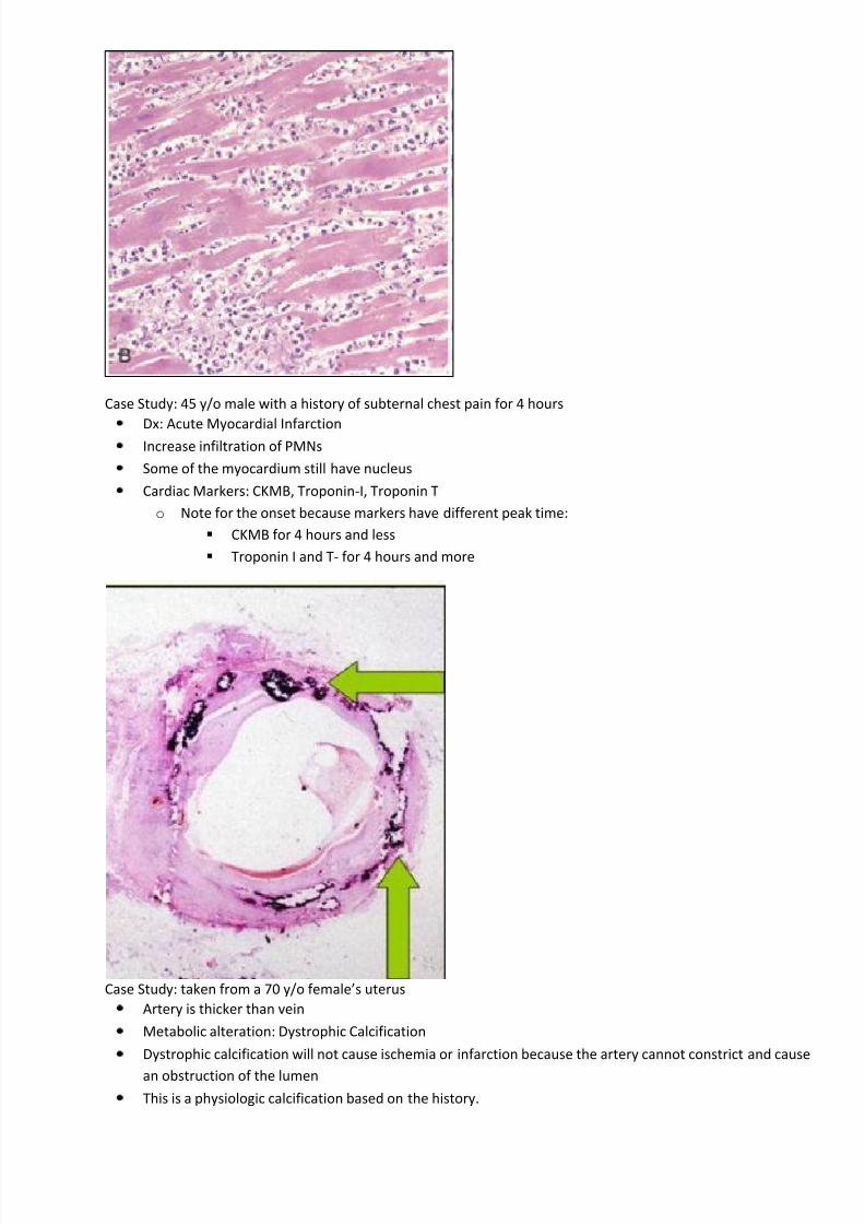
Case Study: 45 y/o male with a history of subternal chest pain for 4 hours
Dx: Acute Myocardial InfarctionIncrease infiltration of PMNs
Some of the myocardium still have nucleus
Cardiac Markers: CKMB, Troponin-I, Troponin T
o Note for the onset because markers have different peak time:
CKMB for 4 hours and less
Troponin I and T- for 4 hours and more
Case Study: taken from a 70 y/o female’s uterus
Artery is thicker than vein
Metabolic alteration: Dystrophic Calcification
Dystrophic calcification will not cause ischemia or infarction because the artery cannot constrict and cause
an obstruction of the lumen
This is a physiologic calcification based on the history.

8/6/2019 PATHOLAB Cellular Growth and Differentiation
http://slidepdf.com/reader/full/patholab-cellular-growth-and-differentiation 4/9
4
Case Study: 65 y/o male with dysuria
Take note of the history
Organ: Prostate Gland
notice the corpora amylacea(C), a distinct feature of prostate gland
Notice the increase in glands and see how closely packed it is.
o This is therefore a Hyperplastic change
Adaptive Mechanism: Hyperplasia
o Increase in cells, in this case there’s an increase in glands
o This is increase in glands, therefore an increase in the size of the organ
Dx: Nodular Prostatic Hyperplasia
glands
8/6/2019 PATHOLAB Cellular Growth and Differentiation
http://slidepdf.com/reader/full/patholab-cellular-growth-and-differentiation 5/9
5
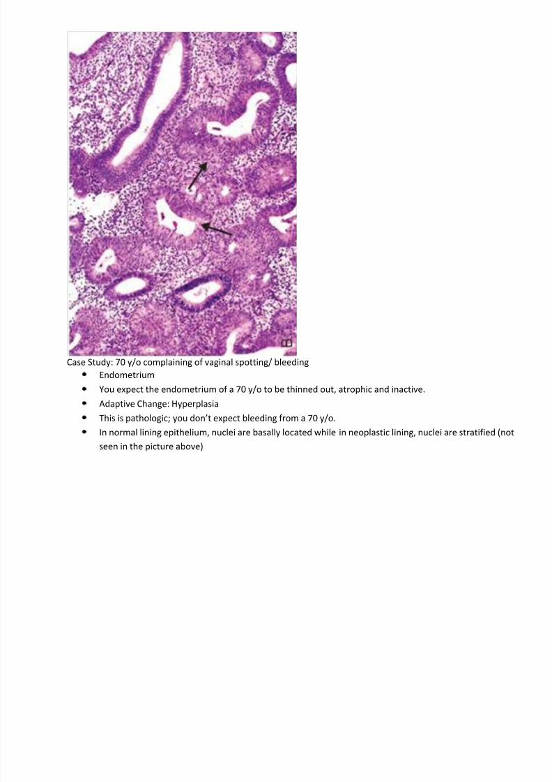
Case Study: 70 y/o complaining of vaginal spotting/ bleeding
Endometrium
You expect the endometrium of a 70 y/o to be thinned out, atrophic and inactive.
Adaptive Change: Hyperplasia
This is pathologic; you don’t expect bleeding from a 70 y/o.
In normal lining epithelium, nuclei are basally located while in neoplastic lining, nuclei are stratified (not
seen in the picture above)

8/6/2019 PATHOLAB Cellular Growth and Differentiation
http://slidepdf.com/reader/full/patholab-cellular-growth-and-differentiation 6/9
6
Case Study:
Transitional zone in cervix: from simple tall columnar to stratified squamous epi.
Adaptive Change: Metaplasia
Notice that the endocervical gland, which is normally beneath the simple columnar epithelium,is already
beneath the stratified squamous epithelium, indicating that metaplasia has taken place probably due toirritation.
If metaplasia persist, it can lead to malignant transformation
o Ex. Barret’s Esophagus
Endocervical
glands
Non-keratinized Stratified Squamous Epithelium
Transition
zone
Simple Columnar
Epithelium
Non-keratinized Stratified Squamous Epithelium

8/6/2019 PATHOLAB Cellular Growth and Differentiation
http://slidepdf.com/reader/full/patholab-cellular-growth-and-differentiation 7/9
7
Case Study: taken from the cervical mass of a 7 y/o male
From Lymph node(ok so the picture [I think] is from the lungs, but you get the point, right?)
This a granuloma, typically seen in a tuberculous infection
Caseous Necrosis
o Grossly: yellow cheesy/chalky material
o Acellular pink amorphous material
o Presence of Epitheloid cells surrounded by lymphocytes
Chronic granulomatous inflammation
o Inflammation seen in necrosis

8/6/2019 PATHOLAB Cellular Growth and Differentiation
http://slidepdf.com/reader/full/patholab-cellular-growth-and-differentiation 8/9
8
Necrosis
Cell death with inflammatory reaction in the host
Apoptosis
cell death by this pathway does not elicit an inflammatory reaction in the hostnuclear changes:
Karyorrhexis
pyknotic nucleus undergoes fragmentation.
Pyknosis
characterized by nuclear shrinkage and increased basophilia
Karyolysis
change that presumably reflects loss of DNA because of enzymatic degradation by
endonucleases.
Metaplasia
reversible change in which one differentiated cell type (epithelial or mesenchymal) is replaced by another
cell type
End of transcription
Pictures from laboratory(thank you Joyce), internet, Wheater’s and Robbin’s.
Study hard.
“I am the light of the world. Whoever follows me will not walk in darkness but will have the light of life.” John
8:12
Adaptive Mechanism Stimuli
Metaplasia Chronic irritation
Hypertrophy/Hyperplasia Increased demand, increased stimulation
Atrophy Decreased nutrients, decreased stimulation
Intracellular Accumulation METABOLIC ALTERATIONS, GENETIC OR ACQUIRED; CHRONIC INJURY
Karyorrhexis
Pyknosis
Karyolysis

8/6/2019 PATHOLAB Cellular Growth and Differentiation
http://slidepdf.com/reader/full/patholab-cellular-growth-and-differentiation 9/9