Embed Size (px)
Citation preview

The association between anomalous pulmonary ve-nous drainage and acquired valvular heart disease hasrarely been reported (1-6). Nonetheless, awareness ofthis entity is important because the appropriate surgicalapproach can then be identified based upon the preoper-ative diagnosis.
The purpose of this case report is to present a case ofrheumatic mitral stenosis and partial anomalous pul-monary venous connection to the left innominate veinaccompanied by the presence of the levoatriocardinalvein. The diagnosis was established preoperatively byCT and MR, thereby making complete surgical correc-
tion feasible.
Case Report
A 67-year-old woman, with a known case of mitralstenosis, underwent chest CT scanning for the preopera-tive evaluation of a palpable soft-tissue lesion on herchest wall. A CT scan was performed using a 16-channelmultidetector CT (Sensation 16, Siemens Healthcare,Forcheim, Germany) without cardiac gating and ob-tained 40 sec after administration of 100 mL of a 300mgI/mL IV contrast medium at a rate of 2.5 mL/sec. TheCT parameters were as follows: 16 × 1.5 mm collima-tion, 100 effective milliamperes (mAs), 120 kVp, a rota-tion time of 0.5 seconds and pitch of 1.5. CT data werereconstructed with a 5 mm slice thickness using a softkernel. The CT scan revealed an abnormal vascularstructure in the left upper mediastinum, which connect-ed the left innominate vein and the left atrium (levoatri-ocardinal vein). At the level of the aortic arch, the abnor-
J Korean Soc Radiol 2010;63:339-343
─ 339─
Partial Anomalous Pulmonary Venous Return via aLevoatriocardinal Vein in Association with Rheumatic
Mitral Stenosis: MR Demonstration and Successful Surgical Repair1
Dongho Hyun, M.D., Eun Jin Chae, M.D., Ph.D., Joon Beom Seo, M.D., Ph.D., Joon-Won Kang, M.D., Kyung-Hyun Do, M.D., Ph.D., Choong Wook Lee, M.D.,
Hyun Joo Lee, M.D., Hye Jeon Hwang, M.D., Tae-Hwan Lim, M.D., Ph.D.
1Department of Radiology and Research Institute of Radiology, AsanMedical Center, University of Ulsan College of MedicineReceived April 12, 2010 ; Accepted June 20, 2010Address reprint requests to : Eun Jin Chae, M.D., Ph.D., Department ofRadiology and Research Institute of Radiology, Asan Medical Center,University of Ulsan College of Medicine, 86 Asanbyeongwon-gil, Songpa-gu, Seoul 138-736, Korea.Tel. 82-2-3010-4400 Fax. 82-2-476-4719 E-mail: [email protected]
The preoperative evaluation of the hemodynamics associated with PAPVR andrheumatic mitral valve stenosis is necessary for successful surgical treatment, eventhough the incidence rate is rare. The purpose of this case report is to present the use-fulness of CT and MRI for diagnosing rheumatic mitral stenosis and partial anomalouspulmonary venous connection to the left innominate vein accompanied by the pres-ence of the levoatriocardinal vein and evaluating its flow dynamics.
Index words : Magnetic Resonance ImagingTomography, X-Ray ComputedMitral stenosisRheumatic heart diseasePartial anomalous pulmonary venousLevoatriocardinal vein
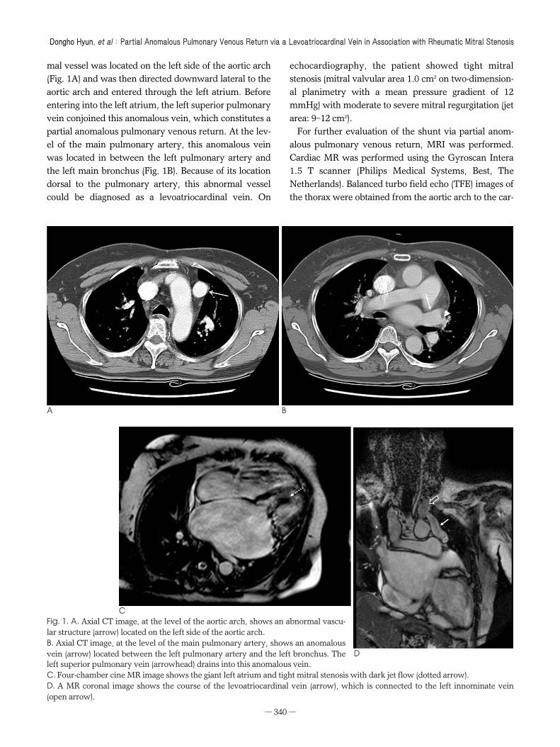
mal vessel was located on the left side of the aortic arch(Fig. 1A) and was then directed downward lateral to theaortic arch and entered through the left atrium. Beforeentering into the left atrium, the left superior pulmonaryvein conjoined this anomalous vein, which constitutes apartial anomalous pulmonary venous return. At the lev-el of the main pulmonary artery, this anomalous veinwas located in between the left pulmonary artery andthe left main bronchus (Fig. 1B). Because of its locationdorsal to the pulmonary artery, this abnormal vesselcould be diagnosed as a levoatriocardinal vein. On
echocardiography, the patient showed tight mitralstenosis (mitral valvular area 1.0 cm2 on two-dimension-al planimetry with a mean pressure gradient of 12mmHg) with moderate to severe mitral regurgitation (jetarea: 9-12 cm2).
For further evaluation of the shunt via partial anom-alous pulmonary venous return, MRI was performed.Cardiac MR was performed using the Gyroscan Intera1.5 T scanner (Philips Medical Systems, Best, TheNetherlands). Balanced turbo field echo (TFE) images ofthe thorax were obtained from the aortic arch to the car-
Dongho Hyun, et al : Partial Anomalous Pulmonary Venous Return via a Levoatriocardinal Vein in Association with Rheumatic Mitral Stenosis
─ 340─
A B
C
Dleft superior pulmonary vein (arrowhead) drains into this anomalous vein.C. Four-chamber cine MR image shows the giant left atrium and tight mitral stenosis with dark jet flow (dotted arrow).D. A MR coronal image shows the course of the levoatriocardinal vein (arrow), which is connected to the left innominate vein(open arrow).
Fig. 1. A. Axial CT image, at the level of the aortic arch, shows an abnormal vascu-lar structure (arrow) located on the left side of the aortic arch.B. Axial CT image, at the level of the main pulmonary artery, shows an anomalousvein (arrow) located between the left pulmonary artery and the left bronchus. The

diac apex for anatomic delineation. The scan parametersof cine images consisted of slice thickness 8 mm, no gap,echo time/repetition time (TE/TR) of 1.7/3.4 ms, flip an-gle of 50 degrees, 169 phase encoding steps, and a scanfield of view (FOV) measuring 320 × 320 mm. Cine im-ages were performed for the evaluation of valvular mo-tion. The scan parameters of cine images consisted of aslice thickness measuring 8 mm, no gap, TE/TR 1.7/3.4ms, a flip angle of 50 degrees, 169 phase encoding steps,a scan FOV measuring 320 × 320 mm, and retrospec-tive gating with 20 calculated phases. The flow of the su-perior vena cava (SVC) and the levoatriocardinal vein
were obtained using velocity-encoded cine (VENC) MRimaging. The scan parameters of cine images consistedof a slice thickness measuring 8 mm, no gap, TE/TR2.8/4.7 ms, a flip angle of 15 degrees, 128 phase encod-ing steps, a scan FOV measuring 320 × 320 mm, andretrospective gating.
Four-chamber cine MR images showed thickeningand doming of the mitral valve leaflets, and also demon-strated a dark jet flow through the stenotic mitral valvearea, which is consistent with tight mitral stenosis (Fig.1C). Coronal MR images grossly determined the courseof the levoatriocardinal vein connecting the left innomi-
J Korean Soc Radiol 2010;63:339-343
─ 341─
E F G
H
Fig. 1. E-G. On sagittal MR images, the left superior pulmonaryvein (arrowhead) drains into the levoatriocardinal vein (arrow).H. Velocity-encoded cine MR image provides information re-garding the flow direction at the level of the superior vena cava(open arrow) and the levoatriocardinal vein (arrow).
nate vein and the left atrium (Fig. 1D). Sagittal MR im-ages demonstrated that the left superior pulmonary veinconnected to the levoatriocardinal vein as a form of par-tial anomalous pulmonary venous return (Figs. 1E-G).Hemodynamically, as the left atrial pressure increased,owing to mitral stenosis, a large amount of the left-to-right shunt was produced from both the left atrium andthe left superior pulmonary vein to the left innominatevein through the levoatriocardinal vein. Assessment ofthe flow dynamics, including quantification of theshunt, was performed by VENC MR imaging. On phasecontrast imaging, the flow direction of the SVC and thelevoatriocardinal vein were opposing (Fig. 1H). On flowmeasurement, the ascending aorta and both central pul-monary arteries showed a flow of 40 ml/min and 100mL/min, respectively. According to the flow measure-ment, the calculated left-to-right shunt was approxi-mately 60% and the ratio of pulmonary to systemicblood flow (Qp/Qs) was approximately 2.51.
The preoperative diagnosis was mitral stenosis andpartial anomalous pulmonary venous connection to theleft innominate vein accompa nied with the presence ofthe levoatriocardinal vein. The patient underwent anopen mitral commissurotomy and ligation of the levoa-triocardinal vein and was ultimately discharged withoutany adverse events.
Discussion
The association of anomalous pulmonary venousdrainage with mitral valve disease has rarely been rec-ognized. Several cases of mitral stenosis with partial pul-monary venous return have been described in previousreports (1-6). However, many of these cases showed thepartial anomalous pulmonary drainage into a systemicvein, but not into an anomalous vein connecting the leftatrium and the systemic vein simultaneously (4-6). Inthe present case, the left superior pulmonary veindrained into the anomalous vasculature, connecting theleft atrium and the left innominate vein.
The levoatriocardinal vein, first described by Edwardet al, is very uncommon and its presence is pathologic(7, 8). It is probably derived from the persistence ofanatomic channels connecting the pulmonary capillaryplexus to the cardinal veins in the embryonic foregut. Itis a pulmonary-systemic connection that provides an al-ternative pathway for pulmonary venous drainage inthe presence of a severe left-sided obstructive lesionsuch as mitral atresia, because this unusual vein con-
nects the left atrium or pulmonary vein to the left in-nominate vein.
Another anomalous vein with many similarities to thelevoatriocardinal vein is a persistent left superior venacava (PLSVC). A PLSVC is usually connected to thecoronary sinus, and rarely to the left atrium or pul-monary vein (3). The levoatriocardinal vein differs froma PLSVC draining into the left atrium in that the levoa-triocardinal vein ascends dorsal to the left pulmonaryartery whereas the PLSVC ascends ventral to it.Moreover, the levoatriocardinal vein may be com-pressed between the left pulmonary artery and the leftbronchus (8).
Partial anomalous pulmonary venous return (PAPVR)is most commonly seen in patients with an atrial septaldefect. When the atrial septum is intact, mitral stenosisof congenital or acquired origin is the next most fre-quent combination (1, 4). The association of PAPVR andrheumatic mitral stenosis appears to be no more thancoincidental (2). Previous studies reported that theamount of left-to-right shunt was insignificant in pa-tients with PAPVR, mitral stenosis, and intact interatrialseptum (9). However, in our patient, the left-to-rightshunt increased to as much as 60% of the pulmonaryblood flow, which is the reason why the surgical ligationof the levoatriocardinal vein was performed.
This case is the first in which the preoperative diagno-sis was established by MR in a patient with partialanomalous pulmonary venous return via a levoatriocar-dinal vein in association with rheumatic mitral stenosis.Preoperative cardiac cine MR with VENC demonstratedthe anatomic configuration of the anomalous vein andallowed a quantitative assessment of the flow dynamicsof the left-to-right shunt via the anomalous vein.Velocity-encoded cine MR imaging is recognized as avaluable technique for the quantitative assessment offlow dynamics in congenital heart diseases. We usedthis technique to measure the left-to-right shunt flow inthis patient as it provided necessary hemodynamic in-formation before surgery.
In summary, we report a rare case of partial anom-alous pulmonary venous return via a levoatriocardinalvein in association with rheumatic mitral stenosis. Weused MR for preoperative imaging to delineate the com-plicated anatomic structure and to determine the propersurgical treatment.
Dongho Hyun, et al : Partial Anomalous Pulmonary Venous Return via a Levoatriocardinal Vein in Association with Rheumatic Mitral Stenosis
─ 342─

References
1. Iga K, Hori K. Abnormal venous connection between the left up-per pulmonary vein and the left brachiocephalic vein, associatedwith rheumatic combined valvular heart disease. Heart Vessels1990;5:113-116
2. Halpern BL, Murray GC, Conti CR, Humphries JO, Gott VL.Continuous murmur due to the combination of rheumatic mitralstenosis and a rare type of anomalous pulmonary venous drainage.Circulation 1970;42:165-170
3. Reid JM, Barclay RS, Stevenson JG, Welsh TM, McSwan N.Rheumatic mitral stenosis in association with partial anomalouspulmonary venous return. Thorax 1968;23:197-199
4. Alpert JS, Dexter L, Vieweg WV, Haynes FW, Dalen JE.
Anomalous pulmonary venous return with intact atrial septum: di-agnosis and pathophysiology. Circulation 1977;56:870-875
5. Bruschke AV, Bloch A. Anomalous pulmonary venous drainageassociated with mitral valve disease. Am Heart J 1969;78:437-443
6. Singh R, McGuire LB, Carpenter M, Dammann JF. Mitral stenosisassociated with partial anomalous pulmonary venous return (withintact atrial septum). An unsolved question. Am J Cariol 1971;28:226-231
7. Beckman CB, Moller JH, Edwards JE. Alternate pathways to pul-monary venous flow in left-sided obstructive anomalies.Circulation 1975;52:509-516
8. Snellen HA, van Ingen HC and Hoefsmit EC. Patterns of anom-alous pulmonary venous drainage. Circulation 1968;38:45-63
9. Shrivastava S, Bhargava S, Tandon R. Partial anomalous pul-monary venous connection with intact arterial septum and mitralstenosis. An evaluation. Indian Heart J 1976;28:145-151
J Korean Soc Radiol 2010;63:339-343
─ 343─
한 상의학회지 2010;63:339-343
류마티스성 승모판 협착에 동반된 Levoatriocardinal 정맥을 통한 부분
폐정맥 환류 이상-자기공명 상 소견 및 성공적인 수술적 치료: 증례 보고1
1울산의 서울아산병원 상의학과
현동호∙채은진∙서준범∙강준원∙도경현∙이충욱∙이현주∙황혜전∙임태환
류마티스성 승모판 협착에 부분 폐정맥 환류 이상이 합병된 증례는 드물게 보고되고 있으며 이런 경우 부분 폐정
맥 환류 이상의 합병으로 인한 혈류 역학적 변화를 수술 전에 평가하는 것이 적절한 치료를 위해서 필수적이다. 저자
들은 류마티스성 승모판 협착에 levoatriocardinal 정맥을 통한 부분 폐정맥 환류 이상이 합병된 환자에서 수술 전
전산화단층촬 과 자기공명 상을 통해 levoatriocardinal 정맥을 진단하고 부분 폐정맥 환류 이상에 의한 혈류역
학적 변화를 정량적으로 평가함으로써 성공적인 수술적 치료가 가능했던 증례를 보고하고자 한다.
![Anomalous “Mutilated Common Trunk” Aortic Arch ......aorta was prevalence up to 16.1%, out of which 11% of the cases were the left VA arose with the left subclavian artery [8]](https://img.dokumen.tips/doc/110x75/5fa5826cb88f1a17f4153354/anomalous-aoemutilated-common-trunka-aortic-arch-aorta-was-prevalence.jpg)