Embed Size (px)
Citation preview

Biomedicine: Human SciencesLecture 7:
Digestive SystemPart One
1Copyright CNM 2016-17: Human Sciences – Digestive System Part One. Last updated 31st August 2016. BQ.

Learning OutcomesIn today’s topic you will learn:
Ø About the digestive system, including its structure and function
Ø Enzymes and hormones involved in digestion.
Ø The types of dietary carbohydrates, fats/oils & proteins.
2http://www.healthpages.org/
Copyright CNM 2016-17: Human Sciences – Digestive System Part One. Last updated 31st August 2016. BQ.
Copyright CNM 2016-17: Human Sciences – Digestive System Part One. Last updated 31st August 2016. BQ.
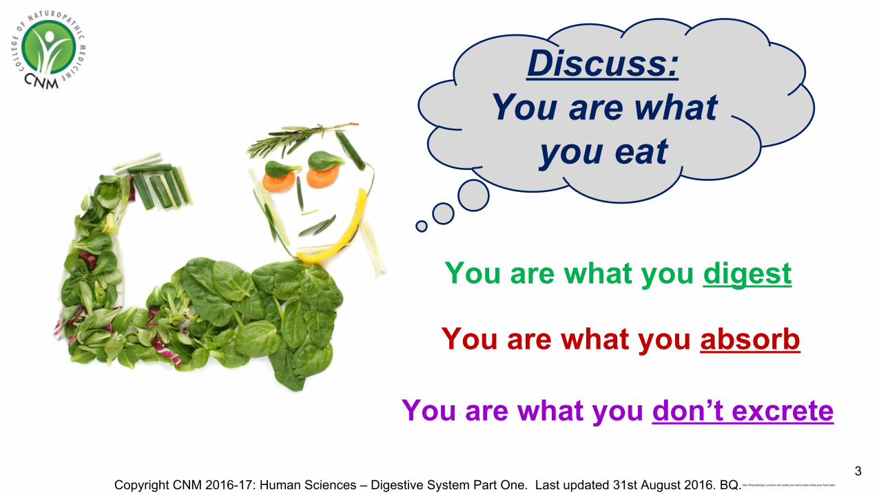
Discuss:You are what
you eat
http://thepaleologic.com/you-are-what-you-eat-but-also-what-your-food-eats/
3
You are what you digest
You are what you absorb
You are what you don’t excrete

Digestive System
Components• Mouth • Oropharynx • Oesophagus • Stomach • Small intestine • Large intestine • Rectum • Anal canal
4Copyright CNM 2016-17: Human Sciences – Digestive System Part One. Last updated 31st August 2016. BQ.
Accessory organs: • Salivary glands• Pancreas • Liver • Gall bladder & biliary
tract
http://intranet.tdmu.edu.ua/data/kafedra/internal/anatomy/classes_stud/en/med/lik/ptn/1/12%20ORGANS%20OF%20DIGESTION.%20MOUTH%20CAV%D0%86TY.%20PHARYNX%20AND%20OESOPHAGUS.%20STOMACH,%20SMALL%20INTESTINE.htm
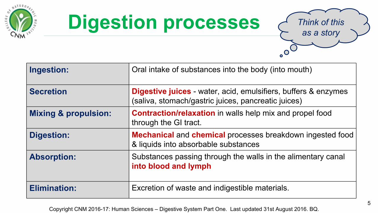
Digestion processes
5Copyright CNM 2016-17: Human Sciences – Digestive System Part One. Last updated 31st August 2016. BQ.
Ingestion: Oral intake of substances into the body (into mouth)
Secretion Digestive juices - water, acid, emulsifiers, buffers & enzymes (saliva, stomach/gastric juices, pancreatic juices)
Mixing & propulsion: Contraction/relaxation in walls help mix and propel food through the GI tract.
Digestion: Mechanical and chemical processes breakdown ingested food & liquids into absorbable substances
Absorption: Substances passing through the walls in the alimentary canal into blood and lymph
Elimination: Excretion of waste and indigestible materials.
Think of this as a story

Gastrointestinal Tract (GIT) Layers
6Copyright CNM 2016-17: Human Sciences – Digestive System Part One. Last updated 31st August 2016. BQ.
http://cellstosymptoms.com/gastrointestinal/tired-all-the-time/
http://cellstosymptoms.com/gastrointestinal/tired-all-the-time/
The entire GIT contains the same basic 4-layer arrangement of tissues:
1. Mucosa
2. Submucosa
3. Muscularis (2 layers)
4. Serosa (perioneum)

Mucosa: Epithelium
• Mouth, pharynx, oesophagus & anus: non-keratinised epithelium for protection (keratin is a tough protein found in skin).
• Stomach & intestines: columnar epithelium with:
v Microvilli – site of absorption.
v Goblet cells – secretion of mucous to lubricate food and protects against digestive juice erosion.
v Enteroendocrine cells - specialised endocrine cells that secrete hormones (serotonin, gastrin, motilin, CCK, into blood) communication
7
Copyright CNM 2016-17: Human Sciences – Digestive System Part One. Last updated 31st August 2016. BQ.
Mucosa layer: inner most layer of the GIT. Divided into 3 layers:
Epithelial cells renew every 5-7
days
entero- = intestine / endocrine = hormones
micro = smallvilli = mucosal projectionscolumnar = cells with height > width

Mucosa: Lamina Propria• Connective tissue containing many blood and
lymphatic vessels that allows absorption of nutrients
• Supports the epithelium and binds it to muscular mucosa
• Contains MALT (mucosa associated lymphoid tissue). Prominent lymphatic nodules contain immune cells (Lymphocytes, macrophages etc.).
• MALT is found in many places in the body. In the GIT, we call this GALT (gut associated lymphoid tissue)
• Especially in tonsils, oesophagus, small intestine, appendix & large intestine.
8Copyright CNM 2016-17: Human Sciences – Digestive System Part One. Last updated 31st August 2016. BQ.
lamina = layerpropria = proper
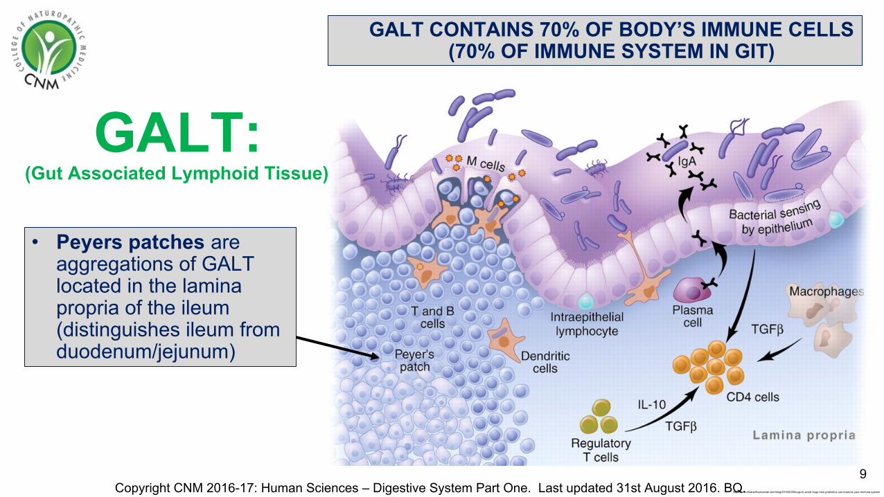
GALT:
9Copyright CNM 2016-17: Human Sciences – Digestive System Part One. Last updated 31st August 2016. BQ.
GALT CONTAINS 70% OF BODY’S IMMUNE CELLS(70% OF IMMUNE SYSTEM IN GIT)
http://www.drkarenfrackowiak.com/blog/2015/8/20/bugs-to-avoid-bugs-how-probiotics-can-improve-your-immune-system
• Peyers patches are aggregations of GALT located in the lamina propria of the ileum (distinguishes ileum from duodenum/jejunum)
(Gut Associated Lymphoid Tissue)

Mucosa: Muscularis Mucosa
• Very thin layer of smooth muscle tissue.
• Creates small folds which increase the surface area for absorption & digestion
• Movement of this ensures that all absorptive cells are fully exposed to the GIT contents
10Copyright CNM 2016-17: Human Sciences – Digestive System Part One. Last updated 31st August 2016. BQ. http://www.dartmouth.edu/~anatomy/Histo/lab_5/GI/DMS127/popup.html

Submucosa Layer• Connective tissue layer that lies between the
mucosa & muscularis
• Contains blood & lymph vessels which receive absorbed food molecules
• Contains network of neurons called the submucosal plexus (‘brain of the gut’)
• May contain glands and lymphatic tissue eg. Peyers patches in ileum
11Copyright CNM 2016-17: Human Sciences – Digestive System Part One. Last updated 31st August 2016. BQ. http://www.pathologyoutlines.com/topic/colonhistology.html
Sub = belowMucosa
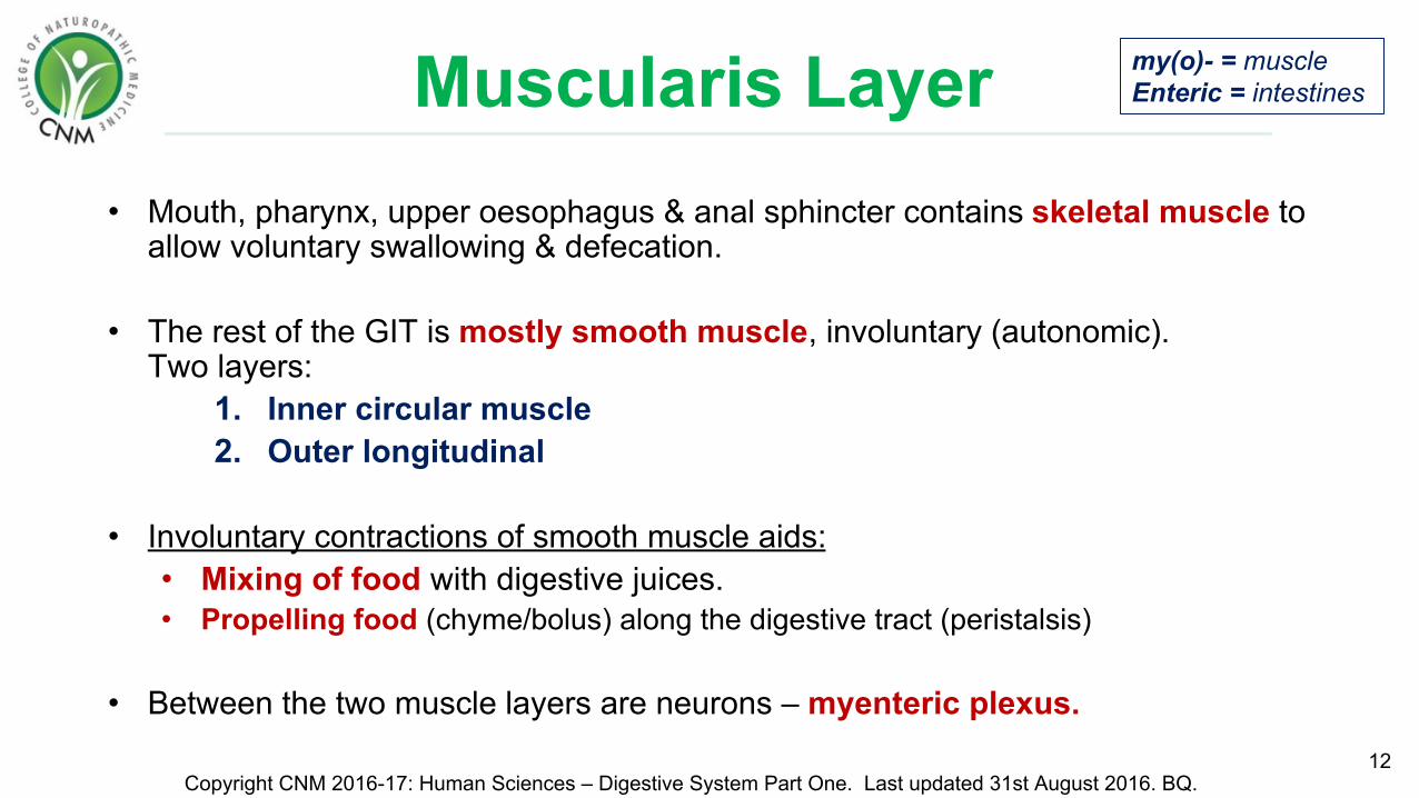
Muscularis Layer• Mouth, pharynx, upper oesophagus & anal sphincter contains skeletal muscle to
allow voluntary swallowing & defecation.
• The rest of the GIT is mostly smooth muscle, involuntary (autonomic).Two layers:
1. Inner circular muscle2. Outer longitudinal
• Involuntary contractions of smooth muscle aids:• Mixing of food with digestive juices.• Propelling food (chyme/bolus) along the digestive tract (peristalsis)
• Between the two muscle layers are neurons – myenteric plexus. 12
Copyright CNM 2016-17: Human Sciences – Digestive System Part One. Last updated 31st August 2016. BQ.
my(o)- = muscleEnteric = intestines

GIT Layers Summary:
13Copyright CNM 2016-17: Human Sciences – Digestive System Part One. Last updated 31st August 2016. BQ. http://medcell.med.yale.edu/systems_cell_biology/gi_tract_lab.php

Peritoneum• Largest serous membrane of the body.
• Weaves between digestive organs.
• Supplied with many blood and lymph vessels.
• A physical barrier to local spread of infection.
• Consists of 2 layers:1. Parietal – covers the wall of the abdomen & pelvic cavity.2. Visceral – covers the organs
• Peritoneal cavity is the space between the 2 layers and contains a lubricating serous fluid.
14Copyright CNM 2016-17: Human Sciences – Digestive System Part One. Last updated 31st August 2016. BQ.
Peritoneum = serous membrane surrounding abdominal organsParietal = Latin for walls of a cavityViscera = Referring to body organs
http://teachmeanatomy.info/abdomen/areas/peritoneum/

Ascites • The accumulation of fluid in the peritoneal cavity (up to a few litres)
• Causes include Liver cirrhosis, GIT malignancy, heart failure, renal disease, pancreatitis
Peritonitis • An acute inflammation of peritoneum.
• Causes of these can be bacterial infection, ruptured appendix, friction, surgical wounds.
Peritoneal Pathologies
15Copyright CNM 2016-17: Human Sciences – Digestive System Part One. Last updated 31st August 2016. BQ.
https://en.wikipedia.org/wiki/Ascites
http://www.digopaul.com/english-word/peritonitis.html
Periton- = peritoneum-itis = inflammation
Greek origin (askos) which means bag/sac

Greater Omentum• The largest fold of the peritoneum.
• Drapes over transverse colon and small intestine like apron
• It’s a double sheet that folds back on itself (hence 4 layers)
• Stores fat: contains a considerable amount of adipose tissue which can greatly expand with weight gain – the ‘beer belly’.
• Has many lymph nodes containing macrophages & plasma cells (produce antibodies) to combat & contain infections of the GIT.
16Copyright CNM 2016-17: Human Sciences – Digestive System Part One. Last updated 31st August 2016. BQ.
Omentum = Latin for apron

Lesser Omentum
• A peritoneal fold that suspends the stomach and duodenum from the liver
• Pathway for blood vessels entering liver: contains hepatic portal vein, common hepatic artery, common bile duct and lymph nodes
17Copyright CNM 2016-17: Human Sciences – Digestive System Part One. Last updated 31st August 2016. BQ. http://www.cram.com/flashcards/jmp-anatomy-abd-cavity-and-organs-2658529
Omentum = Latin for apron
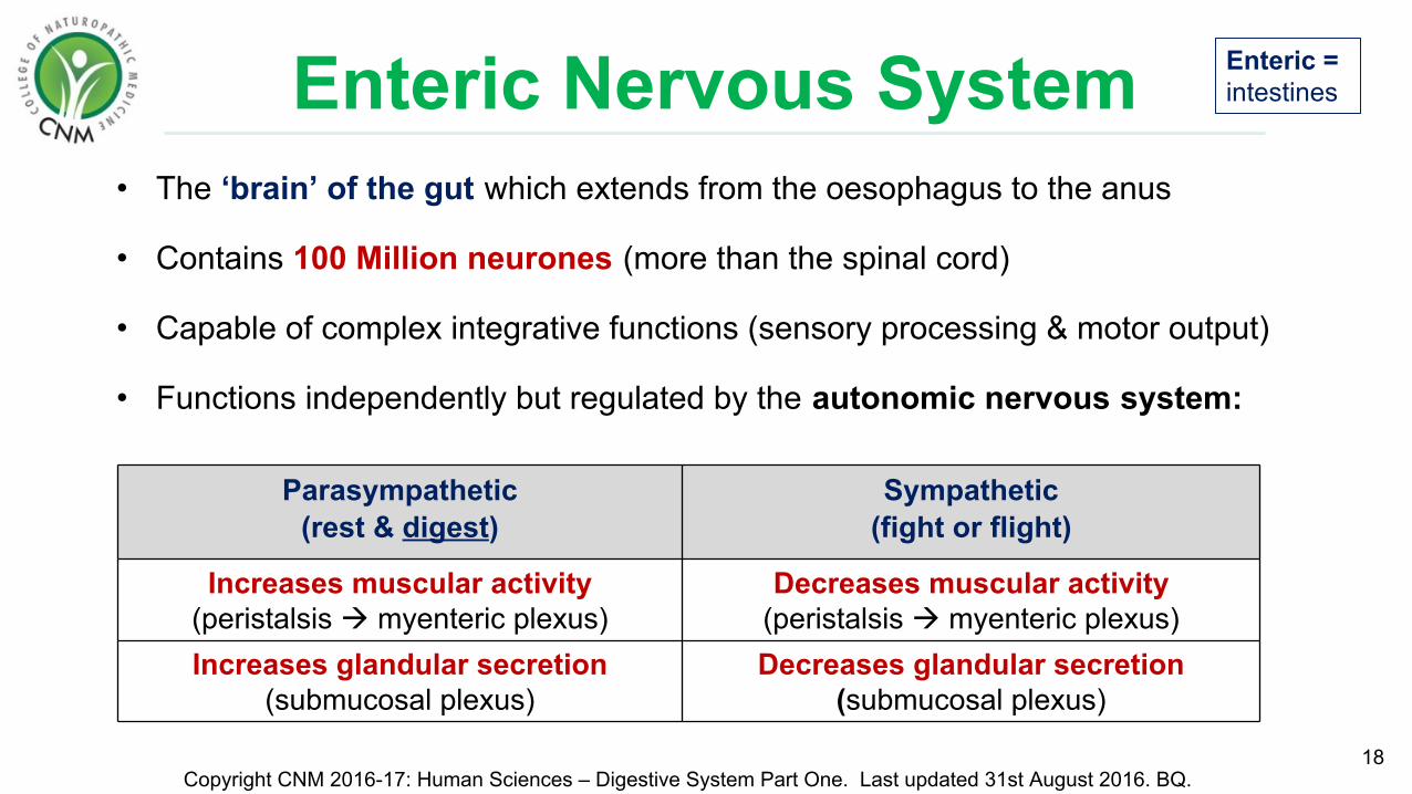
Enteric Nervous System• The ‘brain’ of the gut which extends from the oesophagus to the anus
• Contains 100 Million neurones (more than the spinal cord)
• Capable of complex integrative functions (sensory processing & motor output)
• Functions independently but regulated by the autonomic nervous system:
18
Parasympathetic (rest & digest)
Sympathetic (fight or flight)
Increases muscular activity (peristalsis myenteric plexus)
Decreases muscular activity (peristalsis myenteric plexus)
Increases glandular secretion(submucosal plexus)
Decreases glandular secretion (submucosal plexus)
Copyright CNM 2016-17: Human Sciences – Digestive System Part One. Last updated 31st August 2016. BQ.
Enteric = intestines
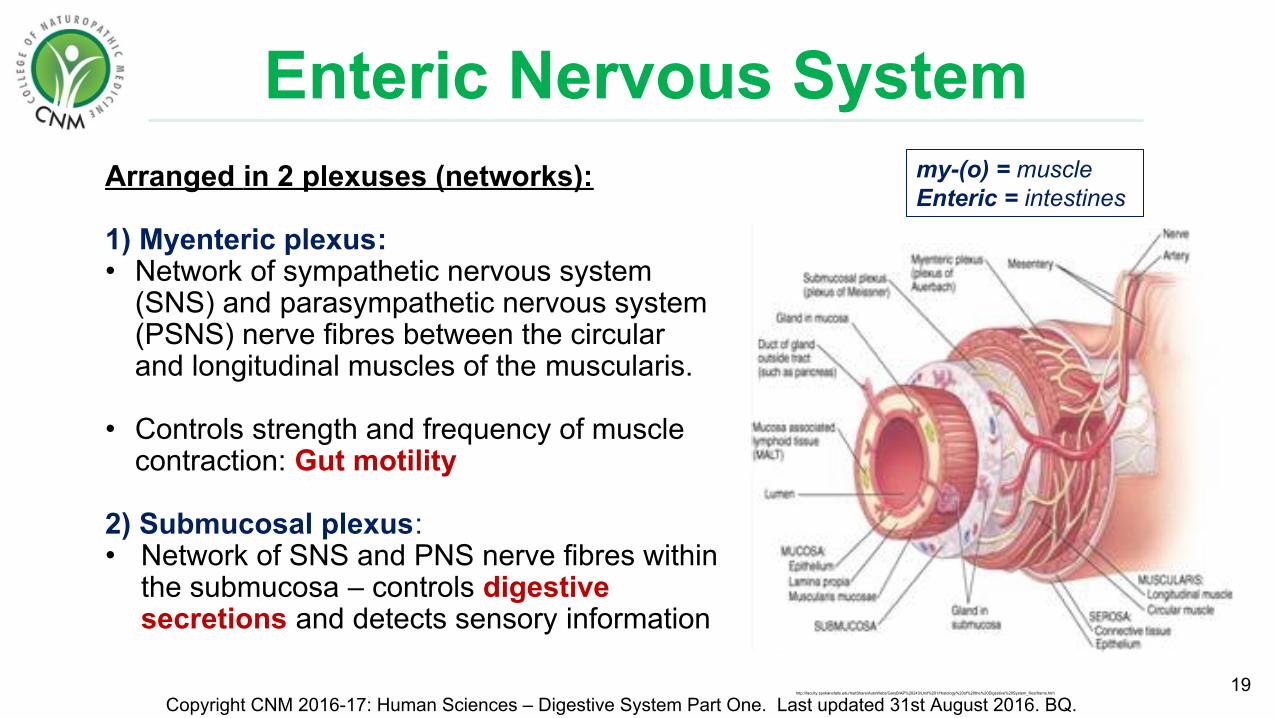
Arranged in 2 plexuses (networks):
1) Myenteric plexus: • Network of sympathetic nervous system
(SNS) and parasympathetic nervous system (PSNS) nerve fibres between the circular and longitudinal muscles of the muscularis.
• Controls strength and frequency of muscle contraction: Gut motility
2) Submucosal plexus:• Network of SNS and PNS nerve fibres within
the submucosa – controls digestive secretions and detects sensory information
19Copyright CNM 2016-17: Human Sciences – Digestive System Part One. Last updated 31st August 2016. BQ.
Enteric Nervous System
http://faculty.spokanefalls.edu/InetShare/AutoWebs/GaryB/AP%20243/Unit%201/Histology%20of%20the%20Digestive%20System_files/frame.htm
my-(o) = muscleEnteric = intestines

Types of neurons:1. Motor neurons (outgoing/action signal)
in the myenteric plexus controls peristalsis & in the submucosal plexus control secretions.
2. Sensory neurons (incoming signal) receive information about the mucosal environment: chemoreceptors & stretch receptors.
3. Interneurons connect the two plexuses.
20Copyright CNM 2016-17: Human Sciences – Digestive System Part One. Last updated 31st August 2016. BQ.
Enteric Nervous System
http://www.nature.com/nrd/journal/v7/n3/fig_tab/nrd2444_F1.html
Video: Intestinal peristalsis: www.youtube.com/watch?v=aNaHj4UlLZM

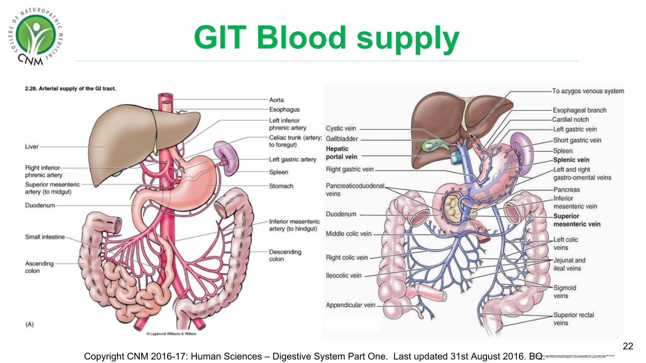
GIT Blood supply• Arterial Blood is supplied from branches of the
abdominal aorta, which include the mesenteric arteries.
• Nutrient rich blood in the intestines is returned by veins via the liver (portal system) or directly from iliac veins:
• The liver filters the blood and processes nutrients which then enter systemic circulation.
• Portal Vein: Lower oesophagus, stomach, pancreas, small & large intestine, upper rectum & spleen.
• Iliac Veins: Lower part of the rectum & anal canal. 21
Copyright CNM 2016-17: Human Sciences – Digestive System Part One. Last updated 31st August 2016. BQ. https://www.studyblue.com/notes/note/n/gi/deck/1938899
GIT Blood supply
22Copyright CNM 2016-17: Human Sciences – Digestive System Part One. Last updated 31st August 2016. BQ. http://medicoreview.blogspot.co.uk/2012/05/blood-supply-of-gi.htmlhttps://www.studyblue.com/notes/note/n/bi-481-study-guide-2011-12-gullo-/deck/9730222

Summary Quiz!1. Name the four basic layers of the GIT?2. Where are the two nerve plexus located?3. What are the two layers of the peritoneum called? What resides
between these layers? 4. Explain what is meant by ‘ascites’5. What is the function of the greater omentum? How many layers
does this have?6. What percentage of the immune system is located in the GIT?7. What vein drains the intestines? Where does this blood vessel go?8. What are ‘peyers patches’?9. Explain what is meant by peristalsis.10. Explain what is meant by “enteric”.
23Copyright CNM 2016-17: Human Sciences – Digestive System Part One. Last updated 31st August 2016. BQ.
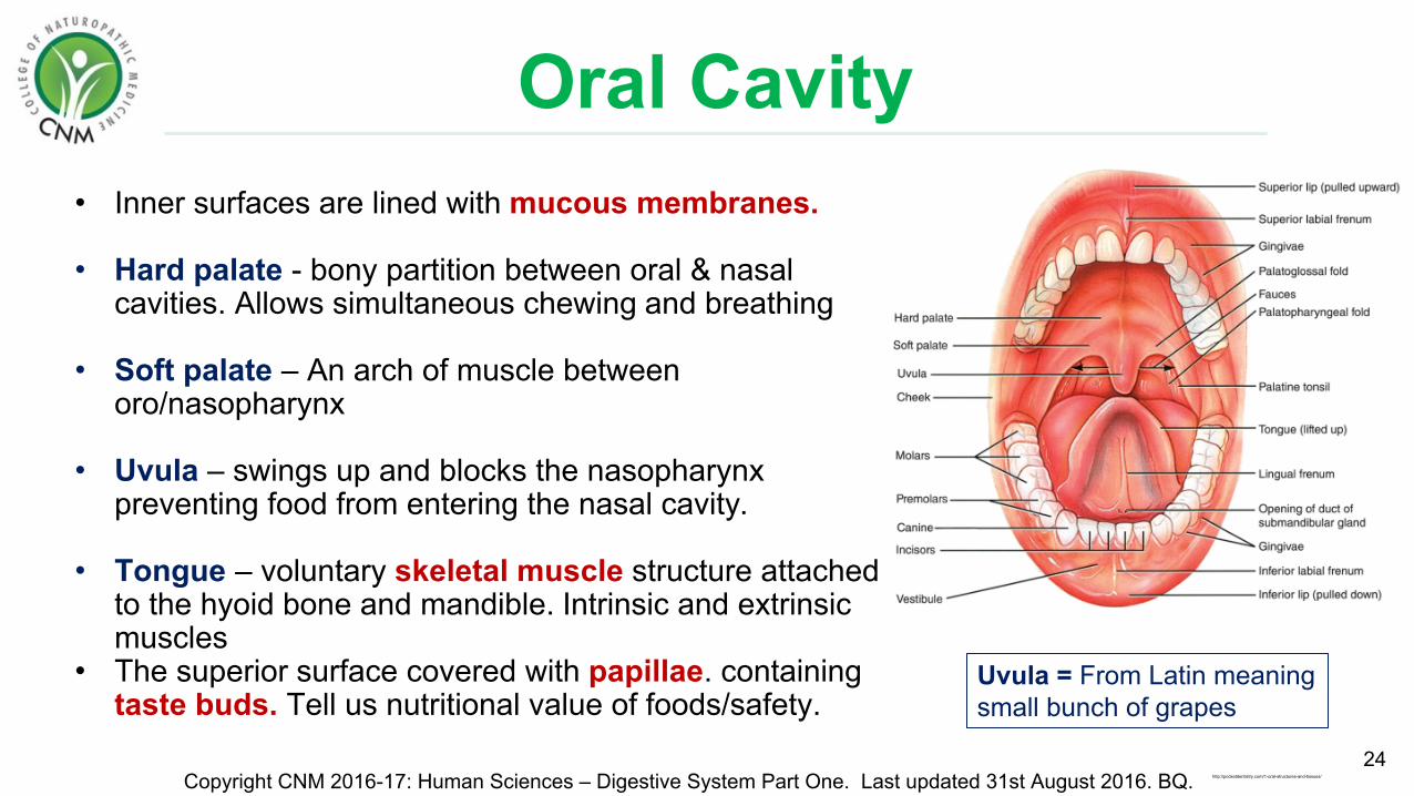
Oral Cavity• Inner surfaces are lined with mucous membranes.
• Hard palate - bony partition between oral & nasal cavities. Allows simultaneous chewing and breathing
• Soft palate – An arch of muscle between oro/nasopharynx
• Uvula – swings up and blocks the nasopharynx preventing food from entering the nasal cavity.
• Tongue – voluntary skeletal muscle structure attached to the hyoid bone and mandible. Intrinsic and extrinsic muscles
• The superior surface covered with papillae. containing taste buds. Tell us nutritional value of foods/safety.
24Copyright CNM 2016-17: Human Sciences – Digestive System Part One. Last updated 31st August 2016. BQ. http://pocketdentistry.com/1-oral-structures-and-tissues/
Uvula = From Latin meaning small bunch of grapes

Oral Cavity
Functions include:
• Mastication
• Speech
• Taste
• Swallowing – bolus (mixed digested food & digestive juices) is pushed by the tongue in to the pharynx closing the nasopharynx - pharynx reflex action.
25Copyright CNM 2016-17: Human Sciences – Digestive System Part One. Last updated 31st August 2016. BQ. http://www.pvsmiles.com/pharyngeal-reflex/

Teeth• Adults have 32 teeth (20 temporary)
• Teeth have a crown, neck & root.
• ‘’Dentin makes up the bulk of a tooth and is present internally. Teeth are covered externally by enamel.
• Sits within the gum (gingiva) and periodontal membrane (a ligament that fixes to bone/connects teeth)
• Functions in Mastication (What chewing does not accomplish mechanically must be completed by the digestive tract chemically)
26Copyright CNM 2016-17: Human Sciences – Digestive System Part One. Last updated 31st August 2016. BQ. https://uk.pinterest.com/pin/50313720809772428/

Saliva• Produced by parotid gland, submandibular gland, sublingual gland
via a reflex controlled by the autonomic nervous system
• Parasympathetic NS stimulates continuous salivation (1-1.5 L/day) which lubricates the mouth & mucous membranes.
• Saliva is swallowed & lubricates oesophagus and is eventually reabsorbed
• During dehydration salivation is stopped, contributing to thirst sensation.
• Sympathetic nervous stimulation (stress response) reduces salivation causing dryness of the mouth (& thicker saliva).
• Touch, taste, smell, sight, sound can stimulate digestive salivation.
27Copyright CNM 2016-17: Human Sciences – Digestive System Part One. Last updated 31st August 2016. BQ.

Saliva Composition• Water (99.5%)
• Mineral Salts (Na, K, Ca, Cl, Bicarbonate, P)
• Enzymes: salivary amylase (parotid), lysozyme (found in many body secretions, breaks down bacterial cell walls)
• Mucous
• Immunoglobulins (IgA)
• Blood clotting factors
• pH 6.35-6.85 (mildly acidic)
28Copyright CNM 2016-17: Human Sciences – Digestive System Part One. Last updated 31st August 2016. BQ. http://www.cancer.gov/types/head-and-neck/patient/salivary-gland-treatment-pdq

Saliva Functions• Digestion - chemical breakdown of polysaccharides.
• Lubricating & dissolving food.
• Cleansing of oral cavity and teeth.
• Defence - non-specific (IgA & lysozymes)
• Taste
• Buffer – acidic foods
• Waste removal – urea / uric acid from the body.
29Copyright CNM 2016-17: Human Sciences – Digestive System Part One. Last updated 31st August 2016. BQ. http://slideplayer.com/slide/5711686/

Oesophagus• A 25cm long muscular tube, attached to cricoid cartilage.
Posterior to trachea. Passes through the diaphragm (T10)
• Lined with stratified squamous epithelium (protection). Mucous membrane lubricated with mucous.
• The superior/middle oesophagus contains skeletal muscle, whilst the lower oesophagus contains smooth muscle
• Food travel to the stomach via muscular contractions (peristalsis)
• Epiglottis – a flap of elastic cartilage which occludes the trachea preventing food entering.
• The lower oesophageal sphincter acts as a seal on the stomach to prevent reflux in to the oesophagus.
30Copyright CNM 2016-17: Human Sciences – Digestive System Part One. Last updated 31st August 2016. BQ.http://www.slideshare.net/brissomathewarackal/oesophagus-anatomy-and-physiology-by-brisso-arackal
Epi- = aboveGlottis = vocal cords

Stomach• A J-shaped organ with 4 main regions: cardia, fundus,
body, pyloric with lesser and greater curvatures
• Same layers as the rest of the GIT, but with 3 layers of muscle (longitudinal, circular and oblique) churning and mixing food with gastric juice.
• 2 sphincters: cardiac and pyloric
Neural feedback: • Food distends the stomach stimulating stretch receptors in
its walls.
• Chemoreceptors monitor pH changes in response to chyme.
• Activates submucosal plexus causing waves of peristalsis and gastric juice flow.
31Copyright CNM 2016-17: Human Sciences – Digestive System Part One. Last updated 31st August 2016. BQ. https://en.wikipedia.org/wiki/Stomach
Chyme = partially digested foodSphincter = ring of muscle that normally maintains constriction of a body passage

Stomach
32Copyright CNM 2016-17: Human Sciences – Digestive System Part One. Last updated 31st August 2016. BQ.https://uk.pinterest.com/pin/352969689518014170/
The stomach secretes 2-3L of highly acidic
gastric juice and mucous a day
Contains simple columnar epithelial cells fast turnover (replace lining every 3 days)

Stomach
33Copyright CNM 2016-17: Human Sciences – Digestive System Part One. Last updated 31st August 2016. BQ.
• Contains glands that extend into the lamina propria. These contain three types of exocrine cells:
Substances secreted FunctionParietal cells • Intrinsic factor (IF)
• Hydrochloric acid (HCl)
• Secretes H+ and Cl- separately which combine in the stomach.
• HCl activates enzymes for digestion• An antimicrobial agent for ingested
microbes and denatures proteins. • IF is necessary for vitamin B12 absorption
Chief cells • Pepsinogen • Gastric lipase
• Protein and lipid digestions• HCl converts pepsinogen to the active
enzyme pepsin for protein digestion.
Goblet cells • Mucous • Secrete mucous for short term protection against acid
Parietal = wall of a cavity

StomachFunctions:• Mixing chamber – churns up food• Holding reservoir – storage• Defence – non-specific • Absorption (limited) – water, alcohol, lipid
soluble drugs (aspirin)• Digestion – limited chemical digestion of proteins
& lipids.• Iron – made more soluble with stomach acid. • Satiation – tells you to stop eating
Hormones:• Ghrelin – stimulates hunger, gastric motility,
growth hormone secretion.• Gastrin (produced by G cells) – responds to
stomach distension, pH and the PNS. Stimulates gastric juice secretion, increases gastric motility
34Copyright CNM 2016-17: Human Sciences – Digestive System Part One. Last updated 31st August 2016. BQ. http://www.shmoop.com/animal-digestion/food-beginning-end.html

Small Intestine• After food combines with stomach secretions the resulting
chyme is pushed through the pyloric sphincter into the small intestine.
• Where most digestion & absorption occurs.
• A long structure (6.5 metres) with villi to maximise surface area
• 3 regions:1. Duodenum – emulsification & most digestion
occurs here.2. Jejunum – most absorption occurs here (1m)3. Ilium – vitamin B12 is absorbed (2m)
• pH approximately 8 (mildly basic).35
Copyright CNM 2016-17: Human Sciences – Digestive System Part One. Last updated 31st August 2016. BQ. http://www.aboutkidshealth.ca/En/ResourceCentres/Nutrition/Digestive-system-conditions-and-special-diets/Digestive-system/Pages/GI-Tract.aspx

Where are nutrients
absorbed?
36Copyright CNM 2016-17: Human Sciences – Digestive System Part One. Last updated 31st August 2016. BQ. http://pamtremble.blogspot.co.uk/2008/05/where-are-nutrients-absorbed.html
..So consider, what might the impact be of damage to the lining of the jejunum? Ileum? Large intestine?
pH through GIT
1-3
6-8
5-7
5-7

Small IntestineCell types include:
37Copyright CNM 2016-17: Human Sciences – Digestive System Part One. Last updated 31st August 2016. BQ. http://www.aboutkidshealth.ca/En/ResourceCentres/Nutrition/Digestive-system-conditions-and-special-diets/Digestive-system/Pages/GI-Tract.aspx
Absorptive cells Digest & absorb nutrientsGoblet cells Secrete mucousPaneth cells Secrete lysozyme Endocrine S cells (secrete the hormone secretin)
CCK cells (secrete the hormone CCK)K cells (secrete glucose dependent insulinotropic peptide)
Duodenal (Brunner’s) glands Secrete alkaline mucous to neutralise acidity.
The total secretions of the small intestine is
approximately 1.5L per day.
Endocrine = hormones

Small Intestine• Villi are finger-like projections with blood
capillaries & lacteals (lymphatic capillaries).
• Absorbed nutrients enter blood vessels and fatty acids enter the lymph.
• Microvilli: (brush border) projections of absorptive cell membranes.
38Copyright CNM 2016-17: Human Sciences – Digestive System Part One. Last updated 31st August 2016. BQ. http://www.aboutkidshealth.ca/En/ResourceCentres/Nutrition/Digestive-system-conditions-and-special-diets/Digestive-system/Pages/GI-Tract.aspx
Brush border enzymes: • Enzymes attached to the intestinal lining
(not free in the lumen). • Includes maltase, sucrose and lactase• Hence enzymatic digestion occurs on absorptive
cell surface. Release when cells slough off.
http://slideplayer.com/slide/7039986/

Brush Border Enzymes
39Copyright CNM 2016-17: Human Sciences – Digestive System Part One. Last updated 31st August 2016. BQ.
Brush Border Enzyme Function
Maltase, Sucrase, Lactase Break down sugars into glucose, fructose, galactose etc
Dipeptidase Break down proteins into amino acids
Aminopeptidase Break down proteins into amino acids
Nucleosidases & Phosphatases Digest RNA & DNA
• Brush border enzymes are imperative for absorption • Enzymes attached to the lining of the intestine (not free in the lumen)
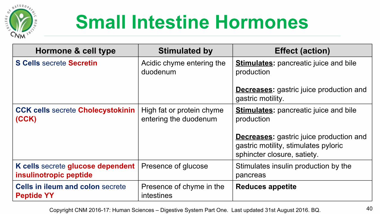
Small Intestine Hormones
40Copyright CNM 2016-17: Human Sciences – Digestive System Part One. Last updated 31st August 2016. BQ.
Hormone & cell type Stimulated by Effect (action)S Cells secrete Secretin Acidic chyme entering the
duodenumStimulates: pancreatic juice and bile production
Decreases: gastric juice production and gastric motility.
CCK cells secrete Cholecystokinin (CCK)
High fat or protein chyme entering the duodenum
Stimulates: pancreatic juice and bile production
Decreases: gastric juice production and gastric motility, stimulates pyloric sphincter closure, satiety.
K cells secrete glucose dependent insulinotropic peptide
Presence of glucose Stimulates insulin production by the pancreas
Cells in ileum and colon secrete Peptide YY
Presence of chyme in the intestines
Reduces appetite
Small Intestine Hormones
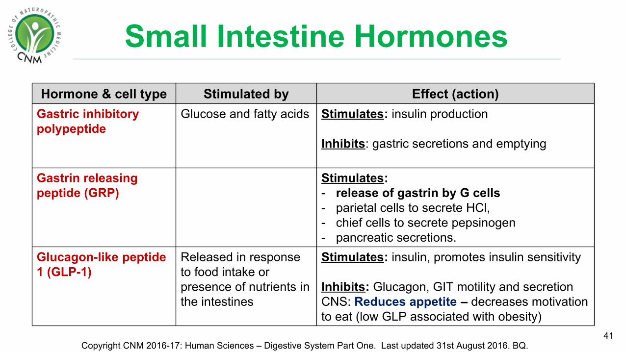
41Copyright CNM 2016-17: Human Sciences – Digestive System Part One. Last updated 31st August 2016. BQ.
Hormone & cell type Stimulated by Effect (action)Gastric inhibitory polypeptide
Glucose and fatty acids Stimulates: insulin production
Inhibits: gastric secretions and emptying
Gastrin releasing peptide (GRP)
Stimulates: - release of gastrin by G cells - parietal cells to secrete HCl, - chief cells to secrete pepsinogen - pancreatic secretions.
Glucagon-like peptide 1 (GLP-1)
Released in response to food intake or presence of nutrients in the intestines
Stimulates: insulin, promotes insulin sensitivity
Inhibits: Glucagon, GIT motility and secretionCNS: Reduces appetite – decreases motivation to eat (low GLP associated with obesity)

Small IntestineFunctions:
• Movement/peristalsis of food
• Digestion.
• Absorption of nutrients & water.
• Hunger / satiety.
• Immunity - peyer's patches & bacterial microflora.
42Copyright CNM 2016-17: Human Sciences – Digestive System Part One. Last updated 31st August 2016. BQ. http://www.myvmc.com/anatomy/gastrointestinal-system/

Small Intestine: Absorption• 90% of absorption occurs in small intestine
• Carbohydrates and amino acids are transported into capillaries:• Monosaccharides: active & passive transport• Amino acids: active transport
• Fatty acids, glycerol, cholesterol and fat soluble vitamins (A,D,E,K) are:
1. Emulsified by bile2. Enter intestinal cells by simple diffusion3. Packaged into chylomicrons and absorbed into
lacteals4. Travel through the lymphatic system and enter
the blood stream at the subclavian vein43
Copyright CNM 2016-17: Human Sciences – Digestive System Part One. Last updated 31st August 2016. BQ. http://www.karelsavry.us/biology/digestion_in_the_small_intestine.html
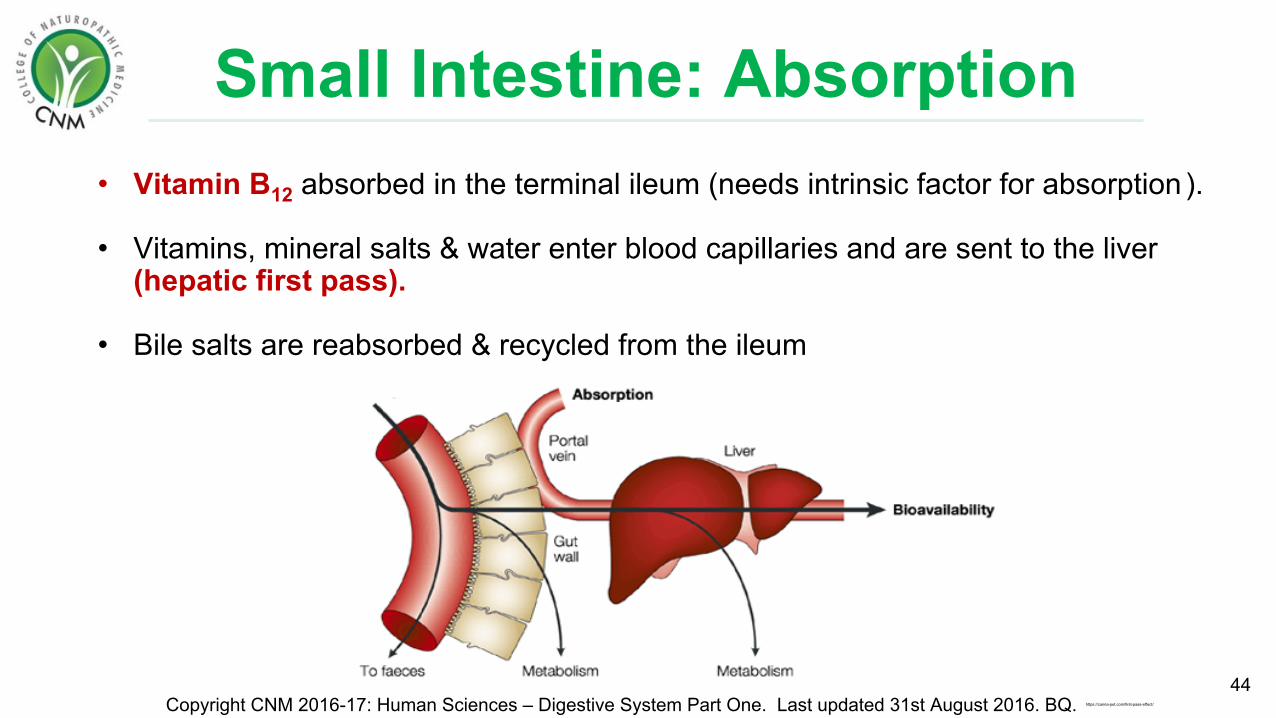
Small Intestine: Absorption• Vitamin B12 absorbed in the terminal ileum (needs intrinsic factor for absorption ).
• Vitamins, mineral salts & water enter blood capillaries and are sent to the liver (hepatic first pass).
• Bile salts are reabsorbed & recycled from the ileum
44Copyright CNM 2016-17: Human Sciences – Digestive System Part One. Last updated 31st August 2016. BQ. https://canna-pet.com/first-pass-effect/
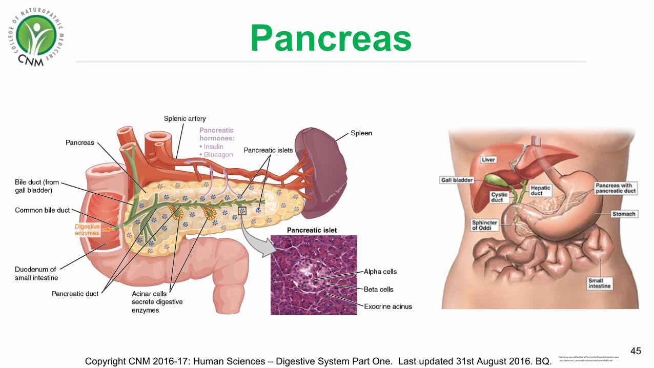
Pancreas
45Copyright CNM 2016-17: Human Sciences – Digestive System Part One. Last updated 31st August 2016. BQ. http://philschatz.com/anatomy-book/contents/m46685.html
http://www.nhs.uk/Conditions/Pancreatitis/Pages/Introduction.aspx

Pancreas• An accessory digestive organ that is approximately 15cm long and retroperitoneal
• Connected to the duodenum via 2 pancreatic ducts.
46Copyright CNM 2016-17: Human Sciences – Digestive System Part One. Last updated 31st August 2016. BQ.
Exocrine Function Endocrine Function• Pancreatic juice is a clear liquid that is
excreted into small intestines (1.2-1.5L/day)• Hormones secreted (into the blood)
• Sodium bicarbonate • Protease enzymes: Trypsin, chymotrypsin,
carboxypeptidase, ribonuclease, deoxyribonuclease.
• Lipase• Pancreatic amylase• Water
• Insulin & Glucagon• Somatostatin (growth-hormone-
inhibiting-hormone)• Pancreatic polypeptide (neuropeptide)
Accessory structure:

Pancreatic Enzymes
47Copyright CNM 2016-17: Human Sciences – Digestive System Part One. Last updated 31st August 2016. BQ.
Pancreatic Enzyme FunctionPancreatic amylase Breakdown starches into sugars
Pancreatic lipase Lipid / fat digestion.
Trypsin Protein digestion
Chymotrypsin Protein digestion
Carboxypeptidase Protein digestion
Ribonuclease Digest RNA
Deoxyribonuclease Digest DNA
Brush Border Enzyme Function
Maltase, Sucrase, Lactase break down sugars into glucose, fructose, galactose etc
Dipeptidase break down proteins into amino acids
Aminopeptidase break down proteins into amino acids
Nucleosidases & Phosphatases digest RNA & DNA
Procarboxypeptidase > Carboxypeptidase digest RNA & DNA
• Pancreatic enzymes are imperative for digestion• Secreted by the pancreas into the lumen of the duodenum
• Proteases are secreted in their inactive form

Gallbladder
• Gallbladder is a pear-shaped 7-10 cm sac in the liver.
• Bile ducts project from the gallbladder and liver meeting at the common bile duct.
• Bile ducts collect bile produced by hepatocytes which pools in the gallbladder.
• Bile enters the small intestine via the common bile duct
48Copyright CNM 2016-17: Human Sciences – Digestive System Part One. Last updated 31st August 2016. BQ. https://www.linkedin.com/pulse/gallbladder-biliary-pancreatic-disorders-jessica-white
Accessory structure:
Hepat- = liver

Gallbladder• Bile is an agent that emulsifies fats.
• Emulsification breaks the lipid into smaller molecules. This increases the surface area for lipid enzymes (lipase) to work
• Bile is composed of bile salts, cholesterol and bilirubin
• pH 7.6-8.6 (mildly basic)
• 90-95% of bile are absorbed & transported back to the liver from the ileum: enterohepatic circulation.
49Copyright CNM 2016-17: Human Sciences – Digestive System Part One. Last updated 31st August 2016. BQ.
Emulsify = to disperseentero-= = intestines-hepatic = liver
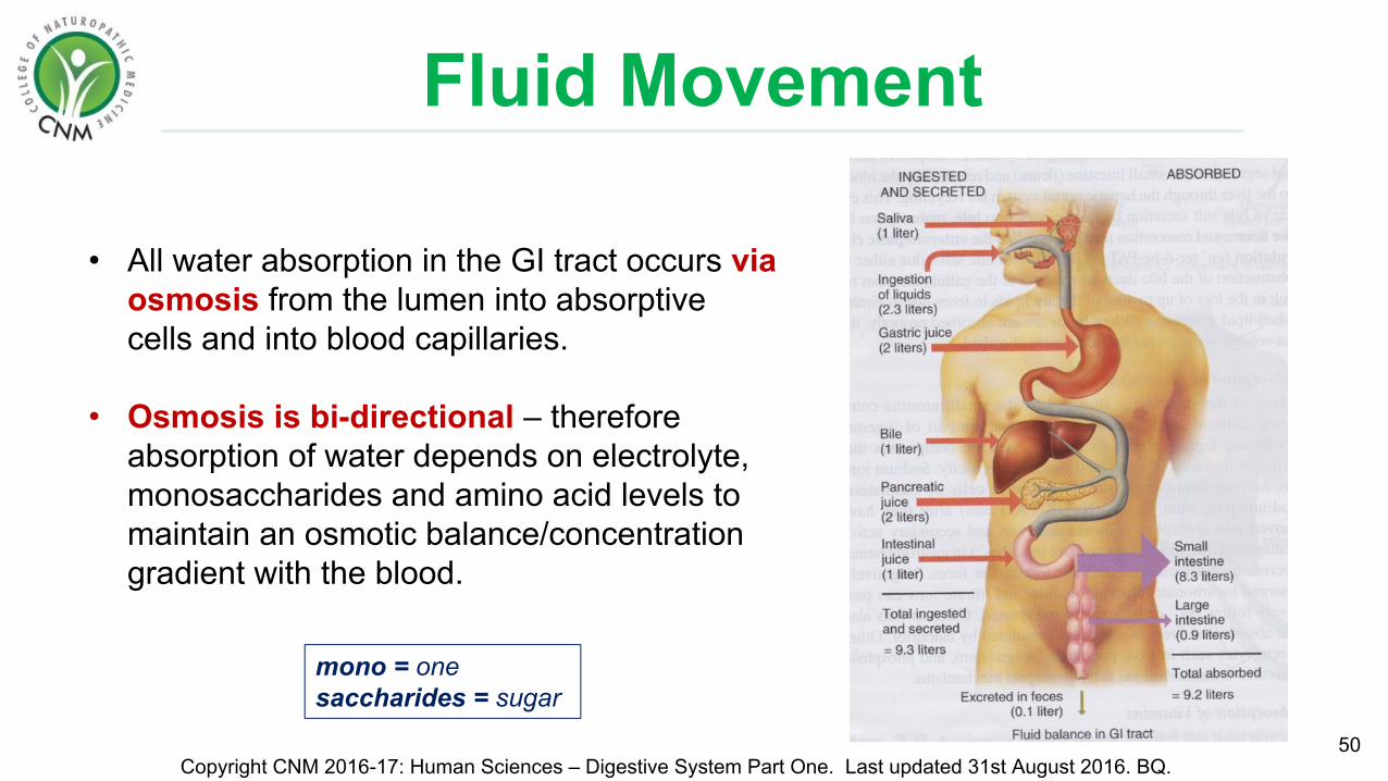
Fluid Movement
50Copyright CNM 2016-17: Human Sciences – Digestive System Part One. Last updated 31st August 2016. BQ.
• All water absorption in the GI tract occurs via osmosis from the lumen into absorptive cells and into blood capillaries.
• Osmosis is bi-directional – therefore absorption of water depends on electrolyte, monosaccharides and amino acid levels to maintain an osmotic balance/concentration gradient with the blood.
mono = onesaccharides = sugar
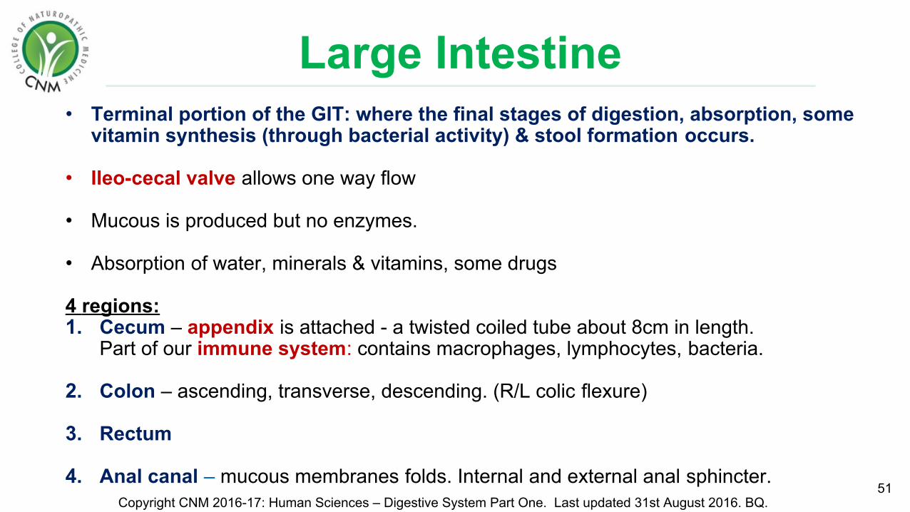
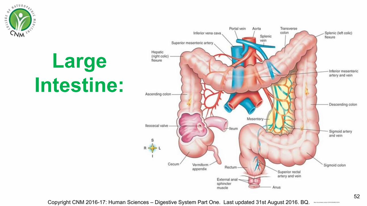
Large Intestine • Terminal portion of the GIT: where the final stages of digestion, absorption, some
vitamin synthesis (through bacterial activity) & stool formation occurs.
• Ileo-cecal valve allows one way flow
• Mucous is produced but no enzymes.
• Absorption of water, minerals & vitamins, some drugs
4 regions:1. Cecum – appendix is attached - a twisted coiled tube about 8cm in length.
Part of our immune system: contains macrophages, lymphocytes, bacteria.
2. Colon – ascending, transverse, descending. (R/L colic flexure)
3. Rectum
4. Anal canal – mucous membranes folds. Internal and external anal sphincter.51
Copyright CNM 2016-17: Human Sciences – Digestive System Part One. Last updated 31st August 2016. BQ.
Large Intestine:
52Copyright CNM 2016-17: Human Sciences – Digestive System Part One. Last updated 31st August 2016. BQ. https://uk.pinterest.com/pin/130745195405310374/

Large Intestine: Microbes• A rich community of microbes:
100,000,000,000,000 (1x10¹⁴)
• Mixture of anaerobic and aerobic bacteria, generally acid-producing
• Mostly symbiotic, but pathogenic species may flourish according to local pH, water content, digestive processes further upstream and antibiotic use
• The final stages of nutrient extraction occur in the colon through microbial fermentation
53Copyright CNM 2016-17: Human Sciences – Digestive System Part One. Last updated 31st August 2016. BQ. http://healthland.time.com/2013/11/05/colon-cancers-newest-culprit-gut-bacteria/
Symbiotic = organisms that live together

Large Intestine: Microbes• Fermentation of remaining carbohydrates producing hydrogen,
CO2 and methane (flatulence)
• Fermentation of residual amino acids to various compounds (including hydrogen sulphide) which contribute to faecal odour.
• Some toxic products of bacterial fermentation are re-absorbed & transported to the liver where they are excreted in urine.
• Bilirubin is decomposed into simpler molecules, these pigments contribute to the colour of faeces.
• Produce some vitamins – vitamin B12, vitamin K & fatty acids
• Faeces are 30-50% bacteria.54
Copyright CNM 2016-17: Human Sciences – Digestive System Part One. Last updated 31st August 2016. BQ. http://ehp.niehs.nih.gov/121-a276/

Defecation• Mass peristaltic movements push fecal
matter into the rectum from sigmoid colon.
• The resulting distension of the rectal wall stimulates stretch receptors & the defecation reflex. This includes sensory impulses to the sacral spinal cord.
• Motor impulses travel down parasympathetic nerves back to the rectum and anus.
• Rectal muscles (with abdominal muscles and diaphragm) contract, increasing abdominal pressure that opens the internal sphincter.
• The external anal sphincter is voluntarily relaxed
55Copyright CNM 2016-17: Human Sciences – Digestive System Part One. Last updated 31st August 2016. BQ.
Number of bowel movements vary depending on diet, health,
stress, exercise, emotions, hydration.
https://www.studyblue.com/notes/note/n/motility/deck/16424950

Summary Questions1) What are the four parts of the large intestine?2) What does the large intestine absorb?3) What are the functions of intestinal symbiotic microbes?4) What body system will fatty acids be absorbed into after entering cells of the
gut wall?5) What do S cells secrete and what is the role of this hormone?6) What do CCK cells secrete and what is the role of this hormone?7) What is a brush border enzyme? Name 2 of them.8) What is the function of villi?9) What are the functions of the small intestine?10) What substance is needed for B12 absorption? What produces this
substance? 56
Copyright CNM 2016-17: Human Sciences – Digestive System Part One. Last updated 31st August 2016. BQ.
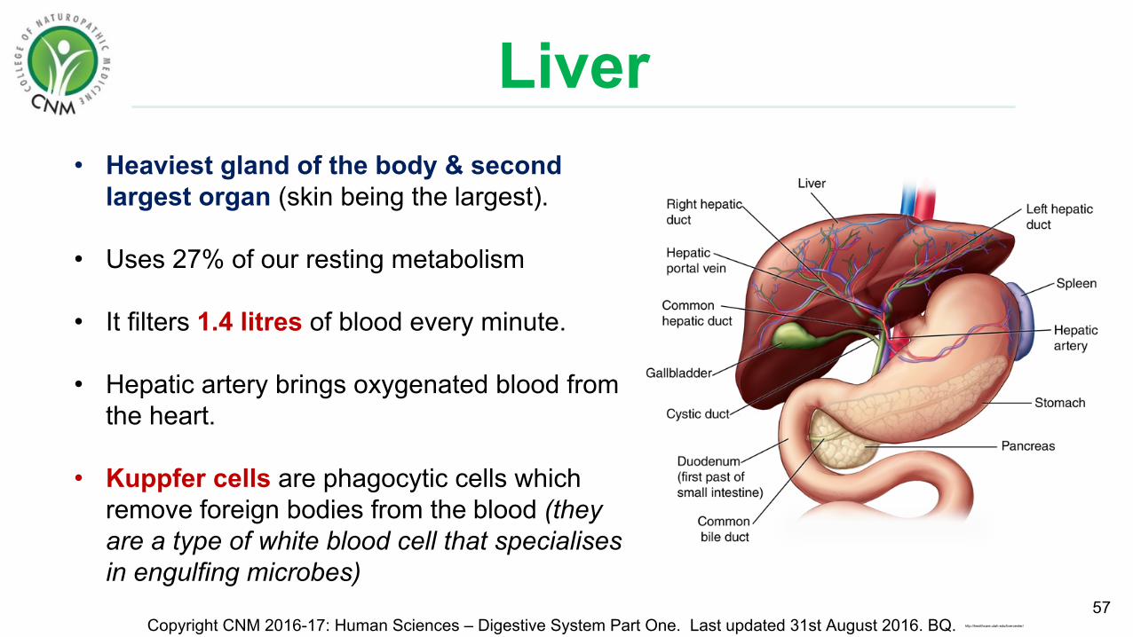
Liver• Heaviest gland of the body & second
largest organ (skin being the largest).
• Uses 27% of our resting metabolism
• It filters 1.4 litres of blood every minute.
• Hepatic artery brings oxygenated blood from the heart.
• Kuppfer cells are phagocytic cells which remove foreign bodies from the blood (they are a type of white blood cell that specialises in engulfing microbes)
57Copyright CNM 2016-17: Human Sciences – Digestive System Part One. Last updated 31st August 2016. BQ. http://healthcare.utah.edu/livercenter/
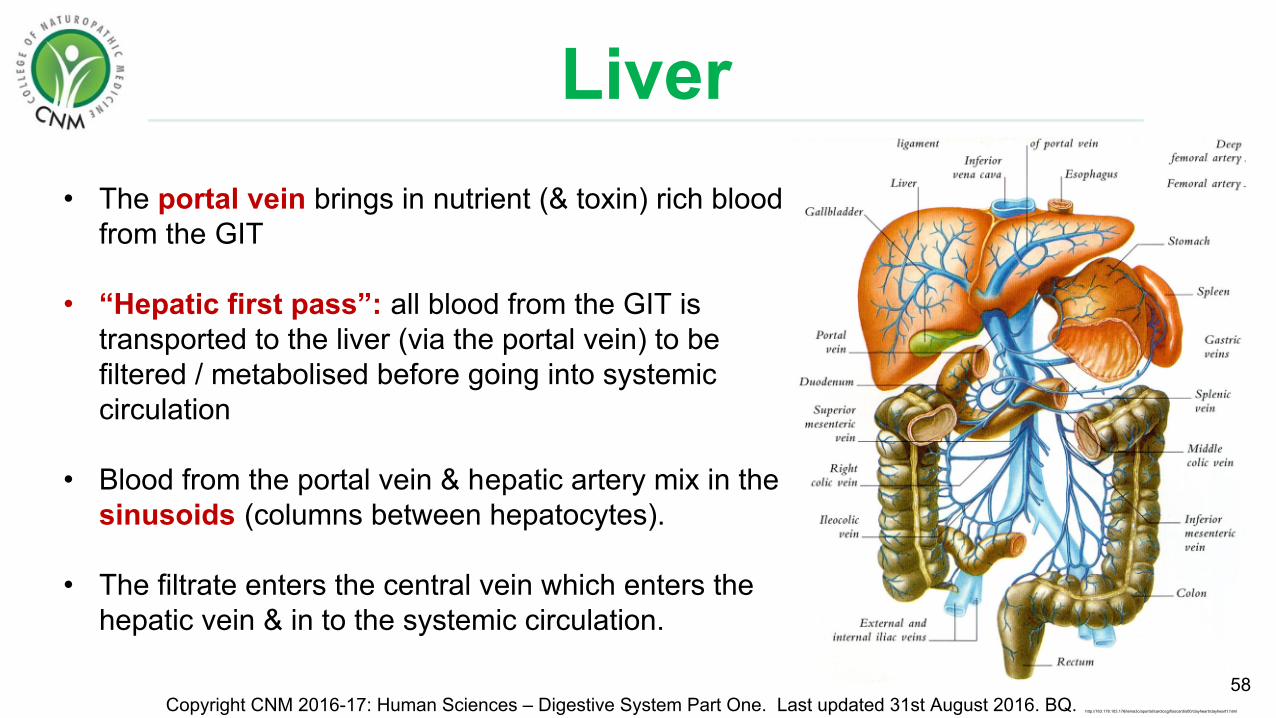
Liver• The portal vein brings in nutrient (& toxin) rich blood
from the GIT
• “Hepatic first pass”: all blood from the GIT is transported to the liver (via the portal vein) to be filtered / metabolised before going into systemic circulation
• Blood from the portal vein & hepatic artery mix in the sinusoids (columns between hepatocytes).
• The filtrate enters the central vein which enters the hepatic vein & in to the systemic circulation.
58Copyright CNM 2016-17: Human Sciences – Digestive System Part One. Last updated 31st August 2016. BQ. http://163.178.103.176/tema3c/aportal/cardiocg/fisocardio00/clayheart/clayheart1.html

Liver Functions• Estimated to have over 500 functions.
1. Cleansing blood of microbes.2. Detoxification (metabolising drugs, toxins & alcohol).3. Bile production & secretion.4. Haemolysis (kuppfer cells)5. Synthesis of plasma proteins (blood clotting & coagulation factors)6. Hormone homeostasis (making, regulating & detoxifying hormones).7. Metabolism of glucose (glycogen), fats (hepatocytes store triglycerides) &
amino acids (de-aminate so can be used for ATP production)8. Heat production – thermogenesis.9. Synthesis: vitamin A (from beta carotene), Co-Q10 & activation of vitamin D.10. Storage (vitamins A,D,E,K, B12), iron, copper, glycogen.
59Copyright CNM 2016-17: Human Sciences – Digestive System Part One. Last updated 31st August 2016. BQ.

LiverMETABOLISM:• Carbohydrate
• Excess glucose is converted to glycogen for storage. • Glycogen to glucose as required
• Fat• Metabolises fat from storage as required.• Synthesises cholesterol & triglycerides
• Protein• Converts essential amino acids into non-essential amino acids . • Removes nitrogen groups from amino acids to form urea to be excreted. • Breaks down nucleotides to form uric acid to be excretion.
• HORMONES:• All hormones (including Insulin & glucagon are deactivated and broken down)
60Copyright CNM 2016-17: Human Sciences – Digestive System Part One. Last updated 31st August 2016. BQ.

Liver Detoxification
61Copyright CNM 2016-17: Human Sciences – Digestive System Part One. Last updated 31st August 2016. BQ.
• Hepatocytes convert toxins into non-toxic metabolites which can then be excreted from the body.
• A healthy liver deals with thousands of toxins each day. These include airbourne pollutants, those in food, drugs etc.
• Highly energy dependent (ATP) and nutrient dependent
• Induced by toxicants, drugs, phytonutrients: enzymes made on the spot

Liver DetoxificationTwo major classifications of chemical compounds:1. Hydrophilic: Excreted in urine or bile2. Lipophilic: Must be chemically altered into hydrophilic
compounds to facilitate elimination
Transforming lipophilic compounds is done in 2 phases:
• Involves cytochrome P450 enzyme complexes (a class of more than 50 enzymes).
• These enzymes are particularly important in metabolising toxins and medications.
• The enzymes are mostly found in liver cells but are also found in the small intestine, lungs, placenta and kidney.
62Copyright CNM 2016-17: Human Sciences – Digestive System Part One. Last updated 31st August 2016. BQ.
Phase I: “Bio-activation”
Lipo = fatHydro = water-philic = loving

Liver Detoxification
• Conjugation reactions – molecules are attached to the toxins to neutralise them making them stable (non-reactive) & water soluble to be excreted.
• Various enzymes are involved to induce many chemical reactions
63Copyright CNM 2016-17: Human Sciences – Digestive System Part One. Last updated 31st August 2016. BQ.
Phase II:
http://www.mayoclinic.org/diseases-conditions/liver-problems/multimedia/the-liver/img-20007443
Conjugation = to join substances together
• Converts water insoluble toxins into water soluble substances to be excreted by the kidneys
• Converts toxins to more reactive substances which can be metabolised in Phase II.
Phase I continued..

Liver Detoxification
64Copyright CNM 2016-17: Human Sciences – Digestive System Part One. Last updated 31st August 2016. BQ. http://www.springmoonfertility.com/detoxification/
Various chemical reactions occur in phase one to modify the toxins structure
Note: you do not need to memorise the names of the chemical reactions

Dietary Carbohydrates
Functions: Energy source (connective tissues, cell to cell recognition)
65
Copyright CNM 2016-17: Human Sciences – Digestive System Part One. Last updated 31st August 2016. BQ.
Monosaccharides One sugar unit Glucose (key source of energy)Fructose (fruits)Galactose(All absorbed in the small intestines)
Disaccharides Two sugar units Maltose – digested into glucose + glucose.Sucrose – digested into glucose + fructoseLactose – digested into glucose + galactose
Polysaccharides Many sugar units Starch – digested into glucose (potatoes, wheat, rice etc.)Glycogen – digested into glucose Cellulose – indigestible. Is indigestible fibre (in cell wall of green plants).
Saccharide = sugar
Dietary Molecules:

Dietary Carbohydrates
66
Copyright CNM 2016-17: Human Sciences – Digestive System Part One. Last updated 31st August 2016. BQ.https://socratic.org/questions/what-are-some-examples-of-monosaccharides
Sucrose
Glucose Starch
http://www.nutrientsreview.com/carbs/polysaccharides.html

Dietary Lipids
67
Copyright CNM 2016-17: Human Sciences – Digestive System Part One. Last updated 31st August 2016. BQ.
Triglycerides • Predominant dietary lipid• Composed of glycerol & 3 fatty acid
chains• Fatty acids are saturated or unsaturated • Digested to glycerol & free fatty acids
(separated into individual chains) which can be absorbed in the small intestine
Phospholipids • Composed of 2 fatty acid tails & a phosphate head
• Digested to free fatty acids and absorbed.
Cholesterol • A steroid particularly in animal foods.• Absorbed undigested.
http://myhome.sunyocc.edu/~weiskirl/lipids_membranes.htm
http://oregonstate.edu/instruct/bb451/451material/lectures/lipidsmemboutline.html
Lipids = fats
Tri- = three-glyceryl = glycerol
Fatty acid chains

Dietary Lipids
Copyright CNM 2016-17: Human Sciences – Digestive System Part One. Last updated 31st August 2016. BQ. http://www.precisionnutrition.com/all-about-healthy-fats
68
• Functions: Energy, insulation, cell membranes, hormones, emulsification
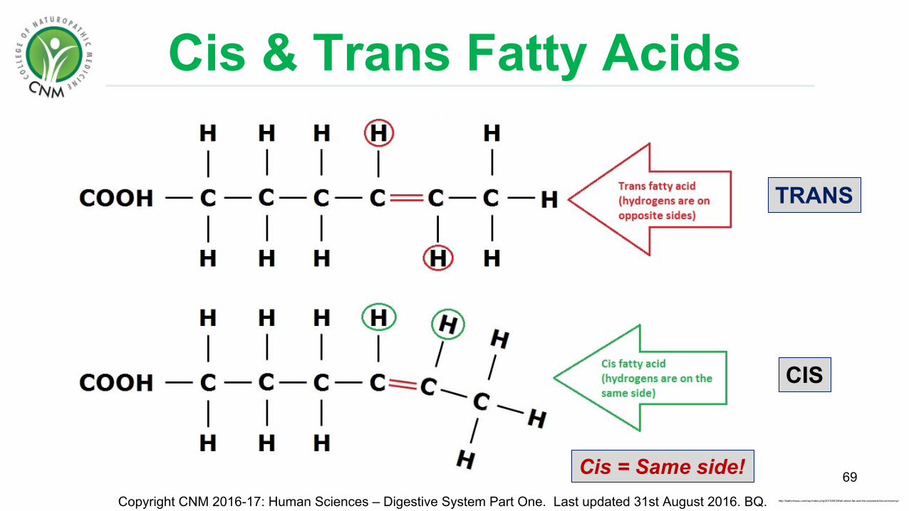
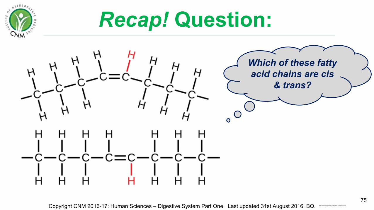
Unsaturated fatty acids have one or more double bonds between carbon atoms (C=C)
Saturated = a molecule containing the greatest number of hydrogen atoms without any double bonds
TRANS
CIS
Copyright CNM 2016-17: Human Sciences – Digestive System Part One. Last updated 31st August 2016. BQ.
69http://katherineau.com/wp/index.php/2015/08/26/all-about-fat-and-the-saturated-fat-controversy/
Cis & Trans Fatty Acids
Cis = Same side!
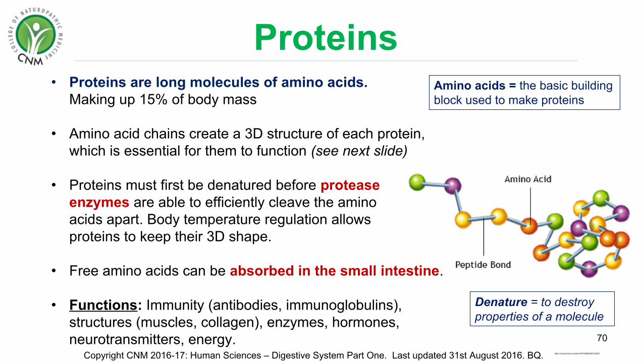
Proteins• Proteins are long molecules of amino acids.
Making up 15% of body mass
• Amino acid chains create a 3D structure of each protein, which is essential for them to function (see next slide)
• Proteins must first be denatured before protease enzymes are able to efficiently cleave the amino acids apart. Body temperature regulation allows proteins to keep their 3D shape.
• Free amino acids can be absorbed in the small intestine.
• Functions: Immunity (antibodies, immunoglobulins), structures (muscles, collagen), enzymes, hormones, neurotransmitters, energy. 70
Copyright CNM 2016-17: Human Sciences – Digestive System Part One. Last updated 31st August 2016. BQ. https://uk.pinterest.com/pin/444378688206139403/
Amino acids = the basic building block used to make proteins
Denature = to destroy properties of a molecule

Protein structure:
71
Copyright CNM 2016-17: Human Sciences – Digestive System Part One. Last updated 31st August 2016. BQ.
Each protein has a unique 3D structure……hence “The lock and
key model”…
The hormone in this diagram is representing a protein (peptide) hormone:
Complex 3D shape of a protein:

Enzymes• Enzymes are biological catalysts which speed up chemical reactions up to
thousands of times.
• They are highly specific with what substrate they react with (lock & key)
• Enzyme names are usually based on the substrate they react with, and end in –ase eg. Lactase.However some enzymes involved in protein digestion end with –in eg. Pepsin
• Enzymes are not changed or consumed in a chemical reaction - they can perform the same reaction many times.
• Are easily denatured by pH & temperature changes.
72Copyright CNM 2016-17: Human Sciences – Digestive System Part One. Last updated 31st August 2016. BQ.
Enzymes are proteins
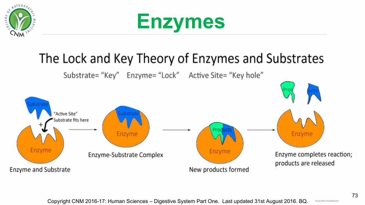
Enzymes
73Copyright CNM 2016-17: Human Sciences – Digestive System Part One. Last updated 31st August 2016. BQ. http://www.desadoc.com/wordpress/enzymes/

Digestion - Summary
74
Carbohydrates Lipids (fats / oils)
Protein
Mouth Amylase: starch to maltose, oligosaccharides & shorter chain polysaccharides.
No digestion No digestion
Stomach No digestion Gastric lipase: fats & oils to fatty acids & mono- & diglycerides.
HCl – denature proteins.pepsinogen converted to active pepsin - breaks proteins (polypeptides) to shorter chain peptides.
Small intestine
Pancreatic amylase – break down shorter chain polysaccharides & oligosaccharides to disaccharides. Sucrase – converts sucrose to glucose & fructose to be absorbed.Lactase - converts lactose to galactose & glucose to be absorbed.Maltase - converts maltose to glucose & glucose to be absorbed.
Bile emulsifies fat
Pancreatic lipase – forms fatty acids & monoglycerides which can be absorbed.
Trypsinogen converted to active trypsinChymotrypsinogen to active chymotrypsin Procarboxypeptidase to active carboxypeptidase Proelasatase to active elastase – Protease enzymes which digest proteins to Dipeptidase (brush border enzymes) – dipeptide to amino acids. Aminopeptidase (brush border enzymes) – peptides to amino acids. Amino acids are absorbed.
Large intestine
Bacterial fermentation to form gases. Bacterial fermentation to gases & other compounds.
Copyright CNM 2016-17: Human Sciences – Digestive System Part One. Last updated 31st August 2016. BQ.
Recap! Question:
75Copyright CNM 2016-17: Human Sciences – Digestive System Part One. Last updated 31st August 2016. BQ.
Which of these fatty acid chains are cis
& trans?
http://www.prettyhealthy.co/tag/the-truth-about-fats/
Exercise!
76Copyright CNM 2016-17: Human Sciences – Digestive System Part One. Last updated 31st August 2016. BQ.
Draw a simplified diagram of the entire GI tract
(beginning with the oral cavity).Include accessory organs!