Embed Size (px)
Citation preview

PAROXYSMAL VENTRICULAR STANDSTILLBY
HAROLD COOKSON
From the General Hospital, Poole
Received January 1. 1952
Among the various rhythms reported in the course of a Stokes-Adams attack, one of the rarestis standstill of the ventricle as an episode during normal rhythm. Such a condition may be calledparoxysmal ventricular standstill, and is to be distinguished from the condition of alternatingcomplete block and normal rhythm to which the term paroxysmal heart block may be applied.Three cases of paroxysmal ventricular standstill have been observed, in two of which there wasparoxysmal heart block also, and ventricular standstill was also provoked by carotid sinus pressure.
Weiss and Baker (1933) described several cases of total cardiac standstill, auricular standstlil,or extreme sinus bradycardia, and one of complete block in auricular fibrillation as a result ofcarotid sinus pressure: all of these had cardiovascular disease. Starling (1921) reported ventricularstandstill on* swallowing but no cardiographic records were made of the attacks. Yater andWilliams (1928-29) described a case of complete cardiac and ventricular standstill with slowirregular P waves. The case of Lewis (1920) showed ventricular standstill with auricular rhythmpreserved; this patient later developed a complete block. A case recorded by Alexander andBauerlein (1936) had variations in the degree of heart block with changes of posture but not withcarotid sinus pressure. Holmes and Weill (1945) recorded mild grades of heart block in healthymen when they lay down; carotid sinus pressure caused a decrease in the P-R interval. Turner(1948) recorded the case of a man of 61 with cardiac asystole with pressure on either carotid sinus,whose attacks ceased after bilateral carotid sinus denervation. Parkinson et. al. (1941) recorded5 examples of ventricular standstill, and collected 28 others that had been reported. Amongall these there was only one in which the standstill was immediately followed by normal rhythmwith unimpaired A-V conduction; this patient showed complete heart block on another day.Isolated cases of ventricular standstill are mentioned in two or three textbooks.
CASE REPORTSThree patients have been observed with attacks of ventricular standstill, two of whom had
normal rhythm and conduction before and after the standstill. In two of them the attacks couldbe provoked in various ways, in addition to those that had no apparent provocation.
Case 1. A woman of 82 had retinal hemorrhage and diabetes for which she was having aninjection of 50 units of insulin daily. For seven months she had been having syncopal and con-vulsive attacks. Physical examination was normal except for the retinal hsemorrhage. A cardio-gram showed normal rhythm, right bundle branch block, but no delay in A-V conduction. Asthe record was being completed with lead IV R, the ventricle suddenly stopped with immediateloss of consciousness and a generalized convulsion after 5 seconds (Fig. 1); the movement of therecord off the paper indicates the beginning of limb movements. The recording, when resumedat the end of the convulsion shows normal rhythm and conduction exactly as before except thatthe rate was slightly quicker. As weeks might elapse between this patient's attacks, it was mostfortunate that one occurred during the only tracing taken. No further information about the sub-sequent history was available except that she died a few months later.
350
on 18 June 2019 by guest. Protected by copyright.
http://heart.bmj.com
/B
r Heart J: first published as 10.1136/hrt.14.3.350 on 1 July 1952. D
ownloaded from

PAROXYSMAL VENTRICULAR STANDSTILL 351
Lea<" IVR --i -----1--------i--- -Sw-vl _._ _ _ X t - --w- __ - _ ~~~~~~~~~~~~~~~~~~~~~~~~~~~. ;.
FIG.10i.A-C1. Wm, e . S a v -s gei c 5 second........ ea.} _...........F ...::.;i
_,IG._I.-Case 1Woa, age 8. Spnaeu venrcua stnsil generalized------co.SnvulsR|io5 ,,4_ U,,,,_asecnd
after the ventricle stops; at end of attack ventricular beating resumed with normal P-R interval.
Case 2. A man, aged 78, with a history of rheumatic fever in childhood and of an illnessdiagnosed as endocarditis when aged 51, gave a history of" black-outs " for ten years, but only atvery long intervals for the first nine years. He developed a gastric ulcer at the age of 77 at whichtime his syncopal attacks became more frequent. At the apex there was an extra heart sound dueto the latent block, but otherwise there was no sign of cardiovascular disease. Normal rhythmwith prolonged A-V conduction was usually present (Fig. 2) but sometimes partial or complete
FIG. 2.-Case 2. Man, aged 78. Normal rhythm; P-R interval about 0 u3sec.
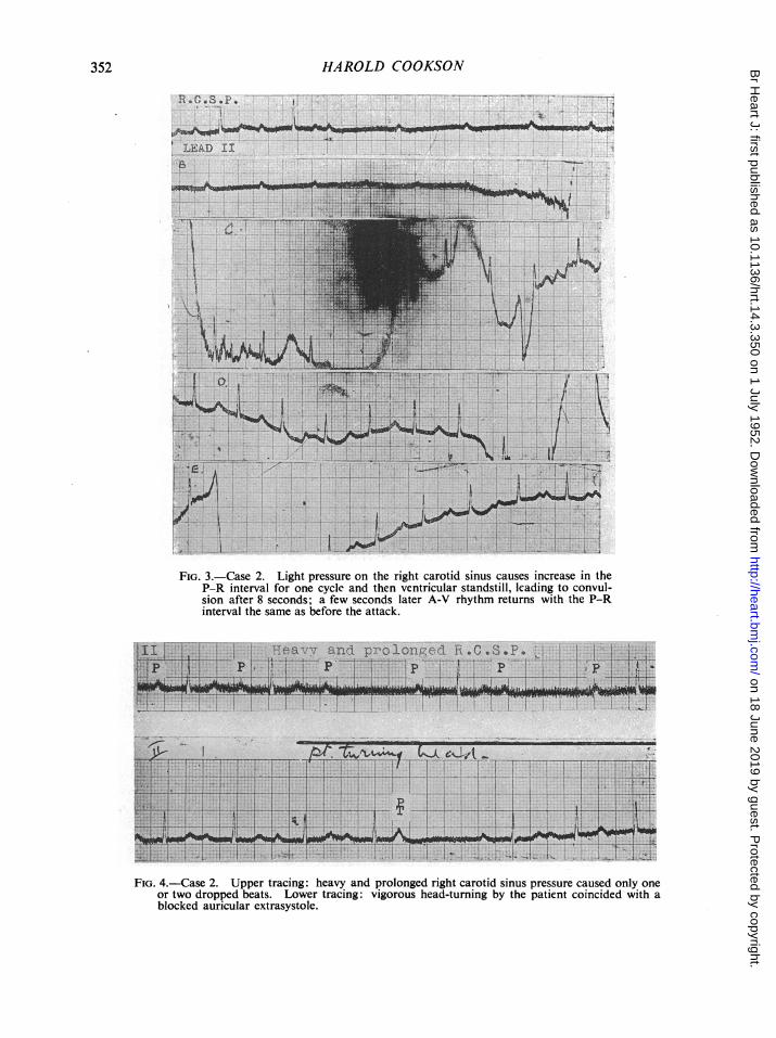
heart block. An attack of unconsciousness with convulsion was witnessed in which theperipheralpulse disappeared. Normal rhythm was recorded before andefter it. Then very light pressureon the right carotid sinus produced an increase in A-V conduction time for one cycle (Fig. 3) andthen ventricular standstill; the auricular rate gradually increased from 65 to 90 a minute, and afterabout 8 seconds, convulsive movements of the limbs were large enough to jerk the recording penoff the paper. After a few seconds, ventricular activity returned with a rate of about 90, the A-Vconduction time being about0h3 sec., the same as before the attack. Ventricular contractionsreturned some time before consciousness was fully regained and before limb movements ceased.On other occasions heavy and prolonged carotid sinus pressure womuld cause only one or twodropped beats with very slight auricular slowing (Fig. 4). This figure also shows a record takenwhile the patient turned his head vigorously from side to side; it coincided with an auricularextrasystole which was blocked. On yet another occasion when left carotid sinus pressure waswithout effect, pressure on the right one caused an extreme sinus bradycardia with ventricularescape (Fig. 5). After a ventricular extrasystole, sinus beating was resumed at almost the usualrate in spite of carotid sinus pressure being maintained. When complete block was present,carotid sinus stimulation seemed to be without any effect. Fig. 6 shows complete block with anoccasional conducted beat; this last occurs when P falls about 0-3 sec. before QRS. Then pressureon both carotid sinuses successively was exerted; the middle tracing shows the independent
on 18 June 2019 by guest. Protected by copyright.
http://heart.bmj.com
/B
r Heart J: first published as 10.1136/hrt.14.3.350 on 1 July 1952. D
ownloaded from
352 HAROLD COOKSON
.~~~~~~~~~~~~~~~~~~
LEAD I.L
_~~~~~~~~~~~~~~~~~_A'sIFIG. 3.-Case 2. Light pressure on the right carotid sinus causes increase in the
P-R interval for one cycle and then ventricular standstill, leading to convul-sion after 8 seconds; a few seconds later A-V rhythm returns with the P-Rinterval the same as before the attack.
eavv and Prolonged R.G.S.P.
____. IW,..'.. Ao . . ........ ... - -- 1 1 - f ................MJ - - ;
u1_ zt .1 i 7-1. .!-
FIG. 4.-Case 2. Upper tracing: heavy and prolonged right carotid sinus pressure caused only oneor two dropped beats Lower tracing: vigorous head-turning by the patient coincided with ablocked auricular extrasystole.
on 18 June 2019 by guest. Protected by copyright.
http://heart.bmj.com
/B
r Heart J: first published as 10.1136/hrt.14.3.350 on 1 July 1952. D
ownloaded from

PAROXYSMAL VENTRICULAR STANDSTILL
R.C.S.P. , 2 KR.C.S.P. . ...........
I.IJL7LIIiII2171 - F'A ....i
*......-.......A.-.2h--l1> ..t-sIiLead '12
vaY J.,j.. I_ .2;' ,i
A B
FIG. 5.-Case 2. (A) Right carotid sinus pressure caused sinus bradycardia and ventricular escape.(B) After a ventricular extrasystole, normal rhythm and rate return in spite of continued rightcarotid sinus pressure.
TI CS*P
FIG. 6.-Case 2. Upper tracing: complete heart block with occasional A-V conduction carotidsinus pressure either left or right has no effect. Lower tracing: return ofnormal rhythm,P-R interval 04 sec., soon after carotid sinus pressure had been stopped.
auricular and ventricular rhythms maintained as before. After all this pressure on the neck hadstopped, the patient was in a distressed condition, but normal rhythm had returned with a P-Rinterval of 04 sec. (lower tracing, Fig. 6). Treatment of this patient with eumydrine, ephedrine,and digitalis in turn had no certain effect on the frequency of the Stokes-Adams attacks.
Case 3. A man, aged 69, subject to attacks of loss of consciousness since 1938, was said tohave had rheumatic fever when 7 years old; there was no evidence of rheumatic heart disease,but some hypertension with left ventricular hypertrophy was present. When first seen in 1945,he had normal rhythm and it was not until 1948 that a record showing complete block was ob-tained. About this time, during the taking of a routine tracing which showed normal rhythm, aventricular extrasystole occurred and this was immediately followed by ventricular standstill, lossof consciousness, and a convulsion (Fig. 7). Immediately after the attack, the tracing was resumedand showed normal sinus rhythm and the A-V conduction within normal limits. The patienthimself preferred his pulse slow (complete block) rather than quick (normal rhythm) as then hefound he had fewer attacks and he had noticed that if he raised his left arm, he would lose con-
. I
FIG. 7.-Case 3. Man, aged 69. Normal rhythm, normal P-R interval. (A) Ventricular standstill follows aventricular extrasystole (lead I), causing a Stokes-Adams attack. (B) Normal rhythm and conduction areresumed immediately after the attack.
353
IA
on 18 June 2019 by guest. Protected by copyright.
http://heart.bmj.com
/B
r Heart J: first published as 10.1136/hrt.14.3.350 on 1 July 1952. D
ownloaded from

3HAROLD COOKSON
BFIG. 8.-Case 3. (A) Ventricular standstill produced immediately by carotid sinus
pressure. (B) Normal rhythm and conduction recorded as soon as the attackwas over.
WS-I.. I I Lfh . 1.1I p. ........ -;......;AL:2z:IiT: rF Al ifMIa-J4l!5
rijLh1ihiLI.jIi -i 1 1' i it .* u
Ex. S. Ex .S.
FIG. 9.-Case 3. Ventricular standstill for three cycles following an extrasystole; the firstbeat after the standstill is also aberrant.
sciousness but not if he raised his right arm. Carotid sinus pressure was tried first on the rightwithout effect, and then on the left with immediate ventricular standstill (Fig. 8). The almostinstantaneous effect of the pressure is shown in the record, pressure on the carotid sinus coincidingwith depression of the lever to start the recording drum. The beginning of limb movements isshown by the unsteadiness of the tracing after about 5 seconds. During this time the auricularrate increased from 75 to 100 a minute. The tracing was resumed as soon as possible afterthe attack and was exactly the same as before it, with sinus rhythm, rate about 75 and normalA-V conduction time. For some months all records showed normal rhythm; then one wasobtained showing ventricular standstill for three cycles after a ventricular premature beat, theresumption of normal rhythm being preceded by another ventricular premature contraction (Fig. 9).
IIT .. _ p _ ,,,,,_ _ - -; _............ _ ;..';- -
Kf- - _ ____---*-r- .-.i-
WeAt'F- v tlvJ s 4 0 we
T _ 'lJ. J.ut.-O..
FIG. 10.-Case 3. Upper tracing: complete heart block. Lower tracing: left carotidsinus pressure causes no auricular or ventricular slowing.
'AFFIL A L... 1wriaw PI
I I m 6 I rlS S ra R - - -354
_idA@_;a- mpq6
I
on 18 June 2019 by guest. Protected by copyright.
http://heart.bmj.com
/B
r Heart J: first published as 10.1136/hrt.14.3.350 on 1 July 1952. D
ownloaded from

PAROXYSMAL VENTRICULAR STANDSTILL
25 min. after inj. atropine. %.; t/Ac a.1
<I I
After exercisef
IC - - -+ - --~~~~~~~~~~~~~~~~~~~~~~~~~~~~~~~~~~~~~~~~~~~~~~~~~~~~~~~~~~~~~~~~~~~~~~~~~~~~~~~~~~~~~~~~~~~~~~~~~~~~~~~~~~~~~~~~~~.....------
- p
p p--=
,,~~~~~~~~~~~~~~~1P~..A. . .. s.h I.i . r , p
FIG. 11.-Case 3. (A) After injection of atropine gr. 1/100, complete heart block with auricularand ventricular rates almost the same; P inverted. (B) After exertion P altered in shape.(C) After more exertion P at first negative becomes upright; 2: 1 sino-auricular block; completeA-V block. (D) Complete block and 2 : 1 sino-auricular block; carotid sinus pressure causestemporary inversion of P; sino-auricular block disappears at end of tracing.
III Under pentothal Intubation.
A BFIG. 12. Case 3. (A) Normal rhythm and normal conduction, rate 105 a minute. (B) Under pentothal anes-
thesia, ventricular rate unchanged, P negative; probably nodal rhythm: passing of tracheal tube (shown byarrow) produced complete block.
The following day, auricular fibrillation was recorded at which time neither arm raising nor carotidsinus pressure had any effect. When complete block was present, carotid sinus pressure whetherleft or right was incapable of causing ventricular standstill, nor did it slow the auricle (Fig. 10).Two to one sino-auricular block occurred from time to time; it would appear after exertion andafter an injection of atropine (Fig. 11); at other times the P wave became inverted or biphasic.While waiting to be given an anesthetic, this patient had sinus tachycardia with normal A-Vconduction (Fig. 12, A). After pentothal P became inverted, the ventricular rate being much thesame. At the moment a McGill's tube was passed through the nose into the trachea, there wasa transition to complete A-V dissociation (Fig. 12, B).
DIsCUSSIONThe commonest causes of the Stokes-Adams attack are believed to be: (1) transition from
normal rhythm to high-grade block; (2) slowing of the idio-ventricular rhythm in the course ofcomplete block; and (3) abnormal ventricular rhythms such as ventricular tachycardia and fibril-
355
on 18 June 2019 by guest. Protected by copyright.
http://heart.bmj.com
/B
r Heart J: first published as 10.1136/hrt.14.3.350 on 1 July 1952. D
ownloaded from

HAROLD COOKSON
lation followed by standstill. Of the three cases of ventricular standstill described here occurringas an episode in the course of normal rhythm, two showed varying degrees of heart block fromtime to time. They had also extreme sensitivity of the carotid sinus at times, but this was onlyfound when normal rhythm was present. Then quite slight pressure on the carotid sinus-oneside was more responsive than the other-was capable of arresting the ventricle within about asecond, followed immediately by loss of consciousness and then within 5 to 8 seconds by toniccontraction of the limbs. The term loss of consciousness is not a precise one, but it is certainthat immediately after the ventricle stopped beating, the patients began to lose consciousness, whilethe onset of tonic limb contractions can be measured accurately from the movement of the base-line of the tracing. The immediate impairment of consciousness may not be entirely due to thefall in cerebral blood flow, and it is possible that a direct stimulation of the cerebral circulationplays a part as suggested by Weiss and Baker (1933) to explain cases in which carotid sinus stimu-lation caused syncope without slowing of the heart rate or fall in blood pressure. The extremesensitivity of the conducting tissues to nerve impulses is shown by the induction of mild attacksby merely raising the arm, or changing posture. These events occurred only when normal rhythmwas present and even then the response to carotid sinus pressure varied much from day to day andeven from hour to hour. However, the position was very different when complete block was present;then carotid sinus stimulation was without effect on the ventricle and usually on the auricle also,though it might slow its rate a little or displace the site of impulse formation. It appears thatwhen normal rhythm is present, the reflex from the carotid sinus acts on the upper part of the bundleof His and is temporarily incapacitated, although immediately before and after it conducts per-fectly. On the other hand, when complete block ic present, the centre governing the ventricle,situated lower in the bundle, is immune from the effects of the reflex. An interesting observationis the onset of ventricular standstill after a ventricular extrasystole. Might this result from fatigueof the bundle by the premature retrograde stimulation, or alternatively might a nervous impulsesimultaneously act upon the ventricle and the bundle to produce both effects?
The diagnosis of epilepsy from loss of consciousness due to paroxysmal ventricular standstillmay be difficult from the history alone, even if a description of the attacks by a good eye-witness isavailable. A short standstill simulates petit mal very closely, but a longer one leading to completeloss of consciousness and convulsion will usually differ from grand mal in the facial pallor ratherthan congestion, in the facial flush as the attack ends, and in the mote rapid return of full con-sciousness. The elderly subject with a history of loss of consciousness, even over many years,and even though his cardiogram at first shows not the least sign of block, may yet be sufferingfrom paroxysmal ventricular standstill or other kind of heart block. For such a patient, carotidsinus pressure might settle the diagnosis, though this is hardly a method to be used indiscriminately.Apart from this, the true diagnosis will probably be revealed by continued observation, as sooneror later ordinary forms of auriculo-ventricular block will make their appearance.
It used to be thought that the heart block of chronic heart disease was usually permanent, butit is now clear that in many the rhythm passes back and forth from block to normal over longperiods of years, the duration of each varying from seconds to months. In addition to the basicstructural lesion, therefore, other factors must be involved and among these are reflexes from thecarotid sinus, pharynx, and cesophagus, and perhaps impulses arising in the brain itself as a resultof emotional disturbance.
REFERENCES
Alexander, H. L., and Bauerlein, T. C. (1936). Amer. Heart J., 11, 223.Holmes, J. H., and Weill, D. R. (1945). Amer. Heart J., 30, 291.Lewis, T. (1920). The Mechanism and Graphic Registration of the Heart Beat. London, p. 358.Parkinson, J., Papp, C., and Evans, W. (1941). Brit. Heart J., 3, 171.Starling, H. J. (1921). Heart, 8, 31.Turner, R. W. D. (1948). Quart. J. Med., 17, 327.Weiss, S., and Baker, J. P. (1933). Medicine, 12, 297.Yater, W. M., and Williams, F. A. (1928-29). Amer. Heart J., 4, 280.
356
on 18 June 2019 by guest. Protected by copyright.
http://heart.bmj.com
/B
r Heart J: first published as 10.1136/hrt.14.3.350 on 1 July 1952. D
ownloaded from