Embed Size (px)
Citation preview
1
Pulmonary Diseases: Structure-Function Correlation II
Parenchymal, Interstitial (Restrictive) and Vascular Diseases
Alain C. Borczuk, M.D.Dept of Pathology
Pulmonary Diseases: Structure-Function Correlation II
Goals:
• To observe the relationship between structural/morphologic manifestation of diseases to measurable functional parameters using prototypical diseases of parenchyma, interstitium and vasculature
• To describe the pathology, Gross and microscopic, of these pulmonary diseases.
2
Pulmonary Diseases: Structure-Function Correlation II
• Disease of the acini and interstitium1) Replacement of air with fluid, inflammatory
cells or cellular debris2) Thickening of alveolar walls and interstitium3) Destruction of acinar walls
• Disease of the conducting airways• Disease of the pulmonary vasculature
Pulmonary Diseases: Structure-Function Correlation II
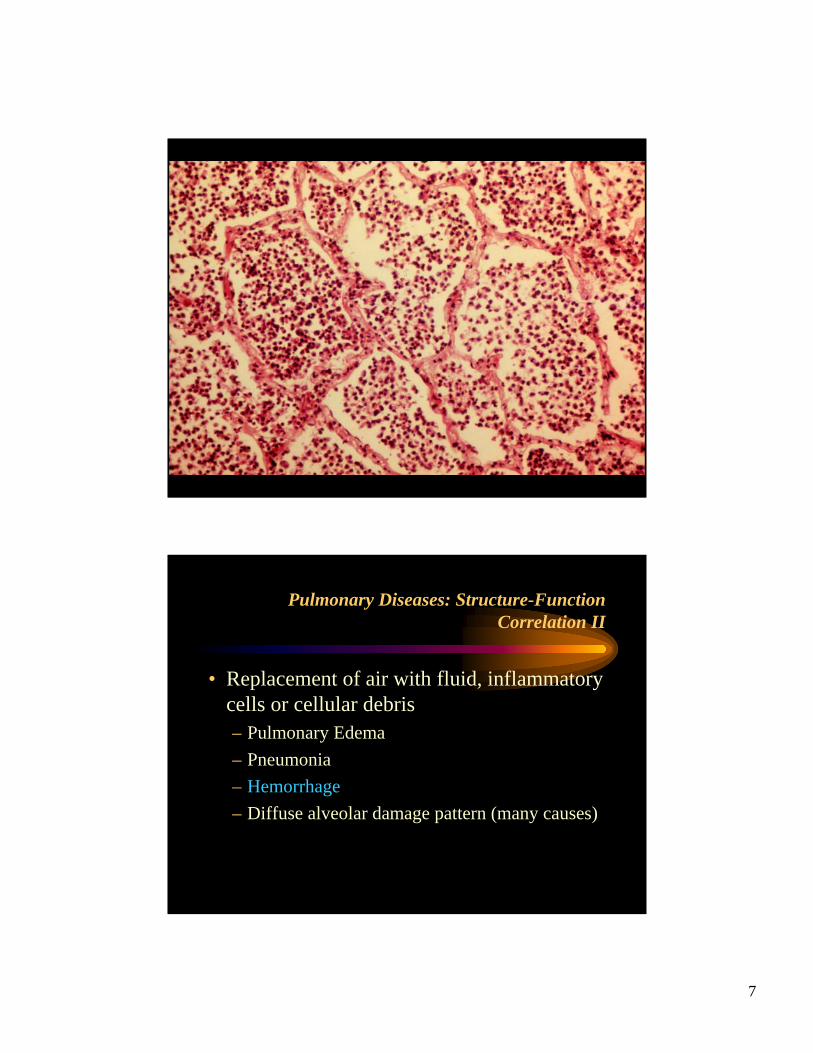
• Replacement of air with fluid, inflammatory cells or cellular debris– Pulmonary Edema– Pneumonia– Hemorrhage– Diffuse alveolar damage pattern (many causes)
3
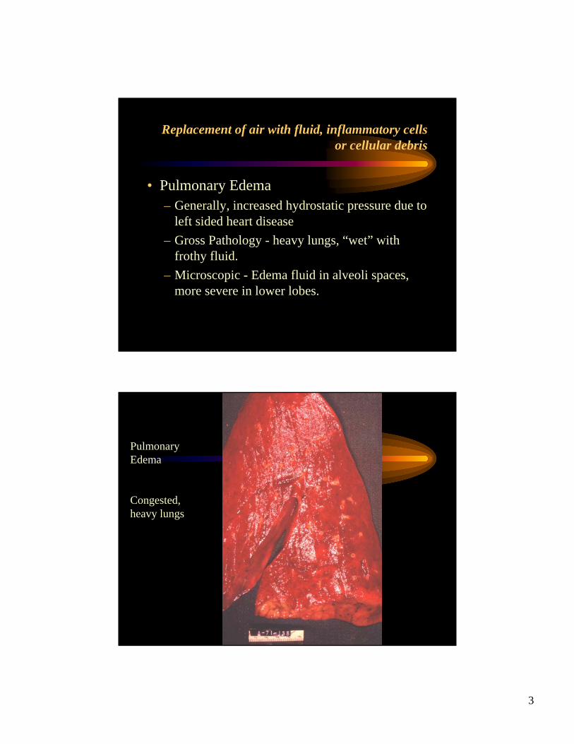
Replacement of air with fluid, inflammatory cells or cellular debris
• Pulmonary Edema– Generally, increased hydrostatic pressure due to
left sided heart disease– Gross Pathology - heavy lungs, “wet” with
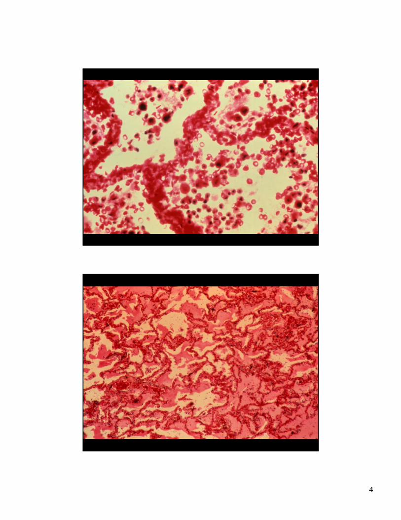
frothy fluid.– Microscopic - Edema fluid in alveoli spaces,
more severe in lower lobes.
Pulmonary Edema
Congested, heavy lungs
4
5
Pulmonary Diseases: Structure-Function Correlation II
• Replacement of air with fluid, inflammatory cells or cellular debris– Pulmonary Edema– Pneumonia– Hemorrhage– Diffuse alveolar damage pattern (many causes)
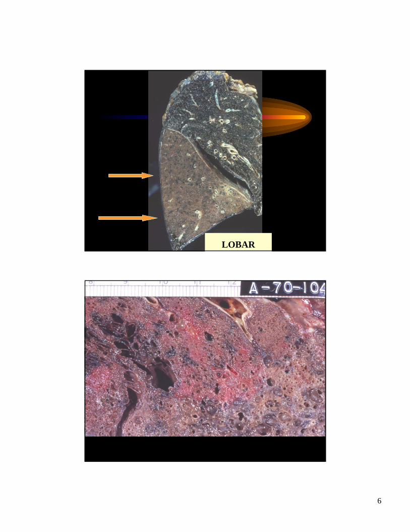
Replacement of air with fluid, inflammatory cells or cellular debris
• Pneumonia– Inflammation of the lung, often infectious– Gross Pathology: Consolidation of lungs
(firmness), either small patches or lobar– Microscopic: Acute bacterial pneumonia,
whether lobar or patchy, is characterized by polymorphonuclear cells filling the alveolar spaces.
6
LOBAR
7
Pulmonary Diseases: Structure-Function Correlation II
• Replacement of air with fluid, inflammatory cells or cellular debris– Pulmonary Edema– Pneumonia– Hemorrhage– Diffuse alveolar damage pattern (many causes)
8
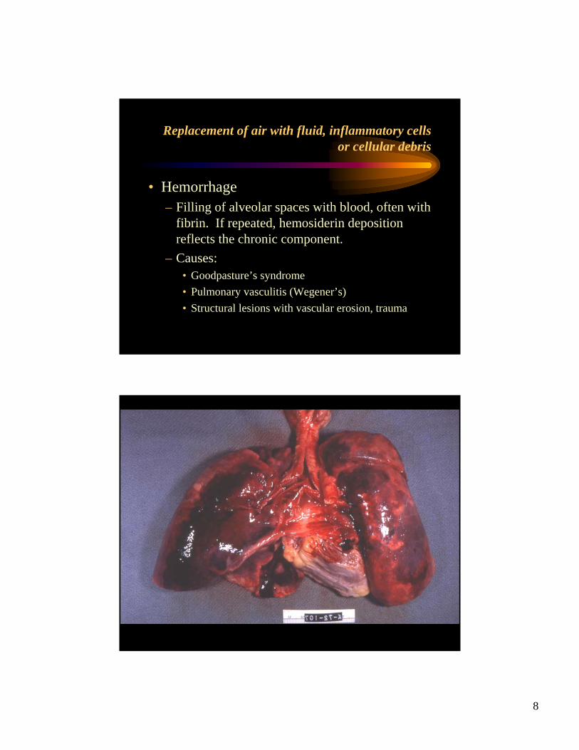
Replacement of air with fluid, inflammatory cells or cellular debris
• Hemorrhage– Filling of alveolar spaces with blood, often with
fibrin. If repeated, hemosiderin deposition reflects the chronic component.
– Causes:• Goodpasture’s syndrome• Pulmonary vasculitis (Wegener’s)• Structural lesions with vascular erosion, trauma
9
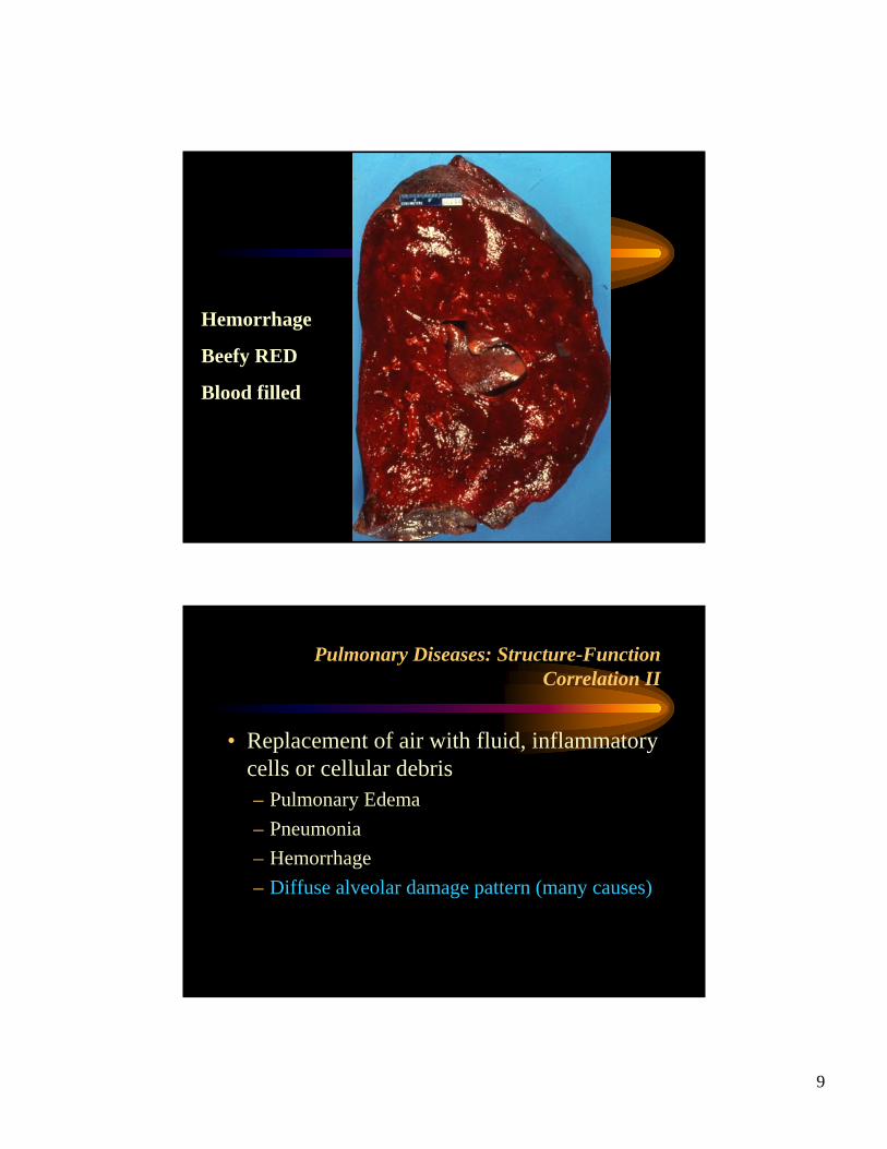
Hemorrhage
Beefy RED
Blood filled
Pulmonary Diseases: Structure-Function Correlation II
• Replacement of air with fluid, inflammatory cells or cellular debris– Pulmonary Edema– Pneumonia– Hemorrhage– Diffuse alveolar damage pattern (many causes)
10
Replacement of air with fluid, inflammatory cells or cellular debris
• Diffuse alveolar damage– Histology of Adult respiratory distress
syndrome (ARDS)– Many causes include pulmonary infection,
shock, sepsis, pancreatitis, burns, toxic inhalations, radiation, near-drowning
– Acute alveolar injury with microvasculardamage leading to edema and tissue injury.
Replacement of air with fluid, inflammatory cells or cellular debris
• Diffuse alveolar damage– Adult Respiratory Distress Syndrome
• Acute onset of shortness of breath• Hypoxemia• Bilateral infiltrates on x-ray• Non-cardiogenic• .
11
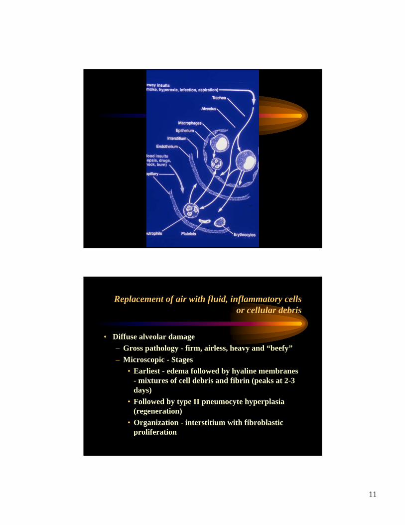
Replacement of air with fluid, inflammatory cells or cellular debris
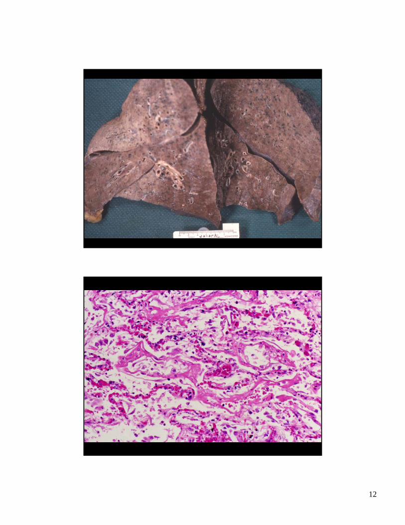
• Diffuse alveolar damage– Gross pathology - firm, airless, heavy and “beefy”– Microscopic - Stages
• Earliest - edema followed by hyaline membranes - mixtures of cell debris and fibrin (peaks at 2-3 days)
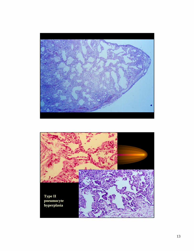
• Followed by type II pneumocyte hyperplasia (regeneration)
• Organization - interstitium with fibroblastic proliferation
12
13
Type II pneumocytehyperplasia

14
Replacement of air with fluid, inflammatory cells or cellular debris
• Alveoli filled with blood, neutrophils, hyaline membranes, or fluid
• Decreased lung volume (atelectasis)
• Increase in shunt flow• In DAD, edema, hypoxic
vasoconstriction and vascular injury may cause pulmonary hypertension
STRUCTURAL VS. FUNCTIONAL
Replacement of air with fluid, inflammatory cells or cellular debris
• Diffuse alveolar damage• .Bridges diseases of alveolar filling and thickening
of interstitium (next section)• After the exudative phase, interstitial changes can
either resolve or lead to fibrosis (interstitial fibrosis)• This acute lung injury is associated with high
mortality, especially in the elderly and when associated with sepsis
15
Pulmonary Diseases: Structure-Function Correlation II
• Disease of the acini and interstitium1) Replacement of air with fluid, inflammatory
cells or cellular debris2) Thickening of alveolar walls and interstitium3) Destruction of acinar walls
• Disease of the conducting airways• Disease of the pulmonary vasculature
Pulmonary Diseases: Structure-Function Correlation II
• Thickening of alveolar walls and interstitium– Idiopathic pulmonary fibrosis/usual interstitial
pneumonia– Sarcoidosis– Hypersensitivity pneumonitis
16
Pulmonary Diseases: Structure-Function Correlation II
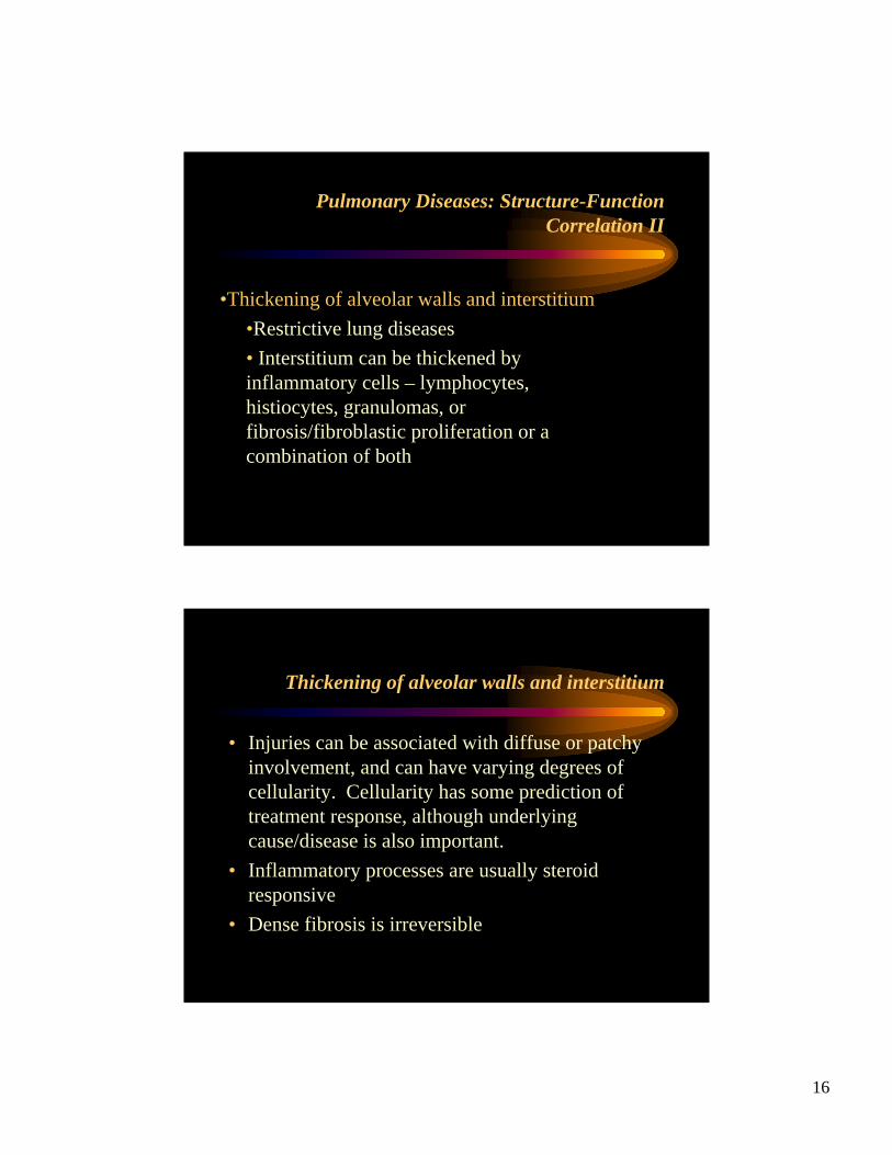
•Thickening of alveolar walls and interstitium•Restrictive lung diseases• Interstitium can be thickened by inflammatory cells – lymphocytes, histiocytes, granulomas, or fibrosis/fibroblastic proliferation or a combination of both
Thickening of alveolar walls and interstitium
• Injuries can be associated with diffuse or patchy involvement, and can have varying degrees of cellularity. Cellularity has some prediction of treatment response, although underlying cause/disease is also important.
• Inflammatory processes are usually steroid responsive
• Dense fibrosis is irreversible
17
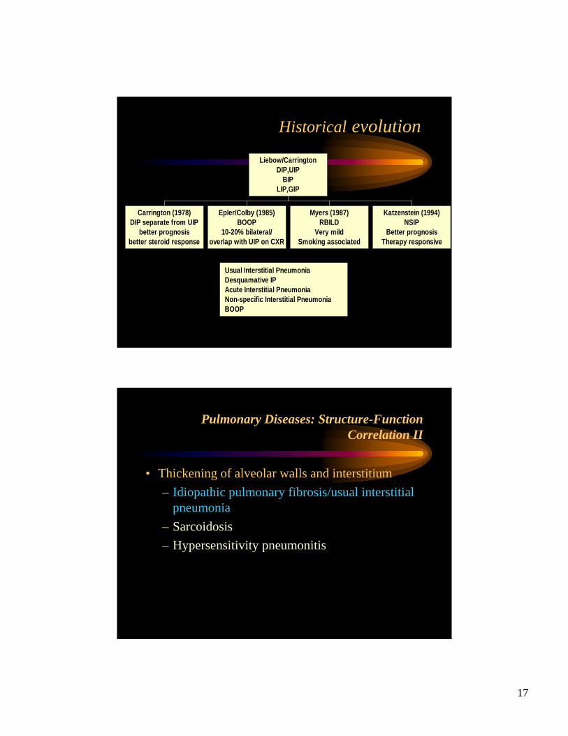
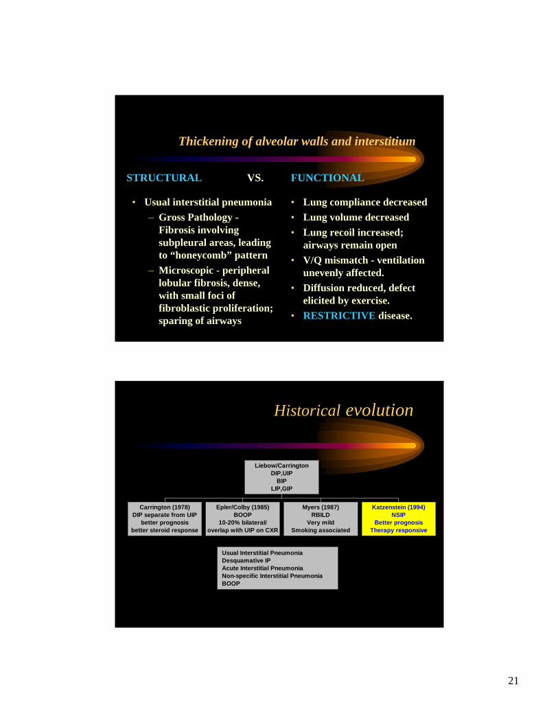
Historical evolution
Usual Interstitial PneumoniaDesquamative IPAcute Interstitial PneumoniaNon-specific Interstitial PneumoniaBOOP
Liebow/CarringtonDIP,UIP
BIPLIP,GIP
Carrington (1978)DIP separate from UIP
better prognosisbetter steroid response
Epler/Colby (1985)BOOP
10-20% bilateral/overlap with UIP on CXR
Myers (1987)RBILD
Very mildSmoking associated
Katzenstein (1994)NSIP
Better prognosisTherapy responsive
Pulmonary Diseases: Structure-Function Correlation II
• Thickening of alveolar walls and interstitium– Idiopathic pulmonary fibrosis/usual interstitial
pneumonia– Sarcoidosis– Hypersensitivity pneumonitis
18
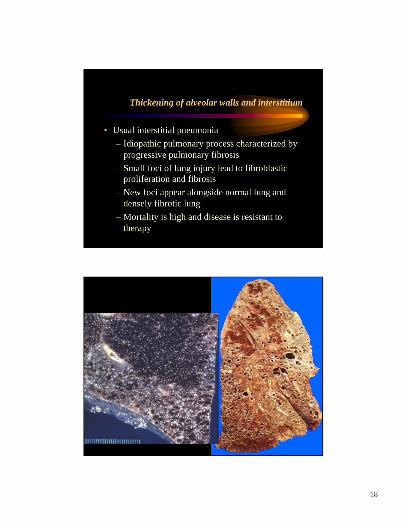
Thickening of alveolar walls and interstitium
• Usual interstitial pneumonia– Idiopathic pulmonary process characterized by
progressive pulmonary fibrosis– Small foci of lung injury lead to fibroblastic
proliferation and fibrosis– New foci appear alongside normal lung and
densely fibrotic lung– Mortality is high and disease is resistant to
therapy
19
20
21
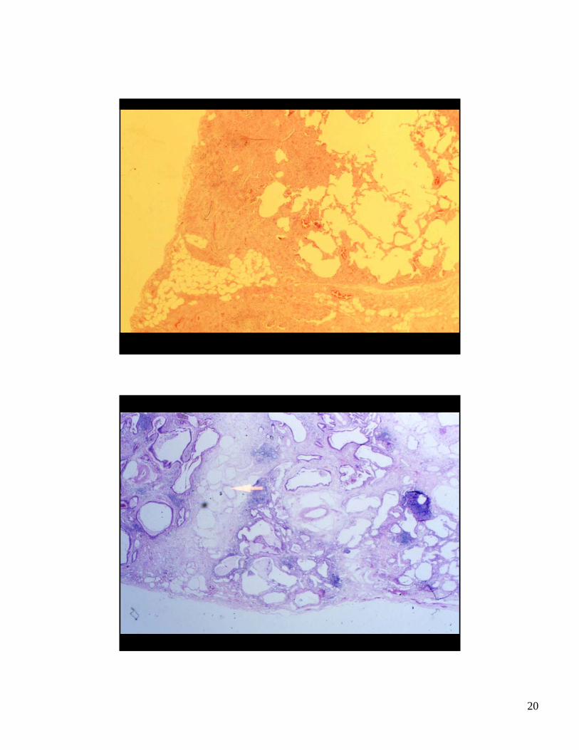
Thickening of alveolar walls and interstitium
• Usual interstitial pneumonia– Gross Pathology -
Fibrosis involving subpleural areas, leading to “honeycomb” pattern
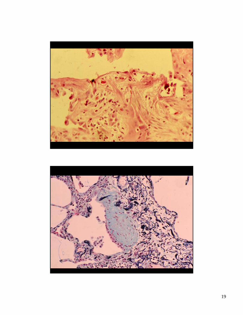
– Microscopic - peripheral lobular fibrosis, dense, with small foci of fibroblastic proliferation; sparing of airways
• Lung compliance decreased• Lung volume decreased• Lung recoil increased;
airways remain open• V/Q mismatch - ventilation
unevenly affected.• Diffusion reduced, defect
elicited by exercise.• RESTRICTIVE disease.
STRUCTURAL VS. FUNCTIONAL
Historical evolution
Usual Interstitial PneumoniaDesquamative IPAcute Interstitial PneumoniaNon-specific Interstitial PneumoniaBOOP
Liebow/CarringtonDIP,UIP
BIPLIP,GIP
Carrington (1978)DIP separate from UIP
better prognosisbetter steroid response
Epler/Colby (1985)BOOP
10-20% bilateral/overlap with UIP on CXR
Myers (1987)RBILD
Very mildSmoking associated
Katzenstein (1994)NSIP
Better prognosisTherapy responsive
22
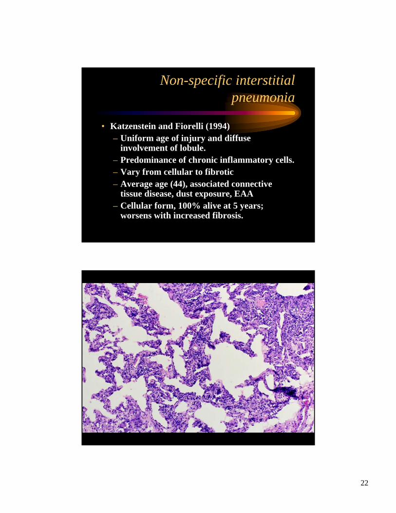
Non-specific interstitial pneumonia
• Katzenstein and Fiorelli (1994)– Uniform age of injury and diffuse
involvement of lobule.– Predominance of chronic inflammatory cells.– Vary from cellular to fibrotic– Average age (44), associated connective
tissue disease, dust exposure, EAA– Cellular form, 100% alive at 5 years;
worsens with increased fibrosis.
23
Why do we classify?
• Some patterns are associated with systemic diseases (e.g. NSIP in collagen vascular disease), or fibrosis due to certain medications
• The idiopathic interstitial pneumonia have different rates of progression to fibrosis
• Steroid responsiveness is high for some diseases (NSIP,BOOP) and low to non-existent for others (UIP)
• Mortality rates, likelihood of progression, decision to treat with cytotoxic agent, and candidacy for transplant may all be affected by the classification
Pulmonary Diseases: Structure-Function Correlation II
• Thickening of alveolar walls and interstitium– Idiopathic pulmonary fibrosis/usual interstitial
pneumonia– Sarcoidosis– Hypersensitivity pneumonitis
24
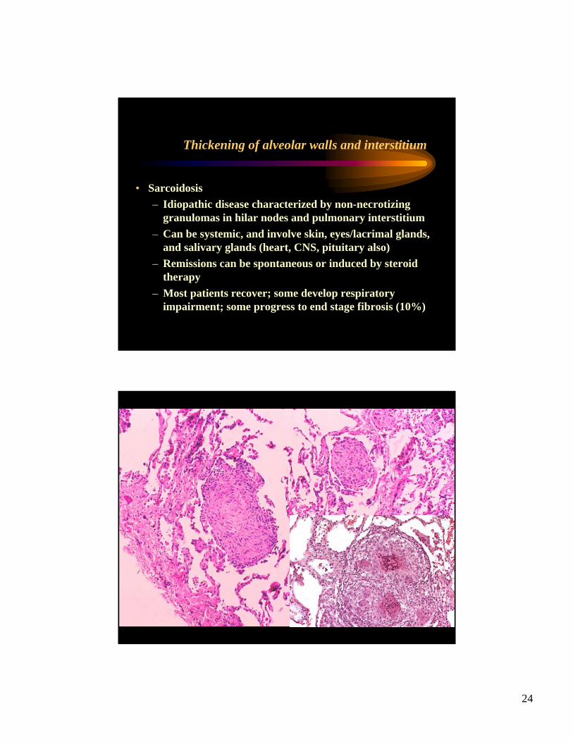
Thickening of alveolar walls and interstitium
• Sarcoidosis– Idiopathic disease characterized by non-necrotizing
granulomas in hilar nodes and pulmonary interstitium– Can be systemic, and involve skin, eyes/lacrimal glands,
and salivary glands (heart, CNS, pituitary also)– Remissions can be spontaneous or induced by steroid
therapy– Most patients recover; some develop respiratory
impairment; some progress to end stage fibrosis (10%)
25
Pulmonary Diseases: Structure-Function Correlation II
• Thickening of alveolar walls and interstitium– Idiopathic pulmonary fibrosis/usual interstitial
pneumonia– Sarcoidosis– Hypersensitivity pneumonitis
Thickening of alveolar walls and interstitium
• Hypersensitivity pneumonitis (extrinsic allergic alveolitis)– Immunologically mediated (type III/typeIV) lung disease
caused by inhalation of organic antigen. Patients have circulating antibodies,complement activation and granuloma formation
– Named after circumstances surrounding antigen exposure:
• Pigeon breeder’s lung • Farmer’s lung - thermophilic actinomycetes• Humidifier lung - thermophilic bacteria• Duck feather fever - duck feather• Maple bark disease• Mushroom picker’s lung
26
Thickening of alveolar walls and interstitium
• Hypersensitivity pneumonitis (extrinsic allergic alveolitis)– Patients experience fever, cough, malaise, dyspnea.– Acutely, patients may link an exposure with
symptoms, but chronic form can be more insidious and therefore detailed history may be needed to make a connection
– Steroid responsive, but can lead to fibrosis in some patients with untreated chronic antigen exposure
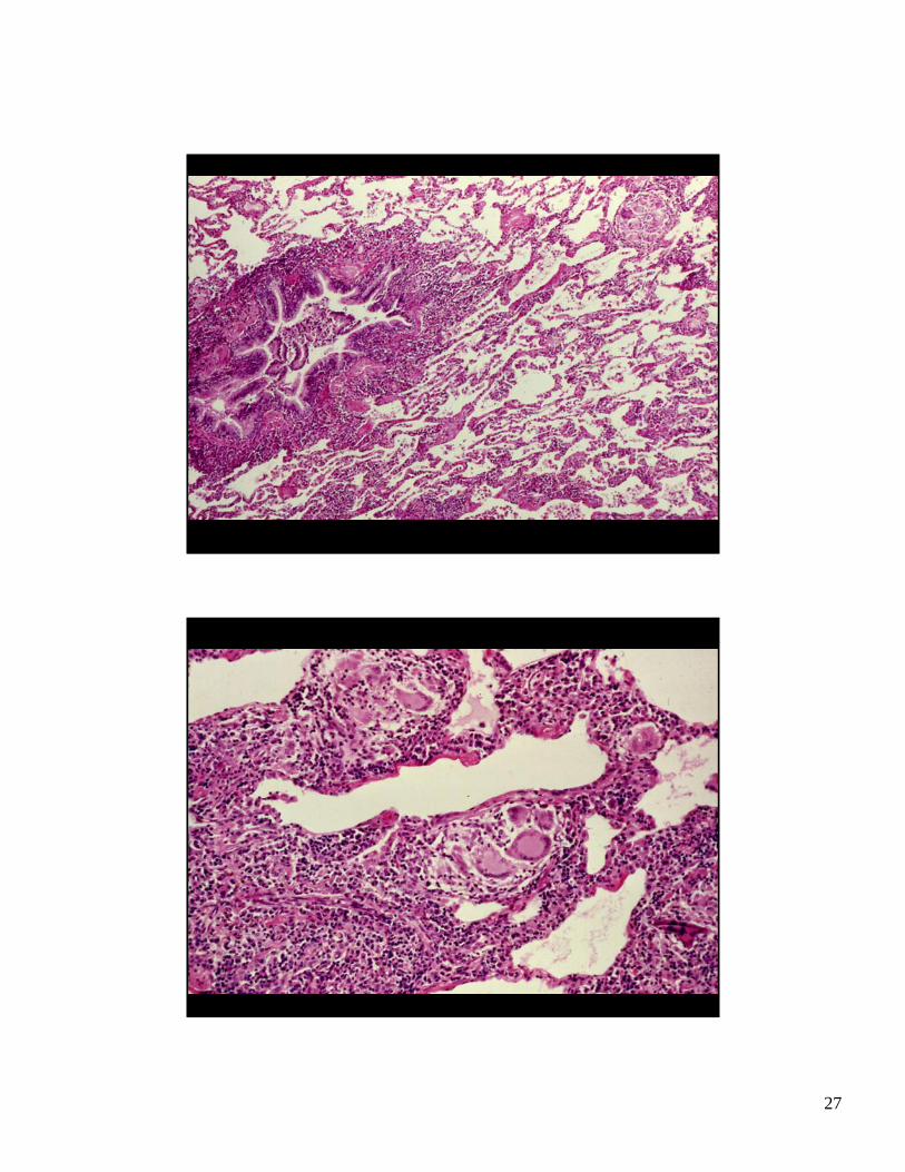
Thickening of alveolar walls and interstitium
• Hypersensitivity pneumonitis (extrinsic allergic alveolitis)– Pathology
• Expansion of peribronchial lymphoid tissue• Mild chronic interstitial pneumonitis• Interstitial histiocytic collections
27

28
Thickening of alveolar walls and interstitium
In summary:
There are a group of restrictive lung diseases characterized by increase in inflammatory cells or fibroblasts in the interstitium/alveolar walls. While they can all lead to fibrosis, some diseases do so invariably (UIP) and others less commonly (NSIP, sarcoid, hypersensitivity). In addition, diseases which are characterized by inflammation are usually steroid responsive. UIP does not respond well to any therapy and therefore has a high mortality.
This is why we attempt to classify these diseases by their inflammation and their patterns of fibrosis.
Thickening of alveolar walls and interstitium
Also of note is that fibrosing lung disease can be caused both by idiopathic processes, as well as by certain known processes such as asbestos exposure or collagen vascular disease, for example scleroderma.
Some use the term cryptogenic, because even though we know that immunologic reaction can lead to fibrosis, we do not know why some patients with identical exposure or diseases do not develop luingdisease
29
Pulmonary Diseases: Structure-Function Correlation II
• Disease of the acini and interstitium1) Replacement of air with fluid, inflammatory
cells or cellular debris2) Thickening of alveolar walls and interstitium3) Destruction of acinar walls
• Disease of the conducting airways• Disease of the pulmonary vasculature
Pulmonary Diseases: Structure-Function Correlation II
• Disease of the pulmonary vasculature– Pulmonary embolism– Pulmonary hypertension
30
Pulmonary Diseases: Structure-Function Correlation II
• Disease of the pulmonary vasculature– Pulmonary embolism– Pulmonary hypertension
Disease of the pulmonary vasculature
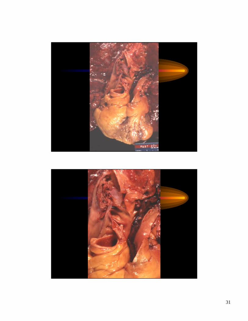
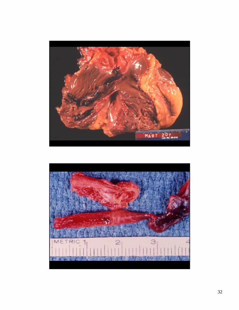
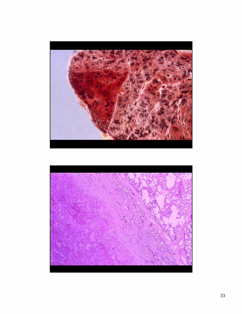
• Pulmonary embolism– Pathology
• The majority of PE arise from deep venous thrombosis of the lower extremity.
• Can occlude pulmonary artery at bifurcation (saddle embolus) or pulmonary artery branches
• Results in infarct only 10% of the time, and infarctions are hemorrhagic when they occur
• Small emboli organize and recanalize. Chronic PE can lead to pulmonary hypertension
31
32
33

34
Pulmonary Diseases: Structure-Function Correlation II
• Disease of the pulmonary vasculature– Pulmonary embolism– Pulmonary hypertension
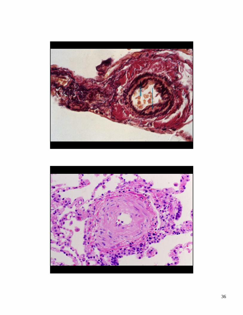
Disease of the pulmonary vasculature
• Pulmonary hypertension– Gross pathology
• Pulmonary artery atherosclerosis and dilatation• Right ventricular hypertrophy and dilatation,
depending on time course of the disease
– Microscopic pathology• Progressive abnormalities reflect severity and
duration of hypertension
35
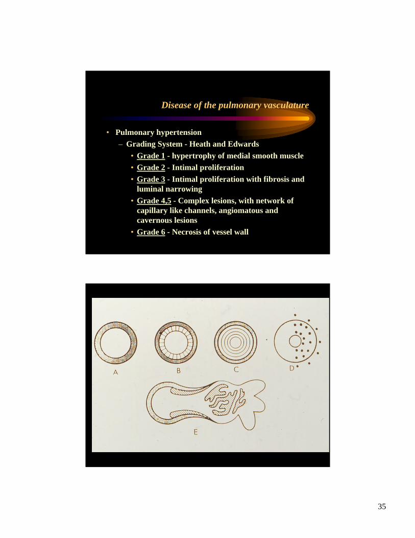
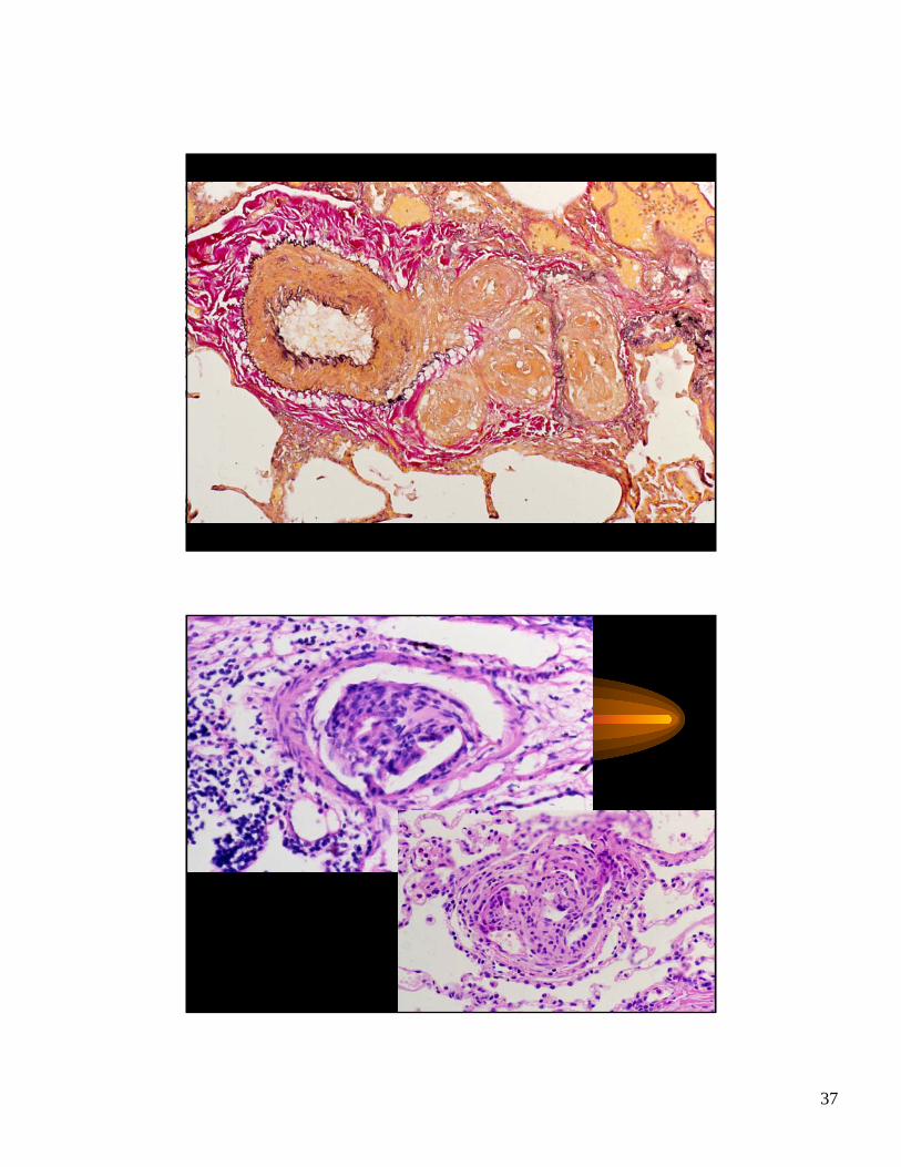
Disease of the pulmonary vasculature
• Pulmonary hypertension– Grading System - Heath and Edwards
• Grade 1 - hypertrophy of medial smooth muscle• Grade 2 - Intimal proliferation• Grade 3 - Intimal proliferation with fibrosis and
luminal narrowing• Grade 4,5 - Complex lesions, with network of
capillary like channels, angiomatous and cavernous lesions
• Grade 6 - Necrosis of vessel wall
36
37


![Interstitial lung disease (ILD), or diffuse parenchymal lung disease … · 2018-10-28 · Interstitial lung disease (ILD), or diffuse parenchymal lung disease (DPLD),[[1] is a group](https://img.dokumen.tips/doc/110x75/5e7d31d2ec5074254471c7d0/interstitial-lung-disease-ild-or-diffuse-parenchymal-lung-disease-2018-10-28.jpg)