Embed Size (px)
Citation preview

University of Nebraska - LincolnDigitalCommons@University of Nebraska - LincolnOther Publications in Zoonotics and WildlifeDisease Wildlife Disease and Zoonotics
December 1999
Parasites and Parisitic Diseases (Field Manual ofWildlife Diseases)Rebecca A. Cole
Milton Friend
Follow this and additional works at: http://digitalcommons.unl.edu/zoonoticspub
Part of the Veterinary Infectious Diseases Commons
This Article is brought to you for free and open access by the Wildlife Disease and Zoonotics at DigitalCommons@University of Nebraska - Lincoln. Ithas been accepted for inclusion in Other Publications in Zoonotics and Wildlife Disease by an authorized administrator ofDigitalCommons@University of Nebraska - Lincoln.
Cole, Rebecca A. and Friend, Milton , "Parasites and Parisitic Diseases (Field Manual of Wildlife Diseases)" (1999). Other Publicationsin Zoonotics and Wildlife Disease. 15.http://digitalcommons.unl.edu/zoonoticspub/15

Introduction to Parasitic Diseases 187
Section 5Parasites andParasitic DiseasesHemosporidiosis
Trichomoniasis
Intestinal Coccidiosis
Renal Coccidiosis
Sarcocystis
Eustrongylidosis
Tracheal Worms
Heartworm of Swans and Geese
Gizzard Worms
Acanthocephaliasis
Nasal Leeches
Miscellaneous Parasitic Diseases
Stained blood smear from a turkey infected with the parasite HaemoproteusmeleagridisPhoto by Carter Atkinson
188 Field Manual of Wildlife Diseases: Birds
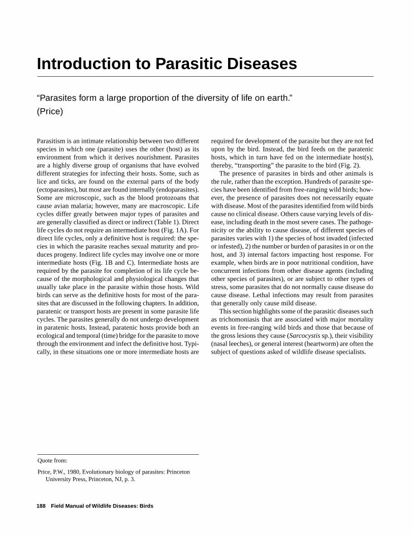
Introduction to Parasitic Diseases
“Parasites form a large proportion of the diversity of life on earth.”
(Price)
Quote from:
Price, P.W., 1980, Evolutionary biology of parasites: PrincetonUniversity Press, Princeton, NJ, p. 3.
Parasitism is an intimate relationship between two differentspecies in which one (parasite) uses the other (host) as itsenvironment from which it derives nourishment. Parasitesare a highly diverse group of organisms that have evolveddifferent strategies for infecting their hosts. Some, such aslice and ticks, are found on the external parts of the body(ectoparasites), but most are found internally (endoparasites).Some are microscopic, such as the blood protozoans thatcause avian malaria; however, many are macroscopic. Lifecycles differ greatly between major types of parasites andare generally classified as direct or indirect (Table 1). Directlife cycles do not require an intermediate host (Fig. 1A). Fordirect life cycles, only a definitive host is required: the spe-cies in which the parasite reaches sexual maturity and pro-duces progeny. Indirect life cycles may involve one or moreintermediate hosts (Fig. 1B and C). Intermediate hosts arerequired by the parasite for completion of its life cycle be-cause of the morphological and physiological changes thatusually take place in the parasite within those hosts. Wildbirds can serve as the definitive hosts for most of the para-sites that are discussed in the following chapters. In addition,paratenic or transport hosts are present in some parasite lifecycles. The parasites generally do not undergo developmentin paratenic hosts. Instead, paratenic hosts provide both anecological and temporal (time) bridge for the parasite to movethrough the environment and infect the definitive host. Typi-cally, in these situations one or more intermediate hosts are
required for development of the parasite but they are not fedupon by the bird. Instead, the bird feeds on the paratenichosts, which in turn have fed on the intermediate host(s),thereby, “transporting” the parasite to the bird (Fig. 2).
The presence of parasites in birds and other animals isthe rule, rather than the exception. Hundreds of parasite spe-cies have been identified from free-ranging wild birds; how-ever, the presence of parasites does not necessarily equatewith disease. Most of the parasites identified from wild birdscause no clinical disease. Others cause varying levels of dis-ease, including death in the most severe cases. The pathoge-nicity or the ability to cause disease, of different species ofparasites varies with 1) the species of host invaded (infectedor infested), 2) the number or burden of parasites in or on thehost, and 3) internal factors impacting host response. Forexample, when birds are in poor nutritional condition, haveconcurrent infections from other disease agents (includingother species of parasites), or are subject to other types ofstress, some parasites that do not normally cause disease docause disease. Lethal infections may result from parasitesthat generally only cause mild disease.
This section highlights some of the parasitic diseases suchas trichomoniasis that are associated with major mortalityevents in free-ranging wild birds and those that because ofthe gross lesions they cause (Sarcocystis sp.), their visibility(nasal leeches), or general interest (heartworm) are often thesubject of questions asked of wildlife disease specialists.
Introduction to Parasitic Diseases 189
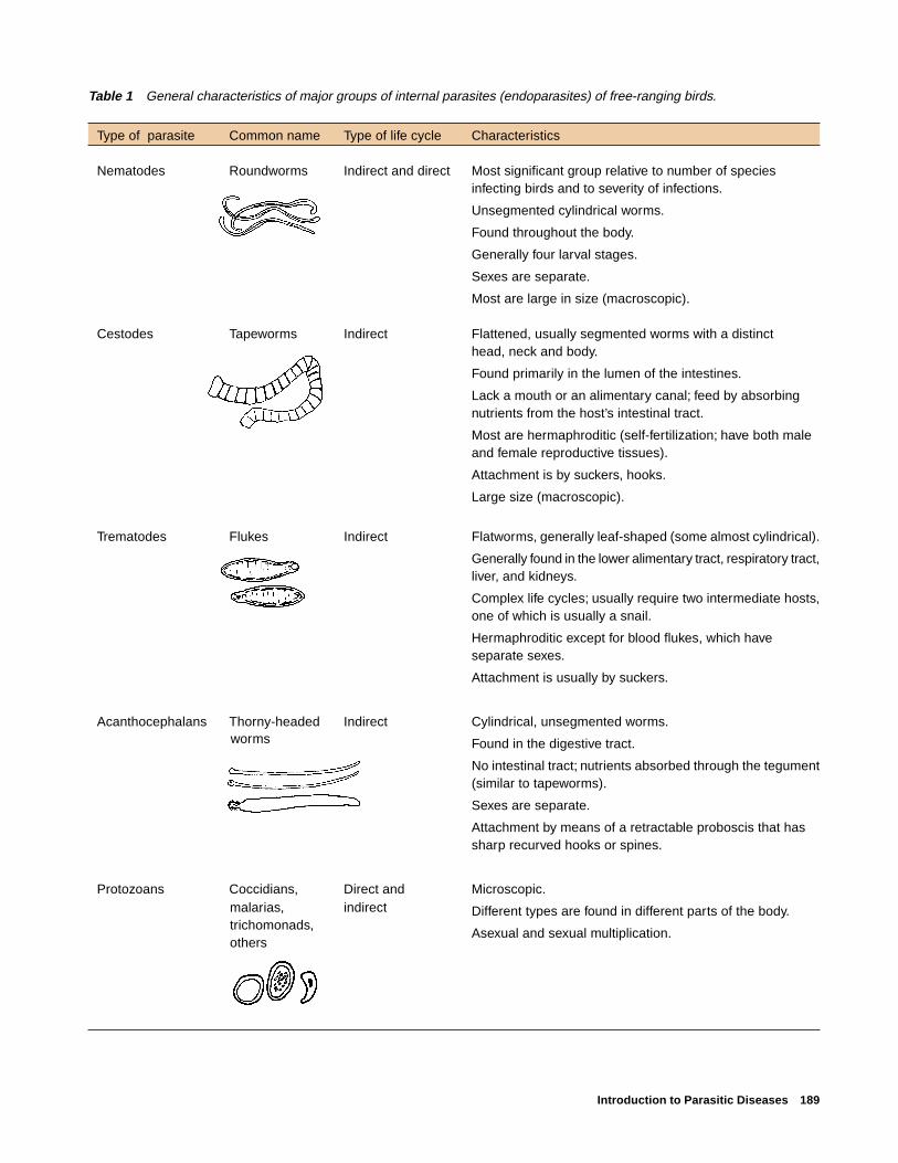
Table 1 General characteristics of major groups of internal parasites (endoparasites) of free-ranging birds.
Type of parasite Common name Type of life cycle Characteristics
Nematodes Roundworms Indirect and direct Most significant group relative to number of speciesinfecting birds and to severity of infections.
Unsegmented cylindrical worms.
Found throughout the body.
Generally four larval stages.
Sexes are separate.
Most are large in size (macroscopic).
Cestodes Tapeworms Indirect Flattened, usually segmented worms with a distincthead, neck and body.
Found primarily in the lumen of the intestines.
Lack a mouth or an alimentary canal; feed by absorbingnutrients from the host’s intestinal tract.
Most are hermaphroditic (self-fertilization; have both maleand female reproductive tissues).
Attachment is by suckers, hooks.
Large size (macroscopic).
Trematodes Flukes Indirect Flatworms, generally leaf-shaped (some almost cylindrical).
Generally found in the lower alimentary tract, respiratory tract,liver, and kidneys.
Complex life cycles; usually require two intermediate hosts,one of which is usually a snail.
Hermaphroditic except for blood flukes, which haveseparate sexes.
Attachment is usually by suckers.
Acanthocephalans Thorny-headed Indirect Cylindrical, unsegmented worms.
Found in the digestive tract.
No intestinal tract; nutrients absorbed through the tegument(similar to tapeworms).
Sexes are separate.
Attachment by means of a retractable proboscis that hassharp recurved hooks or spines.
Protozoans Coccidians, Direct and Microscopic.
Different types are found in different parts of the body.
Asexual and sexual multiplication.
worms
malarias,trichomonads,others
indirect

190 Field Manual of Wildlife Diseases: Birds
Figure 1 Examples of (A) direct, (B)simple indirect, and (C) complex indi-rect parasite life cycles.
A
B
Infected bird
Eggs mature in environment and become infective
Infective embryonated eggs are eaten by bird while feeding
Bird sheds parasiteeggs into the environ-ment in feces
Infected bird
Bird eats sowbug and becomes infected
Sowbug eats eggs of parasite
Eggs hatch in sowbug and infective larvae develop within sowbug
Bird sheds parasiteeggs into the environ-ment in feces

Introduction to Parasitic Diseases 191
CInfected bird
Parasite eggs are passed in feces and hatch in water
Larvae (miracidium) swims to a snail and penetrates it, undergoing further larval developmentwithin the host
New larval stage emerges from snail (cercaria) andswims to new host where itpenetrates and encysts
First intermediate host
Second intermediate host
Other bird species eat secondintermediate host andbecome infected

192 Field Manual of Wildlife Diseases: Birds
Figure 2 Hypothetical parasite life cycle illustrating the role of paratenic (transport) hosts.
Infected bird
Bird sheds parasiteeggs into the environ-ment in feces
Birds feed on fish and become infected to complete life cycle
Second intermediate hostAmphipod is eaten by amphibian where infective stages of larvae develop
First intermediate hostEggs eaten by amphipod where first- and second- stage larvae develop
Paratenic hostFish eats the amphibian and larvae encyst in body of fish. No further development of the parasite
Hemosporidiosis 193
Chapter 24
Hemosporidiosis
SynonymsAvian malaria
CauseHemosporidia are microscopic, intracellular parasitic pro-
tozoans found within the blood cells and tissues of their avianhosts. Three closely related genera, Plasmodium, Haemopro-teus, and Leucocytozoon, are commonly found in wild birds.Infections in highly susceptible species and age classes mayresult in death.
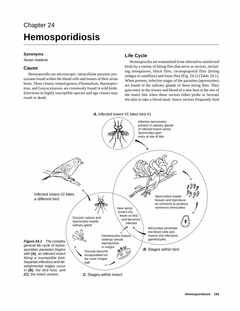
Life CycleHemosporidia are transmitted from infected to uninfected
birds by a variety of biting flies that serve as vectors, includ-ing mosquitoes, black flies, ceratopogonid flies (bitingmidges or sandflies) and louse flies (Fig. 24.1) (Table 24.1).When present, infective stages of the parasites (sporozoites)are found in the salivary glands of these biting flies. Theygain entry to the tissues and blood of a new host at the site ofthe insect bite when these vectors either probe or laceratethe skin to take a blood meal. Insect vectors frequently feed
Figure 24.1 The complexgeneral life cycle of hemo-sporidian parasites beginswith (A), an infected insectbiting a susceptible bird.Separate infectious and de-velopmental stages occurin (B), the bird host, and(C), the insect vectors.
A. Infected insect #1 bites bird #1
Infected insect #2 bites a different bird
C. Stages within insect
Infective sporozoitespresent in salivary glands of infected insect vector.Sporozoites gain entry at site of bite
Sporozoites invade tissues and reproduce as schizonts to produce numerous merozoites
Merozoites penetratered blood cells andmature into infectiousgametocytes
New vector (insect #2) feeds on bird and becomes infected
Gametocytes mature, undergo sexual reproductionin midgut
Oocysts become encapsulated on the outer midgut wall
Oocysts rupture andsporozoites invadesalivary gland
B. Stages within bird

194 Field Manual of Wildlife Diseases: Birds
on exposed flesh around the eyes (Fig. 24.2), the beak, andon the legs and feet, although black flies, ceratopogonid flies,and louse flies can crawl beneath the bird’s feathers to reachthe skin surface. Immediately after they infect a bird, sporo-zoites invade the tissues and reproduce for one or more gen-erations before they become merozoites. Merozoites penetratethe red blood cells and become mature, infectious gameto-cytes. The cycle is completed when the gametocytes in thecirculating blood cells of the host bird are ingested by an-other blood-sucking insect, where they undergo both sexualand asexual reproduction to produce large numbers of sporo-zoites. These invade the salivary glands of the vector and aretransmitted to a new host bird during the vector’s next bloodmeal.
Species AffectedThe avian hemosporidia are cosmopolitan parasites of
birds, and they have been found in 68 percent of the more
than 3,800 species of birds that have been examined. Mem-bers of some avian families appear to be more susceptiblethan others. For example, ducks, geese and swans are com-monly infected with species of Haemoproteus, Leucocyto-zoon, and Plasmodium, and more than 75 percent of water-fowl species that were examined were hosts for one or moreof these parasites. Wild turkeys in the eastern United Statesare also commonly infected by these parasites. Pigeons anddoves have similar high rates of infection, but members ofother families, such as migratory shorebirds, are less fre-quently parasitized.
Differences in the prevalence, geographic distribution, andhost range of hemosporidia are associated with habitat pref-erences of the bird hosts, the abundance and feeding habitswithin those habitats of suitable insect vectors, and innatephysiological differences that make some avian hosts moresusceptible than others. For example, some species of blackflies (Simulium sp.) prefer to feed on waterfowl within a lim-
Table 24.1 Avian hemosporidia parasites and their documented vectors.
Parasite Vector type Common name
Haemoproteus Ceratopogonidae Punkies, no-see-ums, sand flies(Culicoides sp.)Hippoboscidae Hippoboscid or louse flies(Ornithomyia sp.)
Plasmodium Culicidae Mosquitoes(Culex, Aedes sp.)
Leucocytozoon Simulidae Black flies(Simulium sp.)
Figure 24.2 A Culex mosquito feeding on the unfeatheredarea surrounding the eye of an apapane, a native Hawaiianhoneycreeper.
Pho
to b
y Ja
ck J
effre
y, U
.S. F
ish
and
Wild
life
Ser
vice

Hemosporidiosis 195
ited distance of the shoreline. Ducks and geese that spendmore of their time in this zone will be more likely to be ex-posed to bites that carry infective stages of Leucocytozoonsimondi. Biting midges or no-see-ums (Culicoides sp.) thattransmit species of Haemoproteus are more active at dusk inthe forest canopy. Birds that roost here, for example, increasetheir chances for being infected with this parasite. Finally,some avian hosts are more susceptible to hemosporidian para-sites than others, but the physiological basis for this is stillpoorly understood.
Species of Plasmodium and Leucocytozoon are capableof causing severe anemia, weight loss, and death in suscep-tible birds. Young birds are more susceptible than adults, andthe most serious mortality generally occurs within the firstfew weeks of hatching. This is also the time of year whenincreasing temperatures favor the growth of the populationsof insect vectors that transmit hemosporidia. Major outbreaksof L. simondi that caused high mortality in ducks and geesein Michigan and subarctic Canada have been documented.Species of Haemoproteus are generally believed to be lesspathogenic, with only scattered reports of natural mortalityin wild birds.
Penguins and native Hawaiian forest birds are highly sus-ceptible to Plasmodium relictum, a common parasite of song-birds that is transmitted by Culex mosquitoes. This parasitecauses high mortality in both captive and wild populationsof these hosts, and it is a major factor in the decline of nativeforest birds in the Hawaiian Islands.
DistributionSpecies of Plasmodium, Haemoproteus, and Leucocyto-
zoon have been reported from most parts of the world withthe exception of Antarctica, where cold temperatures pre-vent the occurrence of suitable insect vectors. Studies of thedistribution of hemosporidia in North America have shownthat areas of active transmission of the parasites coincidewith the geographic distribution of their vectors. Leuco-cytozoon is most common in mountainous areas of Alaskaand the Pacific Northwest where abundant fast-movingstreams create suitable habitat for aquatic black fly larvae.Species of Haemoproteus and Plasmodium are more evenlydistributed across the continent because their ceratopogonidand mosquito vectors are less dependent on the presence offlowing water for larval development. Migratory birds maywinter in habitats that lack suitable vectors; therefore, thesimple presence of infected birds may not be evidence thatthe parasites are being transmitted to birds at the winteringgrounds.
SeasonalityInfections with Plasmodium, Haemoproteus, and Leuco-
cytozoon are seasonal because transmission depends uponthe availability of vector populations. In temperate NorthAmerica, most birds become infected with hemosporidia
during the spring when conditions for transmission becomeoptimal. Some of these conditions include the onset ofwarmer weather; increases in vector populations; the reap-pearance or relapse of chronic, low-level infections in adultbirds; and the hatching and fledging of susceptible,nonimmune juvenile birds. In warmer parts of the UnitedStates, these parasites may be transmitted at other times ofthe year. In Hawaii, P. relictum in forest bird populationsmay be transmitted throughout the year in warm low-eleva-tion forests, but transmission is more seasonal at elevationsabove 3,000 ft. where cooler winter temperatures limit mos-quito populations.
Field SignsBirds with acute infections of Plasmodium, Haemopro-
teus, and Leucocytozoon, may exhibit similar signs in thefield. These include emaciation, loss of appetite, listlessness,difficulty in breathing, and weakness and lameness in one orboth legs. Survivors develop persistent, low-level infectionsin the blood and tissues that stimulate immunity to reinfec-tion. These survivors do not exhibit any signs of disease, butthey serve as reservoirs of infection, allowing the parasitesto survive droughts and cold winter weather when vectorpopulations have died off.
Gross LesionsGross lesions associated with acute infections include
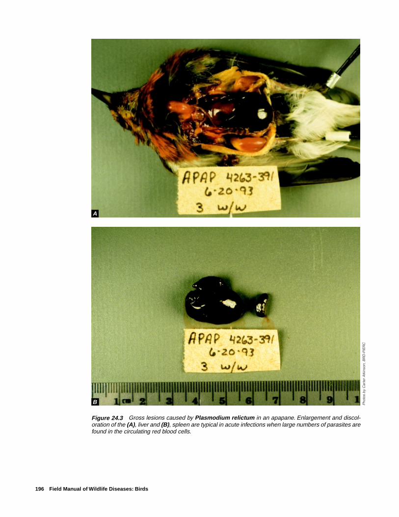
enlargement of the liver and spleen (Fig. 24.3) and the ap-pearance of thin and watery blood as a result of infected bloodcells being destroyed and removed from circulation (Fig.24.4). In Plasmodium and Haemoproteus infections, para-sites within the red blood cells produce an insoluble blackpigment called hemozoin when they digest the host’s oxygen-bearing, iron-laden red blood cell protein or hemoglobin. Thehemozoin is deposited extensively in the host’s spleen andliver tissue as the host’s immune system responds to the in-fection. In very heavy infections, the kidneys may also beaffected. These organs typically appear chocolate brown orblack at necropsy and they may be two or more times theirnormal size (Fig. 24.3). Hemozoin pigment is not producedin Leucocytozoon infections; therefore, organs will not be asdiscolored and dark at necropsy, but they will still appearenlarged. Some species of Haemoproteus form large, cyst-like bodies in muscle tissue that superficially resemble tis-sue cysts produced by species of Sarcocystis (Fig. 24.5).
DiagnosisDefinitive diagnosis of hemosporidian infections is de-
pendent on microscopic examination of a stained blood smearor on an organ impression smear to detect the presence andform of the parasites within the red blood cells (Figs. 24.6,7, 8). Species of Leucocytozoon frequently produce dramaticchanges in the host’s cell structure (Fig. 24.6). Parasitizedred blood cells are often enlarged and elongated so that they
196 Field Manual of Wildlife Diseases: Birds
Figure 24.3 Gross lesions caused by Plasmodium relictum in an apapane. Enlargement and discol-oration of the (A), liver and (B), spleen are typical in acute infections when large numbers of parasites arefound in the circulating red blood cells.
Pho
tos
by
Car
ter
Atk
inso
n, B
RD
-PIE
RC
A
B

Hemosporidiosis 197
Figure 24.4 Thin and watery blood from anapapane infected with Plasmodium relictum before(left) and after (right) centrifugation. In uninfectedsongbirds, approximately half of the blood volume isoccupied by red blood cells. Note that most of theblood cells have been destroyed by the parasite(right).
Figure 24.5 Pectoral muscles of a turkey infected with Haemoproteus meleagridis. Note the whitestreaks and bloody spots in the muscle tissue of this bird (arrows). The tissue stages of this hemosporidianform large, cystlike bodies that may superficially resemble those caused by species of Sarcocystis.
Pho
to b
y C
arte
r A
tkin
son,
BR
D-P
IER
C
Pho
to b
y C
arte
r A
tkin
son,
BR
D-P
IER
C
➡
➡
➡

198 Field Manual of Wildlife Diseases: Birds
Figure 24.8 Stained blood smear from an apapane in-fected with Plasmodium relictum. Some red blood cellscontain multinucleated, asexually-reproducing stages ofthe parasite called schizonts (S). These are diagnosticfor Plasmodium infections and contain one or more cen-trally-located pigment granules (arrows).
Figure 24.6 Stained blood smear from a turkey infectedwith Leucocytozoon smithi. This parasite causes en-largement and distortion of the infected blood cell. Thered blood cell nucleus (N) is divided in two halves that layon either side of the parasite (P). The membrane of theinfected cell is stretched into two hornlike points (arrows).
Figure 24.7 Stained blood smear from a turkey infectedwith Haemoproteus meleagridis. Gametocytes (G) con-tain a single pink-staining nucleus and contain black orgolden brown pigment granules (arrows).
Pho
to b
y C
arte
r A
tkin
son,
BR
D-P
IER
CP
hoto
by
Car
ter
Atk
inso
n, B
RD
-PIE
RC
Pho
to b
y C
arte
r A
tkin
son,
BR
D-P
IER
C
NP
➡
➡
G
➡
➡
S
S
➡
➡
Hemosporidiosis 199
form a pair of horn-like extensions from either end of thecell. Species of Plasmodium and Haemoproteus producefewer changes in their host’s red blood cells, but these para-sites may cause slight enlargement of infected host cells anddisplacement of the red blood cell nucleus to one side (Figs.24.7, 8). Unlike Leucocytozoon, Plasmodium and Haemo-proteus produce golden brown or black deposits of hemozoinpigment in the parasite cell (Figs. 24.7, 8). Further differen-tiation of Plasmodium from Haemoproteus may be difficult.Diagnosis of a Plasmodium infection is dependent on de-tecting the presence of asexually reproducing stages of itslife cycle (schizonts) in the red blood cells of the infectedhost (Fig. 24.8).
ControlControl of the avian hemosporidia is dependent on re-
ducing transmission from infected birds to healthy birdsthrough reduction or elimination of vector populations. Manyof the same techniques that were developed for control ofvector-transmitted human diseases can be used effectively,but few agencies have the resources or manpower to applythem over large areas. Most techniques rely on habitat man-agement to reduce vector breeding sites or depend on theapplication of pesticides that affect larval or adult vectors toreduce vector populations. Large-scale treatment of infectedsurvivor birds could prevent disease outbreaks by reducingsources of infection, but the logistics and practicality of treat-ing sufficient numbers of birds to interrupt transmission areprohibitive. Although some experimental vaccines for theseparasites have been developed, none are currently availablefor general use.
Human Health ConsiderationsThe avian hemosporidia are closely related to the malarial
parasites of humans, but are not capable of infecting people.
Carter T. AtkinsonPacific Island Ecosystems Research CenterKilauea Field Station
Supplementary ReadingAtkinson, C.T., 1991, Vectors, epizootiology, and pathogenicity of
avian species of Haemoproteus: Bulletin of the Society forVector Ecology, vol.16, p. 109–126.
Atkinson, C.T., and van Riper, C., III, 1991, Pathogenicity andepizootiology of avian haematozoa: Plasmodium, Leucocyto-zoon, and Haemoproteus, in Loye, J.E., and Zuk, M., eds.,Bird-parasite interactions, Ecology, evolution, and behavior:New York, Oxford University Press, p. 19–48.
Bennett, G.F., Whiteway, M., and Woodworth-Lynas, C.B., 1982,Host-parasite catalogue of the avian haematozoa: MemorialUniversity of Newfoundland Occasional Papers in BiologyNumber 5, p. 243.
Greiner, E.C., Bennett, G.F., White, E.M., and Coombs, R.F.,1975, Distribution of the avian hematozoa of North America,v. 53, p. 1,762–1,787.
Greiner, E.C., 1991, Leucocytozoonosis in waterfowl and wildgalliform birds: Bulletin of the Society for Vector Ecology,v. 16, p. 84–93.

200 Field Manual of Wildlife Diseases: Birds

Trichomoniasis 201
Chapter 25
Trichomoniasis
SynonymsCanker (doves and pigeons), frounce (raptors), aviantrichomoniasis
CauseAvian trichomoniasis is caused by a single celled proto-
zoan, Trichomonas gallinae. Avirulent T. gallinae strains thatdo not cause disease and highly virulent strains are found innature and circulate within bird populations. The factors thatmake a strain virulent are not known, but they are thought tobe controlled genetically within the parasite. Similarly, thereasons why an avirulent or a virulent form of the parasite isfound within a bird population at any period of time alsoremain unknown. Virulent strains of T. gallinae have causedmajor mortality events or epizootics in doves and pigeons in
addition to less visible, chronic losses (Table 25.1). Infec-tion typically involves the upper digestive tract of doves andpigeons but other species have also been infected (Fig. 25.1).
Trichomoniasis in doves and pigeons, but not in otherspecies, is generally confined to young birds. The parasitewas introduced to the U.S. with the introduction of pigeonsand doves brought by European settlers. It has been reportedthat 80 to 90 percent of adult pigeons are infected, but theyshow no clinical signs of disease. It is speculated that mostof these birds became immune as a result of exposure to aviru-lent strains of the parasite or because they survived mild in-fections. In pigeons and mourning doves, the parasites aretransmitted from the adults to the squabs in the pigeon milkproduced in the crop of the adult. Squabs usually becomeinfected with the first feeding of pigeon milk, which is gen-
Table 25.1 Examples of wild bird mortalities reported in the scientific literature due to trichomoniasis.
Year Magnitude Geographic area Comments
1949–51 Tens of thousands of Southeastern United Trichomoniasis broke out inmourning doves States virtually all States in the
region; the magnitude oflosses focused attention onthe devastation that could becaused by this disease andstimulated research on theecology of this disease.
1950–51 25,000 to 50,000 Alabama Breeding birds were the focusmourning doves each of infection; mortality wasyear thought to have been grossly
underestimated.
1972 Several hundred Nebraska Railroad yards and a grainelevator were focal points ofinfection; birds fed on spilledgrain.
1985 Approximately 800 New Mexico Losses at birdfeeders near Lasmourning doves Cruces.
1988 At least 16,000 California First major epizootic ofband-tailed pigeons trichomoniasis in this species.
1991 Approximately 500 North Carolina —mourning doves

202 Field Manual of Wildlife Diseases: Birds
erally within minutes after hatching. The resulting infectionmay range from asymptomatic or mild disease to a rapidlyfatal course resulting in death within 4–18 days after infec-tion. Other modes for infection are through feed, perhapscontaminated drinking water, and feeding on infected birds(Fig. 25.2).
There is no cyst or resistant stage in the parasite’s lifecycle; therefore, infection must be passed directly from onebird to another, in contaminated feed or water. Feed and waterare contaminated when trichomonads move from the mouthof infected birds, not from their feces. Lesions in the mouthor the esophagus or both of an infected bird (see below) of-ten prevent the passage of ingested grain seeds and cause thebird to regurgitate contaminated food items. Water becomescontaminated by contact with the contaminated bill andmouth. Pigeons that feed among domestic poultry are oftenblamed for contaminating feed and water and passing thedisease to the poultry. Similar transmission has been associ-ated with dove mortality at grain elevators and at birdfeed-ers. Doves and pigeons cross-feed and bill during courtship,and this behavior facilitates direct transmission as does theconsumption of infected birds by raptors. It has been reportedthat some moist grains can maintain viable T. gallinae for atleast 5 days and that parasite survival in water can rangefrom 20 minutes to several hours. These conditions are ad-equate for disease transmission at birdfeeders and waterersbecause of the gregarious habits of doves and pigeons.
Species AffectedTrichomoniasis is considered by many avian disease spe-
cialists to be the most important disease of mourning dovesin North America. Band-tailed pigeons have also sufferedlarge-scale losses from trichomoniasis. This disease has beenreported as a cause of mortality in birds of prey for hundredsof years prior to the causative organism being identified.Songbirds are less commonly reported to be infected, but T.gallinae is reported to be the most important trichomonad ofcaged birds; it is often responsible for epizootics among cap-tive collections. Domestic turkeys and chickens also becomeinfected.
DistributionIt is likely that T. gallinae is found wherever domestic
pigeons and mourning doves are found. Disease in free-ranging wild birds is grossly underreported. Outbreaks atbirdfeeding stations and similar locations reported to theNational Wildlife Health Center have occurred from coast-to-coast within the United States (Fig. 25.3).
SeasonalityEpizootics due to T. gallinae can happen yearround, but
most outbreaks have been reported during late spring, sum-mer, and fall.
Field SignsBecause oral lesions often affect the ability of the bird to
feed, infected birds lose weight, appear listless, and standgrouped together. These birds often appear ruffled. Caseousor cheesy, yellowish lesions may be seen around the beak oreyes of mourning doves and the face may appear “puffy”and distended (Fig. 25.4). Severely infected pigeons may fallover when they are forced to move.
Gross LesionsThe severity and appearance of lesions varies with the
virulence of the strain of the parasite, the stage of infection,and the age of the bird. The most visible lesions from mildlypathogenic strains may simply appear as excess salivationand inflammation of the mucosa or lining of the mouth andthroat. Early oral lesions appear as small, well defined, creamto yellowish spots on the mucosal surface (Fig. 25.5A). Asthe disease progresses the lesions become larger, thicker, and
Pigeons/doves
Falcons/hawks
Owls
Songbirds
Pheasants/quail
Waterfowl
FrequentCommon
OccasionalInfrequentRare
Figure 25.1 Relative frequency of trichomoniasis in free-ranging birds.

Trichomoniasis 203
Birds in nest being fed by dove or pigeon
Billing/feeding courtship
Wild raptor catching or eating an infected dove
Captive raptor being provided infected dove or pigeon
Birds at birdfeederand birdbath
Figure 25.2 Transmission of trichomoniasis.

204 Field Manual of Wildlife Diseases: Birds
EXPLANATIONTrichomoniasis outbreak sites, 1983–97*
Outbreak site
* Outbreaks of trichomoniasis involve doves and pigeons with mortality ranging from tens to hundreds.
Figure 25.3 Locations of outbreaks of trichomoniasis in free-ranging birds,January 1983 through March 1997.
Figure 25.4 Mourning doves at a backyard waterbath. Note the puffy appearance(arrow) of the face of a T. gallinae infected dove.
Pho
to b
y U
.S. F
ish
and
Wild
life
Ser
vice
➡
Trichomoniasis 205
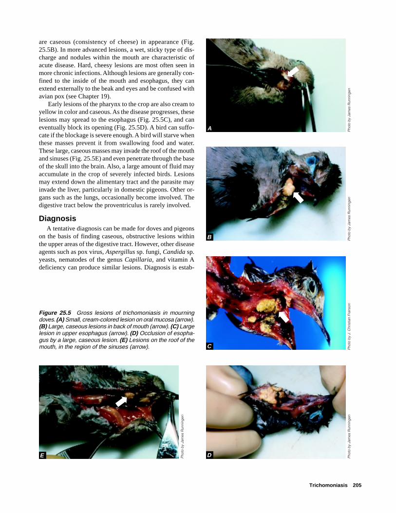
are caseous (consistency of cheese) in appearance (Fig.25.5B). In more advanced lesions, a wet, sticky type of dis-charge and nodules within the mouth are characteristic ofacute disease. Hard, cheesy lesions are most often seen inmore chronic infections. Although lesions are generally con-fined to the inside of the mouth and esophagus, they canextend externally to the beak and eyes and be confused withavian pox (see Chapter 19).
Early lesions of the pharynx to the crop are also cream toyellow in color and caseous. As the disease progresses, theselesions may spread to the esophagus (Fig. 25.5C), and caneventually block its opening (Fig. 25.5D). A bird can suffo-cate if the blockage is severe enough. A bird will starve whenthese masses prevent it from swallowing food and water.These large, caseous masses may invade the roof of the mouthand sinuses (Fig. 25.5E) and even penetrate through the baseof the skull into the brain. Also, a large amount of fluid mayaccumulate in the crop of severely infected birds. Lesionsmay extend down the alimentary tract and the parasite mayinvade the liver, particularly in domestic pigeons. Other or-gans such as the lungs, occasionally become involved. Thedigestive tract below the proventriculus is rarely involved.
DiagnosisA tentative diagnosis can be made for doves and pigeons
on the basis of finding caseous, obstructive lesions withinthe upper areas of the digestive tract. However, other diseaseagents such as pox virus, Aspergillus sp. fungi, Candida sp.yeasts, nematodes of the genus Capillaria, and vitamin Adeficiency can produce similar lesions. Diagnosis is estab-
Figure 25.5 Gross lesions of trichomoniasis in mourningdoves. (A) Small, cream-colored lesion on oral mucosa (arrow).(B) Large, caseous lesions in back of mouth (arrow). (C) Largelesion in upper esophagus (arrow). (D) Occlusion of esopha-gus by a large, caseous lesion. (E) Lesions on the roof of themouth, in the region of the sinuses (arrow). P
hoto
by
J. C
hris
tian
Fran
son
Pho
to b
y Ja
mes
Run
ning
enP
hoto
by
Jam
es R
unni
ngen
Pho
to b
y Ja
mes
Run
ning
en
Pho
to b
y Ja
mes
Run
ning
en
A
B
C
DE➡
➡➡
➡

206 Field Manual of Wildlife Diseases: Birds
lished by finding the trichomonads in the saliva or smears ofthe caseous lesions of infected birds. Specimens are best takenfrom sick birds, or from recently dead birds that are keptchilled and reach the diagnostic laboratory within 48 hoursafter death. Samples of tissues with lesions preserved in 10percent buffered formalin or frozen whole carcasses can beused if fresh carcasses cannot be provided.
ControlThe removal of infected birds is recommended for com-
bating trichomoniasis in poultry and captive pigeons and incaptive collections of wild birds. The focus in both instancesis on birds that harbor virulent strains of the parasite. Elimi-nation of infection from adult birds by drug treatment hasalso been recommended, but this is not a practical approachfor wild birds. Prevention of the build-up of large concentra-tions of doves at birdfeeders and artificial watering areas isrecommended to minimize disease transmission in the wild.Stock tanks, livestock feedlots, grain storage facilities andclusters of urban birdfeeders should be targeted for diseaseprevention activities. Although the environmental persistencefor T. gallinae is rather limited, contaminated feed is sus-pected as a significant source of disease transmission. There-fore, fresh feed should be placed in feeders daily, if it is prac-tical. Platforms and other surfaces where feed may collect,including the area under feeders, should be frequently de-contaminated with 10 percent solution of household bleachin water, preferably just prior to placing clean feed in the
feeder. Pigeons and doves are high risk food sources for birdsof prey; therefore, before they are fed to raptors, pigeonsand doves should be inspected first and found to be free oftrichomoniasis or other infectious diseases.
Human Health ConsiderationsNone. T. gallinae has not been reported to infect humans.
Rebecca A. Cole
Supplementary ReadingConti, J.A., 1993, Diseases, parasites, and contaminants, in
Baskett, T.S., and others, eds., Ecology and management of themourning dove: Harrisburg, Pa., Stackpole Books, p. 205–224.
Levine, N.D., 1985, Flagellates: the trichomonads in veterinaryprotozoology: Ames, Iowa, Iowa State University Press, p. 72–74.
Pokras, M.A., Wheeldon, E.B., and Sedgwick, C.J., 1993, Raptorbiomedicine, in Redig, P.T. and others, eds., Trichomoniasis inowls: report on a number of clinical cases and a survey of theliterature: Minneapolis, Minn., University Minnesota Press,p. 88–91.
Rupier, D.J., and W.M. Harmon, 1988, Prevalence of Trichomonasgallinae in central California mourning doves: California Fishand Game, v. 74, no. 4, p. 471–473.
Stabler, R.M., 1951, A survey of Colorado band-tailed Pigeons,mourning doves, and wild common pigeons for Trichomonasgallinae: Journal of Parasitology, v. 37, p. 471–473.

Intestinal Coccidiosis 207
Chapter 26
Intestinal Coccidiosis
SynonymsCoccidiosis, coccidiasis
CauseCoccidia are a complex and diverse group of protozoan
(single-celled organisms) parasites; the coccidia group con-tains many species, most of which do not cause clinical dis-ease. In birds, most disease-causing or pathogenic forms ofcoccidia parasites belong to the genus Eimeria. Coccidia usu-ally invade the intestinal tract, but some invade other organs,such as the liver and kidney (see Chapter 27).
Clinical illness caused by infection with these parasites isreferred to as coccidiosis, but their presence without diseaseis called coccidiasis. In most cases, a bird that is infected bycoccidia will develop immunity from disease and it willrecover unless it is reinfected. The occurrence of diseasedepends, in part, upon the number of host cells that aredestroyed by the juvenile form of the parasite, and this ismoderated by many factors. Severely infected birds may dievery quickly. Often, tissue damage to the bird’s intestine re-sults in interrupted feeding; disruption of digestive processesor nutrient absorption; dehydration; anemia; and increasedsusceptibility to other disease agents. In cranes, coccidia thatnormally inhabit the intestine sometimes become widely dis-tributed throughout the body. The resulting disease, dissemi-nated visceral coccidiosis (DVC) of cranes, is characterizedby nodules, or granulomas, on the surface of organs and tis-sues that contain developmental stages of the parasite.
Collectively, coccidia are important parasites of domes-tic animals, but, because each coccidia species has a prefer-ence for parasitizing a particular bird species and because ofthe self-limiting nature of most infections, coccidiosis in free-ranging birds has not been of great concern. However, habi-tat losses that concentrate bird populations and the increas-ing numbers of captive-reared birds that are released into thewild enhance the potential for problems with coccidiosis.
Life CycleMost intestinal coccidia have a complex but direct life
cycle in which the infective forms of the parasite invade asingle host animal for development to sexual maturity; thelife cycle is completed in 1–2 weeks (Fig. 26.1). A maturefemale parasite in the intestine of an infected host bird pro-duces noninfective, embryonated eggs or oocysts, which arepassed into the environment in the feces of the host bird. Theoocysts quickly develop into an infective form while theyare in the environment. An uninfected bird ingests the infec-tive oocysts while it is eating or drinking, and the infective
oocysts invade the bird’s intestine. Within the intestine, theoocysts may or may not undergo several stages of develop-ment, depending on the parasite species, before they becomesexually mature male and female parasites. The complex lifecycle for Eimeria (Fig. 26.2) illustrates the exponential rateof infection and destruction of the intestinal epithelial cells,which are the cells that provide the covering of the intestinallining. The mature female parasites release noninfective oo-cysts to the environment, and, thus, the cycle begins anew.
Species AffectedMany animal species, including a wide variety of birds
(Table 26.1) may harbor coccidia. Although disease is notcommon in free-ranging wild birds, several epizootics dueto E. aythyae have been reported among lesser scaup in theUnited States. During those events, predominantly femaleshave died, which suggests that female lesser scaup may bemore susceptible to the disease than male lesser scaup. Le-sions of DVC were first seen in captive sandhill cranes in thelate 1970s. Since then, mortality of captive sandhill andwhooping cranes has been attributed to DVC, and the dis-ease has been found in wild sandhill cranes, including theendangered Mississippi sandhill crane.
Characteristics of Intestinal Coccidiosis
All domestic birds carry more than one species of coc-cidia, and pure infections with a single species are rare.
Different coccidia species are usually found in a spe-cific location within the intestinal tract of the host bird.
After initial exposure to the parasite, the host bird mayquickly develop immunity to it but immunity is notabsolute. A bird can be reinfected by the same or adifferent species of the parasite.
Infections do not generally cause a problem of free-ranging birds; instead, coccidiosis is considered a dis-ease of monoculture and of the raising of birds in con-finement.

208 Field Manual of Wildlife Diseases: Birds
Figure 26.1 Direct life cycle of Eimeria infection in birds.
DistributionCoccidia are found worldwide. The few reported outbreaks
of coccidiosis in free-ranging waterfowl have all occurred inthe Midwestern United States (Fig. 26.3). Recurrent epizoot-ics have broken out at a single reservoir in eastern Nebraska,and coccidiosis is also believed to be the cause of waterfowldie-offs in Wisconsin, North Dakota, Illinois, and Iowa. DVChas been found in migratory sandhill cranes at several loca-tions, and it is a recurring problem in the only free-rangingpopulation of the nonmigratory Mississippi sandhill crane.These birds reside at the Mississippi Sandhill Crane NationalWildlife Refuge in Mississippi.
SeasonalityBirds may be infected with coccidia at any time. Although
little is known about the conditions that may lead to the de-
velopment of clinical disease in wild birds, birds may be-come diseased more frequently during periods of stress. Mostepizootics of intestinal coccidiosis in waterfowl in the Up-per Midwest have broken out in early spring, during a stress-ful staging period of spring migration. Mississippi sandhillcranes also die from DVC most frequently during the spring.
Field SignsField signs for free-ranging wild birds have not been re-
ported. Nonspecific clinical signs reported for captive birdsinclude inactivity, anaemia, weight loss, general unthriftyappearance, and a watery diarrhea that may be greenish orbloody. Tremors, convulsions, and lameness are also occa-sionally seen. Rapid weight loss may lead to emaciation anddehydration followed by death. Young birds that survive se-vere infections may suffer retardation of growth.
lnfected bird
Bird sheds noninfectiveoocysts (eggs) with feces into the environ-ment
Oocysts sporulate within 48 hours and become infective
Susceptible bird ingests infective oocysts while feeding/drinking
Parasite invades intestinal tissue

Intestinal Coccidiosis 209
A. Noninfective parasite oocysts (eggs) containing a single cell referred to as the sporont are passed via feces into the environment.
B. Oocysts become infective after 2 days in the environ-ment at ordinary tempera-tures through sporolation (sporogony), which is a developmental process that results in the sporont dividing and forming four sporocysts each contain-ing two infective sporo-zoites.
C. Infective oocysts are in-gested by birds in conta-minated feed, water, soil, or other ingesta.
D. The oocyst wall breaks within the gizzard of the bird and releases the sporocysts.
E. The sporozoites escape from the sporocysts in the small intestine and enter the epithelial cells, which are cells that line the inter-nal and external surfaces of the body of the intes-tine.
F. The sporozoites develop within the epithelial cells, and asexual multiple fis-sion results in the forma-tion of first-generation meronts, each of which produces about 900 first-generation merozoites.
G. Merozoites break out of the epithelial cells into the intestinal canal about 2.5–3 days after infection. The merozoites enter new host cells and undergo developmental processes resulting in the formation of second-generation mer-onts. By dividing many times, each of these mer-onts produce about
200–350 second-genera-tion merozoites that are 4–8 times larger in size than the first-generation merozoites and that are produced about 5 days after initial oocyst ingestion.
H. The cycle may continue with a third generation of a small number (4–30) of merozoites of intermediate size (between those of the first and second genera-tion). However, many of the second-generation merozoites enter new host cells and begin the sexual phase of the life cycle re-ferred to as gamogony.
I. Most of the second-genera-tion merozoites develop into female gametes or macrogamonts and some become males or microga-monts. The females grow until they reach full size while a large number of tiny microgametes are formed within each of the microgamonts. The macro-gamonts are fertilized by the microgametes and new oocysts result.
J. Seven days after ingestion of infected coccidia, the oocysts break out of their host cells and enter the in-testinal canal to be passed from the body via feces to continue the cycle.
Sporont
Oocyst
SporozoitesOocyst
Sporocysts
MerontEpithelial cell
Cell nucleus
First-generation merozoites
First generationmerozoites
Second -generationmeront
Second- generationmerozoites
Figure 26.2 A typical life cycle of Eimeria sp. in birds. (Adapted from Eimeria tenella in chickens.)

210 Field Manual of Wildlife Diseases: Birds
Table 26.1 Relative occurrence of coccidia in different groups of birds. [Frequency of occurrence: ● occasional, ● common,— not reported]
Bird types Coccidia species
(and examples) Eimeria sp. Isospora sp. Tyzzeria sp. Cryptosporidium sp. Wenyonella sp.
Poultry ● — — ● —(Chicken, turkey)
Anseriformes ● ● ● ● ●
(Ducks, geese)
Charadriiformes ● — — — —(Gulls, shorebirds)
Columbiformes ● — — — ●
(Pigeons, doves)
Coraciiformes — ● — — —(Kingfishers)
Falconiformes — ● — — —(Hawks, falcons)
Galliformes ● ● — ● —(Pheasant, quail)
Gruiformes ● — — — —(Cranes, rails)
Passeriformes — ● — — —(Songbirds)
Pelicaniiformes ● — — — —(Pelicans)
Piciformes — ● — — —(Woodpeckers)
Psittaciformes ● ● — ● —(Parrots)
Strigiformes — ● — — —(Owls)
Struthioniformes — ● — — —(Ostriches)
EXPLANATION
Intestinal coccidiosis outbreaks, by State
Figure 26.3 Location of outbreaks ofintestinal coccidiosis in waterfowl.

Intestinal Coccidiosis 211
Gross LesionsThe location of lesions varies with the species of coccidia
and the severity and intensity of infection. In acutely-affectedlesser scaup, bloody inflammation or enteritis is commonlyseen in the upper small intestine (Fig. 26.4A). In scaup thatsurvive for longer periods, dry crusts form on the mucosal(internal) surface of the intestinal tract. The severity of thislesion decreases from the small intestine to the large intes-tine (Fig. 26.4B). Chronic lesions of intestinal coccidiosistake other forms in different species, sometimes appearingas rather distinct light-colored areas within the intestinal wall(Fig. 26.5).
Lesions of DVC in cranes typically consist of small (usu-ally less than 5 millimeters in diameter), raised, light-col-ored granulomas. These nodules may be found on any sur-face within the body cavity, but they are commonly seen onthe lining of the esophagus near the thoracic inlet area andon the inner surface of the sternum (Fig. 26.6A–C). Light-colored patches may also appear on and within organs suchas the heart and liver (Fig. 26.7A, B).
DiagnosisWhen large numbers of oocysts are found in the feces of
live birds concurrent with diarrhea, emaciation, and palloror pale skin color, coccidiosis should be suspected as thecause of illness. However, a diagnosis of coccidiosis as causeof death requires a necropsy evaluation combined with iden-tification of the causative coccidia. Fecal evaluations are notadequate for a diagnosis of coccidiosis because disease maydevelop before large numbers of oocysts are present in fecesand because oocysts seen in the feces may not be those ofpathogenic species. As with other diagnostic evaluations,submit chilled, whole carcasses for necropsy by qualifiedspecialists. When carcasses cannot be provided, remove in-testinal tracts and submit them chilled. If submissions willbe delayed for several days or longer and carcasses cannotbe preserved by freezing, remove the entire intestinal tractand preserve it in an adequate volume of neutral formalin(see Chapter 3).
ControlOocysts can rapidly build up in the environment when
birds are overcrowded and use an area for a prolonged pe-riod of time. The disease risk increases significantly whenthese conditions result in oocyst contamination of food anddrinking water. In captive situations, good husbandry andsanitation, including continual removal of contaminated feedand litter, can minimize the potential for coccidiosis. Cap-tive birds can be treated with therapeutic agents that control,but that do not eliminate, the level of infection. Therefore,oocyst shedding by those birds after they are removed fromtherapy should be considered if they are to be released ormixed with other birds. Light infections result in a substan-tial level of immunity to that species of coccidia and are use-
Figure 26.4 (A) Hemorrhage in the small intestine of a lesserscaup with acute intestinal coccidiosis (upper part of photo),compared with normal small intestine (lower part of photo).(B) Dry, crust-like lesions in the intestinal tract of a lesser scaupwith chronic intestinal coccidiosis. The lesions are most se-vere in the upper small intestine (top section in photo). Theseverity decreases in lower parts of the intestine (middle andbottom sections in photo).
Pho
tos
by
J. C
hris
tian
Fran
son
Figure 26.5 Intestinal coccidiosis in a common eider fromAlaska, showing distinct light-colored areas within the wall ofthe intestine.
Pho
to b
y J.
Chr
istia
n Fr
anso
n
A
B

212 Field Manual of Wildlife Diseases: Birds
Figure 26.6 Gross lesions of dissemi-nated visceral coccidiosis of cranes. (A)Granulomas on the lining of the esopha-gus (arrows); and (B) in the area of thethoracic inlet [the tip of the forceps isbetween granulomas on the surface ofa vessel and nerve (left) and on the thy-roid gland (right)]; and (C) on the insidesurface of the sternum (arrow).
Pho
tos
by
Jam
es R
unni
ngen
➡
➡➡
A
B
C

Intestinal Coccidiosis 213
ful in preventing epizootics from this disease. Therefore, theobjective is not to completely eliminate infection with coc-cidia; instead, the focus should be on preventing heavyinfections and the establishment and persistence of highlevels of environmental contamination with coccidia. Forfree-ranging birds, flock dispersal may be warranted whenovercrowding continues for prolonged periods of time.
Human Health ConsiderationsNone. Coccidia of birds are not infectious for humans.
Milton Friend and J. Christian Franson
Figure 26.7 Lesions of disseminated vis-ceral coccidiosis also may include lightpatches as seen here on the (A), surfacesof the heart muscle and (B) on the liver(arrows).
Supplementary ReadingCarpenter, J.W., Novilla, M.N., Fayer, R., and Iverson, G.C.,
1984, Disseminated visceral coccidiosis in sandhill cranes:Journal of the American Veterinary Medical Association,v. 185, no. 11, p. 1,342–1,346.
Courtney, C.H., Forrester, D.J., Ernst, J.V., and Nesbitt, S.A.,1975, Coccidia of sandhill cranes, Grus canadensis: TheJournal of Parasitology, v. 61, no. 4, p. 695–699.
Novilla, M.N., Carpenter, J.W., Spraker, T.R., and Jeffers, T.K.,1981, Parenteral development of Eimerian coccidia in sandhilland whooping cranes: Journal of Protozoology, v., 28, no. 2,p. 248–255.
Parker, B.B., and Duszynski, D.W., 1986, Coccidiosis of sandhillcranes (Grus canadensis) wintering in New Mexico: Journal ofWildlife Diseases, v. 22, no. 1, p. 25–35.
Windingstad, R.M., McDonald, M.C., Locke, L.N., Kerr, S.M.,and Sinn, J.A., 1980, Epizootic of coccidiosis in free-flyinglesser scaup: Avian Diseases 24, p. 1,044–1,049.
Pho
to b
y Ja
mes
Run
ning
enP
hoto
by
J. C
hris
tian
Fran
son
➡
➡
A
B
➡
➡

214 Field Manual of Wildlife Diseases: Birds
Renal Coccidiosis 215
Chapter 27
Renal Coccidiosis
CauseRenal coccidiosis is caused by protozoal parasites that
infect the kidneys and associated tissues. Most of the coc-cidia that infect the tissues in birds are Eimeria sp. As withmost other parasitic infections, this infection is not synony-mous with clinical or apparent disease. Asymptomatic in-fections are far more common than those that are severe andcause mortality.
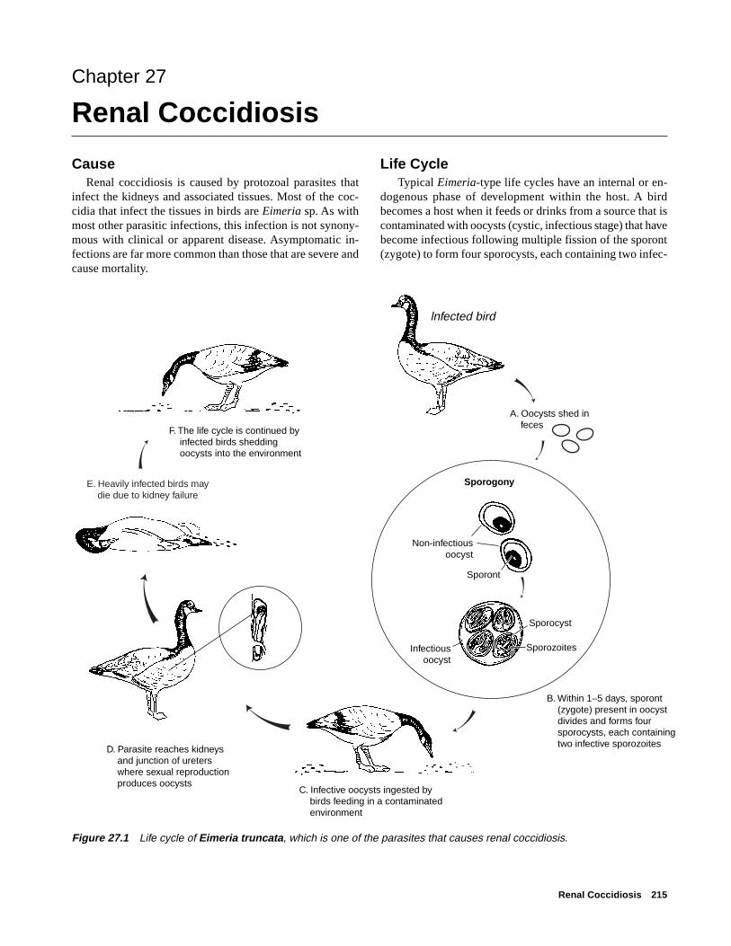
Life Cycle Typical Eimeria-type life cycles have an internal or en-
dogenous phase of development within the host. A birdbecomes a host when it feeds or drinks from a source that iscontaminated with oocysts (cystic, infectious stage) that havebecome infectious following multiple fission of the sporont(zygote) to form four sporocysts, each containing two infec-
lnfected bird
Sporogony
Non-infectiousoocyst
Sporont
Sporozoites
Sporocyst
Infectiousoocyst
E. Heavily infected birds may die due to kidney failure
B. Within 1–5 days, sporont (zygote) present in oocyst divides and forms four sporocysts, each containing two infective sporozoites
F. The life cycle is continued by infected birds shedding oocysts into the environment
D. Parasite reaches kidneys and junction of ureters where sexual reproduction produces oocysts
C. Infective oocysts ingested by birds feeding in a contaminated environment
A. Oocysts shed in feces
Figure 27.1 Life cycle of Eimeria truncata, which is one of the parasites that causes renal coccidiosis.

216 Field Manual of Wildlife Diseases: Birds
tious sporozoites (sporogony) within each oocyst. The in-fective sporozoites within the sporocysts of the oocysts in-vade the bird’s intestinal lining, where they may undergoseveral developmental stages depending on the Eimeria spe-cies. E. truncata, the most well known of the renal coccidia,matures and reproduces only in the kidneys and in the cloacanear its junction with the ureter (Fig. 27.1). It is not knownhow the E. truncata sporozoites get from the intestine to thekidneys; the sporozoites probably undergo asexual reproduc-tion or multiple fission before they reach the kidneys. The
sexual phase of the E. truncata life cycle, or gamogony, takesplace in the kidneys, producing noninfectious oocysts whichare voided with the host bird’s feces into the environment.Sporulated oocysts are resistant to environmental extremes,and their sporozoites can remain infectious for months.
The life cycles of the coccidia that cause renal coccidi-osis are similar to those that cause intestinal coccidiosis (seeChapter 26). However, less is known about the species ofEimeria that cause renal coccidiosis than about those thatcause intestinal coccidiosis.
Types of birds Species of coccidia
Waterfowl
Ducks Eimeria boschadis, E. somatarie, E. sp.
Geese E. truncata, E. sp.
Swans E. christianseni
Fish-eating birds
Gulls E. wobeseri, E. goelandi, E. renicola
Cormorants E. sp.
Loons E. graviae
Marine Birds
Puffins E. fracterculae
Shearwaters E. sp., unidentified coccidia
Land Birds
Owls Unidentified coccidia
Woodcock Unidentified coccidia (in captive colony)
Figure 27.2 Reported occurrences of renal coccidia in wild birds.

Renal Coccidiosis 217
Figure 27.3 Kidneys from double-crested cormorants. Top: normal sizeand color. Bottom: enlarged kidneys withdiffuse pale areas from a bird infectedwith renal coccidia.
Figure 27.4 Cut surfaces from thesame two kidneys as in Fig. 27.3. Bot-tom kidney shows chalky material frombuildup of uric acid salts.
Pho
to b
y J.
Chr
istia
n Fr
anso
nP
hoto
by
J. C
hris
tian
Fran
son
Species AffectedAvian coccidiosis was first reported in France. Canadian
investigators have reported that virtually all species of wildducks they examined are susceptible to renal coccidiosis. Dif-ferent species of renal coccidia are found in different spe-cies of birds (Fig. 27.2). Most reports of renal coccidiosisare of asymptomatic birds or birds that show minor physi-ological or pathological changes due to the parasite. Youngbirds and those that have been stressed by various condi-tions are most likely to have clinical cases of renal coccidi-osis. Mortality has occurred in free-ranging wild geese,eider ducklings, and double-crested cormorants. Disease indomestic geese is usually acute, lasts only 2–3 days, and cankill large segments of the flock.
DistributionRenal coccidiosis is found in birds worldwide.
SeasonalityMortality from renal coccidiosis is most common during
periods of the year when birds are densely aggregated ontheir breeding grounds or wintering areas.
Field SignsThere are no specific field signs that indicate that a bird is
infected with renal coccidia. Young birds will often be ema-ciated and weak, but many other diseases cause similar clini-cal signs.
Gross LesionsInfected birds may be emaciated and have a prominent
keel. In severe infections, kidneys may become enlarged andpale, containing multiple spots or foci of infection that coa-lesce into a mottled pattern (Fig. 27.3). Cutting through thesewhite foci may reveal material that has the consistency ofchalk due to the build up of uric acid salts (Fig. 27.4).

218 Field Manual of Wildlife Diseases: Birds
DiagnosisConfirmation of renal coccidiosis requires microscopic
examination of tissue by the trained staff of a diagnostic labo-ratory. Whole carcasses are generally needed to determinethe cause of death unless kidney damage is so severe that itunquestionably would have caused death. When whole re-frigerated carcasses cannot be provided for evaluation be-cause of field circumstances, the kidneys should be removed,preserved in a 10:1 volume of 10 percent buffered neutralformalin and submitted for diagnosis (see Chapter 2).
ControlControl of renal coccidiosis in free-ranging birds is not
feasible. Crowded conditions facilitate transmission of theparasite through fecal contamination of the environment.Prevention of degradation of habitat quantity and quality onbreeding grounds and wintering areas is needed to minimizedisease risks.
Human Health ConsiderationsThere are no reports of human health concerns with this
disease.
Rebecca A. Cole
Supplementary ReadingGajadhar, A.A., and Leighton, F.A., 1988, Eimeria wobeseri sp. n.
and Eimeria goelandi sp. n. (Protozoa: Apicomplexa) in thekidneys of herring gulls (Larus argentatus): Journal of WildlifeDiseases, v. 24, p. 538–546.
Oksanen, A., 1994, Mortality associated with renal coccidiosis injuvenile wild greylag geese (Anser anser anser): Journal ofWildlife Disease, v. 30, p. 554–556.
Wobeser, G., and Stockdale, P.H.G., 1983, Coccidia of domesticand wild waterfowl (Anseriformes): Canadian Journal ofZoology, v. 61, p. 1–24.
Sarcocystis 219
Chapter 28
Sarcocystis
SynonymsRice breast disease, sarcosporidiosis, sarcocystosis
CauseSarcocystis is a nonfatal, usually asymptomatic infection
that is caused by a parasitic protozoan. Various species ofthis parasite affect mammals, reptiles, and birds. The mostcommonly reported species of the parasite in North Americais Sarcocystis rileyi, the species most commonly found inwaterfowl.
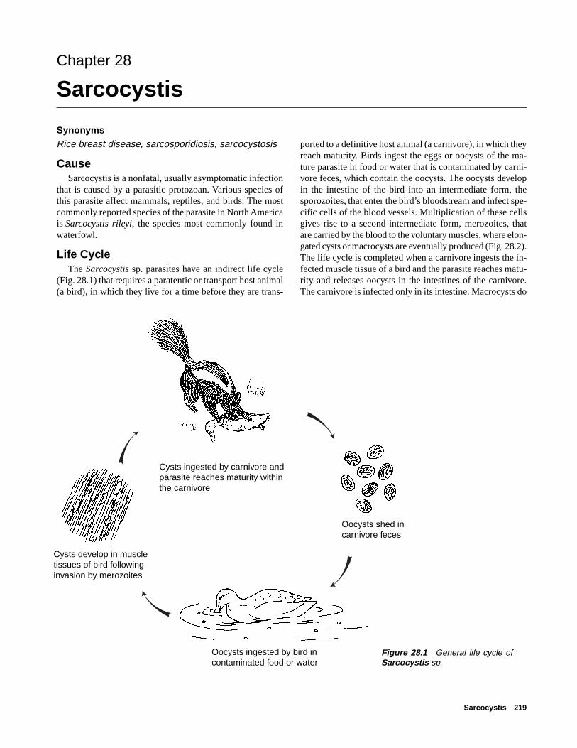
Life CycleThe Sarcocystis sp. parasites have an indirect life cycle
(Fig. 28.1) that requires a paratentic or transport host animal(a bird), in which they live for a time before they are trans-
ported to a definitive host animal (a carnivore), in which theyreach maturity. Birds ingest the eggs or oocysts of the ma-ture parasite in food or water that is contaminated by carni-vore feces, which contain the oocysts. The oocysts developin the intestine of the bird into an intermediate form, thesporozoites, that enter the bird’s bloodstream and infect spe-cific cells of the blood vessels. Multiplication of these cellsgives rise to a second intermediate form, merozoites, thatare carried by the blood to the voluntary muscles, where elon-gated cysts or macrocysts are eventually produced (Fig. 28.2).The life cycle is completed when a carnivore ingests the in-fected muscle tissue of a bird and the parasite reaches matu-rity and releases oocysts in the intestines of the carnivore.The carnivore is infected only in its intestine. Macrocysts do
Figure 28.1 General life cycle ofSarcocystis sp.
Oocysts shed in carnivore feces
Cysts ingested by carnivore and parasite reaches maturity within the carnivore
Oocysts ingested by bird in contaminated food or water
Cysts develop in muscle tissues of bird following invasion by merozoites
220 Field Manual of Wildlife Diseases: Birds
not develop in the carnivore, and the Sarcocystis sp. parasiterarely causes the carnivore illness or other forms of disease.
Species AffectedDabbling ducks (mallard, northern pintail, northern shov-
eler, teal, American black duck, gadwall, and Americanwigeon) commonly have visible or macroscopic forms ofSarcocystis sp.; these forms are far less frequently found inother species of ducks and are infrequently found in geeseand swans. Recent studies of wading birds in Florida havedisclosed a high prevalence of Sarcocystis sp.; similar find-ings have previously been reported from South Africa. Landbirds, such as grackles and other passerine birds, as well asmammals and reptiles can have visible forms of sarcocystis,but it is unlikely that S. rileyi is the species of parasite in-volved. With the exception of waterfowl, this parasite hasreceived little study in migratory birds. This must be takeninto account when considering the current knowledge of spe-cies affected (Fig. 28.3).
DistributionSarcocystis is a common parasitic infection of some water-
fowl species, and it is found throughout the geographic rangeof those species in North America. Less is known about Sar-cocystis sp. in other species of wild birds, but this parasitehas been reported from waterbirds in South Africa, Austra-lia, Canada, and Mexico in addition to the United States.
SeasonalityInfected birds can be found yearround, but waterfowl that
are infected with Sarcocystis sp. are usually observed duringthe hunting season. Infection is not seen in prefledglingwaterfowl, nor is it often seen in juveniles. Two possible rea-sons for these differences between the age classes may be
that the development of visible forms of the parasite requirestime or that birds may not be infected until after they haveleft their breeding grounds. Because visible forms of sarco-cystis are more frequently developed in older birds, hunterdetection tends to be greatest during years of poor waterfowlproduction when the bag contains a greater proportion ofadult birds. A moderate percentage of juvenile mottled ducksthat were collected in Louisiana primarily after the huntingseason were recently found to have light sarcocystis infec-tions. Because this species does not migrate, this suggeststhat the birds were infected within the general geographicarea where they were collected and that the later collectiondate allowed the macrocyst lesions to be visible.
Too little is known about sarcocystis in other groups ofwild birds to evaluate its seasonality.
Field SignsUsually, there is no externally visible sign of this disease
nor is it recognized as a direct cause of migratory bird mor-tality. Severe infections can cause loss of muscle tissue andresult in lameness, weakness, and even paralysis in rare cases.The debilitating effects of severe infections could increasebird susceptibility to predation and to other causes of mor-tality.
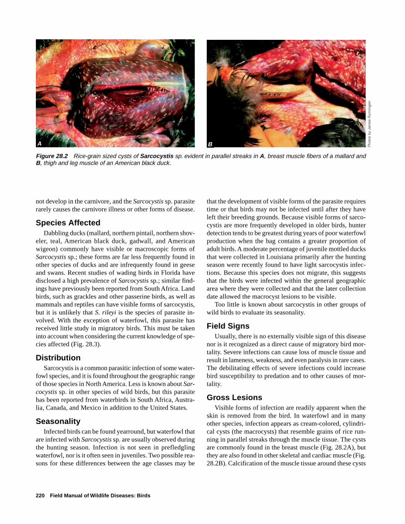
Gross LesionsVisible forms of infection are readily apparent when the
skin is removed from the bird. In waterfowl and in manyother species, infection appears as cream-colored, cylindri-cal cysts (the macrocysts) that resemble grains of rice run-ning in parallel streaks through the muscle tissue. The cystsare commonly found in the breast muscle (Fig. 28.2A), butthey are also found in other skeletal and cardiac muscle (Fig.28.2B). Calcification of the muscle tissue around these cysts
Figure 28.2 Rice-grain sized cysts of Sarcocystis sp. evident in parallel streaks in A, breast muscle fibers of a mallard andB, thigh and leg muscle of an American black duck.
Pho
tos
by
Jam
es R
unni
ngen
BA

Sarcocystis 221
makes them obviously discrete bodies. The degree of calci-fication is often sufficient to give a gritty feeling to the tissuewhen it is cut with a knife.
Lesions that were observed in wading birds differed inappearance; the cysts were white and opaque, and they gen-erally extended throughout the entire length of the infectedmuscle fiber. Cysts were present in the heart muscle and theywere confined to striated muscles.
DiagnosisThe visible presence of sarcosporidian cysts in muscle
tissue is sufficient to diagnose this disease. Visible cysts mayvary in size and shape in different bird species. Good qualitycolor photographs (prints or 35 millimeter slides) of the ex-ternal surface of infected muscle are generally sufficient fora disease specialist to recognize this disease if tissues or awhole carcass cannot be provided. Whole birds should besubmitted if possible. If only tissues can be submitted, thena portion of the infected muscle should be fixed in a 10 per-cent formalin solution. Frozen muscle tissue is also suitablefor diagnosis, and the distinctive appearance of these cystsallows a diagnosis from even partially decomposed carcasses.
ControlThere are no known control methods for this disease, nor
do any seem to be needed or are any being developed. Con-trol of sarcocystis would require interruption of the life cycleof the parasite. Although the life cycles of the Sarcocystissp. that affect wild birds are not precisely known, they areprobably similar to the two-host, indirect life cycle knownfor some other Sarcocystis sp. (Fig. 28.1). The predator-preyrelationship between the intermediate bird hosts and the de-finitive carnivore hosts may be the primary reason that juve-nile birds or some bird species are seldom found to be in-fected. The appropriate carnivores may not be present on thebreeding grounds.
Different species of carnivores seem to be involved in theinfection of different bird species, which suggests that birdsare infected by more than one species of the genus Sarco-cystis sp. If the carnivore-bird cycle is species-specific, thatis, if a specific species of bird can only be infected by oo-cysts that are produced by a parasite in a specific carnivorespecies, then selective control of sarcocystis might be fea-sible. However, current knowledge of the disease does notindicate a need to initiate control because there is little evi-dence that bird health is often compromised by infection.Nevertheless, the role of carnivores in the life cycle of Sar-cocystis sp. infections should be considered when feeding
Figure 28.3 Relative frequency of grossly visible forms ofsarcocystis in selected groups of North American migratorybirds.
Puddle ducks
Grackles
Wading birds
Diving ducks
Mergansers
Sea ducks
Geese
Swans
Shorebirds
Gulls and terns
Pelicans
Raptors
FrequentCommon
OccasionalRare

222 Field Manual of Wildlife Diseases: Birds
uncooked, infected waterfowl to house pets and to farm ani-mals such as hogs.
Human Health ConsiderationsSarcocystis sp. presents no known health hazard to hu-
mans. The primary importance to humans of sarcocystis inwaterfowl is the loss of infected birds for food; the unaes-thetic appearance of parasitized muscle may prompt huntersto discard the carcass. Limited evaluations of hunter responsesto infected carcasses indicate no reduction in carcass con-sumption in areas where the infection is commonly seen.Also, the recognized high prevalence of infection in north-ern shovelers in some areas results in this species often be-ing left unretrieved by some hunters and focuses additionalhunting pressure on other species.
Benjamin N. Tuggle and Milton Friend(Modified from and earlier chapter by Benjamin N. Tuggle)
Supplementary ReadingCawthorn, R.J., Rainnie, D., and Wobeser, G.A., 1981, Experi-
mental transmission of Sarcocystis sp. (Protozoa:Sarcocystidae) between the shoveler (Anas clypeata) duck andthe striped skunk (Mephitis mephitis): Journal of WildlifeDiseases, v. 17, p. 389–394.
Cornwell, G, 1963, New waterfowl host records for Sarcocystisrileyi and a review of sarcosporidiosis in birds: Avian Disease,v. 7, p. 212–216.
Moorman, T.E., Baldassarre, G.A., and Richard, D.M., 1991, Thefrequency of Sarcocystis spp. and its effect on winter carcasscomposition of mottled ducks: Journal of Wildlife Disease,v. 27, no. 3, p. 491–493.
Spalding, M.G., Atkinson, C.T., and Carleton, R.E., 1994,Sarcocystis sp. in wading birds (Ciconiiformes) from Florida:Journal of Wildlife Disease, v. 30, no. 1, p. 29–35.
Eustrongylidosis 223
Chapter 29
EustrongylidosisSynonymsVerminous peritonitis
CauseEustrongylidosis is caused by the nematodes or round-
worms Eustrongylides tubifex, E. ignotus, and E. excisus.Eustrongylides sp. can cause large die-offs of nestlings incoastal rookeries, especially of egrets and other wading birds.
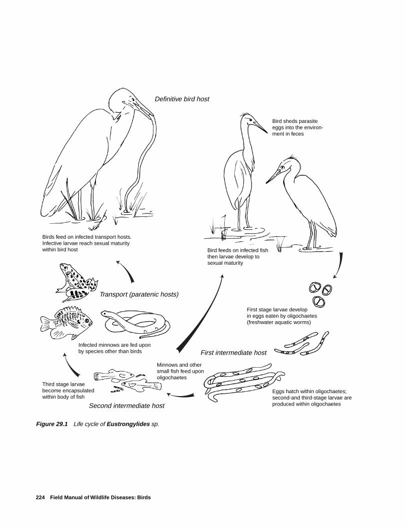
Life CycleThe three species of Eustrongylides that cause disease in
birds have similar indirect life cycles that require two inter-mediate hosts (Fig. 29.1). Four developmental stages of theparasite are required from egg to sexually mature worm. Thefirst larval stage develops within the eggs that are shed in thefeces of the bird host and are eaten by freshwater oligocha-etes or aquatic worms. The oligochaetes serve as the firstintermediate host. The eggs hatch within the oligochaetes,where they develop into second- and third-stage larvae. Min-nows and other small fish, such as species of Fundulus andGambusia, feed upon the infected oligochaetes and serve asthe second intermediate host. The third-stage larvae becomeencapsulated on the internal surface areas of the fish, de-velop into infective fourth-stage larvae, and await ingestionby birds. Predatory fish, which consume infected fish, canserve as paratenic or transport hosts when they are fed uponby birds. Amphibians and reptiles have also been reported assecond-stage intermediate hosts and serve as paratenic hosts.Larvae that are infective for birds can penetrate the ventricu-lus (stomach) within 3–5 hours after a bird ingests an inter-mediate or paratenic host, and the larvae quickly becomesexually mature worms that begin shedding eggs 10–17 dayspostinfection.
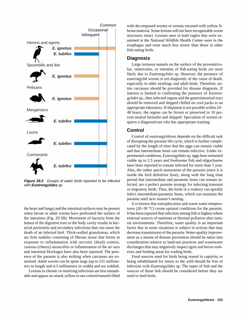
Species AffectedE. tubifex has been reported from four different bird fami-
lies, E. ignotus from three, and E. excisus from three (Fig.29.2). Young wading birds are the most common species tohave large mortalities from eustrongylidosis (Table 29.1).Eustrongylides sp. have also been reported in birds of prey.
DistributionEustrongylides sp. have been reported from birds through-
out much of the world. E. tubifex and E. ignotus are the spe-cies reported within the United States (Table 29.2).Eustrongylid infections within the United States have beenreported from many areas (Fig. 29.3). Typical rookeries wherebirds are infected with Eustrongylides sp. are found in coastal
areas and consist of dense populations of birds nesting onlow islands, often surrounded by canals or ditches. Nestinghabitat often includes stands of low trees, such as willows,with an understory that may be submergent, semisubmergent,or upland mixed-prairie species. Inland rookeries are usu-ally adjacent to lakes or rivers, and nesting trees, particu-larly those used by great blue herons, may be much higherthan those in coastal rookeries. Several wading bird speciesmay nest in these areas, but typically one or two species ac-count for most of the birds in the rookery (Fig. 29.4).
SeasonalityBirds can harbor infections yearround. Mortality usually
is reported in spring and summer and birds less than 4 weeksold are more likely to die than adults. Disease in older birdstends to be of a more chronic nature and infection may beseen at any time of the year.
Field SignsDisease results in a variety of clinical or apparent signs
that are not specific to eustrongylidosis. However, consider-ation of the species affected, the age class of birds involved,and the full spectrum of signs may suggest that eustrongyli-diosis is the cause of mortality. Very early in the infection asthe worm is penetrating the ventriculus, some birds will shaketheir heads, have difficulty swallowing, have dyspnea or dif-ficult or labored breathing and, occasionally, regurgitate theirfood. Anorexia or loss of appetite has been noted in experi-mentally infected nestlings. It has been speculated that anor-exia in combination with sibling competition for food maycontribute to the emaciation seen in naturally infected birds.Infected nestlings also may wander from the nest predisposedto predation or trauma or both. Affected nestlings observedduring one mortality event became progressively weakenedand showed abdominal swelling. Palpation of worms on theventriculus has been useful for detecting infection in livenestlings.
Gross LesionsBirds that have been recently infected often have large,
tortuous, raised tunnels that are visible on the serosal sur-face of the proventriculus, ventriculus, or intestines (Fig.29.5A). The nematodes reside within these tunnels, whichare often encased with yellow, fibrous material, and main-tain openings to the lumen of the organ so that parasite eggsmay be passed out with feces into the environment. A fibrino-peritonitis or fibrin-coated inflammation of the surfaces ofthe peritoneal cavity (the area containing the organs below
224 Field Manual of Wildlife Diseases: Birds
Figure 29.1 Life cycle of Eustrongylides sp.
Birds feed on infected transport hosts. Infective larvae reach sexual maturity within bird host
First stage larvae develop in eggs eaten by oligochaetes(freshwater aquatic worms)
First intermediate host
Second intermediate host
Transport (paratenic hosts)
Minnows and other small fish feed upon oligochaetes
Infected minnows are fed upon by species other than birds
Third stage larvae become encapsulated within body of fish
Bird feeds on infected fishthen larvae develop tosexual maturity
Definitive bird host
Bird sheds parasite eggs into the environ-ment in feces
Eggs hatch within oligochaetes; second-and third-stage larvae are produced within oligochaetes
Eustrongylidosis 225
Herons and egretsE. ignotusE. tubifex
Spoonbills and ibis
E. ignotus Pelicans
E. ignotus Mergansers
E. tubifex
Loons
E. tubifex
Cormorants
E. tubifex
CommonOccasional
Infrequent
Figure 29.2 Groups of water birds reported to be infectedwith Eustrongylides sp.
the heart and lungs) and the intestinal surfaces may be presentwhen larvae or adult worms have perforated the surface ofthe intestines (Fig. 29.5B). Movement of bacteria from thelumen of the digestive tract to the body cavity results in bac-terial peritonitis and secondary infections that can cause thedeath of an infected bird. Thick-walled granulomas, whichare firm nodules consisting of fibrous tissue that forms inresponse to inflammation with necrotic (dead) centers,caseous (cheesy) airsaculitis or inflammation of the air sacsand intestinal blockages have also been reported. The pres-ence of the parasite is also striking when carcasses are ex-amined. Adult worms can be quite large (up to 151 millime-ters in length and 4.3 millimeters in width) and are reddish.
Lesions in chronic or resolving infections are less remark-able and appear as raised, yellow or tan-colored tunnels filled
with decomposed worms or worms encased with yellow fi-brous material. Some lesions will not have recognizable wormstructures intact. Lesions seen in bald eagles that were ex-amined at the National Wildlife Health Center were in theesophagus and were much less severe than those in otherfish-eating birds.
DiagnosisLarge tortuous tunnels on the surface of the proventricu-
lus, ventriculus, or intestine of fish-eating birds are mostlikely due to Eustrongylides sp. However, the presence ofeustrongylid worms is not diagnostic of the cause of death,especially in older nestlings and adult birds. Therefore, en-tire carcasses should be provided for disease diagnosis. Ifinterest is limited to confirming the presence of Eustron-gylides sp., then infected organs and the gastrointestinal tractshould be removed and shipped chilled on cool packs to anappropriate laboratory. If shipment is not possible within 24–48 hours, the organs can be frozen or preserved in 10 per-cent neutral formalin and shipped. Speciation of worms re-quires a diagnostician who has appropriate training.
ControlControl of eustrongylidosis depends on the difficult task
of disrupting the parasite life cycle, which is further compli-cated by the length of time that the eggs can remain viableand that intermediate hosts can remain infective. Under ex-perimental conditions, Eustrongylides sp. eggs have remainedviable up to 2.5 years and freshwater fish and oligochaeteshave been reported to remain infected for more than 1 year.Also, the rather quick maturation of the parasite (once it isinside the bird definitive host), along with the long timeperiod that intermediate and paratenic hosts can remain in-fected, are a perfect parasite strategy for infecting transientor migratory birds. Thus, the birds in a rookery can quicklyinfect intermediate/paratenic hosts, which can maintain theparasite until next season’s nesting.
It is known that eutrophication and warm water tempera-tures (20–30 °C) create optimal conditions for the parasite.It has been reported that infection among fish is highest whereexternal sources of nutrients or thermal pollution alter natu-ral environments. Therefore, water quality is an importantfactor that in some situations is subject to actions that maydecrease transmission of the parasite. Water-quality improve-ment as a means of disease prevention should be taken intoconsideration relative to land-use practices and wastewaterdischarges that may negatively impact egret and heron rook-eries and feeding areas for wading birds.
Food sources used for birds being reared in captivity orbeing rehabilitated for return to the wild should be free ofinfection with Eustrongylides sp. The types of fish and thesources of those fish should be considered before they areused to feed birds.
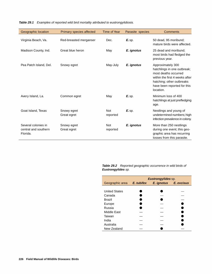
226 Field Manual of Wildlife Diseases: Birds
Table 29.2 Reported geographic occurrence in wild birds ofEustrongylides sp.
Eustrongylides sp.Geographic area E. tubifex E. ignotus E. excisus
United States ● ● —Canada ● — —Brazil ● ● —Europe ● — ●
Russia ● — ●
Middle East — — ●
Taiwan — — ●
India — — ●
Australia — — ●
New Zealand — ● —
Table 29.1 Examples of reported wild bird mortality attributed to eustrongylidiosis.
Geographic location Primary species affected Time of Year Parasite species Comments
Virginia Beach, Va. Red-breasted merganser Dec. E. sp. 50 dead, 95 moribund;mature birds were affected.
Madison County, Ind. Great blue heron May E. ignotus 25 dead and moribund;most birds had fledged theprevious year.
Pea Patch Island, Del. Snowy egret May-July E. ignotus Approximately 300hatchlings in one outbreak;most deaths occurredwithin the first 4 weeks afterhatching; other outbreakshave been reported for thislocation.
Avery Island, La. Common egret May E. sp. Minimum loss of 400hatchlings at just prefledgingage.
Goat Island, Texas Snowy egret Not E. sp. Nestlings and young ofGreat egret reported undetermined numbers; high
infection prevalence in colony.
Several colonies in Snowy egret Not E. ignotus More than 250 nestlingscentral and southern Great egret reported during one event; this geo-Florida. graphic area has recurring
losses from this parasite.

Eustrongylidosis 227
EXPLANATION
Reports of Eustrongylides sp. in birds, by State
Infections reported in birds
Major mortality events reported
Figure 29.3 States where Eustrongylides sp. infections in wild birds have been reported.
Figure 29.4 Although many species may nest in wading bird rookeries, one or two species are oftenpredominant.
Pho
to b
y J.
Chr
istia
n Fr
anso
n

228 Field Manual of Wildlife Diseases: Birds
Human Health ConsiderationsHumans who have consumed raw or undercooked fish that
carry the larval stages of the parasite have experienced gas-tritis or inflammation of the stomach and intestinal perfora-tion requiring surgical removal of worms.
Rebecca A. Cole
Supplementary ReadingFranson, J.C. and Custer, T.W., 1994, Prevalence of
eustrongylidosis in wading birds from colonies in California,Texas, and Rhode Island, United States of America: ColonialWaterbirds, v. 17, p. 168–172.
Measures, L.N., 1988, Epizootiology, pathology and descriptionof Eustrongylides tubifex (Nematoda: Dioctophymatoidea) infish: Canadian Journal of Zoology, v. 66: p . 2212–2222.
Spalding, M.A., Bancroft, G.T., and Forrester, D.J., 1993, Theepizootiology of eustrongylidosis in wading birds(Ciconiiformes) in Florida: Journal of Wildlife Diseases, v. 29,p. 237–249.
Spalding, M.A., and Forrester, D.J., 1993, Pathogenesis ofEustrongylides ignotus (Nematoda: Dioctophymatoidea) inCiconiiformes: Journal of Wildlife Diseases, v. 29, p. 250–260.
Wiese, J.H., Davidson, W.R., and Nettles, V.F., 1977, Large scalemortality of nestling ardeids caused by nematode infection:Journal of Wildlife Diseases, v. 13, no. 4, p. 376–382.
Pho
to b
y Ja
mes
Run
ning
en
Figure 29.5 (A) Raised tunnels caused by Eustrongylidessp. on intestines of a snowy egret. (B) The debris on the intes-tinal surfaces of this snowy egret is characteristic of the peri-tonitis often caused by Eustrongylides sp. infection.
Pho
to b
y J.
Chr
istia
n Fr
anso
n
A
B

Tracheal Worms 229
Chapter 30
Tracheal Worms
SynonymsGape worm, syngamiasis, gapes
CauseInfection by tracheal worms often results in respiratory
distress due to their location in the trachea or bronchi andtheir obstruction of the air passage. Infections by these para-sitic nematodes or roundworms in waterbirds, primarilyducks, geese, and swans, are usually due to Cyathostomabronchialis and infection of land birds are usually due toSyngamus trachea. However, both genera infect a variety ofspecies, including both land and waterbirds. Infections withS. trachea have been more extensively studied than infec-tions with Cyathostoma sp. because of its previous impor-tance as a disease-causing parasite of poultry in many partsof the world. Changes in husbandry practices to modern in-tensive methods for poultry production have essentially elimi-nated S. trachea as an agent of disease in chickens, but it isan occasional cause of disease in turkeys raised on range.
Life CycleTracheal worms have an indirect life cycle (Fig. 30.1) that
requires a paratenic or transport host which transmits the in-fectious larvae to the definitive host bird, where they reachadulthood and reproduce. Adult S. trachea reside within thetrachea. The female releases fertilized eggs, which are swal-lowed by the bird and voided with the feces into the soil.Eggs may also be directly expelled onto the ground from thetrachea. After embryonation (1–2 weeks), infective larvaedevelop within the egg. Birds can become infected by eatinginvertebrate paratenic hosts such as earthworms, snails, slugs,or fly larvae that have consumed the eggs. Infective larvaeare released from the egg and become encysted within thebodies of these invertebrates and can remain infective for upto three and one-half years. Upon ingestion by birds, the lar-vae are believed to penetrate the intestinal wall. Some larvaeenter the abdominal cavity but most enter the bloodstream,where they are carried to the lungs. After further develop-ment in the lungs, the young worms migrate up the bronchito the trachea. Larvae can reach the lungs within 6 hoursafter ingestion and eggs are produced by worms in the tra-chea about 2 weeks after ingestion of those larvae.C. bronchialis is very similar in that earthworms transmitthe infective stage to the bird. Infection of birds withC. bronchialis by direct consumption of fully embryonatedeggs has been documented experimentally; however, wormburdens were extremely low.
Species AffectedDisease caused by tracheal worms is not commonly re-
ported for free-ranging birds within the United States andCanada, but it is common within the United Kingdom and insome other countries. High infection rates within wild birdsin England attest to the potential for this parasite to be aserious pathogen. More than 50 percent of nestling and fledg-ling starlings, more than 85 percent of jackdaws, and 100percent of young rooks were found to be infected in one study.Infection rates in adult birds were considerably lower, butthey still exceeded 30 percent for starlings and rooks. Withinthe United States, S. trachea infections have been reportedfrom wild turkeys, other gamebirds, a variety of passerines,(songbirds), and occasionally from other bird species. Large-scale mortalities have occurred among pheasants and othergamebirds being propagated for sporting purposes. Findingsfrom captive bird collections have led to the conclusion thatalmost any species of cage or aviary bird is susceptible toinfection.
S. trachea has been reported infrequently in waterfowl,but members of the genus Cyathostoma sp. are “characteris-tic” or common parasites of waterfowl. Mortality has beenreported for several species of young geese, leading someinvestigators to suggest that C. bronchialis are potentiallyimportant pathogens for geese. Juvenile free-ranging sand-hill cranes have also been reported to have died fromCyathostoma sp. infection.
DistributionS. trachea and Cyathostoma sp. are found worldwide.
SeasonalityInfected birds can be found yearround. Young birds are
most commonly affected and, therefore, disease is associ-ated with breeding cycles in the spring to summer monthsfor free-ranging birds.
Field SignsMost birds that are infected show no signs of disease. In
general, the severity of disease is dependent upon the degreeof infection and the size of the bird. Small birds are moreseverely affected than larger birds because their narrowertracheal openings result in greater obstruction by the worms.Respiratory distress is the primary clinical sign of disease.Birds with severe infections open their mouth widely and atthe same time stretch out their necks, assuming a “gaping”posture. The adult worms that are attached to the lining of
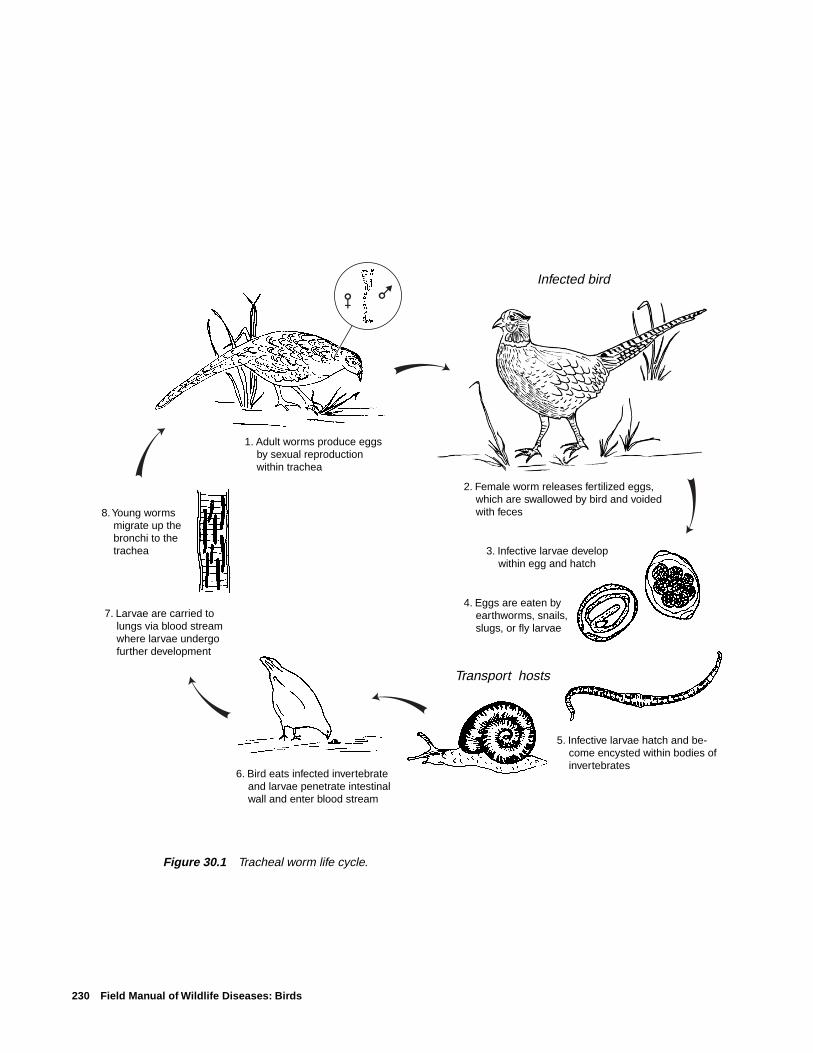
230 Field Manual of Wildlife Diseases: Birds
6. Bird eats infected invertebrate and larvae penetrate intestinal wall and enter blood stream
Transport hosts
Infected bird
1. Adult worms produce eggs by sexual reproduction within trachea
8. Young worms migrate up the bronchi to the trachea
2. Female worm releases fertilized eggs, which are swallowed by bird and voided with feces
4. Eggs are eaten by earthworms, snails, slugs, or fly larvae
3. Infective larvae develop within egg and hatch
7. Larvae are carried to lungs via blood stream where larvae undergo further development
5. Infective larvae hatch and be-come encysted within bodies of invertebrates
Figure 30.1 Tracheal worm life cycle.

Tracheal Worms 231
the trachea cause irritation and excess mucus production. Thisoften results in agitated bouts of coughing, head shaking,and sneezing as the birds attempt to dislodge the parasites.Severely infected birds may have most or all of the trachealopening obstructed by worms, may stop feeding, and mayrapidly lose body condition.
Gross LesionsSeverely affected birds experience severe weight loss and
have poorer development of body mass than uninfected birds,and they often die from starvation (Fig. 30.2). Anemia mayalso be present due to the blood-feeding habits of the para-sites.
DiagnosisIdentification of the worms (Fig. 30.3) and evaluation of
any associated disease signs are required for a diagnosis.Clinical signs are not diagnostic because similar signs canbe seen with some mite infections, aspergillosis, and wet pox.
ControlThere is no feasible method for controlling tracheal worms
in free-ranging birds. Disease prevention should be practicedby minimizing the potential for captive-propagation and re-lease programs to infect invertebrates that are then fed uponby free-ranging birds. Land-use practices that provide directcontact between poultry rearing and wild birds and the dis-posal of bird feces and litter should also be considered be-cause environmental contamination with infective larvae isa critical aspect of the disease cycle.
Human Health ConsiderationsThere are no reports of these nematodes infecting humans.
Rebecca A. Cole
Supplementary ReadingAnderson, R.C., 1992, Nematode parasites of vertebrates: Their
development and transmission: Wallingford, England, CABInternational, 578 p.
Cram, E.B., 1927, Bird parasites of the nematode subordersStrongylata, Ascaridata, and Spirurata: U.S. National MuseumBulletin No. 140, 465 p.
Fernando, M.A., Hoover, I.J., and Ogungbade, S.G., 1973, Themigration and development of Cyathostoma bronchialis ingeese: Journal of Parasitology, v. 59, p. 759–764.
Threlfall, W., 1965, Life-cycle of Cyathostoma lari: Nature,v. 206, p. 1,167–1,168.
Figure 30.3 Syngamus trachea in the trachea of a ring-necked pheasant.
Figure 30.2 Comparative breast muscle mass of noninfectedpheasants (left) and those with Syngamus trachea infections(right).
Pho
to b
y M
ilton
Frie
ndP
hoto
by
Milt
on F
riend

232 Field Manual of Wildlife Diseases: Birds

Heartworm of Swans and Geese 233
Chapter 31
Heartworm of Swans and Geese
SynonymsFilarial heartworm, Sarconema, Sarconema eurycerca
CauseHeartworm in swans and geese is caused by a filarial
nematode or a roundworm of the superfamily Filarioideawhich is transmitted to the bird by a biting louse. The nema-tode and the louse both are parasites. Sarconema eurycercais the only one of several species of microfilaria or the firststage juvenile of the parasite found in the circulating bloodof waterfowl that is known to be pathogenic or cause clinicaldisease.
Life cycleSarconema eurycerca has an indirect life cycle (Fig. 31.1)
that requires the parasite larvae to develop in an intermedi-ate host before they can become infective for and be trans-mitted to a definitive host, where they mature and reproduce.Female adult heartworms release microfilariae into the blood-stream of the definitive host bird. The microfilariae infect abiting louse, Trinoton anserinum, that subsequently feedsupon the bird. The larvae go through three stages of devel-opment within the louse, and the third stage is infectious tobirds. A new host bird becomes infected when the louse bitesit to feed on its blood and the third-stage larvae move into
Worms migrate through the bloodstream to the heart muscle where they mature and release micro-filariae into the blood-stream
Life cycle is continued when biting lice feed upon the bird's blood containing the microfilariae
Three stages of larval development occur within the biting louse
Infective third-stage larvae are transmitted into the bird's blood-stream when the louse takes another blood meal Microfilariae
Figure 31.1 Indirect life cycle of Sarconema eurycerca.

234 Field Manual of Wildlife Diseases: Birds
the bird’s bloodstream. The larvae migrate through the blood-stream to the myocardium, which is the middle and thickestlayer of the heart wall composed of cardiac muscle. Theyare nourished by and develop to sexual maturity within themyocardium. The cycle continues as this next generation ofmature heartworms release microfilariae into the blood-stream.
Infection with the parasite is not synonymous with dis-ease; that is, the parasite may infect and develop within thebird but not debilitate it.
Species AffectedSarconema eurycerca was first identified from a tundra
swan (whistling swan) in the late 1930s. It has since beenreported from trumpeter, Bewick’s, and mute swans and, fromCanada, snow, white-fronted, and bean geese. Varying per-centages of swans (4–20 percent) have been found to be in-fected on the basis of blood smears that were taken fromapparently healthy birds during field surveys. Canadian in-vestigators have reported a prevalence of approximately 10percent of snow geese that were examined at necropsy andwhich had died from other causes. This parasite has not re-ceived sufficient study for its full host range, its relative fre-quency of occurrence in different species, or its significanceas a mortality factor for wild birds to be determined.
DistributionHeartworm is found throughout the range of its swan and
goose hosts.
SeasonalityIt is suspected that while swans and geese are on the breed-
ing grounds, louse infestation and colonization on birds isprevalent. Therefore, the possibility of infection by heart-worm is highest while birds are on the breeding grounds.
Field SignsField signs are not always present in infected birds, and
infection cannot be determined by the presence of clinicalsigns alone. Chronic types of debilitating diseases, such aslead poisoning, may exacerbate louse infestation becausebirds become lethargic and do not preen. No specific fieldsign is diagnostic for infection.
Gross LesionsThe severity of infection dictates the lesions that are seen
at necropsy. Birds may be emaciated or in comparably goodflesh. The heart may be enlarged and have pale foci or spotswithin the myocardium. The thin, long thread-like wormsmay be visible under the surface layer or epicardium of theheart or the worms may be embedded within the deepermuscle tissue of the myocardium (Fig. 31.2).
DiagnosisA diagnosis of heartworm as the cause of death must be
supported by pathologic lesions seen during examination ofthe heart tissues with a microscope and consideration of othercauses. Therefore, whole carcasses should be submitted fordiagnostic assessments. If the transit time is short enough toavoid significant decomposition of the carcass and if the car-cass can be kept chilled during transit, then chilled wholecarcasses should be submitted to qualified disease diagnos-tic laboratories. If those conditions cannot be met, then car-casses should be submitted frozen.
ControlControl of heartworm is not practical for free-ranging
birds. Decreasing the opportunity for heavy infestation ofthe louse intermediate host will result in reduced opportu-nity for heartworm infection.
Human Health ConsiderationsSarconema eurycerca has not been reported to infect humans.
Rebecca A. Cole
Supplementary ReadingCohen, M., Greenwood, T., and Fowler, J.A., 1991, The louse
Trinoton anserinum (Amblycerca: Pthiraptera), an intermedi-ate host of Sarconema eurycerca (Filarioidea: Nematoda), aheartworm of swans: Medical and Veterinary Entomology, v. 5,p. 101–110.
Scheller, E.L., Sladen, W.L., and Trpis, M., 1976, A Mallophaga,Trinoton anserium, as a cyclodevelopmental vector for aheartworm parasite of waterfowl: Science, v. 194, p. 739–740.
Seegar, W.S., 1979, Prevalence of heartworm, Sarconemaeurycerca, Wehr, 1939 (Nematoda), in whistling swan, Cygnuscolumbianus columbianus: Canadian Journal of Zoology, v. 57,p. 1,500–1,502.
Figure 31.2 Heartworms (arrows) on the inner surface ofthe heart of a tundra swan.
Pho
to b
y Ja
mes
Run
ning
en➡
➡
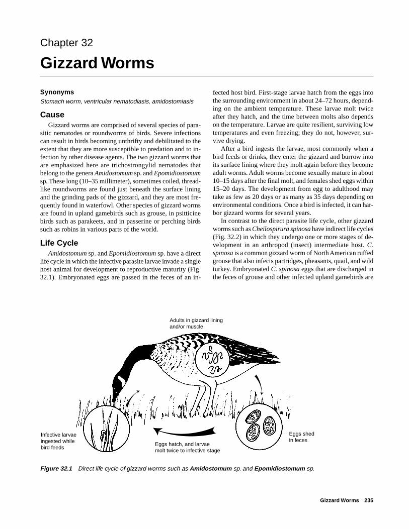
Gizzard Worms 235
Chapter 32
Gizzard Worms
SynonymsStomach worm, ventricular nematodiasis, amidostomiasis
CauseGizzard worms are comprised of several species of para-
sitic nematodes or roundworms of birds. Severe infectionscan result in birds becoming unthrifty and debilitated to theextent that they are more susceptible to predation and to in-fection by other disease agents. The two gizzard worms thatare emphasized here are trichostrongylid nematodes thatbelong to the genera Amidostomum sp. and Epomidiostomumsp. These long (10–35 millimeter), sometimes coiled, thread-like roundworms are found just beneath the surface liningand the grinding pads of the gizzard, and they are most fre-quently found in waterfowl. Other species of gizzard wormsare found in upland gamebirds such as grouse, in psitticinebirds such as parakeets, and in passerine or perching birdssuch as robins in various parts of the world.
Life CycleAmidostomum sp. and Epomidiostomum sp. have a direct
life cycle in which the infective parasite larvae invade a singlehost animal for development to reproductive maturity (Fig.32.1). Embryonated eggs are passed in the feces of an in-
fected host bird. First-stage larvae hatch from the eggs intothe surrounding environment in about 24–72 hours, depend-ing on the ambient temperature. These larvae molt twiceafter they hatch, and the time between molts also dependson the temperature. Larvae are quite resilient, surviving lowtemperatures and even freezing; they do not, however, sur-vive drying.
After a bird ingests the larvae, most commonly when abird feeds or drinks, they enter the gizzard and burrow intoits surface lining where they molt again before they becomeadult worms. Adult worms become sexually mature in about10–15 days after the final molt, and females shed eggs within15–20 days. The development from egg to adulthood maytake as few as 20 days or as many as 35 days depending onenvironmental conditions. Once a bird is infected, it can har-bor gizzard worms for several years.
In contrast to the direct parasite life cycle, other gizzardworms such as Cheilospirura spinosa have indirect life cycles(Fig. 32.2) in which they undergo one or more stages of de-velopment in an arthropod (insect) intermediate host. C.spinosa is a common gizzard worm of North American ruffedgrouse that also infects partridges, pheasants, quail, and wildturkey. Embryonated C. spinosa eggs that are discharged inthe feces of grouse and other infected upland gamebirds are
Figure 32.1 Direct life cycle of gizzard worms such as Amidostomum sp. and Epomidiostomum sp.
Eggs shed in feces
Eggs hatch, and larvae molt twice to infective stage
Infective larvae ingested while bird feeds
Adults in gizzard lining and/or muscle
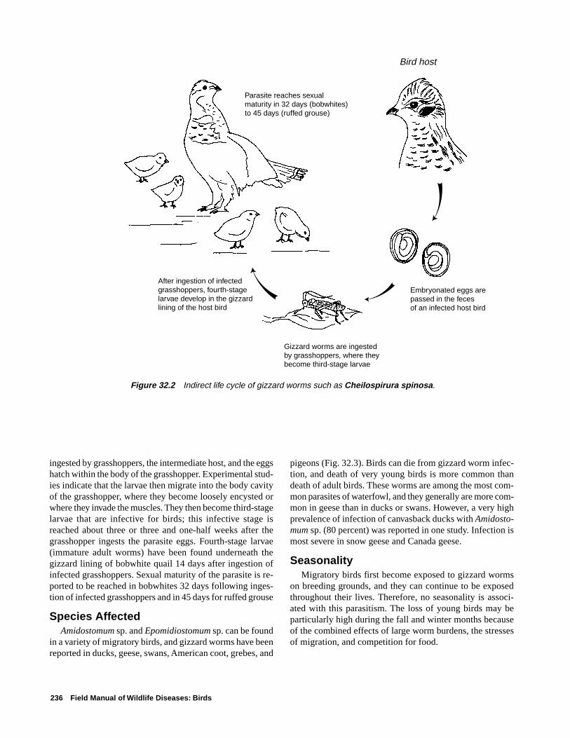
236 Field Manual of Wildlife Diseases: Birds
ingested by grasshoppers, the intermediate host, and the eggshatch within the body of the grasshopper. Experimental stud-ies indicate that the larvae then migrate into the body cavityof the grasshopper, where they become loosely encysted orwhere they invade the muscles. They then become third-stagelarvae that are infective for birds; this infective stage isreached about three or three and one-half weeks after thegrasshopper ingests the parasite eggs. Fourth-stage larvae(immature adult worms) have been found underneath thegizzard lining of bobwhite quail 14 days after ingestion ofinfected grasshoppers. Sexual maturity of the parasite is re-ported to be reached in bobwhites 32 days following inges-tion of infected grasshoppers and in 45 days for ruffed grouse
Species AffectedAmidostomum sp. and Epomidiostomum sp. can be found
in a variety of migratory birds, and gizzard worms have beenreported in ducks, geese, swans, American coot, grebes, and
pigeons (Fig. 32.3). Birds can die from gizzard worm infec-tion, and death of very young birds is more common thandeath of adult birds. These worms are among the most com-mon parasites of waterfowl, and they generally are more com-mon in geese than in ducks or swans. However, a very highprevalence of infection of canvasback ducks with Amidosto-mum sp. (80 percent) was reported in one study. Infection ismost severe in snow geese and Canada geese.
SeasonalityMigratory birds first become exposed to gizzard worms
on breeding grounds, and they can continue to be exposedthroughout their lives. Therefore, no seasonality is associ-ated with this parasitism. The loss of young birds may beparticularly high during the fall and winter months becauseof the combined effects of large worm burdens, the stressesof migration, and competition for food.
Figure 32.2 Indirect life cycle of gizzard worms such as Cheilospirura spinosa.
Embryonated eggs are passed in the feces of an infected host bird
Bird host
Gizzard worms are ingested by grasshoppers, where they become third-stage larvae
After ingestion of infected grasshoppers, fourth-stage larvae develop in the gizzard lining of the host bird
Parasite reaches sexual maturity in 32 days (bobwhites) to 45 days (ruffed grouse)

Gizzard Worms 237
Field SignsThere are no field signs that indicate gizzard worm infec-
tion. Heavy worm burdens can result in poor growth of youngbirds, and birds of all ages are subject to emaciation andgeneral weakness. Severe infections can interfere with fooddigestion by the bird as a result of extensive damage to thegizzard lining and muscle.
Gross LesionsObvious changes from the normal appearance of the giz-
zard result from the development, migration, and feeding ofgizzard worms in that organ. The gizzard lining can sloughoff, become inflamed, hemorrhagic, and become ulceratedas a result of erosion of the grinding pads (Fig. 32.4). Largenumbers (greater than 35) of worms can denude the surfacelining of the gizzard, causing the edges of the grinding padsto degenerate and separate the pads from the underlying tis-sue (Fig. 32.5). In geese, portions of the gizzard muscle candie due to the presence of variable numbers of Epomidio-stomum sp., which migrate through the tissue. Oblong tissuecavities 1–4 centimeters long can also be present (Fig. 32.6),and they can contain granular material that results from tis-sue reaction to worm migration through the muscle.
DiagnosisGizzard worm infection can be determined in live birds
by finding and identifying gizzard worm eggs in the feces.The eggs of Amidostomum sp. and Epomidiostomum sp. aresimilar in size and appearance, and they require speciationby trained personnel.
Large numbers of worms and lesions in the gizzard liningor gizzard muscle of carcasses are highly suggestive of deathcaused by gizzard worms. Submit whole carcasses to dis-ease diagnostic laboratories for more thorough evaluation.If it is not possible to submit a whole carcass and you sus-pect gizzard worms as the cause of mortality, then removethe gizzard (see Chap. 2) and ship it chilled or frozen. If thegizzard has been opened, remove with forceps as many wholeworms as possible and place them in a 10 percent formalinsolution or a 70 percent ethanol solution; do not freeze theseworms. Submit the opened gizzard with the worms or pre-serve slices of the gizzard muscle in 10 percent formalin andforward them for microscopic examination (see Chap. 2).
Figure 32.3 Relative frequency of gizzard worms in selectedgroups of North American migratory birds.
Geese
Swans
Coots
Puddle ducks
Diving ducks
Sea ducks
Wading birds
Shorebirds
Gulls and terns
Pelicans
Raptors
FrequentCommon
OccasionalRare or not reported

238 Field Manual of Wildlife Diseases: Birds
Figure 32.4 Canada goose gizzard showing ulcerations in the gizzard lining caused by gizzard worm(Amidostomum sp.) infection.
Figure 32.5 Closeup of Canada goose gizzard showing A, denuded surface lining, and B, degenerationof the edges of the grinding pads. Note also C, the separation of the pads from the gizzard lining and D, thepresence of worms.
AB
C
D
➡
➡
B
Pho
to b
y Ja
mes
Run
ning
enP
hoto
by
Jam
es R
unni
ngen

Gizzard Worms 239
Benjamin N. Tuggle and Milton Friend(Modified from an earlier chapter by Benjamin N. Tuggle)
Supplementary ReadingBump, R., Darrow, R.W., Edmister, F.C., and Crissey, W.F., 1947,
The ruffed grouse, life history, propagation, management:Buffalo, N.Y., New York State Conservation Department,915 p.
Herman, C.M., and Wehr, E.E., 1954, The occurrence of gizzardworms in Canada geese: Journal of Wildlife Management,v. 18, p. 509–513.
Herman, C.M., Steenis, J.H., and Wehr, E.E., 1955, Causes ofwinter losses among Canada geese: Transactions of the NorthAmerican Wildlife Natural Resources Conference, v. 20,p. 161–165.
Leiby, P.D., and Olsen, O.W., 1965, Life history studies onnematodes of the genera Amidostomum and Epomidiostomumoccurring in the gizzards of waterfowl: Washington, D.C.,Proceedings of the Helminthological Society, v. 32, p. 32–49.
Tuggle, B.N., and Crites, J.L., 1984, The prevalence and pathoge-nicity of gizzard nematodes of the genera Amidostomum andEpomidiostomum (Trichostrongylidae) in the lesser snowgoose (Chen caerulescens caerulescens): Canadian Journal ofZoology, v. 62, p. 1,849–1,852.
Figure 32.6 Areas of tissue destruction and reaction to migrating Epomidiostomum sp. in the gizzardmuscle of a snow goose.
ControlMethods of controlling gizzard worms in free-ranging
birds have not been developed. Attempts to do so would in-volve disruption of the parasite’s life cycle. Amidostomumsp. and Epomidiostomum sp. have a direct life cycle (Fig.32.1), and this suggests that transmission potential is great-est in crowded and continuously used habitat because ofaccumulative fecal contamination, provided that ambient tem-peratures are warm enough (68–77 °F) for larval develop-ment. Newly hatched birds are least resistent to infection,and birds of all ages are susceptible to reinfection.
Gizzard worms such as C. spinosa that have indirect lifecycles could, theoretically, be controlled by reducing theavailability of intermediate hosts to a number that is less thanthat which would allow transmission to be frequent enoughto maintain the parasites. However, such actions, which wouldrequire habitat control or the use of insecticides, are gener-ally not warranted because the parasite does not cause a sig-nificant number of bird deaths. Also, intermediate hosts, suchas grasshoppers, have high food value for birds.
Human Health ConsiderationsGizzard worms are not a threat to humans. Nevertheless,
people who eat waterfowl gizzards should cook them thor-oughly and should discard those that appear unhealthy be-cause other infections may also be present.
➡
➡
Pho
to b
y Ja
mes
Run
ning
en

240 Field Manual of Wildlife Diseases: Birds

Acanthocephaliasis 241
Chapter 33
Acanthocephaliasis
SynonymsThorny-headed worms, acanths
CauseThe phylum Acanthocephala contains parasitic worms re-
ferred to as thorny-headed worms because both the larvaland adult parasites have a retractable proboscis or a tubularstructure at the head, which has sharp, recurved hooks orspines. Much like the cestodes or tapeworms, they lack di-
gestive tracts and absorb nutrients from the bird’s intestinalcanal. This may weaken the bird and may make it more sus-ceptible to other diseases and to predation.
Adult acanthocephalans are found in a variety of bird spe-cies and in other vertebrates. More than 50 species of acan-thocephalans have been reported in waterfowl, but reevalua-tions of acanthocephalan taxonomy are resulting in revisedspeciation. Nevertheless, numerous species within the phy-lum are found in birds.
Figure 33.1 Indirect life cycle of acanthocephalan worms.Intermediate/transport (paratenic) hosts
Cystacanth develops into adult worm in the bird's intestine andproduces eggs
Definitive bird hosts
Infected birds shed parasite eggs, which are developing embryos, into the environment via the feces
Developing embryos are ingested by inter-mediate/transport host
Infective juvenile stage, or cystacanth, develops within intermediate hosts. Birds feed on intermediate or paratenic hosts

242 Field Manual of Wildlife Diseases: Birds
Life CycleAll acanthocephalan species thus far examined have an
indirect life cycle (Fig. 33.1) that requires at least one inter-mediate host. Intermediate hosts tend to be preferred fooditems of the definitive host; thus, the parasite also uses theintermediate host as a means of transport to the definitivehost. Crustacea of the orders Amphipoda, Isopoda, andDecapoda have been identified as common intermediate hostsof acanthocephalans that infect waterfowl. Some acantho-cephalans that affect passerines or perching birds are reportedto use terrestrial insects as intermediate hosts. Fish, snakes,and frogs have been identified as paratenic hosts in the lifecycle of some acanthocephalans that infect birds.
The adult female parasite within the definitive bird hostproduces eggs that are passed with the bird’s feces into theenvironment. When the egg is ingested by the intermediatehost (insect, crustacean, or centipedes and millipedes), theinfective juvenile stage or cystacanth develops within the in-termediate host. In many life cycles of acanthocephalans, ifthis intermediate host is eaten by a vertebrate host which isunsuitable as a definitive host, the cystacanth will penetratethe vertebrates gut, encyst and cease development. This ver-tebrate is now a paratenic host. If the paratenic or intermedi-ate host is eaten by a suitable definitive host the cystocanthwill attach to the definitive host’s intestinal mucosa via thespined proboscis, mature, mate and produce eggs. A changein body coloration has been noted in some crustaceans in-fected with certain species of acanthocephalan. It is thoughtthat this change in color increases predation by definitivehosts. This might be an evolutionary adaptation which in-creases the chances of life cycle completion by the acantho-cephalan.
Species AffectedAcanthocephalans infect all classes of vertebrates and are
common in birds. Ducks, geese, and swans are considered tobe the most commonly infected birds along with birds ofprey, and some species of passerines. All age classes canbecome infected. Severe disease outbreaks have been repeat-edly reported from common eiders. Eider mortality fromacanthocephalans has been documented throughout the arc-tic areas of their range and has been attributed to food habitsrather than to any increased susceptibility of their species.Historical U.S. Fish and Wildlife Service disease diagnosticrecords reported heavy infections of acanthocephalans andmortality in trumpeter swans from Montana.
DistributionWorldwide.
SeasonalityBirds can be infected with acanthocephalans yearround.
Epizootics usually correspond with food shortages, exhaus-tion (resulting from migration or breeding), or stressful cir-
cumstances. Mortality in immature and adult male eidersis commonly seen in late winter and early spring. Adultfemales experience mortality during or after brooding.Eider ducklings often suffer from both acanthocephalansand renal coccidia. Swan cygnets are also susceptible tolethal infections.
Field SignsLethargy and emaciation are nonspecific but common
clinical signs associated with severe infections.
Gross LesionsGross lesions include white nodules on the serosal or ex-
ternal surface of the intestine (Fig. 33.2). Dissection of thenodules will reveal the proboscis of an acanthocephalan.Examination of the intestinal mucosa or internal lining willreveal white-to-orange colored parasites that are firmly at-tached to the mucosa (Fig. 33.3). Some parasites can pen-etrate the gut wall and project into the abdominal cavity.Adhesions between the loops of intestine are not uncommonin the severe cases where the intestine has been penetrated.
Pho
to b
y R
.A. C
ole
Figure 33.3 Acanthocephalans attached to the inner sur-face of the intestine of a bird.
Figure 33.2 Intestinal loops of a bird infected with acantho-cephalans. Parts of the worms protrude through the intestinalwall.
Pho
to b
y J.
Chr
istia
n Fr
anso
n

Acanthocephaliasis 243
DiagnosisPostmortem examinations are required to reach a diagno-
sis of cause of death. When possible, submit chilled carcassesfor evaluation. Fecal evaluations can be used for determin-ing the presence of infection, but they do not provide a de-finitive diagnosis of disease. Evaluation by a parasitologistis required to differentiate the acanthocephalan species. Ifchilled carcasses cannot be submitted, the following alterna-tives in order of preference are: whole refrigerated intestine,frozen intestine, and formalin-fixed intestine. The formalin-fixed intestine may prohibit species identification but it willallow identification to the genus level as well as assessmentsof worm burden and tissue response to the parasites.
ControlControl of acanthocephaliasis in free-ranging birds is not
practical. Captive flocks can be managed so that aggrega-tions of birds and crustaceans are minimized. One approachthat has been suggested for captive flocks is to limit infec-tion in young birds by segregating them on water areas thatare not used by other birds. These segregated water areaswill presumably have much lower numbers of infected inter-mediate hosts for the birds to be exposed to. Acanthoceph-alans that infect mammals have been successfully treated withivermectin or fenbendazole. Thiabendazole has been recom-mended for use in birds, but treatment is acknowledged tobe difficult and success low.
Human Health ConsiderationsNone
Rebecca A. Cole
Supplementary ReadingBowman, D., 1995, Parasitology for veterinarians (6th ed.):
Philadelphia, Pa., W.B. Saunders, 430 p.Crompton, D.W.T. and Nichol, B.B., 1985, Biology of the
Acanthocephala: Cambridge, England, Cambridge UniversityPress, 519 p.
McDonald, M.E., 1988, Key to acanthocephala reported inwaterfowl: U.S. Fish and Wildlife Service, Resource Publica-tion 173, Washington, D.C., 45 p.
Wobeser, G.A., 1997, Acanthocephala, in Diseases of wildwaterfowl (2nd ed): New York, N.Y., Plenum Press,p. 145–146.

244 Field Manual of Wildlife Diseases: Birds

Nasal Leeches 245
Chapter 34
Nasal Leeches
SynonymsDuck leeches
CauseBloodsucking leeches of the genus Theromyzon sp. are
the only leeches in North America known to feed directly inthe nasal passages, trachea, and beneath the nictitating mem-brane of the eyes of migratory birds. Three species of nasalleeches have been reported from North America, T. rude,T. tessulatum, and T. biannulatum. Other genera of leechesfeed on the exposed surfaces of waterfowl.
Species AffectedNasal leeches affect many aquatic bird species (Fig. 34.1).
Affected waterfowl include northern pintail, teal, Americanwigeon, northern shoveler, ring-necked duck, canvasback,redhead, lesser scaup, bufflehead, gadwall, ruddy duck,white-winged scoter, surf scoter, trumpeter swan, and tun-dra swan. Geese may also be parasitized but they are parasit-ized less frequently than ducks and swans.
DistributionNasal leech infestations of waterfowl and other migra-
tory birds have not been reported south of the 30th paralleland are most commonly observed in northern areas becausethese parasites are better adapted to cold-water lakes (Fig.34.2).
SeasonalityPeak parasitism usually occurs during the spring and sum-
mer months when leeches are actively seeking potential hostsand reproducing. During the winter months, the ambient tem-peratures in frozen ponds and marshes considerably slow theirmetabolic rate and, thus, their activity. In wetlands kept freeof ice during the winter, bird activity may stimulate oppor-tunistic feeding by leeches.
Field SignsBirds that have leeches protruding from the nares or at-
tached externally to the mucous membranes of the eyes areeasily recognized from a distance with the aid of binoculars(Fig. 34.3). Leeches may be so blood-engorged that they re-semble small sacks of blood (Fig. 34.4). Infested birds maybe seen vigorously shaking their heads, scratching at theirbills with their feet, or sneezing in an effort to dislodge theleeches and to force air through blocked nasal passages. Theseefforts are usually unsuccessful. Nasal and respiratory tract
Puddle ducks
Swans
Diving ducks
Mergansers
Sea ducks
Geese
Grebes and loons
FrequentCommon
Occasional
Figure 34.1 Relative frequency of nasal leech infestationsin selected groups of migratory birds.

246 Field Manual of Wildlife Diseases: Birds
Figure 34.2 Reported distribution of nasal leeches in North America.
Figure 34.3 External nasal passage infestations of nasal leeches on (A), the eyes and (B), on the nares of a female redheadduck.
Pho
tos
by
Dav
id T
raug
er, B
iolo
gic
al R
esou
rces
Div
isio
n, U
SG
S
A B
PacificOcean
Arctic Ocean
AtlanticOcean
Gulf of Mexico
HudsonBay
C A N A D A
MEXICO
EXPLANATION
Frequency of occurrence of nasal leaches, by State and Province
Frequent
Common
Occasional
Rare or not reported
➡
➡

Nasal Leeches 247
leech infestations can cause labored breathing and gapingsimilar to that seen among birds suffering from aspergillo-sis.
Gross LesionsSevere leech infestations of the eye can result in tempo-
rary blindness. Eye damage may be seen as an accumulationof a stringy, cheeselike material beneath the nictitating mem-brane, as clouding of the cornea, and, in some instances, ascollapse of the globe of the eye. Nasal passages (Fig. 34.5),throat, and trachea can become blocked by engorged leeches.The feeding action of Theromyzon sp. can cause inflamma-tion and extensive damage to the lining of the nasal cavity.
DiagnosisNasal leeches are 10–45 millimeters long when they are
blood-engorged, are amber or olive colored, and have fourpairs of eyes. Those found in the free-living state are green,with variable patterns of spots on the top surface. Diagnosisof parasitism is usually made by seeing blood-engorgedleeches protruding from the nares or attached to the eyes(Fig. 34.3), especially in birds that cannot be handled. Birdswith internal leech infestations cannot be diagnosed by ob-servation. In cases where the suspected cause of death is nasalleech parasitism, submit the entire carcass for examination.Leeches may depart a dead bird, making diagnosis difficult,or they may move to other areas of the body where they maybe overlooked. Therefore, leeches found on carcasses shouldbe collected and submitted with the carcass. They can beshipped alive in pond water and can be maintained in thatcondition for several months if they are kept refrigerated.
If leeches are to be killed before shipment, they must bepreserved in a relaxed state so that species identification canbe made. Straighten specimens between two glass slides (Fig.34.6A) and flood them with a 10 percent formalin solutionfor 3–5 minutes while applying pressure to the top micro-
Figure 34.4 Blood-engorged nasal leeches removed fromthe nares of a trumpeter swan.
Pho
to b
y Ja
mes
Run
ning
en
Figure 34.5 (A), Nasal passages of trumpeter swan infestedwith blood-engorged Theromyzon sp. leeches. (B), Dissectednasal passages of a mallard showing leech infestation insinus passages.
Pho
tos
by
Jam
es R
unni
ngen
A
B
➡
➡
➡
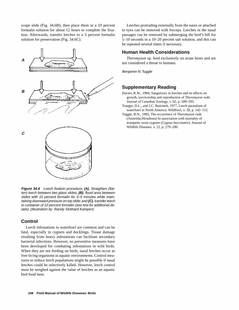
248 Field Manual of Wildlife Diseases: Birds
scope slide (Fig. 34.6B); then place them in a 10 percentformalin solution for about 12 hours to complete the fixa-tion. Afterwards, transfer leeches to a 5 percent formalinsolution for preservation (Fig. 34.6C).
ControlLeech infestations in waterfowl are common and can be
fatal, especially in cygnets and ducklings. Tissue damageresulting from heavy infestations can facilitate secondarybacterial infections. However, no preventive measures havebeen developed for combating infestations in wild birds.When they are not feeding on birds, nasal leeches occur asfree-living organisms in aquatic environments. Control mea-sures to reduce leech populations might be possible if nasalleeches could be selectively killed. However, leech controlmust be weighed against the value of leeches as an aquaticbird food item.
Leeches protruding externally from the nares or attachedto eyes can be removed with forceps. Leeches in the nasalpassages can be removed by submerging the bird’s bill for5–10 seconds in a 10–20 percent salt solution, and this canbe repeated several times if necessary.
Human Health ConsiderationsTheromyzon sp. feed exclusively on avian hosts and are
not considered a threat to humans.
Benjamin N. Tuggle
Supplementary ReadingDavies, R.W., 1984, Sanguivory in leeches and its effects on
growth, survivorship and reproduction of Theromyzon rude:Journal of Canadian Zoology, v. 62, p. 589–593.
Trauger, D.L., and J.C. Bartonek, 1977, Leech parasitism ofwaterfowl in North America: Wildfowl, v. 28, p. 142–152.
Tuggle, B.N., 1985, The occurrence of Theromyzon rude(Annelida:Hirudinea) In association with mortality oftrumpeter swan cygnets (Cygnus buccinator): Journal ofWildlife Diseases, v. 22, p. 279–280.
Figure 34.6 Leech fixation procedure. (A), Straighten (flat-ten) leech between two glass slides; (B), flood area betweenslides with 10 percent formalin for 3–5 minutes while main-taining downward pressure on top slide; and (C), transfer leechto container of 10 percent formalin (see text for additional de-tails). (Illustration by Randy Stothard Kampen)
A
B
C

Miscellaneous Parasitic Diseases 249
Chapter 35
Miscellaneous Parasitic Diseases
Free-ranging wild birds are afflicted with numerous otherparasites that occasionally cause illness and death. Some ofthese parasites, such as two of the trematodes or flukes high-lighted below, can cause major die-offs. This section aboutparasitic diseases concludes with descriptions of some addi-tional parasites that field biologists may encounter in wildbirds. This listing is by no means complete and it is intendedonly to increase awareness of the diversity of types of para-sites that might be encountered during examinations of wildbirds. One should not assume that the parasites found duringthe examination of bird carcasses caused their death. Be-cause parasites of birds vary greatly in size from a protozoaof a few microns in length to tapeworms of several inches inlength and because they can be found in virtually all tissues,body cavities and other locations within the bird, the obser-vation of the parasites will depend on their visibility and thethoroughness of the examination. Therefore, it is generallybeneficial to submit bird carcasses to qualified disease diag-nostic laboratories to obtain evaluations of the significanceof endoparasites or of ectoparasites. The methods that areused to preserve the carcass, tissues, or other specimens canenhance or compromise the ability of specialists to identifythe parasite to species, and even to genera, in some instances.Therefore, whenever possible, it is best to contact the diag-nostic laboratory that will receive the specimens and obtaininstructions for collecting, preserving, and shipping fieldsamples (See Chapters 2 and 3).
Endoparasites
TrematodesMost trematodes or flukes have complex life cycles that
require two intermediate hosts (Fig. 35.1) in which the para-sites develop before they become infective for the definitive,final bird host. In general, a mollusc is the first intermediatehost, and is often a species that lives in the aquatic environ-ment. Therefore, the aquatic environment brings potentialhosts (waterbirds) and these parasites into close proximityand results in infections of waterfowl and other waterbirdsthat sometimes have fatal consequences.
Sphaeridiotrema globulus is a small trematode less than1 millimeter in length that has been reported to cause die-offs in waterfowl, especially in diving ducks and swans, and,occasionally, in coot. Birds become infected by eating snails(the second intermediate host) that are infected with themetacercarial stage of the parasite (Fig. 35.1). This trema-tode feeds on blood, leading to severe blood loss and ane-mia. Birds often die acutely from shock due to an abnor-
mally decreased volume of circulating plasma in the body orhypovolemic shock. Die-offs usually occur in the late sum-mer or early fall and have been reported in the United Statesmost commonly in the Northeast and North Central States,with a few reports from Western States. Die-offs will oftenrecur yearly at the same locations. For example, mortalityfrom S. globulus is suspected as a recurring event on the St.Lawrence River in southern Quebec. Infections by S. globulushave also been found elsewhere in Canada. The trematode isalso present in the Old World and in Australia. Field signsinclude lethargy and bloodstained vents, although these signsare also found with duck plague (See Chapter 16). Grosslesions can appear as an inflammation or enteritis that is char-acterized by obvious hemorrhages in the lower small intes-tine or as inflammation of the intestinal wall with areas ofulcer-like erosions and the presence of a mixture of bloodand fibrin (Fig. 35.2).
Experimental infections of mute swans with S. globulusindicated that as few as 100 metacercariae could be lethal.Juvenile mallard ducks that had no previous exposure toS. globulus died when infected with 550 metacercariae. Someimmunity has been shown to exist in experimental infectionsof mallards. Adult mallards that were given 100 metacer-cariae and that later were challenged with 2,500 metacer-cariae survived, but those that received only 2,500 metacer-cariae died. Depending on how heavily snail populations areinfected, some birds can receive a lethal dose during lessthan 24 hours of feeding. Susceptible waterfowl generallydie 3–8 days postinfection after ingesting a lethal dose ofS. globulus. Younger birds are generally more susceptiblethan older birds.
No parasite control measures have been developed. Anyattempts at control would need to take into account the factthat the intermediate hosts are different within different geo-graphic areas of the United States. There have been no re-ports of this parasite infecting humans.
Cyathocotyle bushiensis is another trematode that infectswaterfowl and coot in the United States and Canada. Thistrematode can be found in the lower intestine, most com-monly in the cecae or blind pouches extending out from thebeginning of the large intestine (Fig. 35.3). The worms areslightly larger than S. globulus, measuring 1.7–1.8 millime-ters in length. The life cycle for this parasite is similar to thatof S. globulus. Birds become infected by consuming snailsharboring the metacercariae. Disease caused by this trema-tode has been reported in black duck, blue-winged teal, green-winged teal, and coot. This trematode was first described inEngland and it is most likely limited by the geographic dis-

250 Field Manual of Wildlife Diseases: Birds
tribution of the snail, Bithynia tentaculata, which serves asthe first intermediate host. This snail is found within the GreatLakes Basin in the United States.
There are no field signs associated with infection byC. bushiensis that have any diagnostic value. Common grosslesions in birds include hemorrhagic areas with plaque for-mation and some cheese-like or caseous core formation inthe lumen of the cecae. Hemorrhage and plaque formationwithin the cecae are often present during early stages of in-fections (5–7 days after infection), whereas semisolid cellu-lar debris that plugs the cecae are found in later infections(day 9 and later). Tissue damage within the birds is directlyrelated to the attachment of the fluke to the mucosa and theeffect of secretions by the fluke on cellular process withinthe intestine. Studies have indicated that there is a negativerelationship between the number of flukes present and weightgain by the bird and a positive relationship between the num-
ber of flukes present and the number of white blood cellsand body temperature. These findings reflect the nutritionalimpacts (reduced weight gain or weight loss) and body re-sponse to infection (increased white blood cells and bodytemperature). It is not known if weight loss in infected birdsis due to cecal and lower intestine dysfunction or if the birdsdo not feed as much as noninfected birds. It has been sug-gested that morbidity and mortality are directly related tovascular leakage in the cecae from tissue damage that leadsto dehydration of the bird. Experiments have shown that afluke burden as low as 32 resulted in deaths of Pekin duck-lings on day 8 postinfection, which indicates that ducks maysuccumb rather quickly to infection.
Control of this parasite, as for all trematode infections,would require preventing birds from feeding on infectedsnails or other invertebrate intermediate hosts. No reports ofhuman infection have been reported for this trematode.
Eggs are shed via bird's feces
Eggs embryonate and produce a free-swimmingmiracidium which isingested by the firstintermediate host
Metacercariae develop into adult trematodes (flukes) within the host andproduce eggs
Bird ingests infectiousmetacercariae while feeding
First intermediate host
Second intermediate host
Further development of the parasite occurs in first intermediate host
Free-swimming cercariae leave first host
Cercariae become encysted as metacercariae on or in second intermediate host
Ingestion of infective parasite by new bird hosts continues the life cycle
Fig. 35.1 General trematode (fluke) life cycle.

Miscellaneous Parasitic Diseases 251
Fig. 35.3 Cyathocotyle bushiensis from the cecae of an American coot.
Fig. 35.2 Small intestine of lesser scaup that died from infection with Sphaeridiotrema globulus. Notethe white flecks, which are S. globulus, and obvious hemorrhage.
Pho
to b
y Ja
mes
Run
ning
enP
hoto
by
Con
stan
ce L
. Rod
eric
k

252 Field Manual of Wildlife Diseases: Birds
Leyogonimus polyoon (Fig. 35.4) is a newly reportedtrematode in North America that caused the deaths of over1,500 and 11,000 coots in Northeastern Wisconsin duringthe falls of 1996 and 1997, respectively (Fig. 35.5). Thistrematode is of similar small size (0.7–1.0 millimeters inlength) as S. globulus (Fig. 35.2). It is known to cause deathin coot and common moorhen in Europe, but the Wisconsinoutbreaks are the first documentation of this parasite caus-ing mortality in North America. The susceptibility of otherNorth American birds, beside coot, remains unknown. At theWisconsin location, various waterfowl species were dyingfrom infections of S. globulus while the coot were dying fromL. polyoon infections. None of the waterfowl were found tobe infected with L. polyoon.
The life cycle for L. polyoon is not known, although thesuspected first intermediate host is the snail Bithnia tentac-ulata. Investigations at the National Wildlife Health Center(NWHC) have disclosed that snails that were collected in
Wisconsin were infected with a cercariae or a larval form ofthe parasite that fits the literature description of L. polyoon.Additional studies are required to confirm that these larvalforms are L. polyoon. Also, the intermediate host for the in-fective larval form or metacacariae has not yet been found.L. polyoon infects primarily the upper and middle areas ofthe small intestine. No significant field signs are associatedwith infection by L. polyoon. Gross lesions seen at necropsyinclude severe enteritis characterized by thickening of theintestinal wall and a fibrous-to-caseous core of necrotic de-bris that blocks the lumen of the intestine (Fig. 35.6).
The location where these outbreaks occurred is a lake thatis drained by a stream, that is underlain with sand, and thathas substantial growths of water weeds. The shoreline hasbeen extensively developed for home sites and other humanuse, and the lake is used for recreation. L. polyoon has notbeen reported to infect humans.
Fig. 35.5 American coot and various species of waterfowlfrom die-off in Shawano Lake, Wisc. Coot mortality was dueto Leyogonimus polyoon, but waterfowl mortality was dueto Sphaeridiotrema globulus.
Fig. 35.4 Leyogonimus polyoon from thesmall intestine of an American coot.
Pho
to b
y K
. Bro
ckm
an-M
eder
as, W
isco
nsin
Dep
artm
ent o
f Nat
ural
Res
our c
es
Pho
to b
y C
onst
ance
L. R
oder
ick
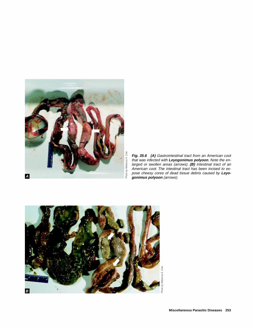
Miscellaneous Parasitic Diseases 253
Fig. 35.6 (A) Gastrointestinal tract from an American cootthat was infected with Leyogonimus polyoon. Note the en-larged or swollen areas (arrows). (B) Intestinal tract of anAmerican coot. The intestinal tract has been incised to ex-pose cheesy cores of dead tissue debris caused by Leyo-gonimus polyoon (arrows).P
hoto
by
Reb
ecca
A. C
ole
Pho
to b
y R
ebec
ca A
. Col
e
A
B
➡
➡
➡➡➡
➡

254 Field Manual of Wildlife Diseases: Birds
CestodesTapeworms are common in wild birds, but they seldom
cause death. Heavy burdens of these parasites may reducethe vigor of the bird and serve as a predisposing factor forother disease agents, or the parasites may occlude the intes-tine (Fig. 35.7). One genus, Gastrotaenia sp., lives in thegizzard and penetrates the keratohyalin lining or the hornycovering of the gizzard pads, causing inflammation and nec-rosis. Cloacotaenia sp. inhabit the ureter or the tubular areathat transports wastes from the kidneys to the cloaca in somewaterfowl.
NematodesTrichostrongylidosis, the disease that is caused by Tri-
chostrongylus tenius, is not currently a significant problemwithin the United States. However it is included becausetrichostrongylidosis, in natural populations of grouse in theirnative habitat in Scotland and elsewhere in the United King-dom, demonstrates the impact that a parasite can have on thepopulation dynamics of the bird host that it infects. T. teniusis a common nematode or roundworm that is found withinthe ceca of some types of wild birds, primarily grouse, geese,and poultry. This parasite has a direct life cycle that is closelyassociated with host food preferences for terrestrial vegeta-
tion (Fig. 35.8). Trichostrongylidosis outbreaks can occurwhenever birds are hatching because of the synchronousphase of the ecology of the parasite and the feeding habits ofbird hosts.
Hatchability of the parasite eggs and survival of the free-living larval stages are moisture and temperature dependent.The parasite eggs do not develop under dry conditions andthe free-living larvae are generally killed by freezing tem-
Figure 35.7 Large numbers of tape worms may occlude theintestine.
Parasite eggs are shed via bird's feces
Eggs mature in the environment and produce larvae that undergo further development within the environment. Larvae crawl to the tips of vegetation where they accumulate in drops of water
Bird hosts
Birds ingest larvae whilefeeding upon moisture-ladenvegetation
Figure 35.8 Life cycle of the nematodeTrichostrongylus tenius.
Pho
to b
y M
ilton
Frie
nd

Miscellaneous Parasitic Diseases 255
peratures. When the feces that contain the parasite eggs arekept moist and when the ambient air temperature is suitable,first-stage larvae develop within the egg and hatch in about2 days; free-living second-stage larvae develop within an-other 1-1/2-to-2 days; and development to infective third-stagelarvae requires an additional 8–16 days. Studies in Scotlandindicate that infective larvae crawl to the tips of moist heatherand accumulate there in drops of water provided by the mistyweather. Grouse that feed on the tips of the heather ingestthe larvae along with their food. Infective larvae molt twicemore within the ceca of the bird before they become sexu-ally mature adults.
Infections have been reported in chicken, turkey, guineafowl, pheasant, quail, pigeons, ducks, and geese; but infec-tions are most notable for red grouse because this parasitehas clearly been shown to regulate natural populations ofthis species. Because adult worms can survive in their birdhost for more than 2 years, all adult birds evaluated in somepopulations have been found to be infected, thereby, provid-ing a reservoir for infection of young. In addition, larvae canarrest development within the ceca, overwinter, and then re-sume development in the spring. The temperature and mois-ture requirements for hatchability of the parasite eggs andsurvival of larvae results in synchronized availability of para-sites during the period of production of young grouse. Thisresults in the primary occurrence of disease and mortality ingrouse during the spring. The hatchlings are exposed to in-fective larvae soon after the diet for chicks changes frominsects to vegetation.
The impacts of infection by T. tenius are greater than justchick mortality. These parasites also decrease available en-ergy for egg laying by adult birds. The resting metabolic rateis increased and there is a decrease in food intake by thebird. The resulting impact is reduced fecundity within thepopulation (fewer chicks) along with high chick mortality.This combination of impacts controls population levels.
Echinuria uncinata is a common nematode that infectsthe proventriculus of various waterfowl species. The life cycleis indirect, and the parasite uses zooplankton, especiallyDaphnia sp., as intermediate hosts (Fig. 35.9). Wormsmature in a duck approximately 51 days after it eats infectedzooplankton. Adult worms, which are approximately 5 mil-limeters long, burrow headfirst into the mucosa and submu-cosa of the proventriculus, causing tissue swelling and in-flammation. Tumor-like nodules form and can be largeenough to obstruct the lumen of the proventriculus. This para-site can be especially dangerous to waterfowl where zoo-plankton blooms coincide with the hatching of young. Oftenbirds that are late to hatch and do not have fully developedimmune systems can consume enough infected zooplanktonin a very short period of time to become severely infected. Inareas where water is shallow, where zooplankton populationsare numerous, and where birds are crowded into the area,this roundworm can be transmitted to many birds during ashort time.
This occurred within a population of the endangeredLaysan duck on Laysan Island, Hawaii during the fall of 1993.A drought had struck the island and the brine flies that the
Parasite eggs are shed via bird's feces
Infection occurs via ingestion of infected zooplankton
Infective larvae develop in zooplankton
Larvae mature in duck and adult worms burrow headfirst into the proventriculus
Bird hosts
Figure 35.9 Life cycle of the nematodeEchinuria uncinata.
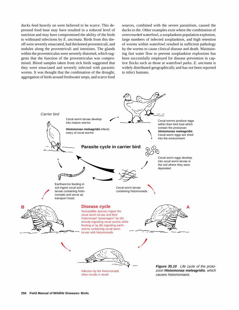
256 Field Manual of Wildlife Diseases: Birds
ducks feed heavily on were believed to be scarce. This de-pressed food base may have resulted in a reduced level ofnutrition and may have compromised the ability of the birdsto withstand infections by E. uncinata. Birds from this die-off were severely emaciated, had thickened proventriculi, andnodules along the proventriculi and intestines. The glandswithin the proventriculus were severely distorted, which sug-gests that the function of the proventriculus was compro-mised. Blood samples taken from sick birds suggested thatthey were emaciated and severely infected with parasiticworms. It was thought that the combination of the drought,aggregation of birds around freshwater seeps, and scarce food
sources, combined with the severe parasitism, caused theducks to die. Other examples exist where the combination ofovercrowded waterfowl, a zooplankton population explosion,large numbers of infected zooplankton, and high retentionof worms within waterfowl resulted in sufficient pathologyby the worms to cause clinical disease and death. Maintain-ing fast water flow to prevent zooplankton explosions hasbeen successfully employed for disease prevention in cap-tive flocks such as those at waterfowl parks. E. uncinata iswidely distributed geographically and has not been reportedto infect humans.
Cecal worm eggs developinto cecal worm larvae inthe soil where they were deposited
Cecal worm larvae containing histomonads
Earthworms feeding insoil ingest cecal wormlarvae containing histo-monads and serve astransport hosts
Histomonas meleagridis infectsovary of cecal worms
Parasite cycle in carrier bird
Cecal worm larvae developinto mature worms
Cecal worms produce eggs within their bird host which contain the protozoanHistomonas meleagridis. Cecal worm eggs are shed into the environment
Carrier bird
Disease cycleSusceptible species ingest the cecal worm larvae and their histomonad "passengers" by (A) directly ingesting cecal worms while feeding or by (B) ingesting earth-worms containing cecal worm larvae with histomonads
AB
Infection by the histomonads often results in death
Figure 35.10 Life cycle of the proto-zoon Histomonas meleagridis, whichcauses histomoniasis.
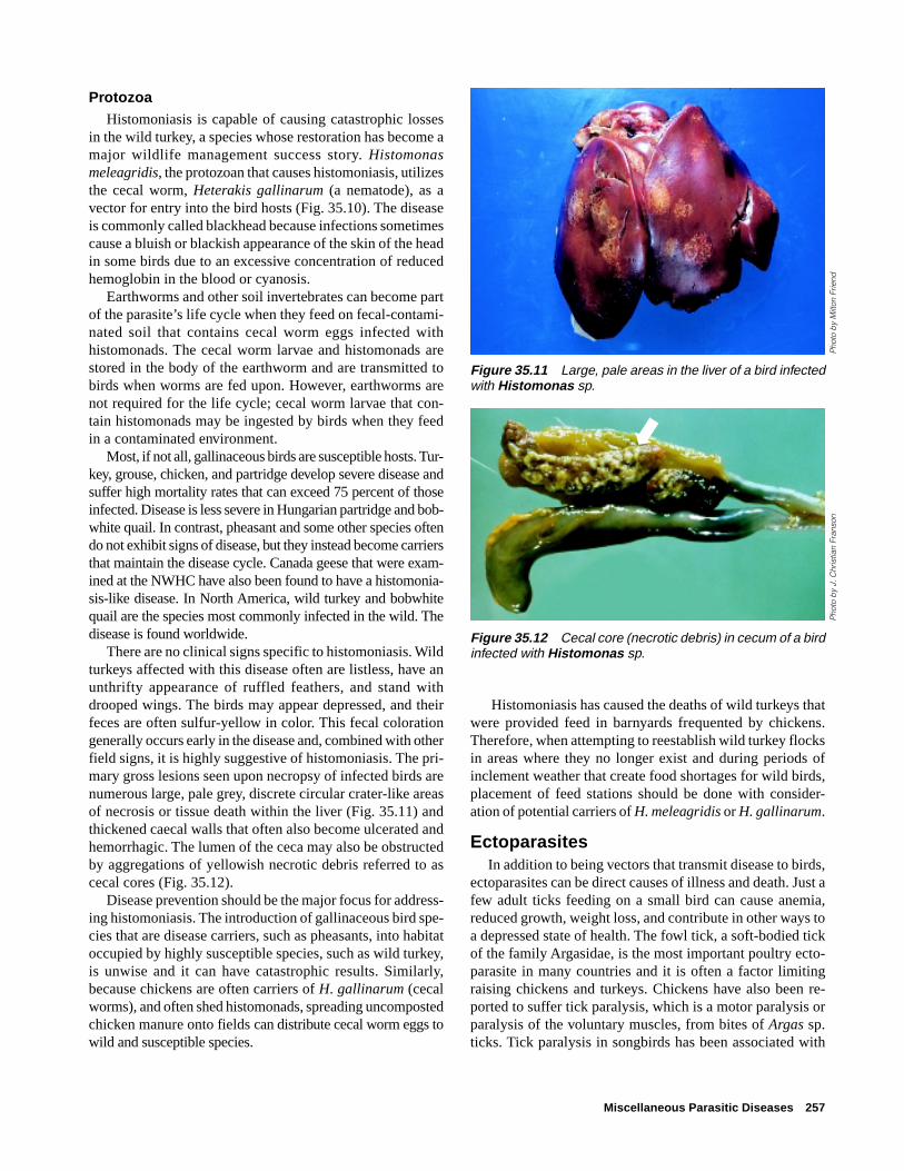
Miscellaneous Parasitic Diseases 257
ProtozoaHistomoniasis is capable of causing catastrophic losses
in the wild turkey, a species whose restoration has become amajor wildlife management success story. Histomonasmeleagridis, the protozoan that causes histomoniasis, utilizesthe cecal worm, Heterakis gallinarum (a nematode), as avector for entry into the bird hosts (Fig. 35.10). The diseaseis commonly called blackhead because infections sometimescause a bluish or blackish appearance of the skin of the headin some birds due to an excessive concentration of reducedhemoglobin in the blood or cyanosis.
Earthworms and other soil invertebrates can become partof the parasite’s life cycle when they feed on fecal-contami-nated soil that contains cecal worm eggs infected withhistomonads. The cecal worm larvae and histomonads arestored in the body of the earthworm and are transmitted tobirds when worms are fed upon. However, earthworms arenot required for the life cycle; cecal worm larvae that con-tain histomonads may be ingested by birds when they feedin a contaminated environment.
Most, if not all, gallinaceous birds are susceptible hosts. Tur-key, grouse, chicken, and partridge develop severe disease andsuffer high mortality rates that can exceed 75 percent of thoseinfected. Disease is less severe in Hungarian partridge and bob-white quail. In contrast, pheasant and some other species oftendo not exhibit signs of disease, but they instead become carriersthat maintain the disease cycle. Canada geese that were exam-ined at the NWHC have also been found to have a histomonia-sis-like disease. In North America, wild turkey and bobwhitequail are the species most commonly infected in the wild. Thedisease is found worldwide.
There are no clinical signs specific to histomoniasis. Wildturkeys affected with this disease often are listless, have anunthrifty appearance of ruffled feathers, and stand withdrooped wings. The birds may appear depressed, and theirfeces are often sulfur-yellow in color. This fecal colorationgenerally occurs early in the disease and, combined with otherfield signs, it is highly suggestive of histomoniasis. The pri-mary gross lesions seen upon necropsy of infected birds arenumerous large, pale grey, discrete circular crater-like areasof necrosis or tissue death within the liver (Fig. 35.11) andthickened caecal walls that often also become ulcerated andhemorrhagic. The lumen of the ceca may also be obstructedby aggregations of yellowish necrotic debris referred to ascecal cores (Fig. 35.12).
Disease prevention should be the major focus for address-ing histomoniasis. The introduction of gallinaceous bird spe-cies that are disease carriers, such as pheasants, into habitatoccupied by highly susceptible species, such as wild turkey,is unwise and it can have catastrophic results. Similarly,because chickens are often carriers of H. gallinarum (cecalworms), and often shed histomonads, spreading uncompostedchicken manure onto fields can distribute cecal worm eggs towild and susceptible species.
Figure 35.11 Large, pale areas in the liver of a bird infectedwith Histomonas sp.
Figure 35.12 Cecal core (necrotic debris) in cecum of a birdinfected with Histomonas sp.
Histomoniasis has caused the deaths of wild turkeys thatwere provided feed in barnyards frequented by chickens.Therefore, when attempting to reestablish wild turkey flocksin areas where they no longer exist and during periods ofinclement weather that create food shortages for wild birds,placement of feed stations should be done with consider-ation of potential carriers of H. meleagridis or H. gallinarum.
EctoparasitesIn addition to being vectors that transmit disease to birds,
ectoparasites can be direct causes of illness and death. Just afew adult ticks feeding on a small bird can cause anemia,reduced growth, weight loss, and contribute in other ways toa depressed state of health. The fowl tick, a soft-bodied tickof the family Argasidae, is the most important poultry ecto-parasite in many countries and it is often a factor limitingraising chickens and turkeys. Chickens have also been re-ported to suffer tick paralysis, which is a motor paralysis orparalysis of the voluntary muscles, from bites of Argas sp.ticks. Tick paralysis in songbirds has been associated with
Pho
to b
y J.
Chr
istia
n Fr
anso
n
➡
Pho
to b
y M
ilton
Frie
nd

258 Field Manual of Wildlife Diseases: Birds
the bite of the hard-bodied bird tick, Ixodes brunneus. Fatalparalysis from bites by this tick has been reported in numer-ous species of small birds. The engorged ticks in fatal casesare generally found on the bird’s head and they may be at-tached to its eyelids. Death results from a powerful neuro-toxin that is secreted by the tick while it feeds on the bird.Other species of Ixodes ticks have been associated with tickparalysis and mortality in marine birds, including albatrossand petrels. An ascending motor paralysis that starts at thefeet, progresses for 7–10 days, and ends in death has beenreported.
Heavy infestations of lice, mites, fleas, flies, and otherbiting insects have also been responsible for causing illnessand even death of wild birds, especially among nestlings.Conditions caused by these insects range from feather lossand skin damage from acariasis or mange, to myiases or in-festation with fly maggots, and anemia. Mites of the genusKnemidocoptes are the primary cause of mange in birds, andthe mites belong to the same family (Sarcoptidae) of mitesthat cause mange in mammals and humans. The Knemido-coptes sp. mites are specific to birds and they are not a hu-man health hazard.
More knowledge is needed about the role of ectoparasitesas causes of bird death. Proper identification of the speciesassociated with bird mortality is an important component ofsuch assessments; therefore, the presence of insects withinbird nests in which freshly dead nestlings are found shouldbe recorded, representative parasite specimens should be col-lected along with any visible parasites on the carcass, andthe parasites submitted should be with the carcass. Ticks andany heavy infestations of fleas, lice, and other insects on livebirds being handled for banding or other purposes shouldalso be noted, and, when practical, samples should be col-lected and submitted for identification to a parasitologist ordisease diagnostic laboratory. An abundance of such para-sites may be indicators of other health problems for the birds.
Rebecca A. Cole and Milton Friend
Supplementary ReadingAnderson, R.C., 1992, Nematode parasites of vertebrates, their
development and transmission: Wallingford, United Kingdom,C•A•B International, University Press, 578 p.
Arnall, L., and Keymer, I.F., 1975, Bird diseases, an introductionto the study of birds in health and disease: Bailliere Tindall,London, T.F.H. Publications, 528 p.
Calnek, B.W. and others, 1997, Diseases of Poultry (10th ed.):Ames, Iowa, Iowa State University Press, 1,080 p.
Delehay, T.M., Speakman, J.R., and Mass, R., 1995, The energeticconsequences of parasitism: effects of a developing infectionof Trichostrongylus tenus (Nematoda) on red grouse (Lagopuslagopus scoticus) energy balance, body weight and condition:Parasitology, v. 110, p. 473–482.
Davidson, W.R., and Nettles, V.F., 1997, Field manual of wildlifediseases in the Southeastern United States (2d ed.): Athens,Ga., University of Georgia, 417 p.
Gagnon, C., Scott, M.E., and McLaughlin, J.D., 1993, Grosslesions and hematological changes in domesticated mallardducklings experimentally infected with Cyathocotylebushiensis (Digenea): Journal of Parasitology, v. 79, p. 757–762.
Hove, J., and Scott, M.E., 1988, Ecological studies on Cyatho-cotyle bushiensis (Digenea) and Sphaeridiotrema globulus(Digenea), possible pathogens of dabbling ducks in southernQuebec: Journal of Wildlife Diseases, v. 24, p. 407–421.
Hudson, P.J., 1986, The effect of a parasitic nematode on thebreeding production of red grouse: Journal of Animal Ecology,v. 55, p. 85–92.
Huffman, J.E., and Roscoe, D., 1989, Experimental infections ofwaterfowl with Sphaeridiotrema globulus (Digenea): Journalof Wildlife Diseases, v. 25, p. 143–146.
Mucha, K.H., and Huggman, J.E., 1991, Inflammatory cellstimulation and wound healing in Sphearidiotrema globulusexperimentally infected mallard ducks (Anas platyrhynchos):Journal of Wildlife Diseases, v. 27, p. 428–434.