Embed Size (px)
Citation preview

Diagn Interv Radiol 2013; 19:471–478
© Turkish Society of Radiology 2013
Papillary lesions of the breast: imaging findings and diagnostic challenges
Pooja Jagmohan, Felicity Jane Pool, Thomas Choudary Putti, Jill Wong
BREAST IMAGINGPICTORIAL ESSAY
ABSTRACT Papillary breast lesions encompass a wide spectrum of pa-thologies ranging from benign lesions, such as solitary in-traductal papilloma, to the uncommon papillary carcinoma. These lesions have various clinical presentations and diverse radiological features. Differentiating benign and malignant papillary lesions based on imaging features may often be difficult. Other benign and malignant pathologies can also mimic papillary lesions on imaging, and tissue diagnosis is essential. Imaging plays an important role in lesion identifi-cation, assessment of extent, tissue sampling, and follow-up. Surgical excision has been recommended for all papillary le-sions due to an increased incidence of high-risk lesions and neoplasia even with percutaneous, biopsy-proven benign papillomas. This review looks at papillary breast lesions from the radiologists’ standpoint and discusses the clinical, imag-ing, and pathological features of these lesions, as well as the role of imaging in their evaluation.
P apillary lesions in the breast are uncommon but arise from a wide range of pathologies and have diverse clinical and im-aging features. A papillary lesion is characterized by an arbo-
rescent structure composed of fibrovascular stalks covered by a lay-er of epithelial cells with or without an intervening myoepithelial cell layer (1). Overlapping features make differentiation of benign and malignant papillary lesions difficult on imaging, and a tissue diagnosis is essential. Definitive histopathologic diagnosis on core biopsy can occasionally be difficult. Additionally, even those lesions shown by percutaneous biopsy to be benign papillomas are associ-ated with an increased likelihood of high-risk lesions and neoplasia. Due to nonspecific findings on imaging and histopathology, as well as varying malignant potential, papillary lesions present significant diagnostic and management challenges for the radiologists, pathol-ogists, and surgeons. We briefly review the types of papillary lesions, multimodality imaging findings, and the radiologist’s role in their evaluation.
The types and clinical features of papillary lesionsPapillary lesions can be broadly categorized as benign or malig-
nant. Benign papillary lesions include a solitary intraductal papillo-ma, multiple intraductal papillomas, and atypical ductal hyperplasia (ADH) within a papilloma. Malignant papillary lesions include duc-tal carcinoma in situ (DCIS) arising in a papilloma, papillary DCIS, intracystic or encapsulated papillary carcinoma, solid papillary car-cinoma, invasive papillary carcinoma arising in an intracystic papil-lary carcinoma, and invasive papillary carcinoma (1).
Intraductal papillomaSolitary papillomas arise from a large central duct, are more
common in perimenopausal women, and present with nipple dis-charge. Multiple papillomas are peripheral lesions arising from the terminal duct lobular unit. These are less common, usually affect a younger age group, and present as a palpable mass. Both can be associated with proliferative and high-risk lesions, such as ra-dial scars, and with an increased risk of cancer. Patients with a solitary papilloma without atypia have a two-fold greater risk of cancer, whereas those with multiple papillomas have a relative risk of three (2).
From the Departments of Diagnostic Imaging (P.J. [email protected], F.J.P.) and Pathology (T.C.P.), National University Health System (NUHS), Singapore, Singapore; Department of Oncologic Imaging (J.W.), National Cancer Centre, Singapore, Singapore.
Received 29 January 2013; revision requested 28 February 2013; revision received 29 March 2013; accepted 9 April 2013.
Published online 28 August 2013.DOI 10.5152/dir.2013.13041
471

472 • November–December 2013 • Diagnostic and Interventional Radiology Jagmohan et al.
ADH and DCIS both describe a neoplastic population of cells within a papilloma. They are more common with multiple papillomas. The atyp-ical component is defined in various ways. Some authors define ADH as a population of such cells measuring ≤3 mm and DCIS as a population of such cells measuring >3 mm (3). Others consider these lesions to be in situ papillary carcinoma (4). Patients with solitary and multiple papillo-mas with atypia have five- and sev-en-fold increased risks of cancer, re-spectively (2).
Papillary DCISAs a variant of DCIS, this lesion is
characterized by neoplastic cells that grow around the internal lining of a duct with papillary projections. Ex-tensive ductal spread and underes-timation of the noncalcified part of the DCIS may make it difficult to ob-tain clear margins on excision. These lesions are associated with higher rates of multicentricity and microin-vasion than other DCIS variants.
Papillary carcinomaPapillary carcinomas are rare, com-
prising 1%–2% of all breast malig-nancies (1). They are more common in the postmenopausal age group and present with a palpable mass and nipple discharge. The absence of an intact myoepithelial cell layer within the papillary structures is an import-ant marker for malignant lesions.
Intracystic or encapsulated papil-lary carcinoma is defined by the pres-ence of papillary carcinoma within a cystically dilated duct. Intracystic papillary carcinomas that are associ-ated with an invasive component are staged according to the size of the in-vasive component (1). A solid papil-lary carcinoma is an indolent tumor composed of circumscribed nodules of ovoid or spindle-shaped epithelial cells with a low nuclear grade. Intra-cystic and solid papillary carcinomas rarely metastasize and are associated with an excellent prognosis. Inva-sive papillary carcinoma is described as an infiltrating breast carcinoma exhibiting an exclusively papillary morphology. It is associated with a better prognosis compared with oth-er forms of invasive carcinoma.
Imaging findingsLike their pathology, the imaging
features of papillary lesions are di-verse.
PapillomaA solitary intraductal papilloma is
usually observed on mammography as a rounded or ovoid, well-circum-scribed retroareolar mass (Figs. 1, 2) that may be associated with ductal dilatation (Fig. 3). Smaller lesions may be occult on mammography. Multiple papillomas are usually pe-ripheral in location and can be bilat-eral. Calcifications are uncommon and include both coarse dense cal-
cifications and microcalcifications (Figs. 2, 4) (5).
The characteristic ultrasonogra-phy (US) finding of a papilloma is a solid mural nodule within a dilated duct (Fig. 5). Other features include an intracystic mass or a well-cir-cumscribed hypoechoic solid mass (Figs. 1, 2, 6). Ductal dilatation may be the only finding in a small papilloma. Color Doppler imaging can depict a vascular pedicle with-in the mural nodule (Figs. 1, 2) (6, 7). Ductography may show an intraluminal filling defect, ductal dilatation, ductal wall irregularity, and distortion. Atypical papillomas may have imaging features similar to benign papillomas, and the diagno-sis is usually based on histopathol-ogy (Figs. 7, 8). Magnetic resonance imaging (MRI) ductography using a microscopic coil has been proposed as a noninvasive alternative for the detection of intraductal papillomas (8).
The current role of MRI for eval-uation of papillomas is unclear, and most papillomas are detected as incidental masses. There may be a role for MRI in the preoperative work-up of multiple nodular lesions to assess the extent of disease prior to surgical excision. MRI may also be useful in the follow-up of recur-rent lesions. Most papillomas are observed as round, ovoid, or lob-ulated well-circumscribed masses with or without ductal dilatation
Figure 1. a, b. A patient with a palpable left breast lump. The mammography in mediolateral-oblique view of the left breast (a) shows a well-circumscribed, ovoid retroareolar opacity. The composite US and color Doppler image (b) shows the palpable lesion as a well-defined hypoechoic solid nodule adjacent to the nipple with prominent internal vascularity. Initial US-guided 14 G core biopsy showed a benign intraductal papilloma. The patient underwent surgical excision, and the diagnosis was confirmed on histopathological examination of the surgical specimen.
a b

Volume 19 • Issue 6 Imaging findings of papillary lesions of the breast • 473
(Figs. 4, 9). Occasionally, a benign papilloma can have spiculated mar-gins on mammography, US, and MRI and can thus mimic malignant disease. Variable enhancement pat-terns have been described, making differentiation from malignancies difficult (7, 8). In atypical papillomas
with DCIS, MRI may play a role in evaluating the extent of DCIS.
Papillary DCISPapillary DCIS can be occult on
imaging. When present, findings include pleomorphic calcifications and architectural distortion on
mammography (Fig. 10), ill-defined hypoechoic mass or calcifications on US, and non-mass-like enhancement on MRI (7).
Papillary carcinomaMammographic findings of pap-
illary carcinomas include round or
Figure 2. a, b. A patient with a palpable, central right breast lump. The mammography in mediolateral-oblique view of the right breast (a) shows a well-circumscribed density in the retroareolar region with few scattered coarse and punctate calcifications (arrowheads). The composite US and color Doppler image (b) shows that this density corresponds to a complex cystic lesion with diffuse, low-level internal echoes and a small solid component (arrows) with vascularity on Doppler insonation. A papillary lesion without atypia within a dilated duct was observed on histopathological examination following a 14 G US-guided core biopsy. A benign papilloma was confirmed on histopathological examination of the surgical excision specimen.
a b
Figure 3. A patient with a screen-detected abnormality in the left breast. The mammography in cranio-caudal view shows a tubular branching structure (arrows) in the left outer breast. On US (not shown), this structure corresponded to a dilated duct with an intraductal nodule. A 14 G core biopsy for this lesion was reported as an intraductal papilloma. Subsequent 11 G vacuum-assisted excision biopsy confirmed this lesion to be a benign intraductal papilloma.

474 • November–December 2013 • Diagnostic and Interventional Radiology Jagmohan et al.
Figure 4. a–c. A patient recalled for assessment of a screen-detected abnormality in the right breast. The mammography in mediolateral-oblique view (a) shows an asymmetric density (arrow) associated with loosely clustered calcifications (arrowhead) in the right lower breast. US (b) shows a poorly defined hypoechoic area from the 3 o’clock to 6 o’clock position in the right breast. MRI (c) performed for further evaluation shows the lesions as lobulated, high-signal nodules in a ductal distribution on the axial vibrant precontrast phase (left-sided image). Subtracted image (right side) of the fifth minute after contrast injection shows mild patchy enhancement in the affected area. MR-guided vacuum-assisted biopsy of the abnormal area yielded an intraductal papilloma. The patient declined surgical excision biopsy.
a b
c
Figure 5. a–c. Benign intraductal papilloma in a patient with bloody discharge from the left nipple. US (a) shows a dilated duct (arrowhead) with an intraluminal hypoechoic lesion (arrow) in the left periareolar region. US-guided 14 G core biopsy showed an intraductal papilloma without atypia. Histopathological examination of the surgical excision specimen (b [H-E, ×40], c [H-E, ×200]) showed a benign intraductal papilloma with an arborescent structure and focal sclerosis.
a b c
Figure 6. A patient with multiple papillomas who had a previous excision biopsy for a left breast nodule that was lobular in situ carcinoma and ductal papilloma on histopathological examination. Follow-up US shows new hypoechoic irregular nodules in both breasts; two of these are shown in the composite US image. Benign papillomas detected on histopathological examination of the US-guided 14 G core biopsy specimens did not show any atypia.

Volume 19 • Issue 6 Imaging findings of papillary lesions of the breast • 475
oval, circumscribed solitary or clus-tered masses, which may be associ-ated with microcalcifications (Figs. 11–13). Spiculations are fairly un-common probably due to the lack of fibrosis. On US, the lesion may be seen as an intraductal mass with or without ductal dilatation, a complex solid cystic mass, or single or multi-ple solid nodules (Figs. 11–14). These lesions are usually vascular and have a tendency to bleed spontaneously, resulting in intracystic fluid-debris levels (7, 9).
On MRI, papillary carcinomas may appear as enhancing nodular lesions or enhancing complex cysts with variable kinetic curves. There are no specific features in terms of mor-phology or kinetic analysis; thus, the usefulness of MRI for differentiation of benign and malignant disease is limited.
Differentiating benign and malignant papillary lesions on imaging
A nonparallel orientation, echo-genic halo, posterior acoustic en-hancement, and associated micro-calcification are reported to be more frequent in malignant lesions (10). Another study evaluating the role of clinicoradiological features in core biopsy proved that benign papillo-mas without atypia showed a high-er postexcision upgrade for patients >50 years, lesions that were ≥1 cm, lesions that were ≥3 cm from the nip-ple, and lesions categorized as Breast Imaging-Reporting and Data System (BI-RADS) 4c and 5 (11). However, due to overlapping findings, imag-ing is neither sensitive nor specific for differentiating benign and malig-nant papillary lesions (12).
Imaging differentials of papillary lesions
Segmental ductal dilatation with no demonstrable ductal mass may also be observed in ductal ectasia. Intraductal content such as blood products, inspissated secretions in ductal ectasia, and neoplastic cells in DCIS can mimic a papillary lesion. Differentials for a complex cystic le-
Figure 7. Atypical papilloma. The composite US and color Doppler image of the right breast shows an irregular hypoechoic mass with angular margins and peripheral vascularity. Histopathological examination of the excision specimen showed a papilloma with focal atypical ductal hyperplasia.
Figure 8. Papilloma with atypia in a patient with left bloody nipple discharge and a normal mammogram. The composite US and color Doppler image shows a distended duct (arrowhead) in the 4 o’clock position with some low-level internal echoes within (left side of image). No internal vascularity was observed on color Doppler insonation (middle image). At the 10 o’clock position (right side of image), there is a small, ovoid, hypoechoic intraductal structure near the nipple; the ductal extension is indicated by an arrowhead. The lesions underwent excision biopsy following hook-wire localization. Both were intraductal papillomas, and the 10 o’clock lesion showed a small focus of DCIS.
Figure 9. Intraductal papilloma on MRI. Axial MRI T1 vibrant fat-saturated postgadolinium image at 1 min postinjection shows an enhancing lobulated mass in the right breast. The mass had rapid initial enhancement and delayed plateau kinetics. Another papilloma with similar imaging features was observed in the left breast (not shown).
476 • November–December 2013 • Diagnostic and Interventional Radiology Jagmohan et al.
sion include hematomas, abscesses, and fat necrosis. A papillary lesion appearing as a well-defined solid nodule may be indistinguishable from a fibroadenoma (Fig. 15). Addi-tionally nonpapillary and papillary carcinomas can have similar appear-ances.
The radiologist’s role and challenges
Imaging is crucial for lesion iden-tification and local staging, guiding tissue diagnosis, and follow-up.
Mammography and US form the mainstay of lesion identification. However, some lesions, such as small papillomas or papillary DCIS, may be occult or have equivocal findings such as nonspecific segmental ductal dilatation. Ductography can help to detect small papillary lesions. Assess-ing the extent of pathology is also critical, particularly for multiple pap-
illomas, DCIS, and carcinomas. In the latter cases, MRI may be useful.
Tissue sampling of papillary le-sions can utilize fine-needle aspira-tion, core biopsy, and vacuum-assist-ed biopsy. US-guided percutaneous core biopsy is used most frequently because it allows for real-time visual-ization, a feature that is essential for sampling the solid component of the lesion.
Nevertheless, definitive histo-pathological categorization of per-cutaneous biopsy samples may be limited by factors such as tissue fragmentation and undersampling. Numerous studies have found a sig-nificant upgrade in the atypia rate (6.9%–27.7%) or malignancy (3.1%–20%) for benign papillomas diag-nosed by core-needle biopsy (13). These findings have resulted in sur-gical excision being recommended even for imaging concordant, percu-
taneous, biopsy-proven benign pap-illomas. Other authors have found a low risk of malignancy for benign papillomas diagnosed by core-nee-dle biopsy and recommend mam-mographic follow-up for benign and excision for atypical papillomas (14). Vacuum assistance has also been uti-lized for the removal of select papil-lary lesions and has the potential to improve lesion sampling and reduce the number of open surgeries (15). Here, again, the potential for recur-rence suggests the need for close fol-low-up.
ConclusionPapillary lesions are an uncommon
group of breast diseases that present unique diagnostic and management challenges due to a wide spectrum of imaging appearances, pathologies, and malignant potential. The radiol-ogist plays an important role in the
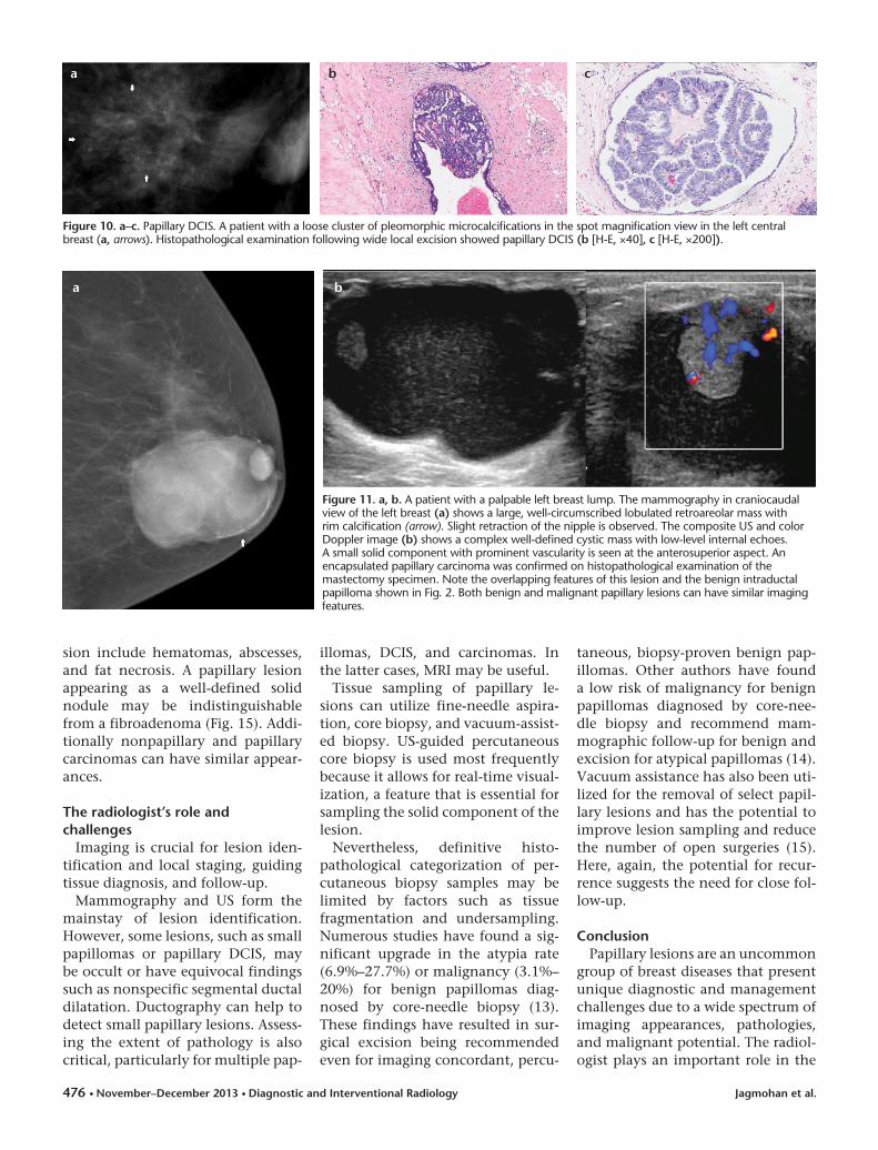
Figure 10. a–c. Papillary DCIS. A patient with a loose cluster of pleomorphic microcalcifications in the spot magnification view in the left central breast (a, arrows). Histopathological examination following wide local excision showed papillary DCIS (b [H-E, ×40], c [H-E, ×200]).
a b c
a
Figure 11. a, b. A patient with a palpable left breast lump. The mammography in craniocaudal view of the left breast (a) shows a large, well-circumscribed lobulated retroareolar mass with rim calcification (arrow). Slight retraction of the nipple is observed. The composite US and color Doppler image (b) shows a complex well-defined cystic mass with low-level internal echoes. A small solid component with prominent vascularity is seen at the anterosuperior aspect. An encapsulated papillary carcinoma was confirmed on histopathological examination of the mastectomy specimen. Note the overlapping features of this lesion and the benign intraductal papilloma shown in Fig. 2. Both benign and malignant papillary lesions can have similar imaging features.
b

Volume 19 • Issue 6 Imaging findings of papillary lesions of the breast • 477
Figure 12. a–c. A patient with a palpable right breast lump. The mammogram in craniocaudal view of the right breast (a) shows a circumscribed lobulated opacity with partly ill-defined margins superiorly in the central breast. US (b) shows an irregular hypoechoic mass with ill-defined margins. This was shown to be an encysted papillary carcinoma with a focus of invasive ductal carcinoma on histopathological examination of the mastectomy specimen (c [H-E, ×200]).
a b
c
Figure 13. a–c. Solid papillary carcinoma. The mammogram in mediolateral-oblique view of the left breast (a) shows a mass with lobulated margins in the upper breast. US (b) shows an ill-defined, irregular hypoechoic mass. Histopathological examination of the wide local excision specimen (c [H-E, ×40]) showed a solid papillary carcinoma.
a b c
Figure 14. a–c. Encysted papillary carcinoma in a male patient presenting with a right breast lump. The composite US and color Doppler image (a) shows a complex solid cystic periareolar mass with lobulated margins and internal vascularity. The patient underwent a mastectomy after an initial US-guided 14 G core biopsy that showed the possibility of low-grade papillary DCIS or encysted papillary carcinoma. The mastectomy specimen confirmed an encysted papillary carcinoma (b [H-E, ×40], c [H-E, ×200]).
a b c

478 • November–December 2013 • Diagnostic and Interventional Radiology Jagmohan et al.
diagnosis and management of these lesions. Knowledge of the types and imaging spectra of various papillary lesions and the role of imaging in their evaluation are thus essential.
Acknowledgement The authors would like to thank the breast
radiologists at the KK Women’s and Children’s Hospital, Singapore for their help with provid-ing some of the included cases.
Conflict of interest disclosure
The authors declared no conflicts of interest.
References
1. Mulligan AM, O’Malley FP. Papillary le-sions of the breast-a review. Adv Anat Path 2007; 14:108–119.
2. Lewis JT, Hartmann LC, Vierkant RA, et al. An analysis of breast cancer risk in women with single, multiple, and atyp-ical papilloma. Am J Surg Pathol 2006; 30:665–672.
3. Page DL, Salhany KE, Jensen RA, Dupont WD. Subsequent breast carcinoma risk af-ter biopsy with atypia in a breast papillo-ma. Cancer 1996; 78:258–266.
4. Ellis IO, Elston CW, Pinder CE. Papillary lesions. In: Elston CW, Ellis IO, eds. The breast. Edinburgh: Churchill Livingstone, 1998; 133–146.
5. Cardenosa G, Eklund GW. Benign pap-illary neoplasms of the breast: mam-mographic appearances. Radiology 1991; 181:751–755.
6. Ganesan S, Karthik G, Joshi M, Damoda-ran V. Ultrasound spectrum in intraductal papillary neoplasms of breast. Br J Radiol 2006; 79:843–849.
7. Eiada R, Chong J, Kulkarni S, Goldberg F, Muradali D. Papillary lesions of the breast: MRI, ultrasound and mammographic ap-pearances. AJR Am J Roentgenol 2012; 198:264–271.
8. Bhattarai N, Kanemaki Y, Kurihara Y, Na-kajima Y, Fukuda M, Maeda I. Intraductal papilloma: features on MR ductography using a microscopic coil. AJR Am J Roent-genol 2006; 186:44–47.
9. Muttarak M, Lerttumnongtum P, Chai-wun B, Peh WCG. Spectrum of papillary lesions of the breast: clinical, imaging and pathological correlation. AJR Am J Roent-genol 2008; 191:700–707.
10. Kim TH, Kang DK, Kim SY, Lee EJ, Jung YS, Yim H. Sonographic differentiation of be-nign and malignant papillary lesions of the breast. J Ultrasound Med 2008; 27:75–82.
11. Youk JH, Kim EK, Kwak JY, Son EJ, Park BW, Lim SI. Benign papilloma without atypia diagnosed at US-guided 14-gauge core-needle biopsy: clinical and US fea-tures predictive of upgrade to malignan-cy. Radiology 2011; 258:81–88.
12. Lam WWM, Chu WCW, Tang APY, Tse G, Ma TKF. Role of radiologic features in the management of papillary lesions of the breast. AJR Am J Roentgenol 2006; 186:1322–1357.
13. Chang JM, Moon WK, Cho N, et al. Man-agement of ultrasonographically detect-ed benign papillomas of the breast at core needle biopsy. AJR Am J Roentgenol 2002; 196:723–729.
14. Syndor M, Wilson JD, Hijaz TA, Massey HD, Shaw de Paredes ES. Underestima-tion of the presence of breast carcinoma in papillary lesions initially diagnosed at core-needle biopsy. Radiology 2007; 242:58–62.
15. Carder PJ, Khan T, Burrows P, Sharma N. Large volume “mammotome “ biopsy may reduce the need for diagnostic sur-gery in papillary lesions of the breast. J Clin Pathol 2008; 61:928–933.
Figure 15. a, b. Mimics of papillary lesions. US (a) shows a cystic lesion with intracystic content; the cyst collapsed completely on fine-needle aspiration. Differentiating debris/clots and solid components within a cyst may be difficult, and demonstration of vascularity on Doppler imaging aids in establishing the solid nature. A biopsy-proven fibroadenoma is observed on US as a well-defined, ovoid hypoechoic retroareolar nodule that mimics a papillary lesion (b).
a b