Embed Size (px)
Citation preview

1
Pancreas Segmentation in CT and MRI Images viaDomain Specific Network Designing and Recurrent
Neural Contextual LearningJinzheng Cai, Le Lu, Fuyong Xing, and Lin Yang
Abstract—Automatic pancreas segmentation in radiology images,e.g., computed tomography (CT) and magnetic resonance imaging(MRI), is frequently required by computer-aided screening,diagnosis, and quantitative assessment. Yet pancreas is a chal-lenging abdominal organ to segment due to the high inter-patientanatomical variability in both shape and volume metrics. Re-cently, convolutional neural networks (CNNs) have demonstratedpromising performance on accurate segmentation of pancreas.However, the CNN-based method often suffers from segmentationdiscontinuity for reasons such as noisy image quality and blurrypancreatic boundary. From this point, we propose to introducerecurrent neural networks (RNNs) to address the problemof spatial non-smoothness of inter-slice pancreas segmentationacross adjacent image slices. To inference initial segmentation, wefirst train a 2D CNN sub-network, where we modify its networkarchitecture with deep-supervision and multi-scale feature mapaggregation so that it can be trained from scratch with small-sizedtraining data and presents superior performance than transferredmodels. Thereafter, the successive CNN outputs are processedby another RNN sub-network, which refines the consistencyof segmented shapes. More specifically, the RNN sub-networkconsists convolutional long short-term memory (CLSTM) unitsin both top-down and bottom-up directions, which regularizesthe segmentation of an image by integrating predictions of itsneighboring slices. We train the stacked CNN-RNN model end-to-end and perform quantitative evaluations on both CT andMRI images.
Index Terms—Pancreas Segmentation, CNN, Bi-directional RNN,Convolutional LSTM, Inter-slice Shape Continuity and Regular-ization.
I. INTRODUCTION
DETECTING unusual volume changes and monitoringabnormal growths in pancreas using medical images
is a critical yet challenging diagnosis task. This wouldrequire to delineate pancreas from its surrounding tissuesin radiology images, e.g., computed tomography (CT) andmagnetic resonance imaging (MRI) scans. Having pancreasaccurately segmented from 3D scans delivers more reliable andquantitative representations than simple cross-section diametermeasurements, which may produce the precision segmenta-tion based biomarkers, such as volumetric measurements and3D shape/surface signatures. Moreover, automated rapid andaccurate segmentation of pancreas on the scale of process-ing thousands of image scans can facilitates new protocols,
J. Cai, F. Xing, and L. Yang are with the J. Crayton Pruitt FamilyDepartment of Biomedical Engineering, University of Florida, Gainesville, FL32611 USA (e-mail: [email protected]; [email protected]; [email protected];[email protected]).
L. Lu is with Nvidia Corporation, 2788 San Tomas Expy, Santa Clara, CA95051, USA (e-mail: [email protected]).
CT\M
RIIm
ages
CNNSub
-Network
RNNSub
-Network
CNNoutput RNNoutput GroundTruth
Fig. 1: Overview of the proposed model architecture. CT/MRIimage sequence is processed by the CNN-RNN segmentationmodel. First, the CNN sub-network inferes probability mapsof the 2D input slices. The RNN sub-network then refines thesegmentations to improve the intra-slice shape continuity. Wenote that the CNN-RNN atacked model is trained end-to-end.
findings, and insights for clinical trials. On the other hand,manual pancreas segmentation is very expensive and some-times even intractable on the dataset at a very large scale.In this paper, to fulfill this practical and important demand,we improve the existing convolutional neural networks (CNN)based segmentation work with novel methodologies, i.e., newCNN module architecture, convolutional-recurrent neural con-textual regularization and a new direct segmentation loss, toachieve significantly boosted performances in both CT andMRI imaging modalities.
One major group of related work on automatic pancreassegmentation in CT images is based on top-down multi-atlasregistration and label fusion (MALF) [1]–[4]. Due to the highdeformable shape and vague boundaries of the pancreas inCT scans from various patients, their reported segmentationaccuracy results (measured in Dice Similarity Coefficient orDSC) range from 69.6±16.7% [3] to 78.5±14.0% [1], [4]under leave-one-patient-out (LOO) evaluation protocol. Onthe other hand, bottom-up deep CNN based pancreas seg-mentation work [5]–[10] have revealed promising results andsteady performance improvements, e.g., from 71.8±10.7% [7],78.0±8.2% [8], to 81.3±6.3% [10] evaluated using the sameNIH 82-patient CT dataset under 4 fold cross-validation (CV).
In comparison, deep CNN approaches appear to demonstratenoticeably higher segmentation accuracies and numericallymore stable results (significantly lower in standard deviation,
arX
iv:1
803.
1130
3v1
[cs
.CV
] 3
0 M
ar 2
018

2
or std) than their MALF counterparts. [8], [10] are built uponthe fully convolutional neural network (FCN) architecture [11]and its variant [12]. However, [8], [10] both rely on post-processing with random forest to further refine CNN’s outputs,which can not propagate errors back to the CNN model.Similarly, for pancreas segmentation on a 79-patient MRIdataset, [5] achieves 76.1±8.7% in DSC, where graph-basedresult fusion is applied. Therefore an end-to-end trainabledeep learning model for pancreas segmentation may be moredesirable to achieve superior results. Additionally, deep CNNbased bottom-up pancreas segmentation methods also havesignificant advantages on run-time computational efficiency,such as 2 ∼ 4 hours in [4] versus 2 ∼ 3 minutes for [10] toprocess a new segmentation case.
From the preliminary version of this paper [13], wepropose a compact-sized CNN architecture with recurrentneural contextual learning for improved pancreas segmenta-tion. In this paper, the segmentation performance of the 2DCNN sub-network is further improved by combining deeply-supervised model training [14] with dedicated multi-scaleaggregation [15] of the low-level features, partially inspiredby the U-Net architecture in [16]. Our implementation impliesthat a much smaller network that is trained from scratch on thetarget dataset [7], e.g., pancreas CT/MRI scans, outperformsthe full-sized UNet [16] model, fine-tuned from the otherimage domain. Subsequently, the sub-network for modelingpancreatic shape continuity is strengthened by taking slicesfrom both the top-down and bottom-up axial directions. Thisis achieved by stacking two layers of convolutional longshort-term memory (CLSTM) [17], which encode neighbor-ing contextual constraints in two opposite directions and wename this structure BiRNN. A similar bi-directional CLSTMarchitecture is also proposed in [18]; however, we applyBiRNN explicitly at the output of 2D CNN sub-network toregularize the shape consistency of segmentation results. Moreimportantly, the implementation of BiRNN is computationallyefficient because it introduces only a small amount of modelparameters (72 variables per CLSTM layer) and requiresconvolution operations on only one channel of the featuremap. On such simplified problem design, the light-weightedBiRNN performs efficient training convergence. Note thatanother major reason why we design stacked 2D CNN-RNNarchitecture to process organ segmentation is that the inter-slice thickness in low-dose CT scans (of screening or repetitiveoncology imaging protocols) and MRI volumes range from2.5 to 5 mm. The out-of-plane spatial smoothness in the axialdirection is more sparse than in-plane resolutions (e.g., ≤1mm) which would significantly limit the effectiveness of 3DCNN models.
Next, we present a novel segmentation-direct loss func-tion to train our end-to-end deep neural network models byminimizing the Jaccard index (JI) between any pair of theground truth annotated pancreas mask and its correspondingsegmentation output mask. The standard practice in FCNimage segmentation models [5], [8], [11], [12] use a lossfunction to sum up the cross-entropy loss at each voxelor pixel. A segmentation-direct loss function can avoid thedata balancing issue during CNN training between the pixel
(a) HNN (b) UNet (c) PNet-MSA (d) ground truth
Fig. 2: Comparison of output probability maps betweenUNet, HNN, and the proposed PNet-MSA, where PNet-MSApresents more boundary details and less false positive detec-tions comparing to HNN and UNet, respectively.
counts residing in positive (pancreas) or negative (background)regions. The pancreas region normally only occupies a verysmall fraction of each CT/MRI slice. Furthermore, there is noneed to calibrate the optimal probability threshold to achievethe best possible binary pancreas segmentation results fromthe FCN’s probabilistic outputs [5], [8], [11], [12]. Similarsegmentation metric based loss functions using Dice similaritycoefficient (DSC) are concurrently proposed and investigatedin [9], [19].
We have extensively and quantitatively evaluated our pro-posed CNN-RNN pancreas segmentation model and its ablatedvariants using both a CT (82 patients) and an MRI (79 patients)dataset, under 4-fold cross-validation (CV). Our completemodel outperforms in DSC by noticeable margins, comparingto previous state-of-the-arts [5], [8]–[10]. Although our com-pact CNN model design and shape continuity/regularizationlearning model is only tested on pancreas segmentation, theapproach is directly generalizable to other organ segmenta-tion tasks using medical imaging. Code and models for ourexperiments will be released upon publication.
II. RELATED WORK
A. Segmentation Model for Object with Complex Shape
With the goal of obtaining the detailed and accurate segmen-tation outcome for complex and difficult objects, a number ofdeep learning approaches have been proposed recently [11],[12], [16], [20], [21]. Some of these models regularize deeplearning output with the original image appearance that the im-age pixels sharing similar colors (or intensity values) probablycome from the same object category. Conditional random fieldor CRF [20], [21] is proposed to handle this scenario. Othermethods propose to learn more localized deep features. Forinstance, deep supervision is proposed in [12], [14], forcingall convolutional layers to learn effective and consistent low-level representations to capture local image features, e.g., edgeand object boundary. Meanwhile, UNet architecture in [16] ispresented to make full use of low-level convolutional featuremaps by projecting them back to the original image size whichdelineates object boundaries with details. To combine virtuesof both network design, we propose to adopt and build uponthe previously proposed UNet [16] and holistically nested net-work (HNN) [12] in this paper due to their end-to-end trainablenature as well as excellent segmentation performances. Asshown in Fig. 2, the output of UNet catches many details

3
implying that the dedicated backward propagation combiningconvolutional layer features of multiple scales is helpful toaccurate pancreas segmentation. However, the probability mapof a well-trained UNet can still be blurring, along with falsepositive detections.
B. Segment Pancreas in 3D
Previous work [5], [8], [9] perform deep 2D CNN segmenta-tion on either CT or MRI image or slice independently. Organsegmentation in 3D CT and MRI scans can also be performedby directly taking cropped 3D sub-volumes as input [19], [22],[23]. Even at the expense of being computationally expensiveand prone-to-overfitting [9], the result of very high segmen-tation accuracy has not been reported for complexly shapedorgans [22], or small anatomical structures [23]. Despite moredemanding memory requirement, 3D CNN approaches deservemore investigation for future work. [18], [24] use hybridCNN-RNN architectures to process/segment sliced CT or MRIimages in sequence. There are no spatial shape continuity orregularization constraints enforced among successive slices.
In this work, we model 3D pancreas segmentation byfirst following the framework by training 2D slice-based CNNmodels for pancreas segmentation. Once this step of CNNtraining converges, inspired by the sequence modeling forprecipitation nowcasting in [17], a convolutional long short-term memory (CLSTM) network is further added to the outputlayer of the deep CNN to explicitly capture and constrainthe contextual segmentation smoothness across neighboringimage slices. We may argue that the CLSTM based sequencemodeling captures more complex contextual segmentationinformation than 3D convolutional kernels. It is empiricallyobserved that the improved segmentation performance bylearning convolution among different CT slices using multi-channel CNN saturates with three successive frames or im-ages [9]. Then the whole integrated network can be end-to-end fine-tuned via stochastic gradient descent (SGD) untilconvergence. The CLSTM module will modify the segmen-tation results produced formerly by CNN alone, by taking theinitial CNN segmentation results of successive axial slices(in either superior or interior direction) into account. Thefinal segmented pancreas shape is constrained to be consistentamong adjacent slices, as a good trade-off between 2D and 3Ddeep models. This sequence learning representation demon-strates the analogy in video content/activity comprehension,from using direct 3D convolutional kernels [25] to captureatomic actionlets towards the more “flexible CNN-encodingfollowed by RNN-decoding” paradigm [26] where long-rangesequential dependencies can be modeled.
III. METHOD
A. CNN Sub-Network
Delineating the pancreas boundary from its surrounding struc-tures in CT/MRI imaging can be challenging due to its com-plex visual appearance and ambiguous outlines. For example,Fig. 2 displays a cross-section area of a pancreas in CT,where the pancreas shares similar intensity values with othersoft tissues and its boundary is blurry where touching with
abdominal organs. Strong existence of visual ambiguity forpancreas segmentation in CT (and MRI) imaging modalitiesdemonstrate very different image statistics from the problem ofsemantic object segmentation in natural images. Direct transferlearning of natural image pre-trained CNNs to medical domainmight be suboptimal. More specifically, when fine-tune anImageNet pre-trained model [27] for pancreas segmentation,we observe that the training loss drops fast, indicating thatthe top CNN model layers can capture the hierarchical (con-ceptual) appearance representation of pancreas sufficiently.However, the magnitudes of gradients (back-propagated fromthe training error) decrease rapidly to leave the bottom CNNlayers not well tuned. This situation becomes even worse whenthe gradient-vanishing problem occurs during model training.To alleviate this undesirable effect, we propose and exploita new method to enable CNN that can be effectively trainedfrom scratch, even with small-sized medical datasets.
To make bottom CNN layers be more discriminativeto pancreatic and non-pancreatic pixels, we train the CNNmodel from scratch. Toward this end, in our preliminarywork [13], we design a CNN network architecture with muchsmaller model size (∼ 10% of parameters) than the modelsin [11], [12] so as to stop the network form over-fitting tothe small-sized training set and accelerate its convergence.Meanwhile, deep-supervision [14] is used introducing strongtraining supervisions at the bottom CNN layers. In preliminary,such network design performs superior to HNN [12] andUNet [16] that transferred from other tasks. To further extendour original network design, in this work, we investigateto upgrade the CNN model with multi-scale feature mapaggregation (MSA), which is largely inspired by the networkarchitecture of UNet [16]. Since we originally present ournetwork design for pancreas segmentation, we refer to it asPNet and its extension proposed in this work as PNet-MSA.
To train the PNet-MSA from scratch, we design the model asa stack of units with the same network architecture. For eachunit, it is first initialized with MSRA initialization [28] andthen added as top layers of the network for end-to-end training.Specifically, each unit module consists a forward branchand a corresponding backward branch. The forward branchcontains 4 layers of (convolution+batch normalization+ReLU)combinations. The backward branch first performs MSA byconcatenating the current forward branch output with formeroutputs via deconvolution, and then the concatenated MSAfeatures are fused to generate the segmentation result. Fig. 3visually depicts 3 stacked unit modules, where between eachpair of the unit modules, a pooling layer, e.g., max-pooling,is inserted into the forward branches to increase the receptivefield size of the top network layers.
To formally present our network design, we first denote thek-th unit module a nested mapping function,
Uk = Bk(F1(X1; θf1 ), . . . , Fk(Xk; θfk ); θbk), (1)
where Fk(·; θfk ) and Bk(·; θbk) represents the k-th forwardand backward branch, and θfk , θbk are the correspondingmodel parameters that can be optimized via training. TheXk ∈ Rd
1k×d
2k×d
3k is the input, which is either output of the
(k − 1)-th forward branch or the input CT/MRI image (i.e.,

4
Conv.(64
,3,1)+
BN+Re
LUCo
nv.(64
,3,1)+
BN+Re
LUCo
nv.(64
,3,1)+
BN+Re
LUCo
nv.(64
,3,1)+
BN+Re
LU
CT\M
RIIm
age
Max-Poo
ling
Conv.(64
,3,1)+
BN+Re
LUCo
nv.(64
,3,1)+
BN+Re
LUCo
nv.(64
,3,1)+
BN+Re
LUCo
nv.(64
,3,1)+
BN+Re
LU
Max-Poo
ling
Conv.(64
,3,1)+
BN+Re
LUCo
nv.(64
,3,1)+
BN+Re
LUCo
nv.(64
,3,1)+
BN+Re
LUCo
nv.(64
,3,1)+
BN+Re
LU
Forw
ardBran
ches
𝐹" 𝐹# 𝐹$
Conv.(1,1,1)𝐹" 𝐿"
𝐹# Deconv.(64,2,1)+BN+ReLU
𝐹"Conv.(64,3,1)+BN+ReLU
𝐿#
𝐹$ Deconv.(64,2,1)+BN+ReLU
𝐹#Conv.(64,3,1)+BN+ReLU
𝐹"
Deconv.(64,2,1)+BN+ReLU
Conv.(1,1,1)
Conv.(64,3,1)+BN+ReLU
𝐿$Conv.(1,1,1)
BB-1
BB-2
BB-3
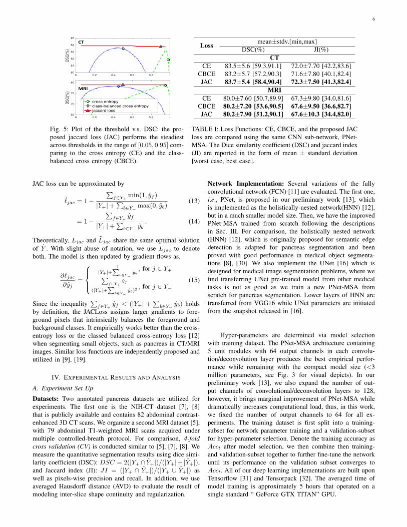
Fig. 3: The main construction units of the proposed PNet-MSA. In practice, we use 5 unit modules for pancreas segmentationand the first 3 are visually depicted here. The Deconv., Conv., BN, ReLU and
⊕present the CNN model structures as the
deconvolution, convolution, batch normalization, rectified linear unit, and concatenation layers, respectively. Hyperparametersfor Deconv. and Conv. are given in the bracket as the number of output channels, kernel size, and kernel stride. The 1st, 2nd,and 3rd forward branches output feature maps F1, F2, and F3 in respective. The “BB” is short for backward branch, and L1,L2, and L3 are the corresponding segmentation losses defined in Eq. 2.
X1), where d1k, d2k, and d3k represent the correponding tensordimensions. And Uk it the output probability map for pancreassegmentation. On Uk, a segmentation loss is measured by,
`k = L(Uk, Y ), (2)
where Y is the ground truth for model training, and we willdefine the loss function L(·, Y ) in Sec. III-C. Thus, each unitmodule has its own update gradient flow starting from itsloss branch Lk. This design is important because it introducesdeep-supervision to the bottom CNN layers, and it enable usto train the PNet-MSA layer by layer from swallow to deep.More specifically, PNet-MSA training starts from k = 1, andk is increased by 1 when Lk-plot converges. The ultimatesegmentation results will be a weighted combination of theunit module outputs,
Y =
K∑k=1
wkUk, (3)
and the overall objective for PNet-MSA is,
` = L(Y , Y ) +
K∑k=1
`k, (4)
where we find K = 5 works the best for pancreas segmenta-tion.
Although PNet-MSA may be extended from processingthe 2D input image to 3D [29], we maintain its 2D archi-tecture as training and inference since the 3D versions can becomputationally expensive, with no significant improvement inperformance [9]. However, our 2D model takes 3-connected-slices as its input when given the segmentation ground truthmask of the middle slice. As explained in Sec. III-B, our PNet-MSA is transformed into a light-weighted 3D model with RNN
stacked to the end of it, which allows our model to capture3D imaging information with minor extra computational loads.This is in a similar spirit to employ RNN to regulate, processand aggregate CNN activations for video classification andunderstanding [26].
B. Recurrent Neural Contextual Learning
From above, the CNN sub-network processes pancreas seg-mentation on individual 2D image slices, delivering remark-able performance on the tested CT and MRI datasets. However,as shown in the first row of Fig. 4, the transition among theresulted CNN pancreas segmentation regions in consecutiveslices may not be smooth, often implying that segmentationfailure occurs. Adjacent CT/MRI slices are expected to becorrelated to each other thus segmentation results from suc-cessive slices need to be constrained for shape continuityand regularization. The model for 3D object segmentation isrequired to be able to detect and recover abnormally lost partinside slices (see Yτ in Fig .4).
To achieve this, we concatenate an RNN sub-network, whichis originally designed for sequential data processing, to theCNN sub-network for modeling inter-slice shape continuityand regularization. That is, we slice any 3D CT (or MRI)volume into an ordered sequence of 2D images and processto learn the segmentation shape continuity among neighboringimage slices with a typical RNN architecture, the long short-term memory (LSTM) unit. However, the standard LSTMrequires vectorized input which would sacrifice the spatialinformation encoded in the output of CNN. We, therefore,utilize the convolutional LSTM (CLSTM) model [17] to pre-serve the 2D image segmentation layout by CNN. As shownin Fig. 4, Hτ and Cτ are the hidden state and cell output
5
of CLSTM in respective at the τ -th slice. The current celloutput Cτ is computed based on both of the former cell hiddenstate Hτ−1 and the current CNN output Yτ . Then, Hτ will becalculated from Cτ and used to produce the next cell outputCτ+1. Contextual information is propagated from slice τ toτ + 1 through convolutional operations.
Our Convolutional LSTM based inter-slice or sequentialsegmentation shape continuity learning is intuitive. Segmen-tation results of the former image slices are encoded in thecell hidden state Hτ−1. Values of Cτ is decided by takingHτ−1 and Yτ together into consideration. If position pi inYτ is predicted as pancreatic tissue by the CNN sub-network,and the same position in Hτ−1 are also encoded as pancreatictissue, then with high confidence that position pi should be apancreatic pixel in Cτ , and vice versa. As a result, CLSTMnot only recovers missing segmentation parts but also outputsmore confident probability maps than the original CNN sub-network. Formally, the CLSTM unit is formulated,
Iτ = σ(Wyi∗Yτ +Whi∗Hτ−1 +Wci�Cτ−1 + bi), (5)
Fτ = σ(Wyf∗Yτ +Whf∗Hτ−1 +Wcf�Cτ−1 + bf ), (6)
Cτ = fτ�Cτ−1+iτ� tanh(Wyc∗Yτ+Whc∗Hτ−1+bc), (7)
Oτ = σ(Wyo∗Yτ +Who∗Hτ−1 +Wco�Cτ + bo), (8)Hτ = oτ� tanh(cτ ), (9)
where ∗ represents convolution operation, and � denotes theHadamard product. Gates Iτ , Fτ , Oτ are the input, forget, andoutput following the original definition of CLSTM. W(·), b(·)are weights and bias in the corresponding CLSTM unit thatneed model optimization. Finally, σ(·) and tanh(·) denote thesigmoid and hyperbolic tangent activation function, respec-tively.Bi-directional Contextual Regularization: We then proposeto use a bi-direction extension of the CLSTM. For pancreas aswell as other organs, its shape in the current slice is constrainedby slices from not only the former ones but also its followings.The contextual information to input could be doubled if theshape regularization is taken in both directions leading toa further improvement. Two layers of CLSTM are stackedworking in two opposite directions as shown in Fig. 4. Then,outputs of the two layers, one in τ−-direction and the otherin τ+-direction, is weighted combined as final segmentationoutput,
Yτ =∑
i∈{−,+}
λiOiτ , (10)
where i represents the τ− and τ+ directions, and λi isthe learned weights when combining CLSTM outputs fromboth directions. Thus, the bi-direction design of shape con-tinuity modeling permits to explicitly enforce the pancreassegmentation spatial smoothness and higher-order inter-sliceregularization.
C. Jaccard Loss
We then propose a new Jaccard (JAC) loss to train the neuralnetwork based image segmentation model. To optimize theJaccard index (JI, a main segmentation metric) directly in net-work training, it makes the learning and inference procedures
(a) Yτ−2 (b) Yτ−1 (c) Yτ (d) Yτ+1 (e) Yτ+2
Hꚍ-2, Cꚍ-2 Hꚍ-1, Cꚍ-1 Hꚍ, Cꚍ
Xꚍ-1 Xꚍ
𝐻'(#, 𝐶'(# 𝐻'(", 𝐶'(" 𝐻', 𝐶'
𝑌,'(" 𝑌,'(f) CLSTM
H ꚍ-1, C ꚍ-1 H ꚍ, C ꚍ H ꚍ+1, C ꚍ+1
Hꚍ-1, Cꚍ-1 Hꚍ+1, Cꚍ+1 Hꚍ, Cꚍ
+ Oꚍ+ Oꚍ+1
+ Oꚍ+1
ꚍ+
ꚍ-
𝑂'(". 𝑂'.".𝑂'.
𝐻'(". , 𝐶'(". 𝐻'., 𝐶'. 𝐻'.". , 𝐶'.".
𝐻'."( , 𝐶'."(𝐻'(, 𝐶'(𝐻'("( , 𝐶'("(
𝑂'."(𝑂'(𝑂'("(𝑌/'(" 𝑌/' 𝑌/'."
(g) Bi-direction CLSTM
(h) Yτ−2 (i) Yτ−1 (j) Yτ (k) Yτ+1 (l) Yτ+2
Fig. 4: The main construction units of the proposed RNNsub-network and its input/output segmentation sequence. Thesequence of CNN sub-network outputs is shown in the firstrow (a-e), is taken as the input of the bi-direction CLSTM(g), which is an RNN architecture composed of 2 layers ofCLSTM (f) working in opposite directions. The third row (h-l) presents the corresponding output sequence, which is sharpand clean. Note that the missing pancreatic part in Yτ (c),in the green dashed box, is recovered by shape continuitymodeling in Yτ (j). For visual clearity, we ommit the inputY(·) in the bi-direction CLSTM (g), which is same as in (f).
consistent and generate threshold-free segmentation. JAC lossis defined as follows:
`jac = L(Y , Y )
= 1− |Y+⋂Y+|
|Y+⋃Y+|
= 1−∑j∈Y yj ∧ yj∑j∈Y yj ∨ yj
(11)
= 1−∑f∈Y+
(1 ∧ yf )
|Y+|+∑b∈Y−(0 ∨ yb)
, (12)
where Y and Y represent the ground truth and PNet-MSAprediction, which can be seamlessly replaced by the BiRNN’soutput Y and we will mainly focus on Y in the followingdiscussion. Respectively, we have Y+ and Y− defined as theforeground background pixel set, and |Y+| is the cardinalityof Y+. Similar definitions are also applied to Y . yj and yj ∈{0, 1} are indexed pixel values in Y and Y . In practice, yj isrelaxed to the probability number in the range of [0, 1] so that
6
0 0.2 0.4 0.6 0.8 180
81
82
83
84
85
DS
C(%
)
0 0.2 0.4 0.6 0.8 165
70
75
80
DS
C(%
)
CT
MRI
0 0.2 0.4 0.6 0.8 1
50
60
70
80
DS
C(%
)
cross entropyclass-balanced cross entropyjaccard loss
Fig. 5: Plot of the threshold v.s. DSC: the pro-posed jaccard loss (JAC) performs the steadiestacross thresholds in the range of [0.05, 0.95] com-paring to the cross entropy (CE) and the class-balanced cross entropy (CBCE).
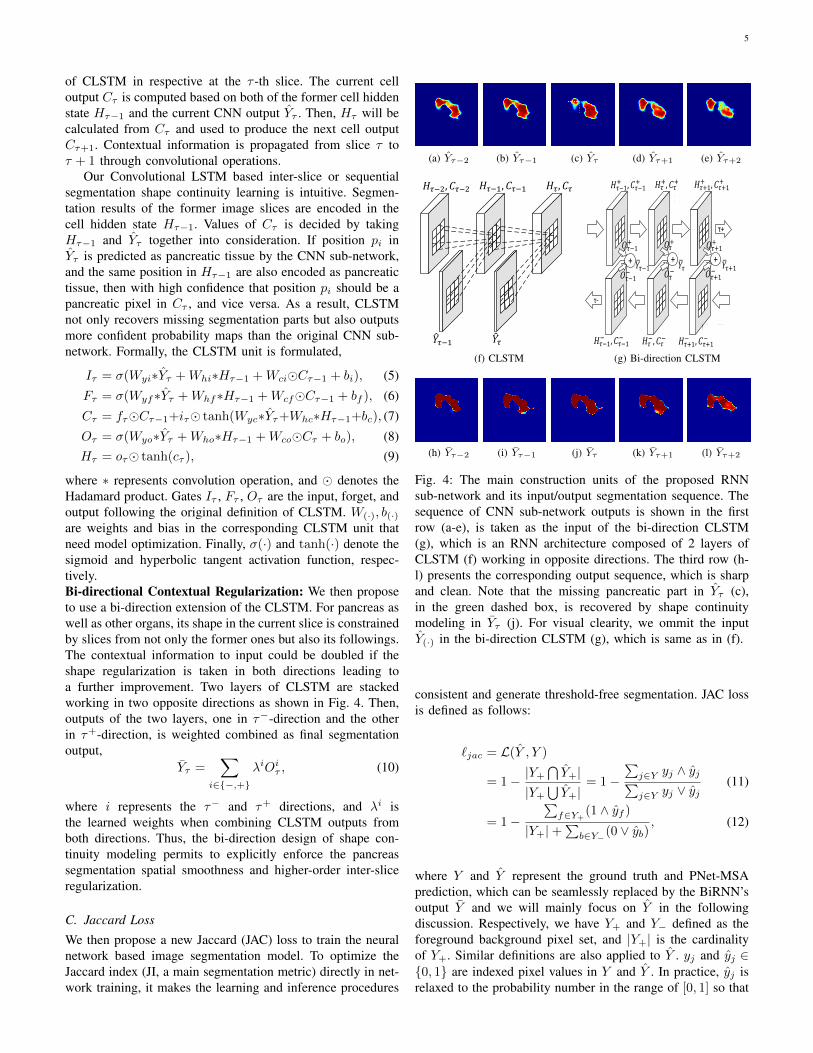
Loss mean±stdv.[min,max]DSC(%) JI(%)
CTCE 83.5±5.6 [59.3,91.1] 72.0±7.70 [42.2,83.6]
CBCE 83.2±5.7 [57.2,90.3] 71.6±7.80 [40.1,82.4]JAC 83.7±5.4 [58.4,90.4] 72.3±7.50 [41.3,82.4]
MRICE 80.0±7.60 [50.7,89.9] 67.3±9.80 [34.0,81.6]
CBCE 80.2±7.20 [53.6,90.5] 67.6±9.50 [36.6,82.7]JAC 80.2±7.90 [51.2,90.1] 67.6±10.3 [34.4,82.0]
TABLE I: Loss Functions: CE, CBCE, and the proposed JACloss are compared using the same CNN sub-network, PNet-MSA. The Dice similarity coefficient (DSC) and jaccard index(JI) are reported in the form of mean ± standard deviation[worst case, best case].
JAC loss can be approximated by
˜jac = 1−
∑f∈Y+
min(1, yf )
|Y+|+∑b∈Y− max(0, yb)
(13)
= 1−∑f∈Y+
yf
|Y+|+∑b∈Y− yb
. (14)
Theoretically, Ljac and Ljac share the same optimal solutionof Y . With slight abuse of notation, we use Ljac to denoteboth. The model is then updated by gradient flows as,
∂`jac∂yj
=
− 1
|Y+|+∑
b∈Y−yb, for j ∈ Y+∑
f∈Y+yf
(|Y+|+∑
b∈Y−yb)2
, for j ∈ Y−(15)
Since the inequality∑f∈Y+
yf < (|Y+| +∑b∈Y− yb) holds
by definition, the JACLoss assigns larger gradients to fore-ground pixels that intrinsically balances the foreground andbackground classes. It empirically works better than the cross-entropy loss or the classed balanced cross-entropy loss [12]when segmenting small objects, such as pancreas in CT/MRIimages. Similar loss functions are independently proposed andutilized in [9], [19].
IV. EXPERIMENTAL RESULTS AND ANALYSIS
A. Experiment Set Up
Datasets: Two annotated pancreas datasets are utilized forexperiments. The first one is the NIH-CT dataset [7], [8]that is publicly available and contains 82 abdominal contrast-enhanced 3D CT scans. We organize a second MRI dataset [5],with 79 abdominal T1-weighted MRI scans acquired undermultiple controlled-breath protocol. For comparison, 4-foldcross validation (CV) is conducted similar to [5], [7], [8]. Wemeasure the quantitative segmentation results using dice simi-larity coefficient (DSC): DSC = 2(|Y+∩ Y+|)/(|Y+|+ |Y+|),and Jaccard index (JI): JI = (|Y+ ∩ Y+|)/(|Y+ ∪ Y+|) aswell as pixels-wise precision and recall. In addition, we useaveraged Hausdorff distance (AVD) to evaluate the result ofmodeling inter-slice shape continuity and regularization.
Network Implementation: Several variations of the fullyconvolutional network (FCN) [11] are evaluated. The first one,i.e., PNet, is proposed in our preliminary work [13], whichis implemented as the holistically-nested network(HNN) [12],but in a much smaller model size. Then, we have the improvedPNet-MSA trained from scratch following the descriptionsin Sec. III. For comparison, the holistically nested network(HNN) [12], which is originally proposed for semantic edgedetection is adapted for pancreas segmentation and beenproved with good performance in medical object segmenta-tions [8], [30]. We also implement the UNet [16] which isdesigned for medical image segmentation problems, where wefind transferring UNet pre-trained model from other medicaltasks is not as good as we train a new PNet-MSA fromscratch for pancreas segmentation. Lower layers of HNN aretransferred from VGG16 while UNet parameters are initiatedfrom the snapshot released in [16].
Hyper-parameters are determined via model selectionwith training dataset. The PNet-MSA architecture containing5 unit modules with 64 output channels in each convolu-tion/deconvolution layer produces the best empirical perfor-mance while remaining with the compact model size (<3million parameters, see Fig. 3 for visual depicts). In ourpreliminary work [13], we also expand the number of out-put channels of convolutional/deconvolution layers to 128,however, it brings marginal improvement of PNet-MSA whiledramatically increases computational load, thus, in this work,we fixed the number of output channels to 64 for all ex-periments. The training dataset is first split into a training-subset for network parameter training and a validation-subsetfor hyper-parameter selection. Denote the training accuracy asAcct after model selection, we then combine then training-and validation-subset together to further fine-tune the networkuntil its performance on the validation subset converges toAcct. All of our deep learning implementations are built uponTensorflow [31] and Tensorpack [32]. The averaged time ofmodel training is approximately 5 hours that operated on asingle standard “ GeForce GTX TITAN” GPU.

7
TABLE II: Comprison to different architectures: we have the proposed PNet-MSA compared with HNN network [12], UNet [16]and its preliminary version PNet-64 [13] under the same experimental setting up that the number of convoultional layer channelis 64. PNet-64 and PNet-MSA are trained from scratch, while HNN and U-Net are fine-tuned from released snapshots of otherimage domains.
Method mean±stdv. [min,max]DSC(%) JI(%) Precision Recall
CTHNN [12] 79.6±7.7 [41.9,88.0] 66.7±9.40 [26.5,78.6] 83.4±6.5 [62.0,94.9] 77.4±11.6 [28.3,92.6]UNet [16] 79.7±7.6 [43.4,89.3] 66.8±9.60 [27.7,80.7] 81.3±7.5 [49.6,97.0] 79.2±11.3 [38.6,94.1]PNet-64 [13] 81.3±8.1 [42.9,89.5] 69.1±10.0 [27.3,81.0] 82.8±6.5 [51.0,95.2] 80.8±11.3 [28.5,93.8]PNet-MSA 83.3±5.6 [59.0,91.0] 71.8±7.70 [41.8,83.5] 84.5±6.2 [60.7,96.7] 82.8±8.37 [56.4,94.6]
MRIHNN [12] 75.9±10.1 [33.0,86.8] 62.1±11.3 [19.8,76.6] 84.4±6.4 [61.0,93.5] 70.6±13.3 [20.7,88.2]UNet [16] 79.9±7.30 [54.8,90.5] 67.1±9.50 [37.7,82.6] 83.7±6.9 [64.6,94.6] 77.3±10.3 [46.1,94.8]PNet-64 [13] 76.3±12.9 [6.30,88.8] 63.1±14.0 [3.30,79.9] 80.0±9.6 [41.6,92.5] 74.6±15.6 [3.40,92.4]PNet-MSA 80.7±7.40 [48.8,90.5] 68.2±9.64 [32.3,82.7] 84.3±7.6 [55.8,95.8] 78.3±10.2 [38.6,95.0]
B. Training Loss Comparison
Table I presents results of three losses, i.e., the cross-entropy(CE), the class-balanced cross entropy (CBCE) [12], and theproposed Jaccard loss (JAC), under 4-fold cross-validationwith the same PNet-MSA segmentation model. On the CTdataset, JAC outperforms CE and CBCE by 0.5% and 0.2%mean DSC, respectively. On the MRI dataset, JAC alsoachieves the best performance referring to the mean DSC andJI. We also evaluate the stability of segmentation performancewith various thresholds. That is because the CNN networkoutputs probabilistic image segmentation maps instead ofbinary masks and an appropriate probability threshold isrequired to obtain the final binarized segmentation outcomes.However, it is often non-trivial to find the optimal probabilitythreshold in practice. Empirically, Naıve cross-entropy lossassigns the same penalty on positive and negative pixels so thatthe probability threshold should be around 0.5. Meanwhile,CBCE gives higher penalty scores on positive pixels (dueto its scarcity), making the resulted “optimal” threshold at arelatively higher value. By contrast, JAC pushes the foregroundpixels to the probability of 1.0 while remains to be stronglydiscriminative against the background pixels. Thus, JAC’splateau around the optimal segmentation performance wouldbe much wider than CE and CBCE so that it could performstably in a wide range of thresholds, i.e., [0.05, 0.95] in ourexperiments. Fig. 5 visually depicts the results of our analysisthat the probability output maps from the JAC loss deliversthe steadiest segmentation results referring to different outputthresholds.
C. CNN Sub-Network Comparison
Before modeling the shape continuity and regularizationamong image slices, it is important to first train an effec-tive CNN sub-network, which presents the base for shapelearning. We test multiple state-of-the-art CNN architectures,i.e., HNN [12], UNet [16], and PNet [13] with the proposedPNet-MSA. Each architecture design would have its ownpros and cons and would perform differently according to
the specific applications. It is possible that when fine-tunedwith large-scale training data, the ImageNet pre-trained large-size networks, e.g., HNN and UNet, might outperform ourcompact-sized PNet and PNet-MSA. However, in this paper,we would focus on the situation that only limited amountof training data is available, which often happens in clinicalpractice.
Table II presents segmentation results of different architec-tures. Without loss of generality, we set the output thresholdfor all CNN outputs to 0.5 basing on the analysis in Sec. IV-B.In comparison, the proposed PNet achieves the best perfor-mance on both of the CT and MRI datasets. We then confirmthe pancreatic volumes that measured by deep learning modelsis consistent with manual measurements in Fig. 6. PNet-MSAmodel trained from scratch output results demonstrate the bestalignment between human and computerized measurements.
D. Modeling Shape Continuity Regularization
Given outputs of PNet-MSA as the best CNN-based segmen-tation results, the bi-direction RNN (BiRNN) sub-network isthen stacked to the top of PNet-MSA and trained end-to-end.In each direction, a one-layer CLSTM is implemented with 1hidden state and 3× 3 convolution filter kernels [17]. Partic-ularly, the number of hidden state is set to 1 since our shapecontinuity learning is inherently simplified by processing onlythe output probability maps of CNN sub-network. CNN outputYτ ∈ Rd
11×d
21×1 (where d11 and d21 are the width and height of
the input image) provides a highly compacted representationof the input CT/MRI image for shape learning. Thus, BiRNNwith the hidden state Hτ ∈ Rd
11×d
21×1 is sufficient to model
and capture the shape continuity regularization among CNNoutputs. We notice that BiRNN could not converge stablyduring model training when a larger hidden state is used. Inaddition, we attempt to employ BiRNN on the feature mapsfrom CNN’s intermediate layers. Subsequently, this causes themodel training process often not to converge. Thus, we mainlyfocus on the current design of BiRNN, which is simplified tolearn the inter-slice shape continuity among successive CNN’ssegmentation outputs.
8
(a) CT: U-Net (b) CT: PNet-MSA
(c) MRI: U-Net (d) MRI: PNet-MSA
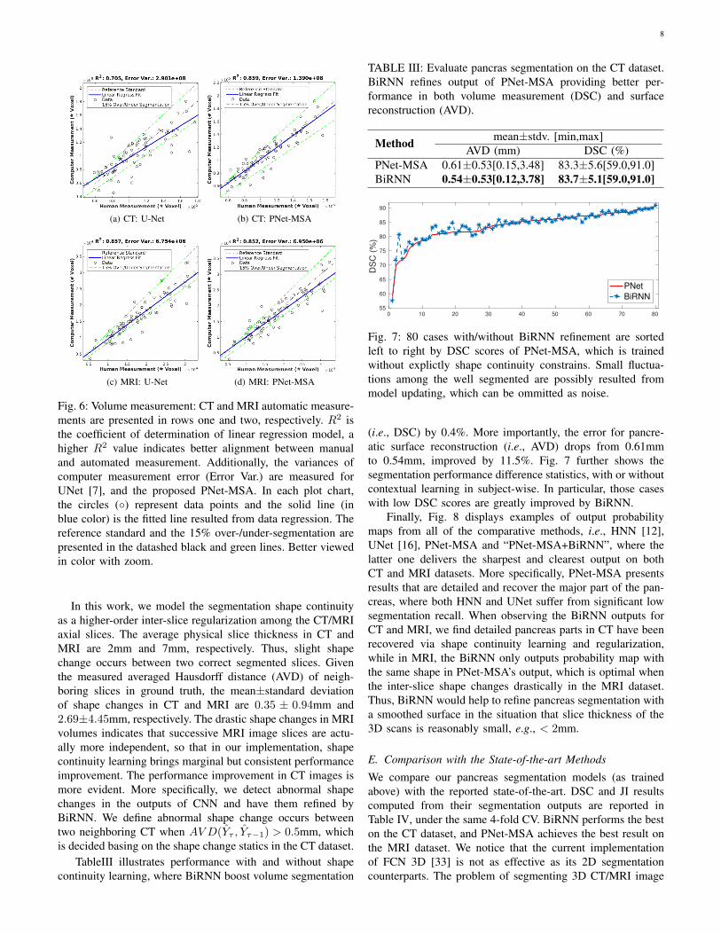
Fig. 6: Volume measurement: CT and MRI automatic measure-ments are presented in rows one and two, respectively. R2 isthe coefficient of determination of linear regression model, ahigher R2 value indicates better alignment between manualand automated measurement. Additionally, the variances ofcomputer measurement error (Error Var.) are measured forUNet [7], and the proposed PNet-MSA. In each plot chart,the circles (◦) represent data points and the solid line (inblue color) is the fitted line resulted from data regression. Thereference standard and the 15% over-/under-segmentation arepresented in the datashed black and green lines. Better viewedin color with zoom.
In this work, we model the segmentation shape continuityas a higher-order inter-slice regularization among the CT/MRIaxial slices. The average physical slice thickness in CT andMRI are 2mm and 7mm, respectively. Thus, slight shapechange occurs between two correct segmented slices. Giventhe measured averaged Hausdorff distance (AVD) of neigh-boring slices in ground truth, the mean±standard deviationof shape changes in CT and MRI are 0.35 ± 0.94mm and2.69±4.45mm, respectively. The drastic shape changes in MRIvolumes indicates that successive MRI image slices are actu-ally more independent, so that in our implementation, shapecontinuity learning brings marginal but consistent performanceimprovement. The performance improvement in CT images ismore evident. More specifically, we detect abnormal shapechanges in the outputs of CNN and have them refined byBiRNN. We define abnormal shape change occurs betweentwo neighboring CT when AVD(Yτ , Yτ−1) > 0.5mm, whichis decided basing on the shape change statics in the CT dataset.
TableIII illustrates performance with and without shapecontinuity learning, where BiRNN boost volume segmentation
TABLE III: Evaluate pancras segmentation on the CT dataset.BiRNN refines output of PNet-MSA providing better per-formance in both volume measurement (DSC) and surfacereconstruction (AVD).
Method mean±stdv. [min,max]AVD (mm) DSC (%)
PNet-MSA 0.61±0.53[0.15,3.48] 83.3±5.6[59.0,91.0]BiRNN 0.54±0.53[0.12,3.78] 83.7±5.1[59.0,91.0]
0 10 20 30 40 50 60 70 8055
60
65
70
75
80
85
90
DS
C (
%)
PNet
BiRNN
Fig. 7: 80 cases with/without BiRNN refinement are sortedleft to right by DSC scores of PNet-MSA, which is trainedwithout explictly shape continuity constrains. Small fluctua-tions among the well segmented are possibly resulted frommodel updating, which can be ommitted as noise.
(i.e., DSC) by 0.4%. More importantly, the error for pancre-atic surface reconstruction (i.e., AVD) drops from 0.61mmto 0.54mm, improved by 11.5%. Fig. 7 further shows thesegmentation performance difference statistics, with or withoutcontextual learning in subject-wise. In particular, those caseswith low DSC scores are greatly improved by BiRNN.
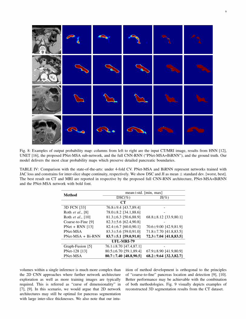
Finally, Fig. 8 displays examples of output probabilitymaps from all of the comparative methods, i.e., HNN [12],UNet [16], PNet-MSA and “PNet-MSA+BiRNN”, where thelatter one delivers the sharpest and clearest output on bothCT and MRI datasets. More specifically, PNet-MSA presentsresults that are detailed and recover the major part of the pan-creas, where both HNN and UNet suffer from significant lowsegmentation recall. When observing the BiRNN outputs forCT and MRI, we find detailed pancreas parts in CT have beenrecovered via shape continuity learning and regularization,while in MRI, the BiRNN only outputs probability map withthe same shape in PNet-MSA’s output, which is optimal whenthe inter-slice shape changes drastically in the MRI dataset.Thus, BiRNN would help to refine pancreas segmentation witha smoothed surface in the situation that slice thickness of the3D scans is reasonably small, e.g., < 2mm.
E. Comparison with the State-of-the-art Methods
We compare our pancreas segmentation models (as trainedabove) with the reported state-of-the-art. DSC and JI resultscomputed from their segmentation outputs are reported inTable IV, under the same 4-fold CV. BiRNN performs the beston the CT dataset, and PNet-MSA achieves the best result onthe MRI dataset. We notice that the current implementationof FCN 3D [33] is not as effective as its 2D segmentationcounterparts. The problem of segmenting 3D CT/MRI image
9
Fig. 8: Examples of output probability map: columns from left to right are the input CT/MRI image, results from HNN [12],UNET [16], the proposed PNet-MSA sub-network, and the full CNN-RNN (“PNet-MSA+BiRNN”), and the ground truth. Ourmodel delivers the most clear probability maps which preserve detailed pancreatic boundaries.
TABLE IV: Comparison with the state-of-the-arts: under 4-fold CV. PNet-MSA and BiRNN represent networks trained withJAC loss and constrains for inter-slice shape continuity, respectively. We show DSC and JI as mean ± standard dev. [worst, best].The best result on CT and MRI are reported in respective by the proposed full CNN-RNN architecture, PNet-MSA+BiRNNand the PNet-MSA network with bold font.
Method mean±std. [min, max]DSC(%) JI(%)
CT3D FCN [33] 76.8±9.4 [43.7,89.4] -Roth et al., [8] 78.0±8.2 [34.1,88.6] -Roth et al., [10] 81.3±6.3 [50.6,88.9] 68.8±8.12 [33.9,80.1]Coarse-to-Fine [9] 82.3±5.6 [62.4,90.8] -PNet + RNN [13] 82.4±6.7 [60.0,90.1] 70.6±9.00 [42.9,81.9]PNet-MSA 83.3±5.6 [59.0,91.0] 71.8±7.70 [41.8,83.5]PNet-MSA + Bi-RNN 83.7±5.1 [59.0,91.0] 72.3±7.04 [41.8,83.5]
UFL-MRI-79Graph-Fusion [5] 76.1±8.70 [47.4,87.1] -PNet-128 [13] 80.5±6.70 [59.1,89.4] 67.9±8.90 [41.9,80.9]PNet-MSA 80.7±7.40 [48.8,90.5] 68.2±9.64 [32.3,82.7]
volumes within a single inference is much more complex thanthe 2D CNN approaches where further network architectureexploration as well as more training images are typicallyrequired. This is referred as “curse of dimensionality” in[7], [9]. In this scenario, we would argue that 2D networkarchitectures may still be optimal for pancreas segmentationwith large inter-slice thicknesses. We also note that our intu-
ition of method development is orthogonal to the principlesof “coarse-to-fine” pancreas location and detection [9], [10].Better performance may be achievable with the combinationof both methodologies. Fig. 9 visually depicts examples ofreconstructed 3D segmentation results from the CT dataset.

10
(a) DSC: 60% (b) DSC: 65% (c) DSC: 70%
(d) DSC: 70% (e) DSC: 75% (f) DSC: 80%
(g) DSC: 80% (h) DSC: 85% (i) DSC: 90%
Fig. 9: 3D visualization of pancreas segmentation resultswhere human annotation shown in yellow and computerizedsegmentation displayed in green. The DSC are 90%, 75%, and60% for three examples from left to right, respectively.
V. CONCLUSION
In this paper, we present a novel CNN-RNN architecture forpancreas segmentation in CT and MRI scans, via our tailor-made CNN module, followed by a bi-directional CLSTMto regularize the segmentation results on individual imageslices. This is different from the independent process assumedin recent previous work [5], [8]–[10]. The shape continuityregularization permits to enforce the pancreas segmentationspatial smoothness explicitly in the axial direction, in analogyto comprehending into videos by parsing and aggregatingsuccessive frames [26]. This may also share some similarityto the human doctor’s way of reading radiology images. Com-bined with the proposed segmentation direct JAC loss functionfor CNN training to generate the threshold-free segmentationresults, our quantitative pancreas segmentation results improvethe previous state-of-the-art approaches [5], [8]–[10] on bothCT and MRI datasets, with noticeable margins. Althoughour proposed method is focused on pancreas segmentation,this approach is generalizable to other organ segmentationin medical image analysis, in especially when the size ofavailable training data is limited.
REFERENCES
[1] M. Oda, N. Shimizu, K. Karasawa, Y. Nimura, T. Kitasaka, K. Misawa,D. Rueckert, and K. Mori, “Regression forest-based atlas localizationand direction specific atlas generation for pancreas segmentation,” inInternational Conference on Medical Image Computing and ComputerAssisted Intervention. Springer, 2016, pp. 556–563.
[2] T. Tong, R. Wolz, Z. Wang, Q. Gao, K. Misawa, M. Fujiwara, K. Mori,J. V. Hajnal, and D. Rueckert, “Discriminative dictionary learning forabdominal multi-organ segmentation,” Medical Image Analysis, vol. 23,no. 1, pp. 92–104, 2015.
[3] R. Wolz, C. Chu, K. Misawa, M. Fujiwara, K. Mori, and D. Rueckert,“Automated abdominal multi-organ segmentation with subject-specificatlas generation,” IEEE Transactions on Medical Imaging, vol. 32, no. 9,pp. 1723–1730, 2013.
[4] K. Karasawa, M. Oda, T. Kitasaka, K. Misawa, M. Fujiwara, C. Chu,G. Zheng, D. Rueckert, and K. Mori, “Multi-atlas pancreas segmenta-tion: Atlas selection based on vessel structure,” Medical Image Analysis,vol. 39, pp. 18–28, 2017.
[5] J. Cai, L. Lu, Z. Zhang, F. Xing, L. Yang, and Q. Yin, “Pancreas segmen-tation in MRI using graph-based decision fusion on convolutional neuralnetworks,” in International Conference on Medical Image Computingand Computer Assisted Intervention. Springer, 2016, pp. 442–450.
[6] A. Farag, L. Lu, H. R. Roth, J. Liu, E. Turkbey, and R. M. Summers,“A bottom-up approach for pancreas segmentation using cascaded super-pixels and (deep) image patch labeling,” IEEE Transactions on MedicalImaging, vol. 26, no. 1, pp. 386–399, 2017.
[7] H. R. Roth, L. Lu, A. Farag, H.-C. Shin, J. Liu, E. B. Turkbey, and R. M.Summers, “Deeporgan: Multi-level deep convolutional networks for au-tomated pancreas segmentation,” in International Conference on MedicalImage Computing and Computer Assisted Intervention. Springer, 2015,pp. 556–564.
[8] H. R. Roth, L. Lu, A. Farag, A. Sohn, and R. M. Summers, “Spatialaggregation of holistically-nested networks for automated pancreas seg-mentation,” in International Conference on Medical Image Computingand Computer Assisted Intervention. Springer, 2016, pp. 451–450.
[9] Y. Zhou, L. Xie, W. Shen, E. Fishman, and A. L. Yuille,“Pancreas segmentation in abdominal CT scan: A coarse-to-fineapproach,” CoRR, vol. abs/1612.08230, 2016. [Online]. Available:http://arxiv.org/abs/1612.08230
[10] H. R. Roth, L. Lu, N. Lay, A. P. Harrison, A. Farag, and R. M. Summers,“Spatial aggregation of holistically-nested convolutional neural networksfor automated pancreas localization and segmentation,” Medical ImageAnalysis, vol. 45, pp. 94–107, 2018.
[11] J. Long, E. Shelhamer, and T. Darrell, “Fully convolutional networks forsemantic segmentation,” in IEEE International Conference on ComputerVision and Pattern Recognition, 2015, pp. 3431–3440.
[12] S. Xie and Z. Tu, “Holistically-nested edge detection,” in IEEE Inter-national Conference on Computer Vision, 2015, pp. 1395–1403.
[13] J. Cai, L. Lu, Y. Xie, F. Xing, and L. Yang, “Improving deep pancreassegmentation in ct and mri images via recurrent neural contextual learn-ing and direct loss function,” in International Conference on MedicalImage Computing and Computer Assisted Intervention, pp. 674–682.
[14] C. Lee, S. Xie, P. W. Gallagher, Z. Zhang, and Z. Tu, “Deeply-supervised nets,” in International Conference on Artificial Intelligenceand Statistics, 2015.
[15] F. Yu and V. Koltun, “Multi-Scale Context Aggregation by Dilated Con-volutions,” in International Conference on Learning Representations,2016.
[16] O. Ronneberger, P. Fischer, and T. Brox, “U-net: Convolutional networksfor biomedical image segmentation,” in International Conference onMedical Image Computing and Computer Assisted Intervention, 2015,pp. 234–241.
[17] X. Shi, Z. Chen, H. Wang, D. Yeung, W. Wong, and W. Woo, “Convo-lutional LSTM network: A machine learning approach for precipitationnowcasting,” in Advances in Neural Information Processing Systems,2015, pp. 802–810.
[18] J. Chen, L. Yang, Y. Zhang, M. S. Alber, and D. Z. Chen, “Combiningfully convolutional and recurrent neural networks for 3d biomedicalimage segmentation,” in Advances in Neural Information ProcessingSystems, 2016, pp. 3036–3044.
[19] F. Milletari, N. Navab, and S. Ahmadi, “V-net: Fully convolutionalneural networks for volumetric medical image segmentation,” in Inter-national Conference on 3D Vision, 2016, pp. 565–571.
[20] L. Chen, G. Papandreou, I. Kokkinos, K. Murphy, and A. L. Yuille,“Deeplab: Semantic image segmentation with deep convolutional nets,atrous convolution, and fully connected crfs,” IEEE Transactions onPattern Analysis and Machine Intelligence, vol. 40, no. 4, pp. 834–848,2018.
[21] S. Zheng, S. Jayasumana, B. Romera-Paredes, V. Vineet, Z. Su, D. Du,C. Huang, and P. H. Torr, “Conditional random fields as recurrent neuralnetworks,” in IEEE International Conference on Computer Vision, 2015,pp. 1529–1537.
[22] J. Merkow, A. Marsden, D. J. Kriegman, and Z. Tu, “Dense volume-to-volume vascular boundary detection,” in International Conference onMedical Image Computing and Computer Assisted Intervention, 2016,pp. 371–379.
[23] K. Kamnitsas, C. Ledig, V. F. J. Newcombe, J. P. Simpson, A. D. Kane,D. K. Menon, D. Rueckert, and B. Glocker, “Efficient multi-scale 3dCNN with fully connected CRF for accurate brain lesion segmentation,”Medical Image Analysis, vol. 36, pp. 61–78, 2017.
[24] M. F. Stollenga, W. Byeon, M. Liwicki, and J. Schmidhuber, “Parallelmulti-dimensional lstm, with application to fast biomedical volumetricimage segmentation,” in Advances in Neural Information ProcessingSystems, 2015, pp. 2998–3006.
11
[25] A. Karpathy, G. Toderici, S. Shetty, T. Leung, R. Sukthankar, andL. Fei-fei, “Large-scale video classification with convolutional neuralnetworks,” in IEEE International Conference on Computer Vision andPattern Recognition, 2014, pp. 1725–1732.
[26] J. Ng, M. Hausknecht, S. Vijayanarasimhan, O. Vinyals, R. Monga,and G. Toderici, “Beyond short snippets: Deep networks for videoclassification,” in IEEE International Conference on Computer Vision,2015, pp. 4694–4702.
[27] K. Simonyan and A. Zisserman, “Very deep convolutional networks forlarge-scale image recognition,” in International Conference on LearningRepresentations, 2015, pp. 1–14.
[28] K. He, X. Zhang, S. Ren, and J. Sun, “Delving deep into rectifiers:Surpassing human-level performance on imagenet classification,” inIEEE International Conference on Computer Vision, 2015, pp. 1026–1034.
[29] O. Cicek, A. Abdulkadir, S. S. Lienkamp, T. Brox, and O. Ronneberger,“3d u-net: learning dense volumetric segmentation from sparse anno-tation,” in International Conference on Medical Image Computing andComputer Assisted Intervention. Springer, 2016, pp. 424–432.
[30] A. P. Harrison, Z. Xu, K. George, L. Lu, R. M. Summers, and D. J.Mollura, “Progressive and multi-path holistically nested neural networksfor pathological lung segmentation from CT images,” in InternationalConference on Medical Image Computing and Computer Assisted Inter-vention. Springer, 2017, pp. 621–629.
[31] M. Abadi, A. Agarwal, P. Barham, and et al., “TensorFlow: Large-scalemachine learning on heterogeneous systems,” arXiv:1603.04467, 2015.
[32] Y. Wu et al., “Tensorpack,” https://github.com/tensorpack/, 2016.[33] H. R. Rotha, H. Odaa, X. Zhoub, N. Shimizua, Y. Yanga, Y. Hayashia,
M. Odaa, M. Fujiwarac, K. Misawad, and K. Moria, “An application ofcascaded 3D fully convolutional networks for medical image segmenta-tion,” ArXiv e-prints, 2018.