Embed Size (px)
Citation preview
PALS Handbook
PALS HANDBOOK
PALS Handbook
Course requirementsBLS competency testingSkills stationsEKG rhythm identificationPALS core case discussion and simulationsPALS core case testing stationsWritten exam
Course outlineSystemic approach to seriously ill or injured childRecognition of respiratory distress and failureManagement of respiratory distress and failureRecognition of shockManagement of shockRhythm identificationPharmacologyVascular accessRecognition and management of bradycardiaRecognition and management of tachycardiaRecognition and management of cardiac arrestPost resuscitation management
Skills stations BLS competency testing (child / infant /AED)Airway competency (ET / LMA / WFC)Vascular access ( IO)Rhythm recognition review
REQUIREMENTS

PALS Handbook
• Kids are not small adults
• Don’t panic– Disorganization is counter productive– It is better to do something right than to undo the
mistake and correct it
• Preventing an arrest is primary since the outcome if an arrest occurs is usually poor.
• A systematic approach is essential
• Remember Kids like – Air ------------- Respiratory– Water ------- Shock– Sugar -------- Glycemic unbalance
PALS BASICS

PALS Handbook
• Problem areas– Respiratory
• Asthma, pneumonia, respiratory failure– Circulatory
• Dehydration, shock– Glucose
• Hyperglycemia, hypoglycemia– Trauma– Cardiac arrest
• Phases of the systemic approach– Initial impression
• Conscious, breathing, color, activity– Evaluate- life threatening / non life threatening
• Primary ABCDE• Secondary – med history, physical exam• Diagnostic tests
– Identify• Respiratory, circulatory, cardiac arrest
– Intervene
SYSTEMIC APPROACH

PALS Handbook
• Assessment• Initial pediatric assessment
– General appearance– Work of breathing– Skin perfusion
• Primary assessment– Airway- patent noiseless– Breathing – present, rate, sounds – Circulation– Disability– Exposure
• Secondary assessment– Physical exam – head to toe– Sample history
• Symptoms, allergies, meds, past history, last meal, events causing– Bedside
• BGL, Vital signs, EKG monitor• Tertiary survey
– Focused history and examination– Diagnostic tests
• ABG and VBG• Hemoglobin concentrations• Central venous oxygen saturation• Arterial lactate• CVP monitoring• Chest X-ray• Peak expiratory flow
Categorize respiratory , circulatory, cardiac
SYSTEMIC APPROAQCH

PALS Handbook
• Airway assessment– Clear – unobstructed– Maintainable – by position or simple airway maneuvers– Not maintainable
• FBAO –Heimlich, blows, CPR• Requires an advanced airway
• Breathing– Respiratory rate– Respiratory effort– Chest expansion and airway movement– Lung and airway sounds– SPO2
• Normal respiratory rates by age– Age Breaths / min– Infant < 1 year 30-60– Toddler (1-3 years) 24-30– Preschooler (4-5 years) 22-34– School age (6-12 years) 18-30– Adolescent 12-16
• Assessment of respiratory rates• Method
– Count respirations for 30 seconds and multiply by 2 for minute rate
• Note Anxiety can raise the respiratory rate– Assess rates frequently if child has respiratory difficulty
RESPIRATORY

PALS Handbook
• Abnormal respiratory rates– Apnea
• Central apnea – suppression of the brain or spinal cord– Tachypnea – causes
• Fever, pain, acidosis, anemia, sepsis• CHF, congenital heart defects
– Bradypnea – causes• Respiratory failure• CNS injury or infection• Hypothermia• Medications• Note: may signal arrest in an acutely ill child
• Respiratory effort– Dyspneic children and infants
• Work hard to maintain adequate oxygenation• Fatigue easily and go into respiratory failure• It is important to recognize and treat respiratory difficulty
– Early signs or respiratory distress• Nasal flaring
– Dilation of the nostrils to increase air intake• Retractions
– Inward movement of the chest wall, neck, and sternum• Head bobbing
– Caused by neck muscles used to assist ventilation• Seesaw respirations
– Chest retracts and abdomen expands during inspiration– Most common in children with neuromuscular disease
RESPIRATORY

PALS Handbook
• Respiratory effort• Chest expansion
– Normal • Symmetrical chest rise (full expansion)• In infants the abdomen may move more than the chest
– Abnormal • Decreased or asymmetrical chest rise
– Inadequate effort– Airway obstruction– Pneumothorax / hemothorax– FBAO– Mucous plug– Pleural effusion
• Lung and airway sounds– Stridor
• Coarse high pitched sound heard during inspiration• Indicates an upper airway obstruction
– Grunting• Short low pitched sound heard during expiration• Sign of lung disease (alveolar collapse)• Early
– Gurgling• Upper airway obstruction• Due to secretions or vomit in the upper airway
– Wheezing• High pitched whistling sound heard on expansion and
inspiration– Crackles – accumulation of alveolar fluid (hairs rubbing together)
RESPIRATORY
PALS Handbook
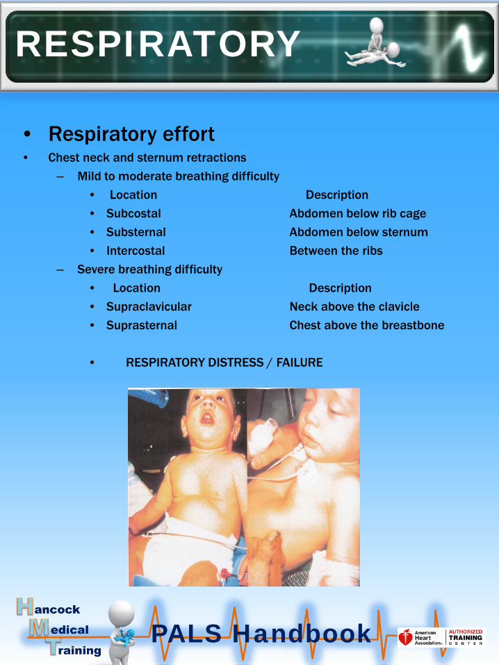
• Respiratory effort• Chest neck and sternum retractions
– Mild to moderate breathing difficulty• Location Description • Subcostal Abdomen below rib cage• Substernal Abdomen below sternum• Intercostal Between the ribs
– Severe breathing difficulty• Location Description• Supraclavicular Neck above the clavicle• Suprasternal Chest above the breastbone
• RESPIRATORY DISTRESS / FAILURE
RESPIRATORY

PALS Handbook
• How to assess circulation and perfusion– Heart rate and rhythm– Pulses– Capillary refill– Skin color and temperature– Blood pressure
• Normal heart rate table– Age Awake Asleep– Infant-3mo. 85-205 80-160– 3 mo.- 2yrs 100-190 75-160– 2 yrs. -10 yrs. 60- 140 60-90– >10 yrs. 60-100 50-90
• Abnormal heart rates– Bradycardia
• Slight bradycardia in athletic children• Usually results from hypoxia (poor perfusion) in ill children• Can result from heart blocks and drug overdoses
– Tachycardia• Mostly related to a cause (i.e. fever, anxiety)• May result from cardiac problems (SVT)
– Cardiac arrest• Poor prognosis
• Pulses– Central
• Femoral, brachial (infants), carotid (older children), axillary– Peripheral
• Radial, dorsal pedis, posterior tibial
CIRCULATION /PERFUSION

PALS Handbook
• Pulse assessment– Normal pulses are strong and equal– Weak or unequal pulses indicate circulatory problems– Pulsus paradoxus
• Fluctuation in volume with the respiratory cycle• Usually associated with asthma and tamponade
– Intubated patients• Reduction in pulse volume indicates hypovolemia
• Capillary refill– The time it takes blood to return to tissue blanched by pressure– Best performed at room temperature– Select an extremity slightly above the level of the heart– Measure the time to return to normal color (< 2 seconds)
• Skin color and temperature– Normal –uniform color over the entire body– Inadequate tissue perfusion
• Pallor– Decreased blood supply to the skin– Decreased number of red cells
• Mottling– Hypoxia, hypovolemia, chock
• Cyanosis – Low oxygen tension– Alveolar hypoventilation (TBI / drug O.D.)– Diffusion effect (pneumonia , PE)– Ventilation /perfusion unbalance (asthma)– Intracardiac shunt
CIRCULATION / PERFISION

PALS Handbook
• Blood pressure• Cuff selection
– Inaccurate readings result from improper cuff selection– The proper cuff should cover 50-75% of the distance from axilla to A.C.
fossa
• Normal pediatric blood pressures– Age Systolic Diastolic– Neonate 60-80 30-53– Infant (1-6 MO.) 73-102 40-68– Infant ( (6-12 MO.) 80-104 44-68– Infant (1 year) 68-103 20-58– Child (2 years) 70-106 25-65– Child (7 years) 79-115 38-79– Adolescent 93-131 45-85
• Hypotension by age– Age Systolic– 0-28 DAYS <60– Infant (1-12 MO.) <70– Children (1-10YR.) <70 + age /2– Children (>10 years) <90
CIRCULATION / PERFUSION
PALS Handbook
• Primary concerns– Inadequate circulatory function
• Decreased level of consciousness• Loss of muscle tone• Centralized seizures• Pupil dilation
– Evaluation of the neurological function
• Assessment of disability– AVPU
• A- Alert• V- Voice (response)• P- Pain (response to)• U- Unresponsive
– Glasgow coma scale• Eye opening
– Spontaneous, speech, pain, none • Verbal response
– Oriented, confused, Inappropriate, Incomprehensible, none• Motor response
– Obeys, Localizes, Withdraws, Flexion, Extension, None • 1 point for each ( maximum score 15 minimum score 3)
– Pupillary response• Size• Equality• Constriction in response to light
–
DISABILITY

PALS Handbook
• Full body exposure– Clothing may cover indications of injuries and physical abnormalities– Clothing also makes it difficult to visualize chest movement – Skin abnormalities such as a rash may be missed by clothing– To correctly assess your patient remove as much clothing as necessary
• Full body assessment (head to toe)– Head
• Skull – fractures, bleeding• Eyes – pupillary response, vision, movement• Ears – hearing, bleeding• Nose and mouth – bleeding, fractures, speech
– Neck and spine• Deformity / fractures / movement• Supple or rigid• Spine –deformity, movement, pain
– Chest• Equal expansion• Bruising deformity or evidence of trauma• Lung sounds• Heart sounds
– Abdomen • Soft, rigid, tender, guarding, pain (check all 4 quadrants)• Bruising, lacerations, swelling, pain, bleeding• Vomiting , bowel movements
– Pelvis • Crepitus, pain, genitalia, bleeding, swelling, hip fracture / dislocation
– Upper extremities and Lower extremities• Range of motion, fractures /dislocation, pain, swelling, bruising, bleeding,
distal pulses, feeling– Skin - Rashes, bites, abnormalities
EXPOSURE

PALS Handbook
• General– Definitions
• Respiratory distress– The body’s increased respiratory efforts to meet
the body’s oxygen demands• Respiratory failure
– Inadequate oxygen and ventilation – Hypoxemia
• Low oxygen tension– Not enough oxygen to meet tissue oxygen
requirements– Does not always create tissue hypoxia– Mechanisms of hypoxemia
» Low atmospheric oxygen• High altitude
» Alveolar hypoventilation• TBI, drug O.D., CNS infection, apnea,
neurological problems» Diffusion effect
• Pulmonary edema, interstitial pneumonia» V/Q unbalance
• Asthma, ARDS, bronchiolitis, FBAO, embolus, atelectasis
» Right to left shunt• Congenital heart disease, extra-cardiac shunt
RESPIRATORYDISTRESS AND FAILURE

PALS Handbook
• General continued– Tissue hypoxia
• Results from inadequate tissue perfusion without compensation
• Signs of tissue hypoxia– Tachycardia– Tachypnea– Nasal flaring– Agitation, anxiety, irritability– Cyanosis (late)– Decreased level of consciousness (late)– Bradycardia (late)
– Hypercarbia • CO2 is the by product of respiration• Retained CO2 creates respiratory acidosis• Causes
– Decreased respiratory rate– CNS dysfunction– Drugs
RESPIRATORY DISTRESS AND FAILURE

PALS Handbook
• Respiratory distress– The body’s increased efforts to meet the oxygen demands
• Physiological factors– Increased airway resistance (upper and lower)– Decreased lung compliance– Use of accessory muscles– CNS disorders that control breathing
• Signs of respiratory distress– Tachypnea– Increased respiratory effort– Nasal flaring– Inadequate respiratory effort (bradypnea)– Abnormal airway sounds (stridor, wheezing, rales– Tachycardia– Pale cool skin– Changes in level of consciousness
RESPIRATORY DISTRESS

PALS Handbook
• Respiratory failure– Inadequate oxygen and ventilation
• Signs of respiratory failure– Marked tachypnea (early)– Bradypnea (late)– Increased , decreased, or no respiratory effort– Poor to absent air movement– Tachycardia (early) – Cyanosis (late)– Stupor / coma (late)
• Examples of respiratory failure
RESPIRATORY FAILURE

PALS Handbook
• Types or respiratory problems– Upper airway obstruction– Lower airway obstruction– Lung tissue disease– Disordered control of breathing
• Upper airway obstruction– Causes
• Croup / epiglottitis• Anaphylaxis• Abscesses / tumors• Foreign body airway obstruction• Secretions • Subglottic stenosis
– Signs • Tachypnea • Increased respiratory effort• Change in voice• Stridor (high pitched sound)
RESPIRATORY PROBLEMS
PALS Handbook
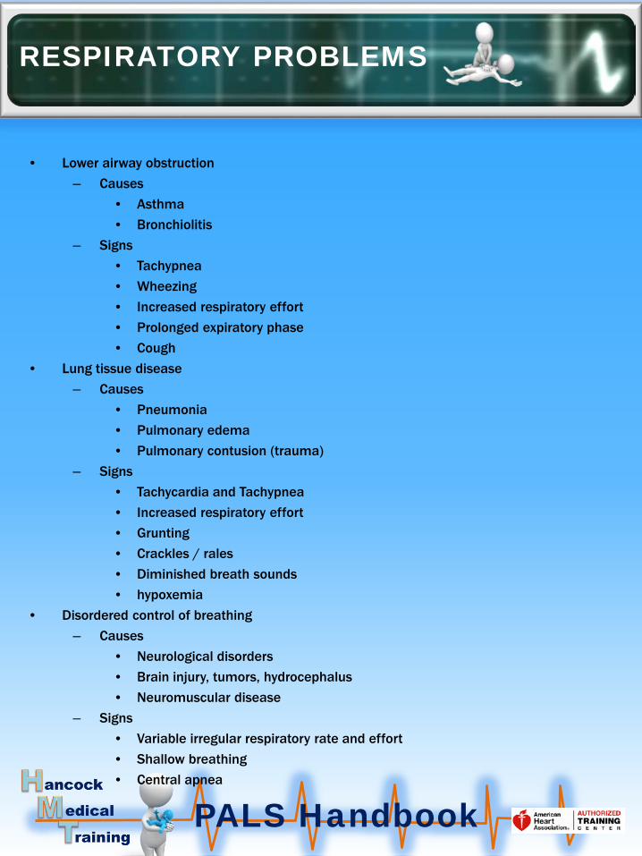
• Lower airway obstruction– Causes
• Asthma • Bronchiolitis
– Signs • Tachypnea• Wheezing• Increased respiratory effort• Prolonged expiratory phase• Cough
• Lung tissue disease– Causes
• Pneumonia• Pulmonary edema• Pulmonary contusion (trauma)
– Signs • Tachycardia and Tachypnea• Increased respiratory effort• Grunting • Crackles / rales• Diminished breath sounds• hypoxemia
• Disordered control of breathing– Causes
• Neurological disorders• Brain injury, tumors, hydrocephalus• Neuromuscular disease
– Signs • Variable irregular respiratory rate and effort• Shallow breathing• Central apnea
RESPIRATORY PROBLEMS

PALS Handbook
• General • Respiratory failure is the major cause of pediatric cardiac arrest
– Procedure • Detect and treat early• Identify the type and severity• Stabilize oxygenation and ventilation• Find the cause after stabilization
– Target management based on 4 types• Upper airway obstruction• Lower airway obstruction• Lung tissue disease• Disordered control of breathing
– For patients with potential for decline• Apply oxygen preferably humidified• Start an IV• Draw labs• Try to identify
– The problem– Type and severity
Life threatening problemsPrimary assessment (ABCDE)Treat life they occurIf there are no life threatening problems proceed to the secondary survey
MANAGING RESPIRATORY DISTRESS AND FAILURE

PALS Handbook
• Upper airway obstruction
– Most common causes• Croup • Anaphylaxis • Foreign body airway obstruction
– Croup • Most common 1 month to 5 years• Signs • Dyspnea, swelling of the vocal cords, barking cough• Causes • Viral, bacterial, allergic, inhaled irritants
– Treatments• Maintain the airway, administer humidified oxygen if SPO2 < 94%• nebulized racemic epinephrine or albuterol• Administer dexamethasone or a steroid• Consider heliox treatment• Severs dyspnea consider advanced or a surgical airway
MANAGING RESPIRATORY DISTRESS AND FAILURE

PALS Handbook
• Upper airway obstruction continued
– Anaphylaxis • High flow oxygen / monitor / IV• Nebulized racemic epinephrine or albuterol every 15 minutes• Administer Benadryl and an H2 blocker• Prepare for intubation• Treat hypotension
– Trendelenburg position– IV fluid boluses 20 ml /kg as needed– Epinephrine infusion if fluids are unsuccessful
• Consider a surgical airway if intubation is not possible
– Foreign body airway obstruction• Object in the trachea / non life threatening
– High flow oxygen / monitor / IV– Prepare for bronchoscopy
• Life threatening obstruction – Heimlich / CPR (back blows and thrusts / CPR infant)– Laryngoscope / Magill forceps– Prepare for a surgical airway–
MANAGING RESPIRATORY DISTRESS AND FAILURE

PALS Handbook
• Lung tissue disease
• Causes– Infectious pneumonia– Chemical and aspiration pneumonitis– Cardiogenic and non cardiogenic pulmonary edema
• Infectious pneumonia– High flow humidified O2 /I.V. / EKG / APO2– Diagnostic tests (ABG, X-ray, labs, cultures)– Antibiotic therapy– Treat wheezing with albuterol– Initiate I.V. fluids– Normalize temperature– CPAP / BIPAP / intubation for severe respiratory distress
• Chemical pneumonitis– Aspiration of a toxic gas or powder– Initiate humidified high flow oxygen initially– Treat wheezing with a nebulized bronchodilator– CPAP / BIPAP / Intubation with a ventilator for severe cases (failure)
• Aspiration pneumonitis– Aspiration of stomach contents– Initiate high floe humidified oxygen initially– Consider CPAP or BIPAP– For severe distress intubation with a ventilator
MANAGING RESPIRATORY DISTRESS AND FAILURE

PALS Handbook
• Lung tissue disease continued• Cardiogenic pulmonary edema
– Causes• Ventricular myocardial dysfunction• Excess pulmonary hydrostatic pressure• Plasma red call diffusion into the alveolar sacks
– Treatment • High flow oxygen, I.V. , EKG• Ventilation support
– CPAP / BIPAP, Ambu bag, intubation (PEEP 6-10)• Consider diuretics• Reduce metabolic demand (temperature and work of breathing)
• Non cardiogenic pulmonary edema (ARDS)– Definition
• Injury to interface between alveoli and pulmonary vessels• Triggers the release of inflammatory mediators
– Causes• Pneumonia, sepsis, pancreatitis, trauma
– Characteristics• acute onset • PaO2 /FiO2 < 200• Bilateral infiltrates (chest x-ray)• No evidence of cardiogenic causes
MANAGING RESPIRATORY DISTRESS AND FAILURE

PALS Handbook
• LUNG TISSUE DISEASE CONTINUED
• ARDS treatment– Cardiac monitor, vital signs, SPO2– Labs ( ABG, blood gas, CBC)– Provide ventilation support
• Ambu bag• CPAP /BIPAP• Intubation• ventilator
– Indications for ventilation support• Worsening clinical and radiographic lung disease• Hypoxia refractory to high FiO2
– Combat hypoxemia• Peep until oxygen saturation is adequate• Low tidal volumes (5-7 ml/kg)• Peak inspiratory pressures < 30-50 cm H2O
MANAGING RESPIRATORY DISTRESS AND FAILURE

PALS Handbook
• Disordered control of breathing• Causes
– Increased ICP• TBI• Subdural /epidural hematoma• Brain tumor, hydrocephalies
– Neuromuscular disease– CNS depression
• Drugs, Infection, Seizures, Metabolic disorders• Interventions for ICP
– Manage ABC’S– Assess breathing problems
• Assist ventilations (Ambu, ET tube hyper-oxygenate)– Poor perfusion or end organ function
• Administer 20 ml/kg boluses of a crystalloid solution– Administer osmotic agents
• Mannitol , dexamethasone– Treat agitation and pain aggressively– Avoid hyperthermia
• Interventions for poisoning and drug overdose– Manage ABC’S– Contact poison control (1-800-222-1222)– Prepare suctioning in case of vomiting– Administer antidote– Perform diagnostic tests
• Interventions with neuromuscular disease– Ventilation support – Manage secretions
MANAGING RESPIRATORY DISTRESS AND FAILURE

PALS Handbook
• Definition – Delivery of oxygen and nutrients do not meet the system’s needs. – This creates inadequate peripheral and end organ perfusion.– The areas of primary concern are the brain, kidneys, heart and liver
• Pathophysiology– Cells need constant oxygen delivery– The primary system is the circulatory system – The secondary system is anaerobic metabolism
• Oxygen is created by acid conversion• This method has a limited ability to sustain oxygen demands• It also creates an undesirable acidic condition in the body
• Oxygen delivery and perfusion requirements– Sufficient oxygen in the blood– Adequate cardiac output– Adequate distribution
SHOCK
PUMP CONTAINER FLUID
HEART ARTERIES AND VEINS
BLOOD

PALS Handbook
• Cardiac output– Cardiac output represents the pumping adequacy of the heart.– Cardiac output (CO) = Stroke volume x Heart rate– The cardiac output is determined by;
• Preload – the volume of blood in the ventricles before contraction• Contractility – the strength of contraction• Afterload – the systemic resistance
• Compensatory mechanisms– The body compensates for shock by the following mechanisms
• Tachycardia• Vasoconstriction• Increased contractility• Increase in smooth venous tone
• Organ response to compensated shock– Heart – tachycardia– Skin – cool, pale, mottled, diaphoretic– Peripheral circulation – delayed capillary refill– Pulses – weak peripheral, narrow pulse pressure– Kidneys – reduced urine output– Intestines – vomiting , ileus
SHOCK

PALS Handbook
• Shock states– Compensated – good perfusion
• Early signs of compensated shock• Normal skin color and temperature
– Compensated poor perfusion• Later signs of compensated shock• Pale, mottled skin, normal blood pressure lower urine output
– Decompensated – blood pressure drops• All signs of late compensated shock• Blood pressure drops
• Response to shock in adults and children– Adults
• Compensate for a shorter time• Decompensate more quickly• Can sustain de-compensation better
– Children• Compensate for a longer period of time• Once they decompensate they crash quickly
• Hypotensive shock• Hypotensive shock is decompensated shock• Hypotension in children
–
SHOCK
Compensated shock Hypovolemic shock Cardiac arrest

PALS Handbook
• Hypovolemic shock (Fluid loss)
– Causes• Diarrhea • Vomiting • Hemorrhage• Osmotic diuresis (DKA)• Inadequate fluid intake• Third space loss (anaphylaxis)• Large burns
– Signs• Tachypnea• Tachycardia• Systolic hypotension / narrow pulse pressure• Weak or absent peripheral pulses• Delayed capillary refill• Cool, pale, mottled, diaphoretic skin• Changes in level of consciousness• Oliguria• Extremities cooler than the trunk
HYPOVOLEMIC SHOCK
PALS Handbook
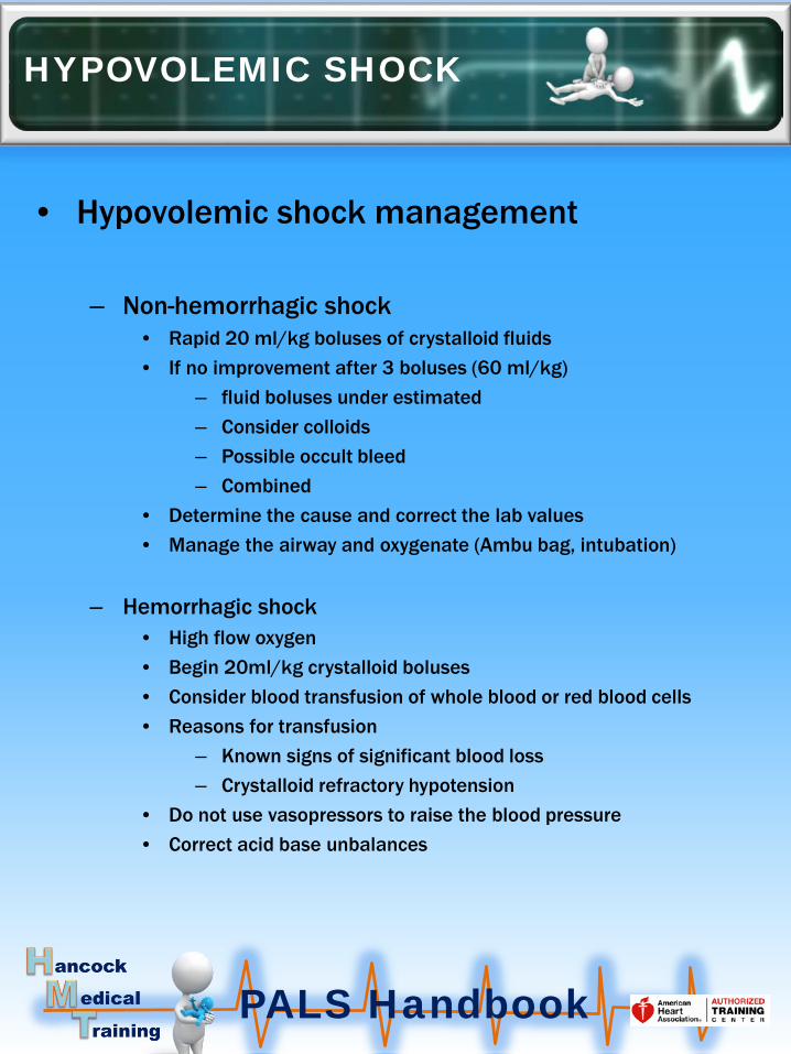
• Hypovolemic shock management
– Non-hemorrhagic shock• Rapid 20 ml/kg boluses of crystalloid fluids• If no improvement after 3 boluses (60 ml/kg)
– fluid boluses under estimated– Consider colloids– Possible occult bleed– Combined
• Determine the cause and correct the lab values • Manage the airway and oxygenate (Ambu bag, intubation)
– Hemorrhagic shock• High flow oxygen• Begin 20ml/kg crystalloid boluses• Consider blood transfusion of whole blood or red blood cells • Reasons for transfusion
– Known signs of significant blood loss– Crystalloid refractory hypotension
• Do not use vasopressors to raise the blood pressure• Correct acid base unbalances
HYPOVOLEMIC SHOCK

PALS Handbook
• Causes– Sepsis – Anaphylaxis– Neurogenic problems
• CNS disorders• Drugs• Spinal cord injuries
• Physiology– High to normal cardiac output – low SVR– Variable blood flow and perfusion (variable SVR)– Increased capillary permeability– Pulmonary hypertension (increased PVR)– Release if inflammatory mediators– Vasodilation causes blood pooling and clotting– Increased lactic acid buildup / acidosis– May lead to hypovolemic shock and cardiac dysfunction– Warm shock – blood shunted to the periphery
• Signs– Tachypnea / tachycardia– Warm shock– Cold shock– Changes in level of consciousness– Oliguria– Fever or hypothermia– Petechial or purpuric rash (septic shock)
DISTRIBUTIVE SHOCK

PALS Handbook
• Septic shock (sepsis)
• Signs – Fever – Presence of infection– Hypotension– Tachycardia– Tachypnea– Acidosis– Metabolic unbalance
• Pathophysiology – Infection / endotoxin stimulates the immune system
• Releases inflammatory mediators• Cytokines create micro clots
– Variable SVR creates mal distribution of flow• Creates blood pooling and localized hypoxia
– Relative hypovolemia• Vasodilation / increased permeability
– Adrenal insufficiency• Cytokines create micro clots causing renal insufficiency• May create myocardial dysfunction
DISTRIBUTIVE SHOCK
PALS Handbook
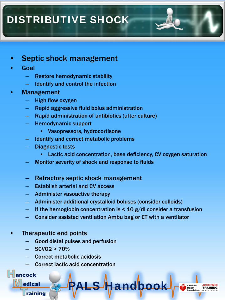
• Septic shock management• Goal
– Restore hemodynamic stability– Identify and control the infection
• Management– High flow oxygen– Rapid aggressive fluid bolus administration– Rapid administration of antibiotics (after culture)– Hemodynamic support
• Vasopressors, hydrocortisone– Identify and correct metabolic problems– Diagnostic tests
• Lactic acid concentration, base deficiency, CV oxygen saturation– Monitor severity of shock and response to fluids
– Refractory septic shock management– Establish arterial and CV access– Administer vasoactive therapy– Administer additional crystalloid boluses (consider colloids)– If the hemoglobin concentration is < 10 g/dl consider a transfusion– Consider assisted ventilation Ambu bag or ET with a ventilator
• Therapeutic end points– Good distal pulses and perfusion– SCVO2 > 70%– Correct metabolic acidosis– Correct lactic acid concentration
DISTRIBUTIVE SHOCK
PALS Handbook
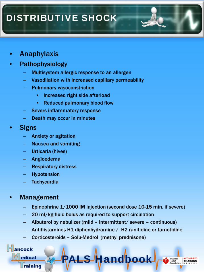
• Anaphylaxis• Pathophysiology
– Multisystem allergic response to an allergen– Vasodilation with increased capillary permeability– Pulmonary vasoconstriction
• Increased right side afterload• Reduced pulmonary blood flow
– Severs inflammatory response– Death may occur in minutes
• Signs– Anxiety or agitation– Nausea and vomiting– Urticaria (hives)– Angioedema– Respiratory distress– Hypotension– Tachycardia
• Management– Epinephrine 1/1000 IM injection (second dose 10-15 min. if severe)– 20 ml/kg fluid bolus as required to support circulation– Albuterol by nebulizer (mild – intermittent/ severe – continuous)– Antihistamines H1 diphenhydramine / H2 ranitidine or famotidine– Corticosteroids – Solu-Medrol (methyl prednisone)
DISTRIBUTIVE SHOCK

PALS Handbook
• Causes– Injuries to the cervical and thoracic spine above T6
• Pathophysiology– Loss of sympathetic nerves that control innervation to the heart
• Signs– Hypotension– Normal heart rate or bradycardia– Increased respiratory rate– Diaphragmatic breathing– Inability to compensate for hypovolemia
• Management – Position the child flat or head down
• Improves venous return– Administer 20 ml/kg crystalloid bolus– Vasopressors if refractory to fluids
• Norepinephrine• epinephrine
– Provide warming or cooling as required
NEUROGENIC SHOCK

PALS Handbook
• Definition • Cardiogenic shock is poor perfusion resulting from cardiac dysfunction
• Causes– Congenital heart disease– Myocarditis – Cardiomyopathy– Arrhythmias– Sepsis– Poisoning or drug overdose– Myocardial injury (trauma)
• Pathophysiology– Increased heart rate and ventricular afterload
• Increased ventricular workload and myocardial oxygen consumption
– Compensatory increase in SVR• Diverts blood from periphery to brain and organs
– Decrease in stroke volume• Decreased contractility and increased afterload
– Increased venous tone• Increases CV and pulmonary capillary wedge pressure
– Diminished renal blood flow (fluid retention)– Pulmonary edema
CARDIOGENIC SHOCK

PALS Handbook
• Signs– Tachypnea– Tachycardia– Low blood pressure / narrow pulse pressure– Weak or absent pulses– Delayed capillary refill , cool extremities– Signs of congestive heart failure
• Pulmonary edema, JVD distension– Changes in level of consciousness– Cold pale diaphoretic skin / cyanosis– oliguria
• Management – Goals
• Improve cardiac function and output • Increase ejection fraction
– Management • Cautious fluid administration / reduce afterload (5-10 ml/kg• Lab and diagnostic studies
– ABG, X-ray, cardiac enzymes, hemoglobin• EKG• Medications
– Diuretics – pulmonary edema– Vasodilators – lower afterload– Inotropes –increase contractility and cardiac output– Analgesics – for pain
CARDIOGENIC SHOCK

PALS Handbook
• Definition – Cardiac output impaired by a physical obstruction to blood flow
• Causes– Cardiac tamponade– Tension pneumothorax– Ductal dependent congenital heart lesions– Massive pulmonary embolism
• Signs– Respiratory failure / pulmonary edema– Rapid deterioration in peripheral perfusion– Congestive heart failure (left, right side or both sides)– Absence of femoral pulses– Metabolic acidosis– Rapid changes in LOC– Tachycardia– Hypotension– Chest pain– Cool extremities and trunk / possible cyanosis
OBSTRUCTIVE SHOCK

PALS Handbook
• Cause – Accumulation of blood in the pericardial sac– Reduces ventricular filling and stroke volume
• Findings – Respiratory distress and increased respiratory effort– Tachycardia– Cool extremities / delayed capillary refill– Muffled heart sounds– Narrow pulse pressure – Distended neck veins– Changes in LOC.
• Treatment – Initially
• Rapid identification• Oxygen and fluid administration
– Pericardial centesis• Perform if impending arrest• Requires a skilled person• Best done with fluoroscopy or electrocardiography
CARDIAC TAMPONADE

PALS Handbook
• Cause– Entry of air into the pleural space that accumulates under pressure
• Trauma or spontaneous• May occur in ventilated patients
• Findings– Respiratory distress with increased respiratory effort– Distended neck veins– Tracheal deviation– Rapid deterioration in perfusion– Rapid evolution tachycardia to bradycardia– Hypotension– Changes in LOC
• Treatment– Immediate needle decompression– Insert an 18-20 gage. Needle
• Over the top of the 3rd rib (second intercostal space)• At the mid clavicular line
– A gush of air will be heard after decompression– More than 1 decompression may be necessary– A chest tube will ultimately be required
TENSION PNEUMOTHORAX

PALS Handbook
• Causes– Cyanotic congenital heart lesions
• Patent ductus arteriosus– Left ventricular outflow obstructive lesions
• Coarctation of the aorta• Aortic valve stenosis• Hypoplastic left heart syndrome
• Findings– Respiratory failure– Cardiomegaly (rapid deterioration in perfusion)– Higher pre ductal versus post ductal pressures– Absence of femoral pulses– Cyanosis– Hypotension with tachycardia
• Treatment – Continuous infusion of prostaglandin E to close ductus arteriosus– Ventilation support with oxygen administration– Electrocardiography– Inotropic agents to improve contractility– Fluids to improve cardiac output (caution)– Correct metabolic derangements
DUCTAL DEPENDENT LESIONS

PALS Handbook
• Cause– Partial or total obstruction of the pulmonary artery and its branches by
a blood clot
• Findings– Respiratory distress with increased respiratory effort– Tachycardia– Cyanosis– Hypotension– Changes in the level of consciousness– Increased ventilation has no effect on SPO2
• Treatment – Initial – ventilation support and fluid therapy– CT angiography / echocardiography (diagnostic)– Anticoagulant therapy (heparin)– Consider thrombolytic agents (TPA)
PULMONARY EMBOLIS

PALS Handbook
• Fundamentals – Optimize the oxygen content of the blood– Improve volume distribution and cardiac output– Reduce the oxygen demand– Correct metabolic derangements
• Successful treatment of shock– Therapeutic end points
• Normal heart rate and blood pressure• Normal pulses• Capillary refill < 2 seconds• Warm extremities• Normal mental status• Urine output > 1 ml/kg/hr• Decreased serum lactate• Reduced base deficit• ScvO2 > 70%
SHOCK MANAGEMENT
PALS Handbook
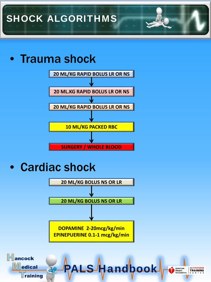
• Trauma shock
• Cardiac shock
SHOCK ALGORITHMS
20 ML/KG RAPID BOLUS LR OR NS
10 ML/KG PACKED RBC
SURGERY / WHOLE BLOOD
20 ML/KG RAPID BOLUS LR OR NS
20 ML.KG RAPID BOLUS LR OR NS
20 ML/KG BOLUS NS OR LR
20 ML/KG BOLUS NS OR LR
DOPAMINE 2-20mcg/kg/min EPINEPUERINE 0.1-1 mcg/kg/min

PALS Handbook
Dehydration shock
SHOCK ALGORITHMS
20ml/kg BOLUS NS OR LR
REPEAT 20ml/kg BOLUSES AS LONG AS LUNGS ARE CLEAR
CONSIDER A MAINTENANCE INFUSION
DO NOTUSE
VASOPRESSORS

PALS Handbook
• Septic shock
SHOCK ALGORITHMS

PALS Handbook
• I.V. Access– Most common I.V. sites
• Antecubital • Hand• Scalp on infants
– Most common I.V. needles• 24 gage• 22 gage• 20 gage
• I.O. access– Any drug or fluid that can be given I.V. can be given I.O– Easier to attain than I.V. access especially in arrest situations– Contraindications
• Fractures in an extremity• Previous attempt in the same place• Infection overlying the bone
MEDICATION / FLUID ACCESS
USE AN ARMBOARD TO SECURE

PALS Handbook
• I.O. INSERTION SITES
MEDICATION / FLUID ACCESS

PALS Handbook
• INFANT / PEDI INSERTION SITE
MEDICATION / FLUID ACCESS

PALS Handbook
• OLDER PEDIATRIC / YOUNG ADULT
MEDICATION / FLUID ACCESS

PALS Handbook
• Insertion procedure using a Jamshidi needle– Identify the tibial tuberosity
– Disinfect the area
– Leave the stylette in the needle
– Stabilize the leg• Do not put your hand behind the leg• Be sure the leg is fully on a hard flat surface
– Insert the needle perpendicular to the leg
– Use a twisting motion with firm pressure
– Insert until you feel a sudden decrease in resistance• A slight popping sound may be heard
– Remove the stylette aspirate and flush with a syringe– Confirm placement
• Pressurized fluids flow freely• There is no infiltration to surrounding tissues
– Stabilize the needle and tape over the flange
– Attach a 3 way stopcock
MEDICATION / FLUID ACCESS

PALS Handbook
• Drugs to control SVT• Adenosine
– Classification - antiarrhythmic– Indications – treatment of SVT– Actions
• Stimulates adenosine receptors• Transiently blocks AV node conduction• Transiently interrupts reentry pathways• Depresses sinus node activity
– Dose • First dose – 0.1 mg/kg rapid push (6 mg max dose) • Second dose – 0.2mg/kg rapid push (12 mg max dose)
• Drugs to control bradycardia• Epinephrine
– Classification –catecholamine, vasopressor, inotrope– Indication – symptomatic bradycardia– Action
• Stimulates beta 1 and 2 adrenergic receptors• Increases contractility, heart rate, blood pressure• Dilates bronchi and arterioles
– Dose • IV / IO 0.1 mg/kg of 1/10000 every 3-5 minutes
RATE CONTROL DRUGS

PALS Handbook
• DRUGS TO CONTROL ASTHMA AND CROUP• Albuterol
– Classification-bronchiole dilator, beta adrenergic agent– Indications – asthma, anaphylaxis, – Action
• Bronchiole and vasodilator,– Dose
• Severe asthma – 0.5 mg/kg/hr continuous nebulizer treatment
• Atrovent (Ipratropium bromide)• Classification - anticholergenic• Indication asthma• Action
– Blocks parasympathetic choline receptors– Inhibits serious mucus secretions
• Dose – Nebulizer – 250-500 mcg every 20 minutes x 2
• Terbutaline• Classification-beta agonist, bronchiole dilator• Indications- asthma• Action
– Dilates bronchioles and arterioles• Dose
– IV /IO – 0.1 – 10 mcg/kg/min. Consider 10 mcg/kg over 5 minutes
RESPIRATORY DRUGS
PALS Handbook
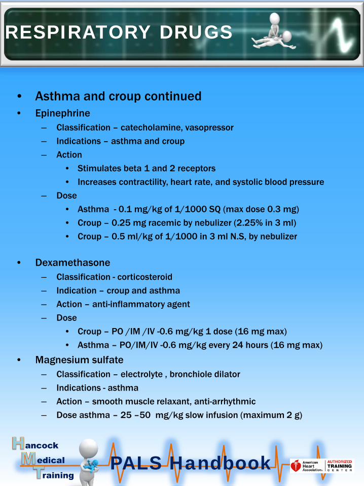
• Asthma and croup continued• Epinephrine
– Classification – catecholamine, vasopressor– Indications – asthma and croup– Action
• Stimulates beta 1 and 2 receptors• Increases contractility, heart rate, and systolic blood pressure
– Dose • Asthma - 0.1 mg/kg of 1/1000 SQ (max dose 0.3 mg)• Croup – 0.25 mg racemic by nebulizer (2.25% in 3 ml)• Croup – 0.5 ml/kg of 1/1000 in 3 ml N.S, by nebulizer
• Dexamethasone– Classification - corticosteroid– Indication – croup and asthma– Action – anti-inflammatory agent– Dose
• Croup – PO /IM /IV -0.6 mg/kg 1 dose (16 mg max)• Asthma – PO/IM/IV -0.6 mg/kg every 24 hours (16 mg max)
• Magnesium sulfate– Classification – electrolyte , bronchiole dilator– Indications - asthma– Action – smooth muscle relaxant, anti-arrhythmic– Dose asthma – 25 –50 mg/kg slow infusion (maximum 2 g)
RESPIRATORY DRUGS

PALS Handbook
• DRUGS FOR ANAPHYLAXIS
• Diphenhydramine– Classification -antihistamine– Indication - anaphylaxis– Action
• Competes with histamines for H1 receptor sites• Decreases allergic response by blocking histamines
– Dose • 1-2 mg/kg every 4-6 hours (maximum dose 50 mg)
Epinephrine– Classification – catecholamine, vasopressor– Indications – asthma and croup– Action
• Stimulates beta 1 and 2 receptors• Increases contractility, heart rate, and systolic blood pressure
– Dose • Asthma - 0.1 mg/kg of 1/1000 SQ (max dose 0.3 mg)
• Dexamethasone– Classification - corticosteroid– Indication – croup and asthma– Action – anti-inflammatory agent– Dose
• Asthma – PO/IM/IV -0.6 mg/kg every 24 hours (16 mg max)
RESPIRATORY DRUGS
PALS Handbook
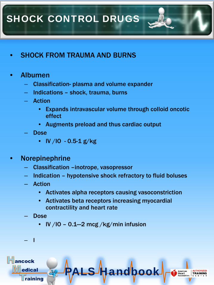
• SHOCK FROM TRAUMA AND BURNS
• Albumen – Classification- plasma and volume expander– Indications – shock, trauma, burns– Action
• Expands intravascular volume through colloid oncotic effect
• Augments preload and thus cardiac output– Dose
• IV /IO - 0.5-1 g/kg
• Norepinephrine – Classification –inotrope, vasopressor– Indication – hypotensive shock refractory to fluid boluses– Action
• Activates alpha receptors causing vasoconstriction• Activates beta receptors increasing myocardial
contractility and heart rate– Dose
• IV /IO – 0.1—2 mcg /kg/min infusion
– I
SHOCK CONTROL DRUGS

PALS Handbook
• SHOCK TRAUMA BURNS• Vasopressin
– Classification – antidiuretic hormone– Indication – septic shock– Action
• Mediated by vasopressin receptors
– Dose • IV /IO -0.0002 – 0.002 units/kg/min continuous infusion
– Monitor • Blood pressure and distal pulses• Water intoxication (headache, drowsiness)
• Dopamine – Classification – catecholamine, vasopressor, inotrope– Indication
• Cardiogenic shock• Distributive shock
– Action• 5 – 15 mcg/kg/min – increases, contractility , decreases SVR• 15 – 20mcg/kg/min – increases SVR and constriction of the
arteries
– Dose • IV/IO -2 – 20 mcg/kg/min
SHOCK CONTROL DRUGS

PALS Handbook
• Alprostadil– Classification – vasodilator, prostaglandin– Indication
• Maintain patency with ductus arteriosus• Tetralogy of Fallot• Aortic stenosis or obstructive lesions
– Action• Acts on FPE receptors to dilate arteries and arterioles• Stimulates uterine and smooth muscles
– Dose• IV /IO – initial - 0.05 – 0.1 mg/kg/min infusion• IV / IO – maintenance – 0.01 - 0.05 mg/kg/min infusion
• Inamrinone (Amrinone) and Milrinone– Classification – phosphodiesterase inhibitor– Indication
• Myocardial dysfunction and increased SVR• Cardiogenic shock with increased SVR• Post cardiac surgery
– Action• Increases myocardial contractility• Reduces preload and afterload (relaxes smooth muscle)
– Dose IV / IO• loading – 0.75 – 1 mg/kg slow bolus -- amrinone• Infusion – 5 – 10 mcg/kg/min -- amrinone
• Loading – 50 mcg /kg (10-60 minutes) - milrinone• Infusion – 0.25 – 0.75mcg/kg/min - milrinone
CARDIAC DRUGS
PALS Handbook
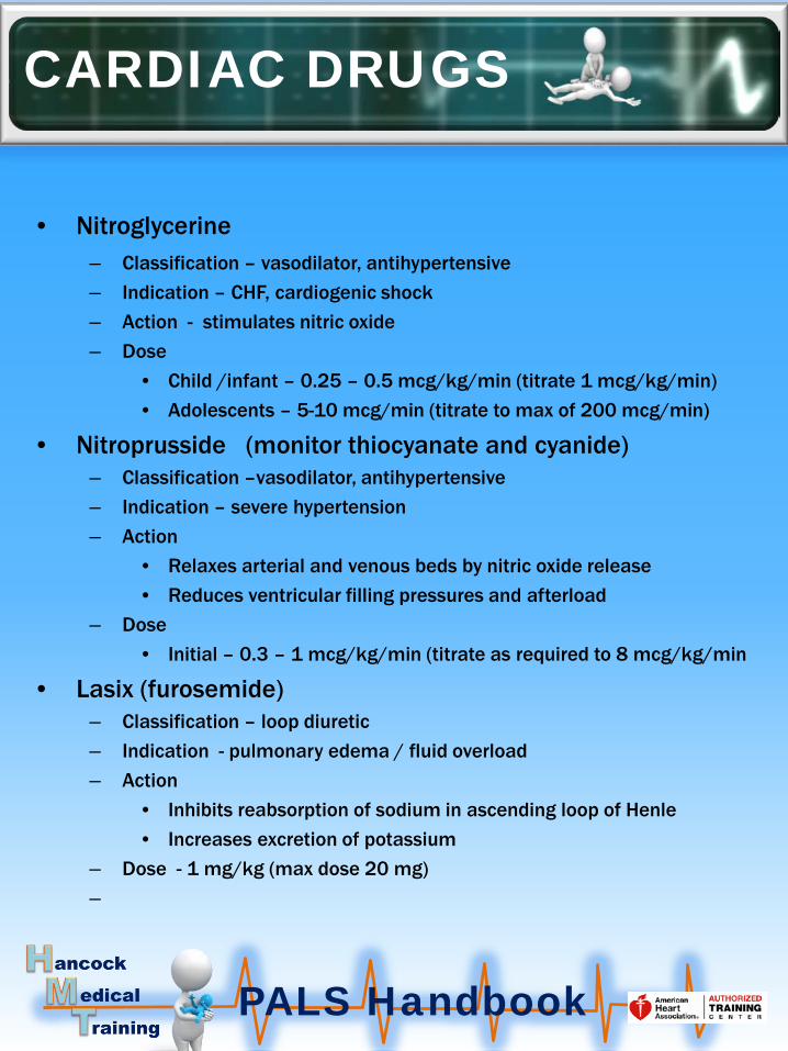
• Nitroglycerine– Classification – vasodilator, antihypertensive– Indication – CHF, cardiogenic shock– Action - stimulates nitric oxide– Dose
• Child /infant – 0.25 – 0.5 mcg/kg/min (titrate 1 mcg/kg/min)• Adolescents – 5-10 mcg/min (titrate to max of 200 mcg/min)
• Nitroprusside (monitor thiocyanate and cyanide)– Classification –vasodilator, antihypertensive– Indication – severe hypertension– Action
• Relaxes arterial and venous beds by nitric oxide release• Reduces ventricular filling pressures and afterload
– Dose • Initial – 0.3 – 1 mcg/kg/min (titrate as required to 8 mcg/kg/min
• Lasix (furosemide)– Classification – loop diuretic– Indication - pulmonary edema / fluid overload– Action
• Inhibits reabsorption of sodium in ascending loop of Henle• Increases excretion of potassium
– Dose - 1 mg/kg (max dose 20 mg)–
CARDIAC DRUGS

PALS Handbook
• Dobutamine– Classification – selective beta adrenergic agent – Indication - ventricular dysfunction– Action
• Stimulates beta 1 receptors• Increases contractility, automaticity, and conduction
velocity• Increases heart rate and vasodilation• Alpha blocking effects (risk of hypotension)
– Dose -IV / IO – 2 – 20 mcg/kg/min
• Lidocaine– Classification - antiarrhythmic– Indication – wide complex tachycardia with a pulse– Action
• Stabilizes cardiac membrane and decreases automaticity• Abolishes reentry
– Dose• 1 mg/kg bolus followed by infusion 1-2 mg/min
CARDIAC DRUGS

PALS Handbook
• Calcium chloride– Classification - electrolyte– Indication
• Hypocalcemia • Hyperkalemia• Calcium channel overdose• Consider for hypomagnesemia
– Action • Maintains nervous, muscular and skeletal functions• Maintains cardiac contractility, coagulation and enzyme functions• Affects secretory function of endocrine and exocrine glands
– Dose • Arrest – 20 mg/kg bolus Non arrest – 20 mg.kg infuse over 30-60 minutes
• Sodium bicarbonate– Classification -electrolyte– Indication
• Severe metabolic acidosis • Hyperkalemia• Sodium channel blocker overdose (tricyclic antidepressants)
– Action – increases plasma bicarbonate– Dose - IV/IO
• Metabolic acidosis / hyperkalemia – 1 mEq slow bolus (50 mEq max)• Sodium channel blocker overdose – 1 – 2 mEq bolus until Ph >7.45
• Magnesium sulfate– Classification – electrolyte, bronchodilator– Indication – asthma, torsades, and hypomagnesemia– Action – smooth muscle relaxant , antiarrhythmic– Dose
• asthma – 25 – 50 mg/kg infusion (20 min) to a max of 2g• Torsades (pulseless) – 25-50 mg/kg bolus to a max of 2g• Torsades (pulse) – 25-50 mg/kg over 20 minutes
ELECTROLYTES
PALS Handbook
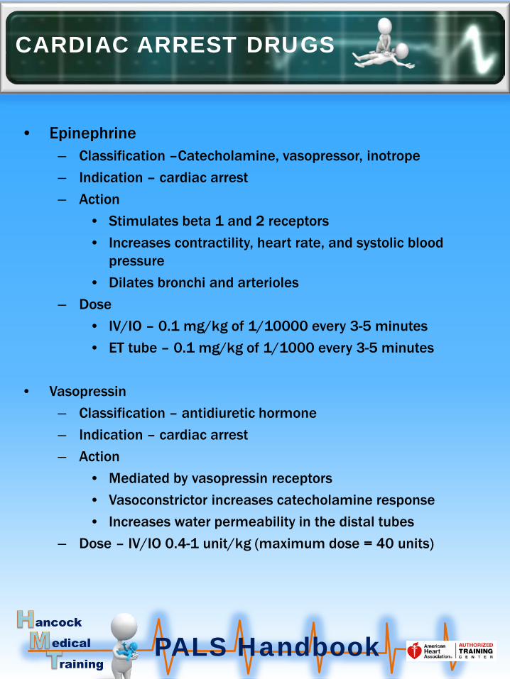
• Epinephrine– Classification –Catecholamine, vasopressor, inotrope– Indication – cardiac arrest– Action
• Stimulates beta 1 and 2 receptors• Increases contractility, heart rate, and systolic blood
pressure• Dilates bronchi and arterioles
– Dose • IV/IO – 0.1 mg/kg of 1/10000 every 3-5 minutes• ET tube – 0.1 mg/kg of 1/1000 every 3-5 minutes
• Vasopressin– Classification – antidiuretic hormone– Indication – cardiac arrest– Action
• Mediated by vasopressin receptors• Vasoconstrictor increases catecholamine response• Increases water permeability in the distal tubes
– Dose – IV/IO 0.4-1 unit/kg (maximum dose = 40 units)
CARDIAC ARREST DRUGS

PALS Handbook
• Amiodarone– Classification - antiarrhythmic– Indication – cardiac arrest– Action
• Works in both the atria and ventricles• Prolongs action potential duration• Slows sinus rate• Inhibits alpha and beta adrenergic receptors
– Dose • IV/IO – 5 mg/kg rapid bolus (maximum dose = 300 mg)• Follow up with a drip infusion
• Lidocaine – Classification – antiarrhythmic, anesthetic– Indication – cardiac arrest– Action
• Works primarily in the ventricles• Increases electrical stimulation stabilizing cardiac membranes• Reduces ICP by inhibiting sodium channels
– Dose • IV/IO -1 mg/kg loading bolus – repeat dose (max 3 mg/kg)• Follow up with a drip• ET – 2-3 mg/kg
CARDIAC ARREST DRUGS

PALS Handbook
• Etomidate– Classification- sedative, hypnotic– Indication
• Sedation for RSI (especially for head injuries)• Sedative for hypotension, trauma, cardiac disease
– Action• Short acting sedative hypnotic agent • Non barbiturate or benzodiazepine• Decreases ICP, cerebral blood flow and cerebral metabolic rate
– Dose - 0.2 – 0.4 mg/kg IV/IO infused over 20 – 30 seconds (max dose 20 mg)
• Atropine
– Classification - anticholinergic
– Indication – RSI• 1 – 5 years receiving succinylcholine• > 5 years receiving a second dose of succinylcholine
– Action• increases heart rate and cardiac output by blocking vagal stimulation• Causes mydriasis (paralysis)
– Dose • IV/IO – 0.01 - 0.02 mg/kg (min dose -01.1 mg / max dose 0.5 mg )• IM - 0.2 mg/kg
• Lidocaine – Classification - antiarrhythmic– Indication- RSI to reduce ICP– Action
• Reduces ICP by inhibiting sodium channels in neurons• Reduces metabolic activity
– Dose – 1 – 2 mg/kg
RSI DRUGS

PALS Handbook
• Narcotic overdose– Naloxone (Narcan)
• Classification – opioid antagonist• Indication – reverse opiate overdose• Action – competes with opioids for receptor site• Dose – 4 mg (repeat until total reversal)
• Anaphylaxis– Epinephrine
• Classification – catecholamine, vasopressor, inotrope• Indication - anaphylaxis• Action
– Beta adrenergic stimulator (particularly beta 2)– Increases heart rate, contractility and automaticity
• Dose – IM -0.01 mg/kg of 1/1000 every 15 min. (0.3 mg max)– IV/IO – 0.01 - 0.1 mg/kg of 1/10000 (max dose 1 mg)– Hypotension refractory to fluids – 0.1 – 1 mcg/kg/min
• Hypoglycemia– Dextrose (glucose)
• Classification -carbohydrate• Indication - hypoglycemia• Action – increases blood glucose• Dose - 0.5 – 1 g/kg• Concentrations
– D50W – 1- 2 ml/kg– D25W – 2-4 ml/kg – dilute D50 1:1with N.S.– D12.5W – 5-10 ml/kg dilute D25 1:1 with N.S. (newborns)
SPECIAL DRUGS
Follow bolus with infusion if required
Monitor after administration

PALS Handbook
BROSLOW TAPE

PALS Handbook
• General– Starlings law
• CO = STROKE VOLUME X HEART RATE– Primary bradycardia (cardiac causes)
• Congenital abnormality in the pacemaker system• Surgical injury to the conduction system• Cardiomyopathy• myocarditis
– Secondary bradycardia (non cardiac causes)• Hypoxia • Acidosis• Hypotension• Hypothermia• Drug effects
• Signs and symptoms– Hypotension– Decreased level of consciousness– Shock– Poor organ perfusion– Respiratory distress or failure– Sudden collapse
• Types of bradycardia– Sinus bradycardia– First degree heart block– Mobitz 1 (Wenckebach)– Mobitz 2 (second degree type 2)– Third degree (complete heart block)
BRADYCARDIAS
PALS Handbook
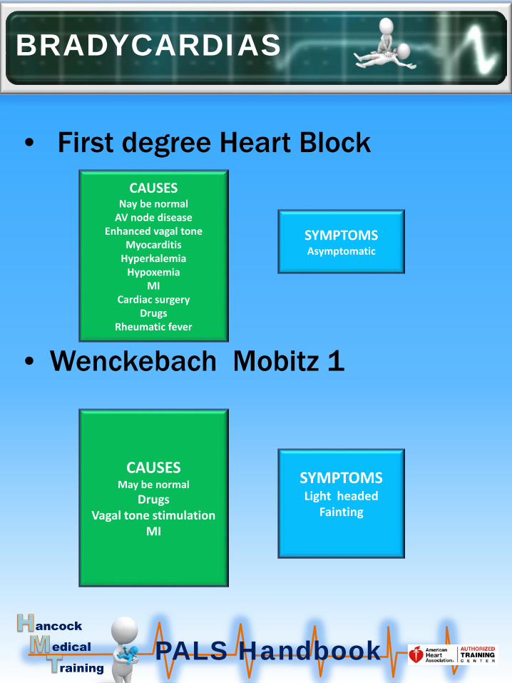
• First degree Heart Block
• Wenckebach Mobitz 1
BRADYCARDIAS
CAUSESNay be normal
AV node diseaseEnhanced vagal tone
MyocarditisHyperkalemia
HypoxemiaMI
Cardiac surgeryDrugs
Rheumatic fever
SYMPTOMSAsymptomatic
CAUSESMay be normal
DrugsVagal tone stimulation
MI
SYMPTOMSLight headed
Fainting

PALS Handbook
• Second degree Mobitz 2
• Third degree (complete HB)
BRADYCARDIAS
CAUSESAbnormal conduction
Parasympathetic controlDrugs (rare)
MICardiac surgery
SYMPTOMSIrregular heart beat
Light headedness
Syncope
CAUSESConduction deficiencies
MyocarditisCardiac surgery
MIParasympathetic tone
Drug toxicitySevere hypoxiaSevere acidosis
SYMPTOMSFatigue
Light headednesssyncope
PALS Handbook
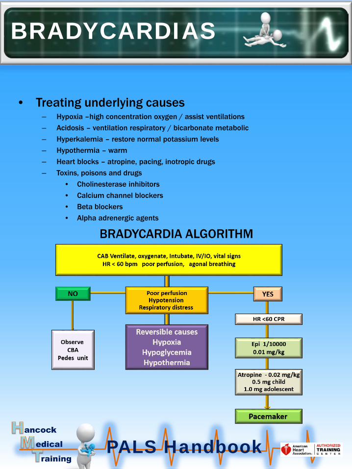
• Treating underlying causes– Hypoxia –high concentration oxygen / assist ventilations– Acidosis – ventilation respiratory / bicarbonate metabolic– Hyperkalemia – restore normal potassium levels– Hypothermia – warm – Heart blocks – atropine, pacing, inotropic drugs– Toxins, poisons and drugs
• Cholinesterase inhibitors • Calcium channel blockers• Beta blockers • Alpha adrenergic agents
BRADYCARDIA ALGORITHM
BRADYCARDIAS

PALS Handbook
• General– Tachycardias are fast rhythms originating in either the atria or ventricles– Sinus tachycardias usually do not create hemodynamic compromise but
actually increase cardiac output (stress and fever)– Rapid tachyarrhythmias reduce cardiac output by not allowing
ventricular filling during diastole.– Severe hemodynamic compromise can occur if these rhythms are
allowed to continue– .
• Types of tachycardias– Sinus tachycardia– Supraventricular tachycardia (SVT) – stable / unstable– Wide complex tachycardia – stable / unstable
• Signs of hemodynamic instability– Poor feeding and irritability– Respiratory distress and failure– Signs of shock (poor perfusion and hypotension)– Altered mental status– Sudden collapse (weak rapid pulses)
TACHYCARDIAS

PALS Handbook
• Common causes– Pain– Anxiety– Hypoxia– Hypovolemia– shock– Fever– Metabolic stress– Injury (pneumothorax, tamponade, embolism)– Toxins– Anemia
• SINUS TACHYCARDIA ALGORITHM
SINUS TACHYCARDIA
TREAT THE CAUSETREAT THE ARRHYTHMIA
CAB, OXYGEN,VITAL SIGNS, EKG, HX, LABS, 12 LEAD
TREAT THE CAUSEANXIETY, FEVER, HYPOXIA,
HYPOVOLENIAEXERTION, PAIN, INJURY,
TOXINS

PALS Handbook
• General– May be episodic (PSVT)– Tolerated better in infants– Prolonged SVT can lead to CHF resulting in severe hemodynamic
compromise
• Signs and symptoms– Tachypnea– Wheezes, crackles, grunting associated with CHF– Delayed capillary refill time (cool extremities)– Diaphoresis hypotension – Altered mental status– irritability– lethargy
• STABLE SVT ALGORITHM
SUPRAVENTRICULAR TACHYCARDIA

PALS Handbook
• SUPRAVENTRICULAR ALGORITHM (UNSTABLE)
SUPRAVENTRICULAR TACHYCARDIA
CAB OXYGEN VITALS IV /IO SPO2 EKG PREPARE CODE EQ
VAGAL MANEUVERS
ADENOSINE 0.1 mg/kgRAPID WITH FLUSH
SYNC CARDIOVERSION0.5-1.0 J/kg
SYNC CARDIOVERSIONUP TO 2 J/kg
SYNC CARDIOVERSIONUP TO 2 J/kg
12 LEADCONSULT

PALS Handbook
• STABLE WIDE COMPLEX TACHYCARDIA ALGORITHIM
WIDE COMPLEX TACHYCARDIA
CAB OXYGEN EKG SPO2 VITAL SIGNS IV/IO LABS
HISTORY 12 LEAD
AMIODARONE 5 mg/kg (20-60 min)OR
Lidocaine 1.0 mg/kg
SYNC CARDIOVERSION0.5 – 1 J/kg MAY INCREASE TO 2 J/kg
PEDIATRIC CARDIOLOGIST
SUCCESSFUL
PALS Handbook
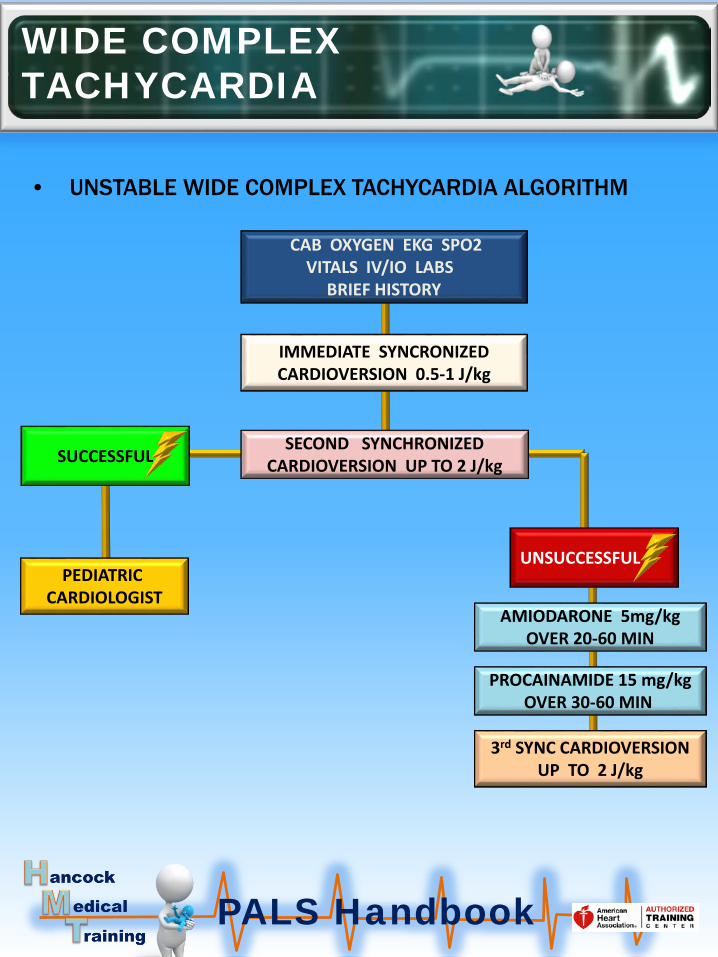
• UNSTABLE WIDE COMPLEX TACHYCARDIA ALGORITHM
WIDE COMPLEX TACHYCARDIA
CAB OXYGEN EKG SPO2VITALS IV/IO LABS
BRIEF HISTORY
IMMEDIATE SYNCRONIZEDCARDIOVERSION 0.5-1 J/kg
SECOND SYNCHRONIZEDCARDIOVERSION UP TO 2 J/kg
AMIODARONE 5mg/kgOVER 20-60 MIN
PROCAINAMIDE 15 mg/kgOVER 30-60 MIN
3rd SYNC CARDIOVERSIONUP TO 2 J/kg
PEDIATRIC CARDIOLOGIST
SUCCESSFUL
UNSUCCESSFUL

PALS Handbook
• PATHWAYS
• CAUSES OF CARDIAC ARREST
CARDIAC ARREST
RESPIRATORYFAILURE
HYPOTENSIVESHOCK
CARDIO / PULMONARY FAILURE
HYPOXIC / ASPHICAL ARREST
SUDDEN ARRHYTHMIA
SUDDEN CARDIAC ARREST
HYPOVOLEMIA
HYPOXIA
HYDROGEN ION
HYPOGLYCEMIA
HYPO – HYPER KALEMIA
HYPOTHERMIA
TENSION PNEUMOTHORAX
TAMPONADE CARDIAC
TOXINS
THROMBUS PULMONARY
THROMBOSIS CORONARY

PALS Handbook
• CAUSES OF CARDIAC ARREST
CARDIAC ARREST
CARDIACARREST
RESPIRATORYUPPER AIRWAY
LOWER AIRWAY
DISORDERED CONTROL BREATHING
LUNG TISSUE DISEASE
HYPOTENSION
HYPOVOLEMIC SHOCK
CARDOOGENIC SHOCK
DISTRIBUTIVE SHOCK
SIDS ARRHYTHMIASTRAUMA DROWNING
OUT OF HOSPITAL IN HOSPITAL
HYPOTENSIONMETABOLIC /ELECTROLYTE
HYPOVOLEMIC SHOCKCARDOOGENIC SHOCKTOXICOLOGIC SHOCKDISTRIBUTIVE SHOCK
ARRHYTHMIAS
RESPIRATORYUPPER AIRWAY
LOWER AIRWAY
DISORDERED CONTROL BREATHING
LUNG TISSUE DISEASE

PALS Handbook
• Non shock able rhythms
– PEA Asystole Torsades
• Shock able rhythms– Pulseless Ventricular tachycardia– Ventricular tachycardia
• Ventricular tachycardia and Torsades
CARDIAC ARREST RHYTHMS

PALS Handbook
• Intravenous– Give the drug by bolus injection– Give while continuing CPR– Follow with a 5-10 ml bolus of normal saline– Raise the extremity
• Intraosseous– IO access can be established for all age groups– IO access can be achieved in 30 – 60 seconds– IO is preferred over the endotracheal route– Any drug given IV can also be given IO
• Endotracheal– Drug absorption is unpredictable– Optimal dose of most drugs is unknown – Recommended dose is higher– A limited number of drugs can be given
CARDIAC ARREST DRUG ADMINISTRATION
PALS Handbook
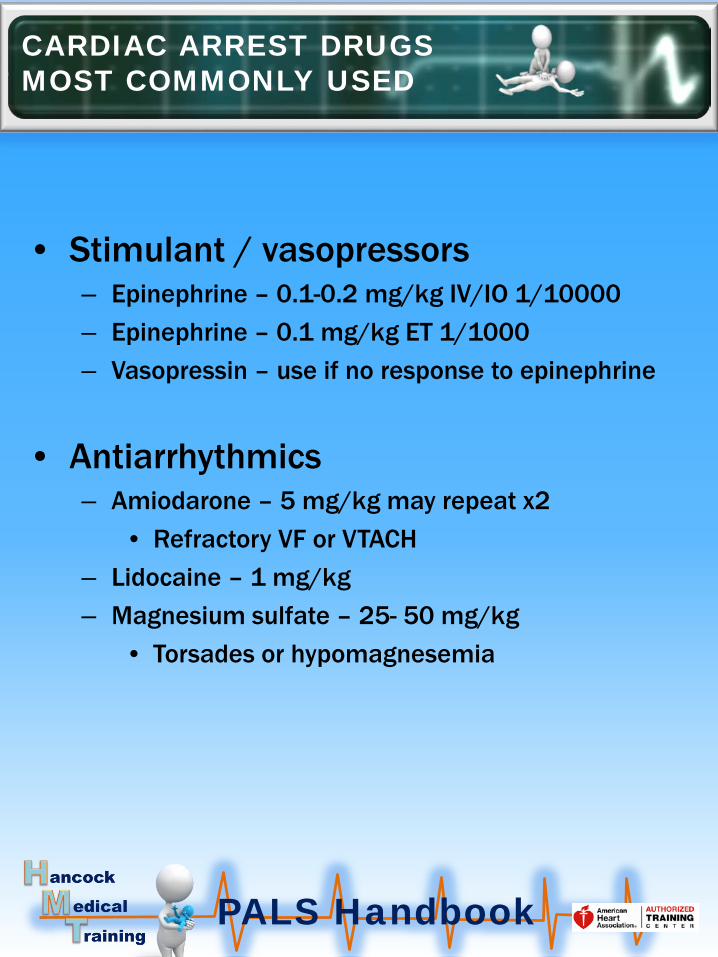
• Stimulant / vasopressors– Epinephrine – 0.1-0.2 mg/kg IV/IO 1/10000– Epinephrine – 0.1 mg/kg ET 1/1000– Vasopressin – use if no response to epinephrine
• Antiarrhythmics – Amiodarone – 5 mg/kg may repeat x2
• Refractory VF or VTACH– Lidocaine – 1 mg/kg– Magnesium sulfate – 25- 50 mg/kg
• Torsades or hypomagnesemia
CARDIAC ARREST DRUGS MOST COMMONLY USED
PALS Handbook
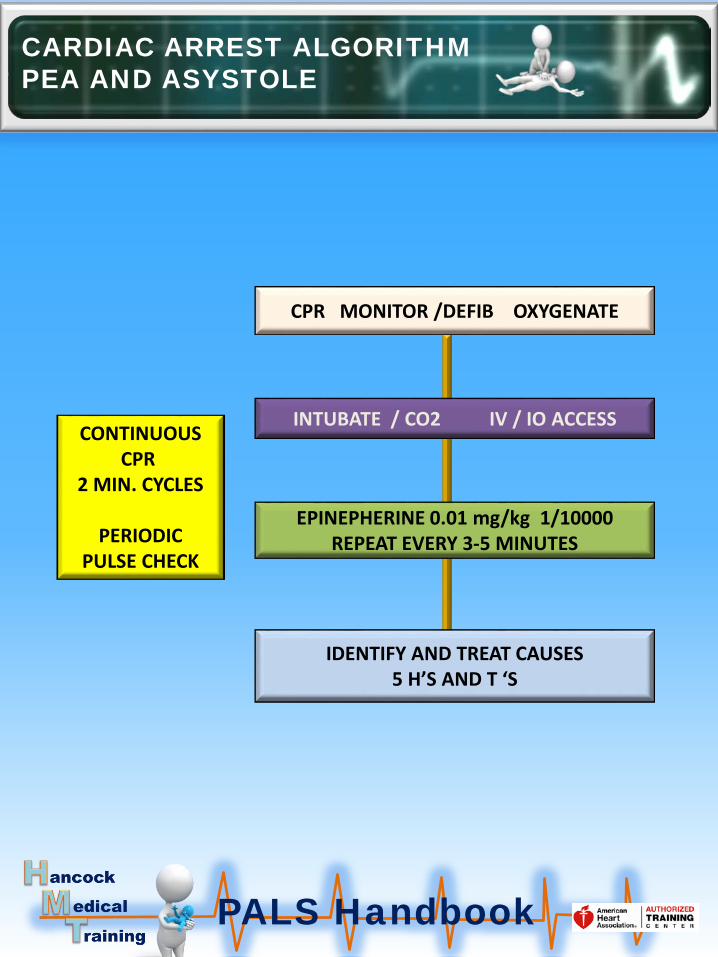
CARDIAC ARREST ALGORITHM PEA AND ASYSTOLE
CPR MONITOR /DEFIB OXYGENATE
INTUBATE / CO2 IV / IO ACCESS
EPINEPHERINE 0.01 mg/kg 1/10000REPEAT EVERY 3-5 MINUTES
IDENTIFY AND TREAT CAUSES5 H’S AND T ‘S
CONTINUOUSCPR
2 MIN. CYCLES
PERIODICPULSE CHECK

PALS Handbook
CARDIAC ARREST ALGORITHM PULSELESS VTACH AND VF
CPR MONITOR/DEFIB OXYGENATEDEFIBRILLATE AT 2 J/Kg
INTUBATE / CO2 IV /IOI ACCESS
DEFIBRILLATE AT 4J/Kg
EPINEPHERINE IV/IO 0.01 mg/kg (1/10000)EVERY 3-5 MINUTES
DEFIBRILLATE AT 4J/Kg
AMIODARONE 5mg/kg REPEAT TO 15mg//kg
DEFIBRILLATE AT 4 J/Kg
TREAT REVERSIBLE CAUSES
PALS Handbook
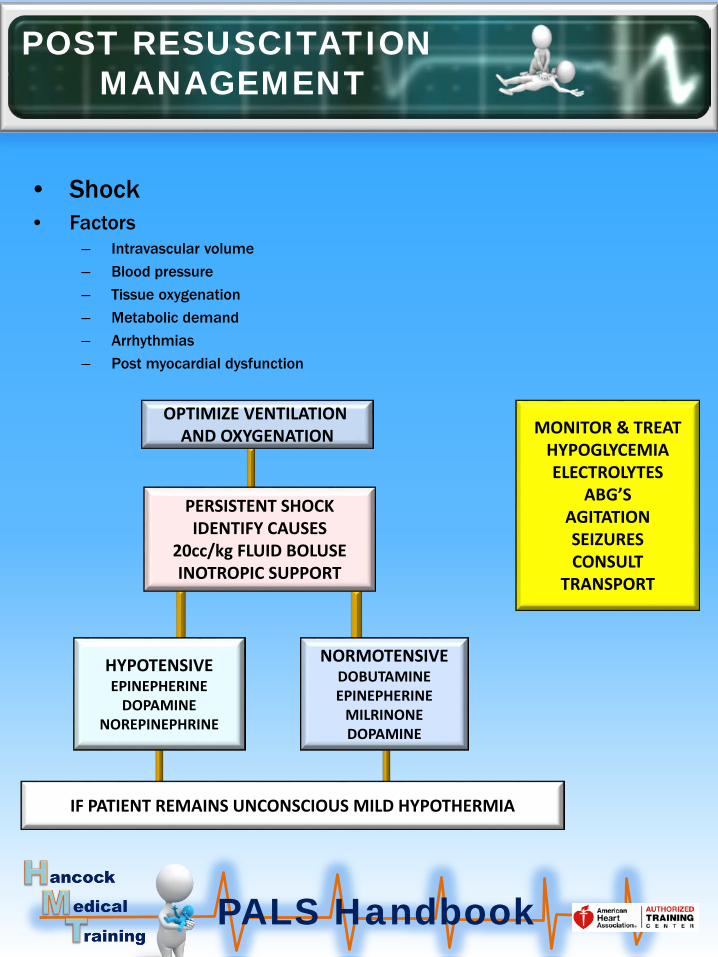
• Shock• Factors
– Intravascular volume – Blood pressure– Tissue oxygenation– Metabolic demand– Arrhythmias – Post myocardial dysfunction
POST RESUSCITATIONMANAGEMENT
OPTIMIZE VENTILATION AND OXYGENATION
PERSISTENT SHOCKIDENTIFY CAUSES
20cc/kg FLUID BOLUSEINOTROPIC SUPPORT
HYPOTENSIVEEPINEPHERINE
DOPAMINENOREPINEPHRINE
NORMOTENSIVEDOBUTAMINEEPINEPHERINE
MILRINONEDOPAMINE
IF PATIENT REMAINS UNCONSCIOUS MILD HYPOTHERMIA
MONITOR & TREATHYPOGLYCEMIAELECTROLYTES
ABG’SAGITATIONSEIZURESCONSULT
TRANSPORT

PALS Handbook
• Respiratory– Factors
• Oxygenation• Intubation• Ventilation• Analgesia and• Sedation• Neuromuscular blockade
• Cardiovascular – Factors
• Intravascular volume• Blood pressure• Tissue oxygenation• Metabolic demand• Arrhythmias• Post arrest myocardial dysfunction
POST RESUSCITATION MANAGEMENT

PALS Handbook
• Neurologic – Factors
• Brain perfusion• Blood pressure• Temperature control• Increased ICP• Seizures
• Gastrointestinal– Factors
• Gastric distension – Naso or orogastric tube
• Ileus• Hepatic failure
• Hematologic system– Factors
• Blood component therapy• Coagulopathy• Platelets• RBC and WBC
POST RESUSCITATIONMANAGEMENT

PALS Handbook
• History and assessment
• Typical newborn vital signs– Heart rate – 100 – 180– Respiratory rate 30 – 60 bpm– Systolic blood pressure – 55 – 90 mmHg– Diastolic blood pressure – 25 – 55 mmHg
NEWBORNS
HISTORYMULTIPLE BIRTHS
PREMATUREMERCONIUM
NARCOTIC USE
ASSESSMENTGESTATION
AMNIOTIC FLUIDBREATHING
CRYINGMUSCLE TONE
Sign 0 1 2
Heart rate Absent < 100 > 100
Muscle tone Limp Some flexion Active motion
Respiratory effort Absent Slow/irregular Good exchange
Response to stimulation
No response Grimace Cough, sneeze or cry
Color Blue/pale Peripheral cyanosis Pink
APGAR Score: 0-3=critical 4-7=mild-moderate distress 7-10=normal Assess @ 1 and 5 minutes

PALS Handbook
• Meconium – Suction mouth and nose if obstructed– Intubate– Suction using a meconium aspirator– Repeat with new tubes until clear
NEWBORNS