Embed Size (px)
Citation preview

RESEARCH Open Access
Palmitoleic acid (16:1n7) increases oxygenconsumption, fatty acid oxidation and ATPcontent in white adipocytesMaysa M. Cruz1, Andressa B. Lopes4, Amanda R. Crisma2, Roberta C. C. de Sá1, Wilson M. T. Kuwabara2, Rui Curi2,3,Paula B. M. de Andrade3 and Maria I. C. Alonso-Vale1*
Abstract
Background: We have recently demonstrated that palmitoleic acid (16:1n7) increases lipolysis, glucose uptake andglucose utilization for energy production in white adipose cells. In the present study, we tested the hypothesis thatpalmitoleic acid modulates bioenergetic activity in white adipocytes.
Methods: For this, 3 T3-L1 pre-adipocytes were differentiated into mature adipocytes in the presence (or absence)of palmitic (16:0) or palmitoleic (16:1n7) acid at 100 or 200 μM. The following parameters were evaluated: lipolysis,lipogenesis, fatty acid (FA) oxidation, ATP content, oxygen consumption, mitochondrial mass, citrate synthaseactivity and protein content of mitochondrial oxidative phosphorylation (OXPHOS) complexes.
Results: Treatment with 16:1n7 during 9 days raised basal and isoproterenol-stimulated lipolysis, FA incorporationinto triacylglycerol (TAG), FA oxidation, oxygen consumption, protein expression of subunits representing OXPHOScomplex II, III, and V and intracellular ATP content. These effects were not observed in adipocytes treated with 16:0.
Conclusions: Palmitoleic acid, by concerted action on lipolysis, FA esterification, mitochondrial FA oxidation, oxygenconsumption and ATP content, does enhance white adipocyte energy expenditure and may act as local hormone.
Keywords: Lipogenesis, Lipolysis, Triglyceride/fatty acid cycle, Bioenergetics, Mitochondria, Beta-oxidation
BackgroundWhite adipose tissue (WAT) stores triacylglycerol (TAG)and its metabolic feature involves lipogenesis and lipolysis,which are associated with changes in the volume of ma-ture adipocytes. The activities of these two metabolicpathways vary with the need to incorporate or release fattyacids (FA), which depends on the nutritional status of theindividuals, energy expenditure, hormone levels (e.g. insu-lin), activities of the enzymes involved in these processes(e.g. ATGL and HSL, FAS and G6PDH) and the hetero-geneity existing among white adipose tissue depots [1–3].Mitochondria play an important role in cellular func-
tion, not only as a major site of ATP production, but alsoby regulating energy expenditure, apoptosis signaling and
production of reactive oxygen species [4]. The impact ofmitochondria function and dysfunction on pre-adipocyteand adipocyte metabolism has attracted a growing body ofinterest over the last decade [5–7]. There are severalreports linking mitochondrial dysfunction with impairedWAT energetic metabolism and the development of obes-ity, insulin resistance and type 2 diabetes mellitus (T2DM)[6, 8, 9]. Mitochondria are the main site for aerobic glu-cose and fatty acid oxidation, oxygen consumption, andgeneration of reactive oxygen species and ATP, which areassociated with enhanced basal metabolic rate [10, 11].Therefore, mitochondria function in WAT might play akey role to control basal metabolic rate and should beconsidered as a target to be investigated for the develop-ment of alternative therapies to treat/prevent obesity andrelated metabolic disorders.Among several types of lipids produced and released
by adipocytes, palmitoleic acid, a ω-7 monounsaturatedfatty acid (16:1n7, n7) synthesized by the desaturation of
* Correspondence: [email protected] of Biological Sciences, Institute of Environmental Sciences,Chemical and Pharmaceutical, Federal University of São Paulo, 210, SaoNicolau St, Diadema 09913-030, BrazilFull list of author information is available at the end of the article
© The Author(s). 2018 Open Access This article is distributed under the terms of the Creative Commons Attribution 4.0International License (http://creativecommons.org/licenses/by/4.0/), which permits unrestricted use, distribution, andreproduction in any medium, provided you give appropriate credit to the original author(s) and the source, provide a link tothe Creative Commons license, and indicate if changes were made. The Creative Commons Public Domain Dedication waiver(http://creativecommons.org/publicdomain/zero/1.0/) applies to the data made available in this article, unless otherwise stated.
Cruz et al. Lipids in Health and Disease (2018) 17:55 https://doi.org/10.1186/s12944-018-0710-z

palmitic acid (16:0) by stearoyl-CoA desaturase 1 (SCD1)activity, has been shown to act systemically in peripheraltissues modulating important metabolic processes. Caoand cols [12] showed that palmitoleic acid improves insu-lin resistance in skeletal muscle and liver and preventshepatosteatosis. As palmitoleic acid is produced andsecreted by WAT, Cao and cols., 2008 [12] named it “lipo-kine”. Palmitoleic acid treatment leads to increased glu-cose uptake, AKT phosphorylation and raises GLUT1and GLUT4 protein levels in plasma membrane ofskeletal muscle cells [13, 14]. Moreover, palmitoleicacid treatment also enhances AKT, insulin receptor,insulin receptor substrate-1 and 2 protein phosphoryl-ation in the liver [12, 15] and exerts cytoprotectiveeffects in pancreatic β-cells [16, 17].We investigated herein the effects of palmitoleic acid
on WAT bioenergetics. Recently, our group has shownthat palmitoleic acid treatment increases adipocyte lip-olysis and the content of the major lipases (ATGL andHSL) through a PPARα-dependent mechanism [18]. Be-sides, this lipokine enhanced glucose uptake and GLUT4content associated with AMPK activation in these cells,favoring cellular glucose utilization towards energy pro-duction [19]. Our hypothesis is that palmitoleic acidincreases white adipocyte basal metabolism. To test thisproposition, 3 T3-L1 adipocytes were treated with pal-mitoleic acid (100 μM) for 9 days. The results were com-pared with those obtained with palmitic acid and vehicleunder similar experimental protocol. Lipolysis, FA in-corporation into lipids and FA conversion into CO2, aswell as parameters of mitochondrial bioenergetics (fattyacid oxidation, oxygen consumption and ATP content)and proteic analysis of subunits representing OXPHOScomplex (I, II, III, IV e V) were investigated. Treatmentwith palmitoleic acid did enhance mitochondrial activityas indicated by enhanced ATP generation, proteic ex-pression of II, III, and V complexes of the mitochondrialelectron transport chain, FA oxidation and oxygen con-sumption. Taken together, boosted mitochondrial bio-energetics in combination with raised lipolysis and FAreesterification leads to increased adipocyte energy ex-penditure via TAG/FA cycle stimulation (futile cycle).Therefore, palmitoleic acid plays a relevant role in WATmetabolism and should be considered as a candidate tobe tested in obesity-related therapies.
MethodsCell culture3 T3-L1 preadipocytes were cultured in Dulbeccos Modi-fied Eagle Medium (DMEM) containing 10% calf serumand 1% penicillin-streptomycin until confluence. Differen-tiation was induced 2 days post-confluence by addition ofdexamethasone (1 μM), isobutylmethylxanthine (0.5 mM),insulin (1.67 μM), and 10% fetal bovine serum (FBS). After
48 h, medium was replaced by DMEM containing 10%FBS and 0.41 μM insulin [20]. Cells were cultivated for9 days in the presence of palmitic acid (16:0), palmitoleicacid (16:1n7), each at 100 μM dissolved in ethanol0.05% (vehicle), starting from the first day of differenti-ation (day 0). This dose of palmitoleic acid was foundto have no cytotoxic or deleterious effects on cells asevaluated by plasma membrane integrity and DNAfragmentation during differentiation until the day 9[21]. As we demonstrated before (18) that the palmito-leic acid effects in adipocytes are structure specific,here, we did not add more additional control such asoleic acid (18:1n9) to the cell cultures. In some experi-ments, etomoxir (Sigma-Aldrich), an inhibitor of CPT-1 activity, was used to inhibit fatty acid oxidation at aconcentration of 40 μM, added 24 h before the experi-ments. Cell culture medium was changed every 2 days.All reagents and drugs were purchased from SigmaChemical Company (St. Louis, MO, USA). Similar pro-cedure was used in our previous studies [18].
Lipolysis measurementLipolysis was estimated as the rate of glycerol (FreeGlycerol Determination Kit, Sigma) and free fatty acid(FFA) (NEFA Kit RH series, Wako Diagnostics, CA,USA) released from differentiated 3 T3-L1 cells after9-day treatment with the fatty acids or vehicle (insome experiments with or without etomoxir) during30 min of incubation. Similar procedure was used inour previous studies [18, 22]. Results were expressedas nanomoles of glycerol per 106 cells and μEq/L offree FA per well for glycerol and FFA, respectively.
Incorporation of [1-14C]-palmitate into triacylglycerolKRH (Krebs Ringer Hepes bicarbonate) buffer, pH 7.4,containing 1% BSA and 2 mM glucose plus palmitate(200 μM), saturated with a gas mixture of 95% O2 and5% CO2, was added to 3 T3-L1 cells after 9-day treat-ment with FA or vehicle. [1-14C]-Palmitate was thenadded to the buffer (1850 Bq/tube or well) and left for2 h at 37 °C. Cells were then washed three times withphosphate buffered saline (PBS) and Dole’s reagentcontaining isopropanol:n-heptane:H2SO4 (4:1:0.25 vol/vol/vol) was added to the remaining reaction mixture forlipid extraction.The mixture was transferred to polypropylene tubes,
which were vortexed three times during the following30 min. After adding n-heptane (1.5 mL) and distilledwater (1.5 mL), tubes were vortexed and the mixturewas decanted for additional 5 min. The upper phase wascollected (in duplicates) and transferred to a scintillationvial for determination of radioactivity trapped into TAGusing a β-counter (1450 LSC, Counter MicroBeta, Trilux;PerkinElmer). Results were expressed as nanomoles of FA
Cruz et al. Lipids in Health and Disease (2018) 17:55 Page 2 of 12

per 106 cells. Similar procedure has been used in our pre-vious studies [18].
Decarboxylation of [1-14C]-palmitate (fatty acid oxidation)Differentiated 3 T3-L1 cells (after 9-day treatment withFA or vehicle) were incubated in KRH buffer (pH 7.4)containing BSA (1%) and [1-14C]-palmitate (50 μM,1850 Bq/tube or well), saturated with a gas mixture of95% O2 and 5% CO2, for 2 h, at 37 °C. Prior to 2 h incu-bation period, each well was covered with a piece ofWhatman filter paper and the plate was sealed with par-afilm to maintain the atmosphere saturated with the gasmixture. Following the 2 h incubation, the filter paperwas soaked with 0.1 mL of ethanolamine to trap theCO2 produced, and 0.2 mL of 8 N H2SO4 was injectedinto the wells with the aid of a needle to rupture thecells. After 45 min of CO2 trapping, the filter paper wasremoved and transferred to scintillation vials for radio-activity counting [23, 24]. Results were expressed asnanomoles of oxidized [1-14C]-palmitate per 106 cells.
Oxygen consumptionOxygen consumption rates in intact cells were measuredas an indication of mitochondrial respiratory activity.After 9-day treatment with palmitic acid (100 μM), pal-mitoleic acid (100 μM) or vehicle, 3 T3-L1 cells weregently tripsinised, re-suspended in KRH (pH 7.4) con-taining BSA (0.1%), and transferred to the oxygraph(OROBOROS Oxygraph-2 k). The oxygraph chamberswere previously equilibrated with KRH containing BSA0.1% at 37 °C. Carbonyl cyanide m-chlorophenyl hydra-zine (CCCP, 1 μM f.c.) was added as positive control formaximal respiratory rate (uncoupling) determination.Oxygen consumption rates were normalized by cellnumber and expressed as % of the control [25]. Oxygenconsumption was also measured in mature 3 T3-L1adipocytes (9 days after differentiation) treated for 24 hwith palmitoleic acid (200 μM) or vehicle.
ATP content determinationATP content was determined in 3 T3-L1 cell lysates after9 days of treatment with FA or vehicle (in the presence orabsence of etomoxir) using an ATP bioluminescence assaykit (Roche, Mannheim, Germany). Measurements were per-formed in a luminometer (Biotech, model: Synergy HT).Results were normalized by cell number and expressed aspercentage of control [26].
Western blot analysisAfter treatment with FA or vehicle for 9 days, 3 T3-L1 cellswere homogenized and processed in buffer composed inmM by: 50 HEPES, 40 NaCl, 50 NaF, 2 EDTA, 10 sodiumpyrophosphate, 10 sodium glycerophosphate, 2 sodiumorthovanadate, and 1% Triton-X100 and EDTA-free
protease inhibitors. Identical amounts of protein aliquotsfrom 3 T3-L1 cell lysates were resolved on Nupage gradi-ent gels (4–12%, Invitrogen Life Technologies) and trans-ferred to nitrocellulose membranes. After blockage with5% milk for 1 h, membranes were overnight incubated at4 °C with the following primary antibodies: mitochondrialcomplex I subunit NDUFB8 (20 kDa), complex II subunit30 kDA (30 kDa), complex III subunit Core 2 (48 kDa),COXIV subunit I (40 kDa), complex V ATP synthasesubunit alpha (53 kDa) (OXPHOS kit, MitoSciences, Inc.)and gamma-tubulin (~ 50 kDa #5886, Cell Signaling,Beverly, MA, USA) in 5% milk (1:1000). After wash-ing, membranes were subsequently incubated withappropriate peroxidase-conjugated secondary antibody(1:5000) for 1 h and developed using the ECL en-hanced chemiluminescence substrate (GE Healthcare LifeSciences, Björkgatan, Uppsala). Densitometric analyseswere performed using the ImageJ software (NationalInstitutes of Health, Bethesda, MD).
RNA extraction, reverse transcription and quantitativereal-time PCR (real-time qRT-PCR)Total RNA from 3 T3-L1 cell lysates was extracted usingTrizol (Invitrogen Life Technologies), analyzed for qualityon agarose gel and absorbance ratios of 260/280 nm and260/230 nm, and reverse transcribed to cDNA using theSuperScript III cDNA kit (Invitrogen Life Technologies).Gene expression was evaluated by real-time qRT-PCRusing a Rotor Gene (Qiagen, Roermond, Netherlands) andSYBR Green as fluorescent dye (Qiagen) with 36B4/Rplp0as housekeeping gene. The reaction conditions were asfollows: 95 °C for 5 min, then 40 cycles of 95 °C for 5 sand 60 °C for 10 s. PCR products were run on agarose gelto confirm the size of the fragment and specificity ofamplification. Primers used were: Pnpla2 (5′-3′ sense:GGTCCTCTGCATCCCTCCTT; 5′-3’antisense: CTGTCCTGAGGGAGATGTC), aP2 (5′-3′ sense: AAGGTGAAG AGCATCATAACCCT; 5′-3’antisense: TCACGCCTTTCATAACACATTCC) and 36B4 (5′-3′ sense: TAAAGACTGGAGACAAGGTG; 5′-3’antisense: GTGTACTCAGTCTCCAC AGA). Data were obtained as ct values (ct =cycle number at which logarithmic PCR plots cross acalculated threshold line) and used to determine Δctvalues (Δct = (ct of the target gene) - (ct of the housekeep-ing gene). Data were expressed as arbitrary units using thefollowing calculation: [expression = 1000×(2-Δct) arbitraryunits (AU)].
Mitochondrial mass determinationThe methodology was based on the protocol used byShen et al. [25]. A fluorescent probe (Mito-TrackerGreen FM; Molecular Probes, Eugene, OR, USA) wasused to determine the mitochondrial mass of adipocytes[27]. Mature adipocytes (9 days of differentiation) were
Cruz et al. Lipids in Health and Disease (2018) 17:55 Page 3 of 12

treated with palmitic or palmitoleic acid, trypsinised andcentrifuged at 1500×g, 4 °C, for 5 min, resuspended inKRH buffer containing 0.1% BSA (w/v) and then incu-bated with 0.1 μmol/L MitoTracker Green FM in KRHbuffer for 30 min at 37 °C. Cells were centrifuged at1500×g, 4 °C, for 5 min and resuspended in 400 μL offresh KRH buffer. Fluorescence measurements werecarried out in CALIBUR cytometer (BD) in the FL1channel. Ten thousand events were analyzed per experi-ment. Data were analyzed using Cell Quest software.
Citrate synthase activity assayCitrate synthase (EC 4.1.3.7) maximum activity was de-termined as previously described by Alp et al. [28].Briefly, after 9 days treatment with FA or vehicle, 3 T3-L1 cells were homogenized (vortex) in extraction buffercontaining Tris-HCl (50 mM), EDTA (1 mM), leupep-tin (50 μM), and aprotinin (5 μM), pH 7.4, and centri-fuged (16,000 g, 30 s, 4 °C). The assay buffer consistedof Tris HCl (100 mM), DTNB (0.2 mM), acetyl-CoA(100 mM), and Triton (1%), pH 6.5. The reaction wasstarted by adding 10 uL of oxaloacetic acid (500 mM)25 °C. The absorbance was monitored for 10 min at420 nm. Protein concentration in the supernatants wasdetermined using a BCA® protein assay kit (PIERCEBiotechnology, Rockford, IL). The maximal enzymeactivity was expressed as micromoles per minute permicrogram of protein.
Statistical analysisResults are expressed as mean ± SEM. One-Way ANOVAfollowed by Tukey post-hoc test were used to comparethe effects of different treatments. Analysis was performedusing GraphPad Prism 5.0 software (GraphPad Software,Inc., San Diego, CA, USA). The level of significance wasset at p ≤ 0.05.
ResultsFirst, we examined the effect of 9-day treatment withpalmitoleic or palmitic acid on 3 T3-L1 adipocyte lipoly-sis. Palmitoleic acid increased both basal (by 3-fold) andstimulated (by 3.5-fold) lipolysis as measured by theglycerol released to the medium [mean ± SEM, Basal (ve-hicle: 828.9 ± 95.4; 16:1n7: 3027.2 ± 511) and stimulated(vehicle: 1760.3 ± 174; 16:1n7: 6077.7 ± 822) nMol/106
cells, p < 0.05] (Fig. 1a and b, respectively). Pnpla2mRNA levels were significantly increased (by 30%) incells treated with palmitoleic acid (Fig. 1c). We furtherinvestigated whether 16:1n7 would also promote an in-crease in free fatty acid (FFA) release by these cells (dic-tated by a glycerol/FA proportion of 1:3, as products ofTAG hydrolysis). Palmitoleic acid did not cause signifi-cant increase of FFA release into the medium, that is, nodifference was observed among the groups. Interestingly,this profile was slightly altered by pretreatment with eto-moxir (a potent inhibitor of fatty acid mitochondrial up-take and oxidation), since 16:1n7 + etomoxir treated cellspresented an increase by 5% in the FFA release into themedium (mean ± SEM, 16:1n7: 59.2 ± 0.8; 16:1n7 + eto-moxir: 63.3 ± 0.44 μMol/L, p < 0.05) (Fig. 3b). We did notobserve any effect of palmitic acid on lipolytic activity.We hypothesized that palmitoleic acid should boost
TAG lipolysis/FA re-esterification cycle. To test thishypothesis, we analyzed if FA esterification was raisedin cells treated with palmitoleic acid. Indeed, an in-crease of [1-14C]-palmitate incorporation into TAG(by 40%) was observed in cells treated with palmito-leic acid (mean ± SEM, vehicle: 23.42 ± 0.5; 16:1n7:28.73 ± 0.8 nMol/106 cells, p < 0.05) (Fig. 2a). More-over, aP2 mRNA levels were positively regulated bypalmitoleic acid (Fig. 2b). These results show that FAincorporation into TAG is increased in 3 T3-L1adipocytes treated with palmitoleic acid. No effect onthe synthesis of TAG was observed in cells treatedwith palmitic acid.
a b c
Fig. 1 Basal (a) and isoproterenol-stimulated (b) lipolysis measuring by release of glycerol (nanomoles per 106 cells). c mRNA levels of Pnpla2gene (arbitrary units). Experiments performed in differentiated 3 T3-L1 adipocytes treated for 9 days with vehicle, palmitic acid (16:0, 100 μM) orpalmitoleic acid (16:1n7, 100 μM). Results are means ± SEM. * P < 0.05 16:1n7 vs. all groups. The results are the average of 3independent experiments (n = 4/experiment)
Cruz et al. Lipids in Health and Disease (2018) 17:55 Page 4 of 12

Next, we investigated if palmitoleic acid could en-hance FA beta-oxidation. The conversion of [14C]-palmitate to CO2 was raised by 30% in cells treatedwith palmitoleic acid when compared to control cells(mean ± SEM, vehicle: 0.8 ± 0.033; 16:1n7: 1.03 ± 0.05nMol/106 cells, p < 0.05) (Fig. 3a). No change in FAOwas observed in cells treated with palmitic acid whencompared to control. Disruption of fatty acid oxida-tion by etomoxir in a dose of 40 μM was confirmedin this experiment by partial attenuation (~ 25%) offatty acid oxidation.Other mitochondrial parameters, such as ATP levels
and oxygen consumption, were also measured in 3 T3-L1cells. FA oxidation is a metabolic pathway that requiresoxygen to take place. We performed oxygen consump-tion measurements in 3 T3-L1 adipocytes treated withfatty acids using a modular system for high-resolutionrespirometry (HRR), the Oroboros Oxygrapk-2 k (O2k).
Oxygen consumption was raised in cells treated withpalmitoleic acid for 9 days (by 13%, Fig. 4a) and 24 h(by 10%, Fig. 4b). So, palmitoleic acid stimulates basalenergetic metabolism in white adipocytes. Palmitic acidtreatment did not affect oxygen consumption in any ofthe conditions studied.We further investigated the palmitoleic effects on ATP
content. 16:1n7, but not 16:0 treatment of the cells, raisedATP levels by 17% (mean ± SEM, vehicle: 100 ± 1.72;16:1n7: 119.8 ± 1.51 percentage of control, p < 0.05) (Fig. 5).We hypothesized that raised FAO, secondary to raisedlipolysis promoted by 16:1n7, elevates ATP production(corroborating oxygen consumption) in a magnitude cap-able of overcome its consumption (raised by the exacerba-tion of TAG/FA cycle). This hypothesis is supported here,by the experiments performed under pretreatment of thecells with etomoxir, that completely prevented the incre-ment on ATP content induced by palmitoleic acid (Fig. 5).
a b
Fig. 2 a [1-14C]-palmitate incorporation into TAG (nanomoles of incorporated [1-14C]-palmitate per 106 cells) and b mRNA levels of aP2 (arbitraryunits). Experiments performed in differentiated 3 T3-L1 adipocytes treated for 9 days with vehicle, palmitic acid (16:0, 100 μM) or palmitoleic acid(16:1n7, 100 μM). Results are means ± SEM. * P < 0.05 16:1n7 vs. all groups. The results are the average of 3 independentexperiments (n = 6/experiment)
a b
Fig. 3 a Conversion of [1-14C]-palmitate into CO2 (nanomoles of converted [1-14C]-palmitate into CO2 per 106 cells) and b Free fatty acids
released under lipolysis (μMol per liter per well). Experiments performed in 3 T3-L1 adipocytes under 9-days treatment with vehicle, etomoxir(40 μM), palmitic acid (16:0, 100 μM) or palmitoleic acid (16:1n7, 100 μM). Results are means ± SEM. * P < 0.05 vs. all groups. The results are theaverage of 3 independent experiments (n = 4/experiment)
Cruz et al. Lipids in Health and Disease (2018) 17:55 Page 5 of 12

Since the augmented ATP concentration observed inour studies suggest an increment in mitochondrialOXPHOS efficiency, we next evaluated protein expres-sion of subunits representing complex II, III, and V ofthe mitochondrial electron transport chain. Complex IIand III were positively modulated by palmitoleic acid (by54% and by 36%, respectively), but not by palmitic acid,when compared with the control group (Fig. 6b and c).Likewise, complex V was positively modulated by palmi-toleic acid (by 40%) when compared to the controlgroup (Fig. 7e). However, palmitoleic acid did not elicitany change in the content of mitochondrial complexes Iand IV (Fig. 6a and d, respectively).
Mitochondria mass was evaluated by flow cytometry,according to the methodology proposed by Shen and col-leagues [25]. A decrease of ~ 10% in mitochondria masswas observed in cells treated with palmitoleic acid. Therewas no effect of palmitic acid on this parameter (Fig. 7a).Another parameter to measure mitochondria content iscitrate synthase activity. Palmitoleic acid (but not palmiticacid) led also to a reduction of ~ 10% in citrate synthaseactivity (mean ± SEM, vehicle: 0.031 ± 0.0005; 16:1n7:0.024 ± 0.0008 μMol/min/μg of protein, p < 0.05) (Fig. 7b),when compared with the control group.
Discussion3 T3-L1 adipocytes were treated with palmitic acid or pal-mitoleic acid, being the latter recently described to induceimportant effects on carbohydrate and lipid metabolism inliver and skeletal muscle [12, 15, 29, 30]. We have previ-ously demonstrated that palmitoleic acid controls import-ant metabolic processes also in adipose tissue. Wereported increased lipolysis and glucose uptake, togetherwith changes in expression of related genes and proteins(ATGL, HSL, AMPK and GLUT4) in white adipocytes[18, 19]. In the present work, we observed that palmitoleicacid modulates other aspects of adipocyte metabolism,mainly mitochondria bioenergetics. Palmitoleic acid (butnot palmitic acid) enhanced energy expenditure in adipo-cytes through TAG/FA cycle stimulation, FA oxidation,oxygen consumption and increased protein expression ofsubunits representing complex II, III, and V of the mito-chondrial electron transport chain. The treatment of theadipocytes with palmitic acid (16:0), different from palmi-toleic (16:1n7), showed no marked effect on the parame-ters herein studied.An increase in both basal and isoproterenol-stimulated
lipolysis, together with augmented gene expression ofthe Pnpla2, were observed in 3 T3-L1 cells treated withpalmitoleic acid (9 days, from the induction of preadipo-cyte differentiation) when compared to the palmitic acid
a b
Fig. 4 Oxygen consumption by 3 T3-L1 adipocytes under (a) chronic (9 days) and b acute (24 h) treatments with vehicle, palmitic acid (16:0,100 μM, respectively) or palmitoleic acid (16:1n7, 200 or 100 μM, respectively). Results expressed as percentage of control. Results are presentedas means ± SEM. * P < 0.05 16:1n7 vs. all groups. The results are the average of 3 independent experiments (n = 6/experiment)
Fig. 5 ATP content on 3 T3-L1 adipocytes under 9-day treatmentwith vehicle, etomoxir (40 μM), palmitic acid (16:0, 100 μM) orpalmitoleic acid (16:1n7, 100 μM). Results expressed as percentage ofcontrol. Results are means ± SEM. * P < 0.05 16:1n7 vs. all groups.The results are the average of 3 independentexperiments (n = 4/experiment)
Cruz et al. Lipids in Health and Disease (2018) 17:55 Page 6 of 12

and control groups. These findings corroborate previousex vivo results obtained by our group [18]. We haveshown that there is an increase of lipolysis in primaryadipocytes extracted from epididymal fat pads of micetreated with palmitoleic acid (by gavage) for 10 days andalso, in mature (6 days post-differentiation) 3 T3-L1 cellstreated with palmitoleic acid (by 24 h), via a PPARα-dependent mechanism. The increment in lipolysis pro-moted by palmitoleic acid was estimated according tothe concentration of glycerol released into the medium.A concomitant increment of FFA released from adipo-cytes [dictated by a glycerol/FA proportion of 1:3, asproducts of TAG hydrolysis] treated with palmitoleicacid was not observed. This suggests that the fatty acids
originated from lipolysis of cells treated with palmitoleicacid could be used in other metabolic pathways such as:(1) re-esterification, (2) mitochondrial oxidation or (3)mitochondrial uncoupling, including the possibility ofoverlapping pathways. We ruled out mitochondrial un-coupling due to the fact that cellular ATP levels wereaugmented upon palmitoleic acid treatment (Fig. 5) andUCP1/2 mRNA levels are not altered under this condi-tion (data not shown).To test the hypothesis 1, we analyzed if FA esterifica-
tion was increased in cells treated with palmitoleic acidfor 9 days. Palmitoleic acid did increase [1-14C]-palmi-tate incorporation into TAG as well as aP2 mRNA levelsin adipocytes. aP2 gene encodes a protein involved in
a
d
b c
e
Fig. 6 Total content of subunits representing OXPHOS proteins complexes analyzed by western blot. a Complex I. b Complex II. c Complex III. dCoxIV. e Complex V. Experiments were performed in differentiated 3 T3-L1 adipocytes treated for 9 days with vehicle, palmitic acid (16:0, 100 μM)or palmitoleic acid (16:1n7, 100 μM). Results expressed as arbitrary units. Results are presented as means ± SEM. * P < 0.05 16:1n7 vs. all groups,# P < 0.05 16:1n7 vs. vehicle. The results are the average of 3 independent experiments (n = 4/experiment)
Cruz et al. Lipids in Health and Disease (2018) 17:55 Page 7 of 12

FA uptake and transport to be then associated to coen-zyme A (a reaction catalyzed by acyl-CoA synthetase)to generate acyl-CoA. Corroborating these findings, wedemonstrated an increase in the generation of glycerol3-phosphate (G3-P) via glycolysis (required for esterifi-cation of fatty acids to TAG), as evidenced by thehigher rates of glucose incorporation into the glycerolfraction of TAG [18]. In another recent work publishedby our group [19] an increase of glucose uptake byadipocytes, via a mechanism that involves AMPK, pro-moted by palmitoleic acid was demonstrated. This ob-servation is compatible with increased G3-P formationfrom glucose, since there was not seen any increase ingene expression of the glyconeogenic regulatory enzymePEPCK or in glycerol kinase activity in this study, the twoother possible sources of G3-P [31]. These results suggestthat palmitoleic acid promotes FA re-esterification andTAG/FA futile cycle stimulation.A concomitant increase in lipolysis and FA re-
esterification implies in stimulation of the substrate cycle(or “futile” cycle) TAG/FA cycle [32, 33]. In mammals, anincrease in this cycle activity together with raised fatty acidoxidation can lead to augmented basal metabolic rate(BMR) and total energy expenditure [34] as well as greatersensitivity to hormonal control of energy metabolism (e.g.lipolytic and lipogenic activities). Additionally, the stimula-tion of this cycle is a mechanism associated with inductionof thermogenesis during cold exposure [35] and also whenplasma leptin levels are high [32].In the next step, we investigated if palmitoleic acid
could enhance FA beta-oxidation and oxygen con-sumption. Maassen, Romijn and Heine [36] suggestedthat FA oxidation in adipocytes might also contributeto the retention of FA in these cells, thus avoidingexcessive release of FA into the circulation when lip-olysis is stimulated reducing lipotoxicity. We observedan increase (by 30%) in FA oxidation in 3 T3-L1
adipocytes treated with palmitoleic acid. This effectmaybe underestimated due to the fact that palmitoleicacid can compete with labeled [1-14C]-palmitate(50uM) added to the medium during this test for thebeta-oxidation pathway.Despite the fact that some studies describe FA oxidation
as a minor metabolic pathway in white adipocytes, espe-cially when compared to FA re-esterification [37–39], Dib,Bugge and Collins [40] claim that FA oxidation in WAT isnot negligible and contributes from 5 to 10% of the BMRin lean and obese adults, respectively. Noteworthy, thepercentage of endogenous FA oxidized in primary ratwhite adipocyte was calculated and found to be around0.2%, what seems small when compared to re-esterified(~ 50%) and released endogenous FFA (~ 50,1%) [41]. Weestimated herein the amount of FFA released by the cells,which were around 40 nmoles/106 cells. The esti-mated FFA released is much lower than the amountof glycerol released (by 70-fold) but it is much higherthan the amount of CO2 generated (~ 1 nmol/106
cells). This finding corroborates the data that FFA re-esterification and endogenously released FFA are themajor pathways when compared to FFA oxidation.Anyway, pretreatment with etomoxir, that has beenshown to bind CPT-1 with high affinity, thereby pre-venting mitochondrial uptake of fatty acids [42],caused an increment in the FFA release into themedium by the cells treated with palmitoleic acid.The disruption of fatty acid oxidation by etomoxirwas confirmed here, by the attenuation of fatty acidoxidation.Wang and cols [41] claim that FFA oxidation assays
are usually performed during a short period of time (a 2-hperiod or so) and, if extrapolated to years, this oxidativepathway could reduce more than 1 kg of fat mass per50 kg of fat mass per year in humans depending on ifthere is combined fasting or not. Therefore, WAT FAO
a b
Fig. 7 a Mitochondrial mass (percentage of florescence). b Citrate synthase activity (micromoles per minute per microgram of protein).Experiments performed in differentiated 3 T3-L1 adipocytes treated for 9 days with vehicle, palmitic acid (16:0, 100 μM) or palmitoleic acid(16:1n7, 100 μM). Results are means ± SEM. * P < 0.05 16:1n7 vs. all groups. The results are the average of 3–4 independentexperiments (n = 6/experiment)
Cruz et al. Lipids in Health and Disease (2018) 17:55 Page 8 of 12

might be not so negligible as previously thought to be. Ac-tually, efficient mitochondrial WAT FAO may increase re-spiratory capacity, reduce adipocyte size, enhance lipolysisand reduce lipotoxicity [6, 38, 43]. A recent study withhuman twins showed that adiposity is correlated withdownshifting of FAO and that, indeed, mitochondrial bio-genesis, oxidative metabolic pathways and OXPHOS pro-teins in subcutaneous adipose tissue are downregulated inacquired obesity [44]. Altogether these studies corroboratethe hypothesis that mitochondrial WAT FAO stimulationhas relevant effects on body energy balance.Together with increased FAO, an increment in basal
O2 consumption was observed in 3 T3-L1 cells treatedwith palmitoleic acid, which suggests an increase inBMR of these cells. This enhancement in oxygen con-sumption could be explained by an increase in FAO,mitochondrial uncoupling or both [45].The augmentation in O2 consumption observed in
the present study supports the consistent raise in FAOcatabolic pathway. In addition, cellular ATP levels wereraised by 17% as well as protein levels of the subunitATP-5A of the OXPHOS complex V (ATP synthase,Fig. 6e) upon palmitoleic acid treatment. Pretreatmentwith etomoxir completely prevented this effect, dem-onstrating the dependent effect of FAO increase forthe palmitoleic effects here observed. Altogether, theraise in all these mitochondrial parameters: oxygenconsumption, FAO and ATP generation, points out toenhanced mitochondrial oxidative phosphorylationefficiency [46] upon palmitoleic acid treatment. Al-though the palmitoleic acid effects seems mild,according Gao and colleagues [47] mitochondria donot require major changes to reflect a prominentmetabolic effect. In contrast, abrupt mitochondrialchanges are toxic and induce cell death. This workshowed that both sudden increases or decreases causemitochondrial damage by high levels of glucose andfree fatty acids in 3 T3-L1 adipocytes.At first, palmitoleic acid-induced increase in cellular
ATP levels seems odd since raised AMP/ATP ratio isthe main factor that leads to AMPK phosphorylationand activation. In addition, given the crucial role ofthe acyl-CoA synthetase in the re-esterification of FAreleased from lipolysis [48], this finding looks indeedunexpected. Acyl-CoA synthetase catalyzes the activa-tion of 1 FA in 1 acyl-CoA, consuming 1 ATP (1 AMPand pyrophosphate). In fact, it utilizes two equivalentsof ATP since pyrophosphate is cleaved into 2 inor-ganic phosphate molecules, breaking a high-energyphosphate bond. Nevertheless, FAO generates NADHand FADH2, which enter the electron transport chain toproduce ATP. Our hypothesis is that raised FAO (second-ary to raised lipolysis) promoted by 16:1n7, elevates ATPproduction (corroborating oxygen consumption increase)
in a magnitude capable of overcome its consumption(raised by the exacerbation of TAG/FA cycle). This hy-pothesis is supported here, by the experiments performedunder pretreatment of the cells with etomoxir, that com-pletely prevented the increment on ATP content inducedby palmitoleic acid. Actually, Baldwin et al. [49] calculatedthat the energy required for TAG/FA cycle is around 8molecules of ATP per release and re-esterification of 3molecules of FA (2,7 ATP/FA). This energetic demandcould be provided by mitochondria FAO (fasting) and glu-cose oxidation (fed state) [50]. Indeed, the effectiveness offuel oxidation by mitochondrial relies on mitochondriacontent, mitochondrial activity and mitochondrial effi-ciency to synthesize ATP from the oxidation of substrates[46]. Noteworthy, relative to their dry mass, FA providetwice as much ATP as carbohydrates: six times morewhen comparing stored FA to stored glycogen [51]. It isimportante to emphasize that cell lines in culture, includ-ing 3 T3-L1 cells, display a high rate of glycolysis. How-ever, we can assume herein that glycolysis is notresponsible for the increament in ATP levels, since theresults showed that when CPT1 was inhibited in cellstreated with palmitoleic acid, the ATP levels increase wascompletely abolished.The augmented ATP concentration observed corrob-
orates our data of an increment in mitochondrialOXPHOS efficiency (increased protein expression ofsubunits representing OXPHOS complex II, III and IV)in cells treated with 16:1n7. Raised ATP consumption isobserved when lipolysis occurs simultaneously with FAre-esterification (TAG/FA cycle) [52]. As a paradox,augmented basal and isoproterenol stimulated lipolysis(promoted by 16:1n7) increases AMP/ATP ratio, whichis known to promote AMPK activation [53], that, inturn, would stimulate catabolic pathways in order to in-crease ATP production, corroborating the data of raisedbeta-oxidation and oxygen consumption demonstratedherein. These findings also support the previous datashown by our group that 16:1n7 led to an increase onglucose uptake and oxidation associated with AMPKactivation [19].WAT mitochondria are potential targets in the search
for the development of therapies to prevent and treatobesity, inflammation, insulin resistance and relateddisorders [6, 40, 54]. Ahmadian et al. (56) suggested thataugmented lipolysis could result in a change in adipocytemetabolism via greater use of FA and enhanced energyexpenditure, protecting against obesity [55]. The fact thatpalmitoleic acid reduced citrate synthase activity corrobo-rates the results of decreased mitochondrial mass underthe effect of this fatty acid, suggesting more efficient mito-chondria, although fewer in number, in these adipocytestreated with palmitoleic acid. Indeed, relevant metabolicparameters of these cells were increased, such as oxygen
Cruz et al. Lipids in Health and Disease (2018) 17:55 Page 9 of 12
consumption, beta-oxidation, lipolysis and lipogenesis,which corroborates an increased intrinsic mitochondrialfunction as a physiologic adaptation. It is currently ac-cepted that mitochondria are dynamic organelles, whichadjust according to the cell’s energy demand.Mitochondrial function (particularly OXPHOS un-
coupling and FA oxidation) is considered a crucialfactor to improve systemic insulin sensitivity [56–59].Mitochondrial oxidative capacity in WAT correlatesnegatively with adiposity [50]. For example, a WATknockout mouse for Liver Receptor X alpha (ATaKOmouse) was generated and presented more weight gainand fat mass on a high-fat diet compared with wild-type controls as a result of the decrease in WAT lipo-lytic and oxidative capacities [60]. Accordingly, LXRactivation in vivo and in vitro led to decreased WATadipocyte size and increased glycerol release fromprimary adipocytes, respectively, with a concomitant in-crease in oxygen consumption [40]. Thereby, novelcompounds with potential to stimulate lipolysis, TAG/FA cycle, FA oxidation in WAT are desired in therapiesfocusing obesity and related diseases.
ConclusionsIn conclusion, our data show that palmitoleic acidplays an important role on WAT metabolic and mito-chondrial function, suggesting an increase in energyexpenditure of adipocytes by TAG/FA cycle acceler-ation, FA oxidation and oxygen consumption and,thus, raised cell energy metabolism. We presented
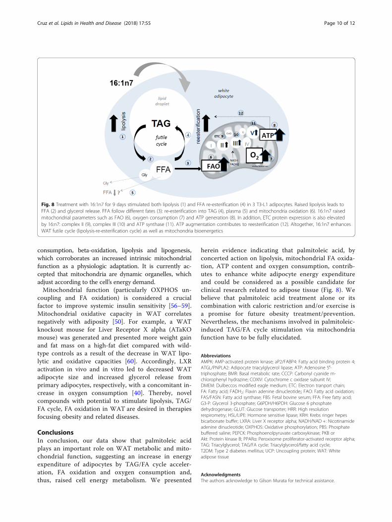
herein evidence indicating that palmitoleic acid, byconcerted action on lipolysis, mitochondrial FA oxida-tion, ATP content and oxygen consumption, contrib-utes to enhance white adipocyte energy expenditureand could be considered as a possible candidate forclinical research related to adipose tissue (Fig. 8). Webelieve that palmitoleic acid treatment alone or itscombination with caloric restriction and/or exercise isa promise for future obesity treatment/prevention.Nevertheless, the mechanisms involved in palmitoleic-induced TAG/FA cycle stimulation via mitochondriafunction have to be fully elucidated.
AbbreviationsAMPK: AMP-activated protein kinase; aP2/FABP4: Fatty acid binding protein 4;ATGL/PNPLA2: Adipocyte triacylglycerol lipase; ATP: Adenosine 5′-triphosphate; BMR: Basal metabolic rate; CCCP: Carbonyl cyanide m-chlorophenyl hydrazine; COXIV: Cytochrome c oxidase subunit IV;DMEM: Dulbeccos modified eagle medium; ETC: Electron tranport chain;FA: Fatty acid; FADH2: Flavin adenine dinucleotide;; FAO: Fatty acid oxidation;FAS/FASN: Fatty acid synthase; FBS: Fetal bovine serum; FFA: Free fatty acid;G3-P: Glycerol 3-phosphate; G6PDH/H6PDH: Glucose 6 phosphatedehydrogenase; GLUT: Glucose transporter; HRR: High resolutionrespirometry; HSL/LIPE: Hormone sensitive lipase; KRH: Krebs ringer hepesbicarbonate buffer; LXRA: Liver X receptor alpha; NADH/NAD +: Nicotinamideadenine dinucleotide; OXPHOS: Oxidative phosphorylation; PBS: Phosphatebuffered saline; PEPCK: Phosphoenolpyruvate carboxykinase; PKB orAkt: Protein kinase B; PPARα: Peroxisome proliferator-activated receptor alpha;TAG: Triacylglycerol; TAG/FA cycle: Triacylglycerol/fatty acid cycle;T2DM: Type 2 diabetes mellitus; UCP: Uncoupling protein; WAT: Whiteadipose tissue
AcknowledgmentsThe authors acknowledge to Gilson Murata for technical assistance.
Fig. 8 Treatment with 16:1n7 for 9 days stimulated both lipolysis (1) and FFA re-esterification (4) in 3 T3-L1 adipocytes. Raised lipolysis leads toFFA (2) and glycerol release. FFA follow different fates (3): re-esterification into TAG (4), plasma (5) and mitochondria oxidation (6). 16:1n7 raisedmitochondrial parameters such as FAO (6), oxygen consumption (7) and ATP generation (8). In addition, ETC protein expression is also elevatedby 16:n7: complex II (9), complex III (10) and ATP synthase (11). ATP augmentation contributes to reesterification (12). Altogether, 16:1n7 enhancesWAT futile cycle (lipolysis-re-esterification cycle) as well as mitochondria bioenergetics
Cruz et al. Lipids in Health and Disease (2018) 17:55 Page 10 of 12

FundingThis work was supported by grants from FAPESP (2011/51627–8 and2011/51701–3).
Availability of data and materialsAll data generated or analyzed during this study are included in thispublished article.
Authors’ contributionsMMC, ABL, ARC, RCCS and WMTK performed the experiments, analyzed theresults and revised the manuscript. RC and PBMA designed the study,analyzed the results and revised the manuscript. MICAV designed the study,analyzed the results, wrote the manuscript and supervised the study. Allauthors read and approved the final manuscript.
Ethics approval and consent to participateNot applicable.
Consent for publicationNot applicable.
Competing interestsThe authors declare that they have no competing interests.
Publisher’s NoteSpringer Nature remains neutral with regard to jurisdictional claims inpublished maps and institutional affiliations.
Author details1Department of Biological Sciences, Institute of Environmental Sciences,Chemical and Pharmaceutical, Federal University of São Paulo, 210, SaoNicolau St, Diadema 09913-030, Brazil. 2Department of Physiology andBiophysics, Institute of Biomedical Sciences, University of São Paulo, SãoPaulo, Brazil. 3Interdisciplinary Postgraduate Program in Health Sciences,Institute of Physical Activity Sciences and Sports, Cruzeiro do Sul University,São Paulo, Brazil. 4Department of Nursing , Health Sciences Center, FederalUniversity of Espírito Santo, Vitória, Brazil.
Received: 11 January 2018 Accepted: 13 March 2018
References1. Jensen MD. Lipolysis: contribution from regional fat. Annu Rev Nutr. 1997;
17:127–39. https://doi.org/10.1146/annurev.nutr.17.1.127. PubMed PMID:9240922.
2. Ruge T, Hodson L, Cheeseman J, Dennis AL, Fielding BA, Humphreys SM, etal. Fasted to fed trafficking of fatty acids in human adipose tissue reveals anovel regulatory step for enhanced fat storage. J Clin Endocrinol Metab.2009;94(5):1781–8. https://doi.org/10.1210/jc.2008-2090. PubMed PMID:19223522.
3. Sethi JK, Vidal-Puig AJ. Thematic review series: adipocyte biology. Adiposetissue function and plasticity orchestrate nutritional adaptation. J Lipid Res.2007;48(6):1253–62. https://doi.org/10.1194/jlr.R700005-JLR200. PubMedPMID: 17374880; PubMed Central PMCID: PMC4303760.
4. Lanza IR, Nair KS. Functional assessment of isolated mitochondria in vitro.Methods Enzymol. 2009;457:349–72. https://doi.org/10.1016/S0076-6879(09)05020-4. PubMed PMID: 19426878; PubMed Central PMCID:PMC2782617.
5. Carriere A, Fernandez Y, Rigoulet M, Penicaud L, Casteilla L. Inhibition ofpreadipocyte proliferation by mitochondrial reactive oxygen species. FEBSLett. 2003;550(1–3):163–7. PubMed PMID: 12935904.
6. De Pauw A, Tejerina S, Raes M, Keijer J, Arnould T. Mitochondrial (dys)function in adipocyte (de)differentiation and systemic metabolic alterations.Am J Pathol. 2009;175(3):927–39. https://doi.org/10.2353/ajpath.2009.081155.PubMed PMID: 19700756; PubMed Central PMCID: PMC2731113.
7. Kita T, Nishida H, Shibata H, Niimi S, Higuti T, Arakaki N. Possible role ofmitochondrial remodelling on cellular triacylglycerol accumulation. JBiochem. 2009;146(6):787–96. https://doi.org/10.1093/jb/mvp124. PubMedPMID: 19671539.
8. Kaaman M, Sparks LM, van Harmelen V, Smith SR, Sjolin E, Dahlman I, et al.Strong association between mitochondrial DNA copy number and
lipogenesis in human white adipose tissue. Diabetologia. 2007;50(12):2526–33. https://doi.org/10.1007/s00125-007-0818-6. PubMed PMID: 17879081.
9. Sutherland LN, Bomhof MR, Capozzi LC, Basaraba SA, Wright DC. Exerciseand adrenaline increase PGC-1{alpha} mRNA expression in rat adiposetissue. J Physiol. 2009;587(Pt 7):1607–17. https://doi.org/10.1113/jphysiol.2008.165464. PubMed PMID: 19221126; PubMed Central PMCID:PMC2678229.
10. Duchen MR. Mitochondria in health and disease: perspectives on a newmitochondrial biology. Mol Asp Med. 2004;25(4):365–451. https://doi.org/10.1016/j.mam.2004.03.001. PubMed PMID: 15302203.
11. Geisler JG. Targeting energy expenditure via fuel switching and beyond.Diabetologia. 2011;54(2):237–44. https://doi.org/10.1007/s00125-010-1932-4.PubMed PMID: 20953861.
12. Cao H, Gerhold K, Mayers JR, Wiest MM, Watkins SM, Hotamisligil GS.Identification of a lipokine, a lipid hormone linking adipose tissue to systemicmetabolism. Cell. 2008;134(6):933–44. https://doi.org/10.1016/j.cell.2008.07.048.PubMed PMID: 18805087; PubMed Central PMCID: PMC2728618
13. Dimopoulos N, Watson M, Sakamoto K, Hundal HS. Differential effects ofpalmitate and palmitoleate on insulin action and glucose utilization in ratL6 skeletal muscle cells. Biochem J. 2006;399(3):473–81. https://doi.org/10.1042/BJ20060244. PubMed PMID: 16822230; PubMed Central PMCID:PMC1615906.
14. Obanda DN, Cefalu WT. Modulation of cellular insulin signaling and PTP1Beffects by lipid metabolites in skeletal muscle cells. J Nutr Biochem. 2013;24(8):1529–37. https://doi.org/10.1016/j.jnutbio.2012.12.014. PubMed PMID:23481236; PubMed Central PMCID: PMC4509740.
15. Yang ZH, Miyahara H, Hatanaka A. Chronic administration of palmitoleicacid reduces insulin resistance and hepatic lipid accumulation in KK-Aymice with genetic type 2 diabetes. Lipids Health Dis. 2011;10:120. https://doi.org/10.1186/1476-511X-10-120. PubMed PMID: 21774832; PubMedCentral PMCID: PMC3155149.
16. Diakogiannaki E, Dhayal S, Childs CE, Calder PC, Welters HJ, Morgan NG.Mechanisms involved in the cytotoxic and cytoprotective actions ofsaturated versus monounsaturated long-chain fatty acids in pancreatic beta-cells. J Endocrinol. 2007;194(2):283–91. https://doi.org/10.1677/JOE-07-0082.PubMed PMID: 17641278; PubMed Central PMCID: PMC1994570.
17. Morgan NG, Dhayal S. Unsaturated fatty acids as cytoprotective agents in thepancreatic beta-cell. Prostaglandins Leukot Essent Fatty Acids. 2010;82(4–6):231–6. https://doi.org/10.1016/j.plefa.2010.02.018. PubMed PMID: 20206490.
18. Bolsoni-Lopes A, Festuccia WT, Farias TS, Chimin P, Torres-Leal FL, DerogisPB, et al. Palmitoleic acid (n-7) increases white adipocyte lipolysis and lipasecontent in a PPARalpha-dependent manner. Am J Physiol EndocrinolMetab. 2013;305(9):E1093–102. https://doi.org/10.1152/ajpendo.00082.2013.PubMed PMID: 24022867.
19. Bolsoni-Lopes A, Festuccia WT, Chimin P, Farias TS, Torres-Leal FL, Cruz MM,et al. Palmitoleic acid (n-7) increases white adipocytes GLUT4 content andglucose uptake in association with AMPK activation. Lipids Health Dis. 2014;13:199. https://doi.org/10.1186/1476-511X-13-199. PubMed PMID: 25528561;PubMed Central PMCID: PMC4364637.
20. Park BH, Qiang L, Farmer SR. Phosphorylation of C/EBPbeta at a consensusextracellular signal-regulated kinase/glycogen synthase kinase 3 site isrequired for the induction of adiponectin gene expression during thedifferentiation of mouse fibroblasts into adipocytes. Mol Cell Biol. 2004;24(19):8671–80. https://doi.org/10.1128/MCB.24.19.8671-8680.2004. PubMedPMID: 15367685; PubMed Central PMCID: PMC516726.
21. Cruz MM. Efeitos dos ácidos graxos palmítico e palmitoleico sobreparâmetros metabólicos de adipócitos 3T3-L1 [Mestrado]: UniversidadeFederal de São Paulo; 2015.
22. de Sa RD, Crisma AR, Cruz MM, Martins AR, Masi LN, do Amaral CL, et al.Fish oil prevents changes induced by a high-fat diet on metabolism andadipokine secretion in mice subcutaneous and visceral adipocytes. J Physiol.2016;594(21):6301–17. https://doi.org/10.1113/JP272541. Epub 2016/11/02.PubMed PMID: 27558442; PubMed Central PMCID: PMCPMC5088242.
23. Amengual J, Petrov P, Bonet ML, Ribot J, Palou A. Induction of carnitinepalmitoyl transferase 1 and fatty acid oxidation by retinoic acid in HepG2cells. Int J Biochem Cell Biol. 2012;44(11):2019–27. https://doi.org/10.1016/j.biocel.2012.07.026. PubMed PMID: 22871568.
24. Mercader J, Madsen L, Felipe F, Palou A, Kristiansen K, Bonet ML. All-transretinoic acid increases oxidative metabolism in mature adipocytes. CellPhysiol Biochem. 2007;20(6):1061–72. https://doi.org/10.1159/0000110717.PubMed PMID: 17975308.
Cruz et al. Lipids in Health and Disease (2018) 17:55 Page 11 of 12

25. Shen W, Liu K, Tian C, Yang L, Li X, Ren J, et al. R-alpha-lipoic acid andacetyl-L-carnitine complementarily promote mitochondrial biogenesis inmurine 3T3-L1 adipocytes. Diabetologia. 2008;51(1):165–74. https://doi.org/10.1007/s00125-007-0852-4. PubMed PMID: 18026715.
26. de Andrade PB, Rubi B, Frigerio F, van den Ouweland JM, Maassen JA,Maechler P. Diabetes-associated mitochondrial DNA mutation A3243Gimpairs cellular metabolic pathways necessary for beta cell function.Diabetologia. 2006;49(8):1816–26. https://doi.org/10.1007/s00125-006-0301-9.PubMed PMID: 16736129.
27. Tourniaire F, Musinovic H, Gouranton E, Astier J, Marcotorchino J, ArreguinA, et al. All-trans retinoic acid induces oxidative phosphorylation andmitochondria biogenesis in adipocytes. J Lipid Res. 2015;56(6):1100–9.https://doi.org/10.1194/jlr.M053652. Epub 2015/04/29. PubMed PMID:25914170; PubMed Central PMCID: PMCPMC4442868.
28. Alp PR, Newsholme EA, Zammit VA. Activities of citrate synthase and NAD+-linked and NADP+-linked isocitrate dehydrogenase in muscle fromvertebrates and invertebrates. Biochem J. 1976;154(3):689–700.
29. Hodson L, Karpe F. Is there something special about palmitoleate? CurrOpin Clin Nutr Metab Care. 2013;16(2):225–31. https://doi.org/10.1097/MCO.0b013e32835d2edf. PubMed PMID: 23324899.
30. Talbot NA, Wheeler-Jones CP, Cleasby ME. Palmitoleic acid prevents palmiticacid-induced macrophage activation and consequent p38 MAPK-mediatedskeletal muscle insulin resistance. Mol Cell Endocrinol. 2014;393(1–2):129–42.https://doi.org/10.1016/j.mce.2014.06.010. PubMed PMID: 24973767; PubMedCentral PMCID: PMC4148479.
31. Forest C, Tordjman J, Glorian M, Duplus E, Chauvet G, Quette J, et al. Fattyacid recycling in adipocytes: a role for glyceroneogenesis andphosphoenolpyruvate carboxykinase. Biochem Soc Trans. 2003;31(6):1125–9.Portland Press Ltd. https://doi.org/10.1042/bst0311125.
32. Reidy SP, Weber JM. Accelerated substrate cycling: a new energy-wastingrole for leptin in vivo. Am J Physiol Endocrinol Metab. 2002;282(2):E312–7.https://doi.org/10.1152/ajpendo.00037.2001. PubMed PMID: 11788362.
33. Wolfe RR, Klein S, Carraro F, Weber JM. Role of triglyceride-fatty acid cycle incontrolling fat metabolism in humans during and after exercise. Am J Phys.1990;258(2 Pt 1):E382–9. PubMed PMID: 2106269.
34. Klaman LD, Boss O, Peroni OD, Kim JK, Martino JL, Zabolotny JM, et al.Increased energy expenditure, decreased adiposity, and tissue-specific insulinsensitivity in protein-tyrosine phosphatase 1B-deficient mice. Mol Cell Biol. 2000;20(15):5479–89. PubMed PMID: 10891488; PubMed Central PMCID: PMC85999.
35. Vallerand AL, Zamecnik J, Jones PJ, Jacobs I. Cold stress increases lipolysis,FFA Ra and TG/FFA cycling in humans. Aviat Space Environ Med. 1999;70(1):42–50. PubMed PMID: 9895020.
36. Maassen JA, Romijn JA, Heine RJ. Fatty acid-induced mitochondrialuncoupling in adipocytes as a key protective factor against insulinresistance and beta cell dysfunction: a new concept in the pathogenesis ofobesity-associated type 2 diabetes mellitus. Diabetologia. 2007;50(10):2036–41. https://doi.org/10.1007/s00125-007-0776-z. PubMed PMID: 17712547;PubMed Central PMCID: PMC2039833.
37. Baht HS, Saggerson ED. Comparison of triacylglycerol synthesis in rat brownand white adipocytes. Effects of hypothyroidism and streptozotocin-diabetes on enzyme activities and metabolic fluxes. Biochem J. 1988;250(2):325–33. PubMed PMID: 3355527; PubMed Central PMCID: PMC1148859.
38. Frayn KN, Langin D, Karpe F. Fatty acid-induced mitochondrial uncouplingin adipocytes is not a promising target for treatment of insulin resistanceunless adipocyte oxidative capacity is increased. Diabetologia. 2008;51(3):394–7. https://doi.org/10.1007/s00125-007-0901-z. PubMed PMID: 18097647.
39. Harper RD, Saggerson ED. Factors affecting fatty acid oxidation in fat cellsisolated from rat white adipose tissue. J Lipid Res. 1976;17(5):516–26.PubMed PMID: 965842.
40. Dib L, Bugge A, Collins S. LXRalpha fuels fatty acid-stimulated oxygenconsumption in white adipocytes. J Lipid Res. 2014;55(2):247–57. https://doi.org/10.1194/jlr.M043422. PubMed PMID: 24259533; PubMed Central PMCID:PMC3886663.
41. Wang T, Zang Y, Ling W, Corkey BE, Guo W. Metabolic partitioning ofendogenous fatty acid in adipocytes. Obes Res. 2003;11(7):880–7. https://doi.org/10.1038/oby.2003.121. PubMed PMID: 12855758.
42. den Hartigh LJ, Han CY, Wang S, Omer M, Chait A. 10E,12Z-conjugatedlinoleic acid impairs adipocyte triglyceride storage by enhancing fatty acidoxidation, lipolysis, and mitochondrial reactive oxygen species. J Lipid Res.2013;54(11):2964–78. https://doi.org/10.1194/jlr.M035188. Epub 2013/08/21.PubMed PMID: 23956445; PubMed Central PMCID: PMCPMC3793601.
43. Vankoningsloo S, Piens M, Lecocq C, Gilson A, De Pauw A, Renard P, et al.Mitochondrial dysfunction induces triglyceride accumulation in 3T3-L1 cells:role of fatty acid beta-oxidation and glucose. J Lipid Res. 2005;46(6):1133–49. https://doi.org/10.1194/jlr.M400464-JLR200. PubMed PMID: 15741651.
44. Heinonen S, Buzkova J, Muniandy M, Kaksonen R, Ollikainen M, Ismail K, etal. Impaired mitochondrial biogenesis in adipose tissue in acquired obesity.Diabetes. 2015;64(9):3135–45. https://doi.org/10.2337/db14-1937. PubMedPMID: 25972572.
45. Garlid KD, Jaburek M, Jezek P. Mechanism of uncoupling protein action.Biochem Soc Trans. 2001;29(Pt 6):803–6. PubMed PMID: 11709078.
46. Crescenzo R, Bianco F, Mazzoli A, Giacco A, Liverini G, Iossa S. Skeletalmuscle mitochondrial energetic efficiency and aging. Int J Mol Sci. 2015;16(5):10674–85. https://doi.org/10.3390/ijms160510674. PubMed PMID:25970752; PubMed Central PMCID: PMC4463669.
47. Gao CL, Zhu C, Zhao YP, Chen XH, Ji CB, Zhang CM, et al. Mitochondrialdysfunction is induced by high levels of glucose and free fatty acids in 3T3-L1 adipocytes. Mol Cell Endocrinol. 2010;320(1–2):25–33. https://doi.org/10.1016/j.mce.2010.01.039. Epub 2010/02/11. PubMed PMID: 20144685.
48. Lobo S, Wiczer BM, Bernlohr DA. Functional analysis of long-chain acyl-CoAsynthetase 1 in 3T3-L1 adipocytes. J Biol Chem. 2009;284(27):18347–56.https://doi.org/10.1074/jbc.M109.017244. PubMed PMID: 19429676; PubMedCentral PMCID: PMC2709349.
49. Baldwin RL. Metabolic functions affecting the contribution of adipose tissueto total energy expenditure, Fed. Proc. 1970;29:1277–83.
50. Flachs P, Rossmeisl M, Kuda O, Kopecky J. Stimulation of mitochondrialoxidative capacity in white fat independent of UCP1: a key to leanphenotype. Biochim Biophys Acta. 2013;1831(5):986–1003. https://doi.org/10.1016/j.bbalip.2013.02.003. PubMed PMID: 23454373.
51. Carracedo A, Cantley LC, Pandolfi PP. Cancer metabolism: fatty acidoxidation in the limelight. Nat Rev Cancer. 2013;13(4):227–32. https://doi.org/10.1038/nrc3483. PubMed PMID: 23446547; PubMed Central PMCID:PMC3766957.
52. Gauthier MS, Miyoshi H, Souza SC, Cacicedo JM, Saha AK, Greenberg AS,et al. AMP-activated protein kinase is activated as a consequence of lipolysisin the adipocyte: potential mechanism and physiological relevance. J BiolChem. 2008;283(24):16514–24. https://doi.org/10.1074/jbc.M708177200.PubMed PMID: 18390901; PubMed Central PMCID: PMC2423258.
53. Liu Q, Gauthier MS, Sun L, Ruderman N, Lodish H. Activation of AMP-activated protein kinase signaling pathway by adiponectin and insulin inmouse adipocytes: requirement of acyl-CoA synthetases FATP1 and Acsl1and association with an elevation in AMP/ATP ratio. FASEB J. 2010;24(11):4229–39. https://doi.org/10.1096/fj.10-159723. PubMed PMID: 20667975;PubMed Central PMCID: PMC2974418.
54. Rogge MM. The role of impaired mitochondrial lipid oxidation in obesity.Biol Res Nurs. 2009;10(4):356–73. https://doi.org/10.1177/1099800408329408.PubMed PMID: 19190032.
55. Ahmadian M, Duncan RE, Sul HS. The skinny on fat: lipolysis and fatty acidutilization in adipocytes. Trends Endocrinol Metab. 2009;20(9):424–8. https://doi.org/10.1016/j.tem.2009.06.002. PubMed PMID: 19796963; PubMedCentral PMCID: PMC2764815.
56. Harms M, Seale P. Brown and beige fat: development, function andtherapeutic potential. Nat Med. 2013;19(10):1252–63. https://doi.org/10.1038/nm.3361. PubMed PMID: 24100998.
57. Roman S, Agil A, Peran M, Alvaro-Galue E, Ruiz-Ojeda FJ, Fernandez-VazquezG, et al. Brown adipose tissue and novel therapeutic approaches to treatmetabolic disorders. Transl Res. 2015;165(4):464–79. https://doi.org/10.1016/j.trsl.2014.11.002. PubMed PMID: 25433289.
58. Rousset S, Alves-Guerra MC, Mozo J, Miroux B, Cassard-Doulcier AM,Bouillaud F, et al. The biology of mitochondrial uncoupling proteins.Diabetes. 2004;53(Suppl 1):S130–5. PubMed PMID: 14749278.
59. Surwit RS, Wang S, Petro AE, Sanchis D, Raimbault S, Ricquier D, et al. Diet-induced changes in uncoupling proteins in obesity-prone and obesity-resistant strains of mice. Proc Natl Acad Sci U S A. 1998;95(7):4061–5.PubMed PMID: 9520493; PubMed Central PMCID: PMC19963.
60. Bottcher H, Furst P. Decreased white fat cell thermogenesis in obeseindividuals. Int J Obes Relat Metab Disord. 1997;21(6):439–44. PubMedPMID: 9192226.
Cruz et al. Lipids in Health and Disease (2018) 17:55 Page 12 of 12


![€¦ · Web view2009. 4. 23. · [Cr2O72-] Reverse Rate. A. increases increases. B. increases decreases. C. decreases decreases. D. decreases increases. 31. A small amount of H2SO4](https://img.dokumen.tips/doc/110x75/608f2c47b9e3f5096f2e5efc/web-view-2009-4-23-cr2o72-reverse-rate-a-increases-increases-b-increases.jpg)

![PlantAcyl-CoA:LysophosphatidylcholineAcyltransferases (LPCATs ... et... · 2015. 6. 1. · Chemicals—[1-14C]18:1 and [1-14C]palmitoleic acid (16:0) were purchased from PerkinElmer](https://img.dokumen.tips/doc/110x75/602cd5d3378486433432c434/plantacyl-coalysophosphatidylcholineacyltransferases-lpcats-et-2015.jpg)


