Embed Size (px)
Citation preview

JOURNAL OF MEDICALCASE REPORTS
Niessen et al. Journal of Medical Case Reports 2013, 7:128http://www.jmedicalcasereports.com/content/7/1/128
CASE REPORT Open Access
Palliative treatment of presacral recurrence ofendometrial cancer using irreversibleelectroporation: a case reportChristoph Niessen1*, Ernst-Michael Jung1, Andreas G Schreyer1, Walter A Wohlgemuth1, Benedikt Trabold2,Joachim Hahn3, Michael Rechenmacher3, Christian Stroszczynski1 and Philipp Wiggermann1
Abstract
Introduction: Irreversible electroporation (IRE) is a new minimally invasive tumor ablation technique which inducesirreversible disruption of cell membrane integrity by changing the transmembrane potential resulting in cell death.Irreversible electroporation is currently undergoing clinical investigation as local tumor therapy for malignant liverand lung lesions. This is the first case report to describe the successful palliative ablation of a presacral recurrenceof an endometrial cancer to achieve locoregional tumor control and pain relief.
Case presentation: A 56-year-old Caucasian woman was referred for interventional treatment of an advanced localrecurrence of endometrial cancer (11.9 × 11.6 × 14.9cm) with infiltration of the sacral bone and nerve plexus.Due to the immediate proximity to the sacral plexus, the patient could neither undergo surgical therapy nor asecond radiation therapy. Due to its ablation mechanism irreversible electroporation was deemed to be the besttherapy option.
Conclusion: We showed in this case that a large tumor mass adjacent to a bundle of neural structures, the sacralplexus, can be widely ablated by irreversible electroporation with only minor temporary impairment of the neuralfunction, even though a large infiltrating tissue volume (941cm3) was ablated.
IntroductionIrreversible electroporation (IRE) is a new non-thermallocal ablative treatment procedure which induces the ir-reversible permeabilization of a membrane lipid bilayerby creation of nanopores resulting in cell death [1]. IREis currently undergoing clinical investigation as a locallyablative tumor therapy for different organ systems, suchas kidney, lung or liver lesions [2].Endometrial cancer represents the seventh most fre-
quent tumor worldwide in women with an annual inci-dence of 142,000 patients worldwide [3]. The standardtherapy for endometrial cancer is total removal of theuterus, cervix, as much as the parametrial tissue aspossible, and a wide margin of the vagina (Wertheim-Meigs operation). At advanced stage endometrial cancer
* Correspondence: [email protected] of Radiology, University Hospital Regensburg, Franz-JosefStrauss Allee 11, 93042 Regensburg, GermanyFull list of author information is available at the end of the article
© 2013 Niessen et al.; licensee BioMed CentraCommons Attribution License (http://creativecreproduction in any medium, provided the or
adjuvant radiation is performed. Radiation is consideredstandard therapy of inoperable tumor stages [4].This article reports the successful palliative ablation of
a presacral recurrence of endometrial cancer as an indi-vidual palliative therapy trial in order to achieve locore-gional tumor control and pain relief.
Case presentationA 56-year-old Caucasian woman was referred for treat-ment of a local recurrence of endometrial cancer withinfiltration of her sacral bone and nerve plexus. Afterinitial diagnosis of endometrial cancer in 2005 (pT1B N0MX G1 L0 V0), a Wertheim-Meigs operation was per-formed with subsequent radiotherapy with the initial re-sult of complete tumor remission. In 2009 the patientpresented with histologically confirmed pulmonary me-tastases. Under anti-hormonal therapy (medroxyproges-terone acetate), again complete remission was achieved.At the start of 2012, 7 years after the initial diagnosis, thepatient was admitted to our hospital due to a presacral
l Ltd. This is an Open Access article distributed under the terms of the Creativeommons.org/licenses/by/2.0), which permits unrestricted use, distribution, andiginal work is properly cited.
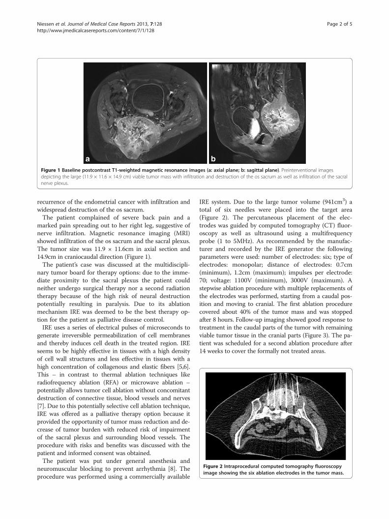
Figure 1 Baseline postcontrast T1-weighted magnetic resonance images (a: axial plane; b: sagittal plane). Preinterventional imagesdepicting the large (11.9 × 11.6 × 14.9 cm) viable tumor mass with infiltration and destruction of the os sacrum as well as infiltration of the sacralnerve plexus.
Figure 2 Intraprocedural computed tomography fluoroscopyimage showing the six ablation electrodes in the tumor mass.
Niessen et al. Journal of Medical Case Reports 2013, 7:128 Page 2 of 5http://www.jmedicalcasereports.com/content/7/1/128
recurrence of the endometrial cancer with infiltration andwidespread destruction of the os sacrum.The patient complained of severe back pain and a
marked pain spreading out to her right leg, suggestive ofnerve infiltration. Magnetic resonance imaging (MRI)showed infiltration of the os sacrum and the sacral plexus.The tumor size was 11.9 × 11.6cm in axial section and14.9cm in craniocaudal direction (Figure 1).The patient’s case was discussed at the multidiscipli-
nary tumor board for therapy options: due to the imme-diate proximity to the sacral plexus the patient couldneither undergo surgical therapy nor a second radiationtherapy because of the high risk of neural destructionpotentially resulting in paralysis. Due to its ablationmechanism IRE was deemed to be the best therapy op-tion for the patient as palliative disease control.IRE uses a series of electrical pulses of microseconds to
generate irreversible permeabilization of cell membranesand thereby induces cell death in the treated region. IREseems to be highly effective in tissues with a high densityof cell wall structures and less effective in tissues with ahigh concentration of collagenous and elastic fibers [5,6].This – in contrast to thermal ablation techniques likeradiofrequency ablation (RFA) or microwave ablation –potentially allows tumor cell ablation without concomitantdestruction of connective tissue, blood vessels and nerves[7]. Due to this potentially selective cell ablation technique,IRE was offered as a palliative therapy option because itprovided the opportunity of tumor mass reduction and de-crease of tumor burden with reduced risk of impairmentof the sacral plexus and surrounding blood vessels. Theprocedure with risks and benefits was discussed with thepatient and informed consent was obtained.The patient was put under general anesthesia and
neuromuscular blocking to prevent arrhythmia [8]. Theprocedure was performed using a commercially available
IRE system. Due to the large tumor volume (941cm3) atotal of six needles were placed into the target area(Figure 2). The percutaneous placement of the elec-trodes was guided by computed tomography (CT) fluor-oscopy as well as ultrasound using a multifrequencyprobe (1 to 5MHz). As recommended by the manufac-turer and recorded by the IRE generator the followingparameters were used: number of electrodes: six; type ofelectrodes: monopolar; distance of electrodes: 0.7cm(minimum), 1.2cm (maximum); impulses per electrode:70; voltage: 1100V (minimum), 3000V (maximum). Astepwise ablation procedure with multiple replacements ofthe electrodes was performed, starting from a caudal pos-ition and moving to cranial. The first ablation procedurecovered about 40% of the tumor mass and was stoppedafter 8 hours. Follow-up imaging showed good response totreatment in the caudal parts of the tumor with remainingviable tumor tissue in the cranial parts (Figure 3). The pa-tient was scheduled for a second ablation procedure after14 weeks to cover the formally not treated areas.

Figure 3 Follow-up postcontrast T1-weighted magnetic resonance images at 24 hours after first intervention. Viable enhancingtumor tissue (arrow) in the cranial and peripheral part of the lesion. Central parts of the tumor are necrotic (arrow heads). (a: axial plane,b: sagittal plane).
Niessen et al. Journal of Medical Case Reports 2013, 7:128 Page 3 of 5http://www.jmedicalcasereports.com/content/7/1/128
During the two IRE procedures the patient did not haveany cardiovascular events, in particular no supraventricu-lar tachycardia and no atrial fibrillation. Complications,especially postinterventional paralysis or bleeding, werenot observed. After the first ablation procedure the patientdid not complain about aggravated back pain; neither sen-sory deficit, nor loss of strength in her legs, nor paresthe-sia were observed. A neurological examination after thesecond ablation session revealed a mild 4+ paresis of theright extensor hallucis longus (L4 to S1) with com-plete resolution after 4 weeks. No sensory loss or im-pairment of bladder function occurred. After the secondintervention opiate medication could be withdrawn. Usingcarbamazepine (200mg twice a day) and Polamidone(levomethadone) (5mg three times a day) pain control wasachieved. The patient’s 24 hour follow-up imaging afterthe second ablation as well as follow-up imaging (Figure 4)
Figure 4 Follow-up postcontrast T1-weighted magnetic resonanceparts of the tumor mass show viable enhancing tumor tissue (arrow). Cb: sagittal plane).
after 8 weeks (consisting of contrast-enhanced ultrasound,MRI and a CT scan) showed wide ablation of the tumorwith necrosis of most portions of the tumor and reductionof tumor volume to 791cm3.
DiscussionAmong the different tumor ablation techniques RFA isthe most widespread technique [9]. Even though percu-taneous ablation techniques are used as possibly curativetherapies, palliative tumor ablation can be useful toachieve locoregional control of tumor growth, pain reliefor pain control, especially in patients with unresectabletumor manifestations [10].Due to heat dissipation to adjacent structures there is
an inherent risk of thermal damage of adjacent organs,blood vessels and, of course, nerves. Thus, lesions closeto adjacent structures with high risk of unintended heat
images at 8 weeks after second intervention. Only peripheralentral parts of the tumor are necrotic (arrow head). (a: axial plane,

Niessen et al. Journal of Medical Case Reports 2013, 7:128 Page 4 of 5http://www.jmedicalcasereports.com/content/7/1/128
destruction still pose a challenge for percutaneous ther-mal ablation techniques. IRE, in contrast to RFA, is anon-thermal ablation technique of soft tissue and offersa possibility to overcome the aforementioned limitationsof thermal ablations. Instead of using heat, IRE uses aseries of electrical pulses for microseconds to generateirreversible permeabilization of cell membranes, presum-ably through the formation of nanoscale defects in thecell membrane, and thereby induces cell death in thetreated tissue.IRE originally was viewed as an undesirable side effect
of reversible electroporation and therefore was studiedonly to define the upper limit of electrical parametersthat induce reversible electroporation. Due to its variousfeatures, for example transdermal delivery or introduc-tion of drugs and genes into cells, or electrochemo-therapy, reversible electroporation is an importantmethod in biotechnology and medicine [11]. Davalos,Mir and Rubinsky in 2005 reported that IRE can be usedas an independent modality for ablation of substantialtissue volumes [12]. Their findings were subsequentlyconfirmed in experimental studies on cells and in largeanimal models [13,14].In a series of studies, Lee could show that IRE pro-
duces irreversible tissue damage, which earlier was at-tributed only to thermal effects [15]. Furthermore, IREproved to be especially effective in tissues with a highdensity of cell wall structures and less effective in tissueswith a high concentration of collagenous and elastic fi-bers, which is suggestive of a cell selective effect [5,6].There appears to be complete ablation up to the marginof blood vessels without compromising the functionalityof the blood vessels [16]. This – in contrast to thermalablation – would allow tumor cell ablation without con-comitant destruction of connective tissue, blood vesselsand nerves, which means ablation of tumor cells in thoseareas where thermal ablation was not possible before. Inthe proximity of larger blood vessels thermal ablationtechniques are also hindered by the heat-sink effect. Dueto its cooling effect blood flow is an important determi-nant as much as a limiting factor of thermal ablationtechniques [17,18]. IRE seems to be unaffected by theblood flow and conversely does not potentially affect themacrovascularization of the ablation zone [19].
ConclusionDue to its more selective and non-thermal ablation ef-fect IRE widens the field of minimally invasive treatablelesions. We showed in this case report that a large ma-lignant lesion adjacent to a bundle of neural structures,that is the sacral plexus, can be widely ablated by IREwith only minor, temporary impairment of the neuralfunction, even though a large infiltrating tissue volumewas ablated.
ConsentWritten informed consent was obtained from the patientfor publication of this case report and accompanying im-ages. A copy of the written consent is available for re-view by the Editor-in-Chief of this journal.
Competing interestsThe authors declare that they have no competing interests.
Authors’ contributionsPW, EMJ, BT, CN and CS performed the IRE procedure. AGS, JH, MR andWW were major contributors in writing the manuscript. All authors read andapproved the final manuscript.
Author details1Department of Radiology, University Hospital Regensburg, Franz-JosefStrauss Allee 11, 93042 Regensburg, Germany. 2Department ofAnaesthesiology, University Hospital Regensburg, Franz-Josef Strauss Allee 11,93042 Regensburg, Germany. 3Department of Hematology and Oncology,University Hospital Regensburg, Franz-Josef Strauss Allee 11, 93042Regensburg, Germany.
Received: 10 January 2013 Accepted: 27 March 2013Published: 13 May 2013
References1. Freeman SA, Wang MA, Weaver JC: Theory of electroporation of planar
bilayer membranes: predictions of the aqueous area, change incapacitance, and pore-pore separation. Biophys J 1994, 67(1):42–56.
2. Thomson KR, Cheung W, Ellis SJ, Federman D, Kavnoudias H, Loader-Oliver D,Roberts S, Evans P, Ball C, Haydon A: Investigation of the safety of irreversibleelectroporation in humans. J Vasc Interv Radiol 2011, 22:611–621.
3. Parkin DM, Pisani P, Ferlay J: Global cancer statistics. CA Cancer J Clin 1999,49:33–64.
4. Amant F, Moerman P, Neven P, Timmermann D, Van Limbergen E, VergoteI: Treatment modalities in endometrial cancer. Curr Opin Oncol 2007,19:479–485.
5. Lee EW, Loh CT, Kee ST: Imaging guided percutaneous irreversibleelectroporation: ultrasound and immunohistological correlation. TechnolCancer Res Treat 2007, 6:287–294.
6. Lee EW, Chen C, Prieto VE, Dry SM, Loh CT, Kee ST: Advanced hepaticablation technique for creating complete cell death: irreversibleelectroporation. Radiology 2010, 255(2):426–433.
7. Onik G, Rubinsky B, Mikus P: Irreversible electroporation: Implications forprostate ablation. Technol Cancer Res Treat 2007, 6:295–300.
8. Deodhar A, Dickfeld T, Single GW, Hamilton WC Jr, Thornton RH, SofocleousCT, Maybody M, Gonen M, Rubinsky B, Solomon SB: Irreversibleelectroporation near the heart: ventricular arrhythmias can be preventedwith ECG synchronization. AJR Am J Roentgenol 2011, 196:330–335.
9. Lencioni R, Crocetti L: Local-regional treatment of hepatocellularcarcinoma. Radiology 2012, 262:43–58.
10. Mylona S, Karagiannis G, Patsoura S, Galani P, Pomoni M, Thanos L:Palliative treatment of rectal carcinoma recurrence using radiofrequencyablation. Cardiovasc Intervent Radiol 2012, 35:875–882.
11. Heller R: Overview of electroporation. 2002. Technol Cancer Res Treat 2002,1:317–318.
12. Davalos R, Mir L, Rubinsky B: Tissue ablation with irreversibleelectroporation. Annals Biomed Eng 2005, 33:223–231.
13. Miller L, Leor J, Rubinsky B: Cancer cells ablation with irreversibleelectroporation. Technol Cancer Res Treat 2005, 4:699–706.
14. Ben-David E, Appelbaum L, Sosna J, Nissenbaum I, Goldberg SN:Characterization of irreversible electroporation ablation in in vivoporcine liver. Am J Roentgenol 2012, 198:W62–W68.
15. Lee RC: Cell injury by electric forces. Ann NY Acad Sci 2005, 1066:85–91.16. Maor E, Ivorra A, Leor J, Rubinsky B: The effect of irreversible electroporation
on blood vessels. Technol Cancer Res Treat 2007, 6:307–312.17. Patterson EJ, Scudamore CH, Owen DA, Nagy AG, Buczkowski AK:
Radiofrequency ablation of porcine liver in vivo: effects of blood flowand treatment time on lesion size. Ann Surg 1998, 227:559–565.

Niessen et al. Journal of Medical Case Reports 2013, 7:128 Page 5 of 5http://www.jmedicalcasereports.com/content/7/1/128
18. Charpentier KP: Irreversible electroporation for the ablation of livertumors: are we there yet? Arch Surg – Chicago 2012, 147:1053–1061.
19. Kingham TP, Karkar AM, D’Angelica MI, Allen PJ, Dematteo RP, GetrajdmanGI, Sofocleous CT, Solomon SB, Jarnagin WR, Fong Y: Ablation ofperivascular hepatic malignant tumors with irreversible electroporation.J Am Coll Surg 2012, 215(3):379–387.
doi:10.1186/1752-1947-7-128Cite this article as: Niessen et al.: Palliative treatment of presacralrecurrence of endometrial cancer using irreversible electroporation: acase report. Journal of Medical Case Reports 2013 7:128.
Submit your next manuscript to BioMed Centraland take full advantage of:
• Convenient online submission
• Thorough peer review
• No space constraints or color figure charges
• Immediate publication on acceptance
• Inclusion in PubMed, CAS, Scopus and Google Scholar
• Research which is freely available for redistribution
Submit your manuscript at www.biomedcentral.com/submit