Embed Size (px)
Citation preview

Palaeontologia Electronica palaeo-electronica.org
Investigation of claims of late-surviving pterosaurs: the cases of Belon’s, Aldrovandi’s, and
Cardinal Barberini’s winged dragons
Phil Senter and Darius M. Klein
ABSTRACT
Here we investigate claims that pterosaurs survived into the sixteenth and seven-teenth centuries. In 1557, 1640, and 1651 the European naturalists Pierre Belon,Ulisse Aldrovandi, and Giovanni Faber, respectively, published illustrations of winged,bipedal specimens that had been stuffed and mounted. Some recent young-Earth cre-ationist authors claim that these specimens were recently-killed pterosaurs. The draw-ings and descriptions are detailed enough to test the pterosaur hypothesis as well asthe alternative hypothesis that the specimens are taxidermic composites of parts of dif-ferent animals. However, before now, no one has investigated these three cases orattempted to test these hypotheses. Here we report an investigation in which thesehypotheses are tested. By comparing the specimens with pterosaurs, we found that inall three specimens, all regions of the body are inconsistent with pterosaur anatomy.Comparison with extant animals reveals that Belon’s and Aldrovandi’s dragons aredecapitated snakes with attached mammal heads. Their wings are the pectoral fins offlying gurnards (Dactylopterus volitans). Their “legs” are the forelimbs of rabbits orcanids in reptile-skin sleeves. The dragon illustrated by Faber and owned by CardinalFrancesco Barberini includes the skull of a weasel (Mustela nivalis), the belly skin of asnake, the dorsal and lateral skin of a lizard, and the tail skeleton of an eel (Anguillaanguilla). These hoaxes now join the list of discredited “proofs” of human-pterosaurcoexistence.
Phil Senter. Department of Biological Sciences, Fayetteville State University, 1200 Murchison Road, Fayetteville, North Carolina 28301, U.S.A. [email protected] M. Klein. Department of Comparative Literature, Ballantine Hall 914, Indiana University, Bloomington, Indiana 47405, U.S.A. [email protected]
Keywords: Pterosauria; young-Earth creationism; Ulisse Aldrovandi; Pierre Belon; Giovanni Faber; hoax
PE Article Number: 17.3.41ACopyright: Palaeontological Association November 2014Submission: 26 May 2014. Acceptance: 22 October 2014
Senter, Phil and Klein, Darius M. 2014. Investigation of claims of late-surviving pterosaurs: the cases of Belon’s, Aldrovandi’s, and Cardinal Barberini’s winged dragons. Palaeontologia Electronica Vol. 17, Issue 3;41A; 19p; palaeo-electronica.org/content/2014/967-late-surviving-pterosaurs
SENTER,AND KLEIN: LATE-SURVIVING PTEROSAURS?
INTRODUCTION
Here we investigate claims that certain taxi-dermic specimens displayed during the EuropeanRenaissance were carcasses of recently-killedpterosaurs. These claims were made by Bill Coo-per (1992), John Goertzen (1993, 1998), DaveWoetzel (2006, 2012), and James Gilmer (2011) inorder to cast doubt on the separation of humansand pterosaurs by millions of years and, by exten-sion, to cast doubt on evolutionary theory. Althoughthe specimens no longer exist, sixteenth- and sev-enteenth-century naturalists who examined thespecimens described and illustrated them. Thesedescriptions and illustrations contain sufficientdetail to allow tests of the pterosaur hypotheses bymeans of comparison with pterosaur anatomy.
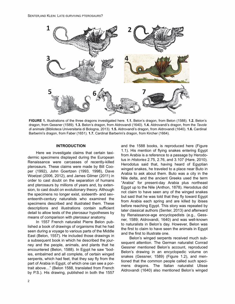
In 1557 French naturalist Pierre Belon pub-lished a book of drawings of organisms that he hadseen during a voyage to various parts of the MiddleEast (Belon, 1557). He included those drawings ina subsequent book in which he described the jour-ney and the people, animals, and plants that heencountered (Belon, 1588). In Egypt he saw “bod-ies, embalmed and all complete, of certain wingedserpents, which had feet, that they say fly from thepart of Arabia in Egypt, of which one can see a por-trait above…” (Belon 1588, translated from Frenchby P.S.). His drawing, published in both the 1557
and the 1588 books, is reproduced here (Figure1.1). His mention of flying snakes entering Egyptfrom Arabia is a reference to a passage by Herodo-tus in Histories 2.75, 2.76, and 3.107 (Hare, 2010).Herodotus said that, having heard of Egyptianwinged snakes, he traveled to a place near Buto inArabia to ask about them. Buto was a city in theNile delta, and the ancient Greeks used the term“Arabia” for present-day Arabia plus northeastEgypt up to the Nile (Anthon, 1878). Herodotus didnot claim to have seen any of the winged snakesbut said that he was told that they fly toward Egyptfrom Arabia each spring and are killed by ibisesbefore reaching Egypt. This story was repeated bylater classical authors (Senter, 2013) and afterwardby Renaissance-age encyclopedists (e.g., Gess-ner, 1589; Aldrovandi, 1640) and was well-knownto naturalists in Belon’s day. However, Belon wasthe first to claim to have seen the animals in Egyptand the first to illustrate one.
Belon’s winged serpents received much sub-sequent attention. The German naturalist ConradGessner mentioned Belon’s account, reproducedBelon’s drawing in an encyclopedic volume onsnakes (Gessner, 1589) (Figure 1.2), and men-tioned that the common people called such speci-mens dragons. The Italian naturalist UlisseAldrovandi (1640) also mentioned Belon’s winged
FIGURE 1. Illustrations of the three dragons investigated here. 1.1. Belon’s dragon, from Belon (1588). 1.2. Belon’sdragon, from Gessner (1589). 1.3. Belon’s dragon, from Aldrovandi (1640). 1.4. Aldrovandi’s dragon, from the Tavoledi animale (Biblioteca Universitaria di Bologna, 2013). 1.5. Aldrovandi’s dragon, from Aldrovandi (1640). 1.6. CardinalBarberini’s dragon, from Faber (1651). 1.7. Cardinal Barberini’s dragon, from Kircher (1664).
2

PALAEO-ELECTRONICA.ORG
serpents and included an illustration of one in asection on winged dragons in his posthumousencyclopedia of snakes, Serpentum et DraconumHistoriae (Figure 1.3). The English author EdwardTopsell copied Gessner’s copy of Belon’s picture inhis own encyclopedic volume on snakes, but didnot mention Belon (Topsell, 1608).
Cooper (1992) reproduced Topsell’s copy ofBelon’s drawing in an article in Creation Ex NihiloTechnical Journal and cited it as an example of adrawing of a dinosaur. Unaware that the originaleyewitness (Belon) had seen the animals up close,in his figure caption he asked, “Could the ratherridiculous looking wings of Topsell’s monster havecome about through the original eyewitnessaccount having mistaken from a distance the mark-ings of armour-plating for wings?...The wingsapart, note the woodcut’s surprising similarity to amodern reconstruction of a Tyrannosaurus” (Coo-per, 1992). Goertzen corrected Cooper in a letter tothe editor in the following issue of the journal, iden-tifying the picture as a copy of Belon’s illustration ofa winged specimen that Belon had observed, andimplying that it was a pterosaur (Goertzen, 1993).In a subsequent publication, Goertzen identifiedBelon’s winged animal as the pterosaur Dimorpho-don macronyx (Goertzen, 1998). Woetzel (2006)also claimed that Belon’s winged serpents werepterosaurs.
Gilmer (2011) later expressed a unique viewon Belon’s flying serpents. He reproduced Aldro-vandi’s (1640) copy of Belon’s winged dragon illus-tration and called it a “land dinosaur”. In his view,the appendages that appeared to be wings wereactually a frill that was spread for defensive oraggressive display, as in the frilled lizard (Chlam-ydosaurus kingii), or were appendages that were“moved like wings to lighten the creature’s body forfaster running.”
Aldrovandi had his illustrators produce aseries of paintings of which line-drawing copieswere included in his books. This series of paint-ings, which is now called the Tavole di animali,remains unpublished in print, but photos of theentire collection are now posted online (BibliotecaUniversitaria di Bologna, 2013). Painting 140 ofvolume 4 of the Tavole (Figure 1.4) is of a speci-men that Aldrovandi described, with an accompa-nying drawing (Figure 1.5), in Serpentum etDraconum Historiae (Aldrovandi, 1640). Accordingto Aldrovandi’s description, “in the year of our Lord1600, a true mummified African [aethiopicus: in ref-erence to lands south of Egypt] dragon was givenas gift by Francisco Centensis to the most illustri-
ous Ulisse Aldrovandi…it had, moreover, fiveprominent and conspicuous protuberances on itsback, which were lacking in the dragon of Belon; italso had two feet armed with claws, and wasdepicted with small ears. The entire body was dec-orated with green and blackish scales. It bore twowings suitable for flying, and a long and flexible tail,straight with dull yellow scales, such as were visi-ble on the stomach and throat. The mouth wasarmed with sharp teeth. The lower part of the headwas flat next to the small ears. The pupils of theeyes were black with a pale yellow circle. Finally,the two nostrils were visible and open” (Aldrovandi,1640, translated from Latin by D.K.).
Aldrovandi insisted that his illustrators illus-trate specimens directly, depicting what they saw infront of them so as to ensure accuracy rather thantaking artistic liberties (Olmi, 2007). The illustrationof the specimen that Centensis gave him is there-fore likely painted directly from the original speci-men. Aldrovandi’s published drawing (Figure 1.3)of Belon’s specimen bears suspicious resem-blance to that of the specimen from Centensis (Fig-ure 1.5). This can probably be explained byAldrovandi’s insistence on depicting actual speci-mens. Because Belon did not purchase a wingedserpent specimen (or, at least, did not record hav-ing done so) but simply illustrated one, the illustra-tor did not have a specimen from Belon toillustrate. In order to draw directly from a specimen,therefore, the illustrator had little choice but toredraw the specimen from Centensis and modifythe drawing to match Belon’s illustration. It appearsthat Aldrovandi’s drawing of Belon’s specimen isreally of the Centensis specimen with the five dor-sal lumps omitted and with the tail configured tomatch Belon’s illustration.
Neither Cooper, Goertzen, Gilmer, nor Woet-zel specifically mentioned Aldrovandi’s specimen.However, it was included by Aldrovandi in a chap-ter on winged dragons, which Woetzel (2006) men-tioned. Woetzel claimed that the winged animals towhich Aldrovandi referred were pterosaurs.
The third alleged pterosaur to be consideredhere is a specimen owned by Cardinal FrancescoBarberini (1597 – 1679), nephew of Pope UrbanVIII (reigned 1623 – 1644). The specimen, whichBarberini considered a dragon, was a gift from KingLouis XIII of France (Bartholin, 1678). Barberiniwas a patron of certain members of the LynceanAcademy, a scientific association that producedseveral noteworthy monographs (Freedberg,2002). One of these was the multi-authored RerumMedicarum Novae Hispaniae Thesaurus (Medici-
3

SENTER,AND KLEIN: LATE-SURVIVING PTEROSAURS?
nal Treasures of New Spain), on the natural historyof Mexico (Lyncean Academy, 1651). Under politi-cal pressure (Freedberg, 2002), Lyncean anato-mist Giovanni Faber added a description ofBarberini’s dragon to Tesoro Messicano (MexicanTreasures), a section in Rerum Medicarum NovaeHispaniae Thesaurus. An English translation byD.K. of Faber’s Latin description of the specimen isgiven here in Appendix.
Faber’s description of Cardinal Barberini’sdragon included a detailed drawing of the speci-men (Figure 1.6). The German Jesuit scholar Atha-nasius Kircher (1601/2 – 1680) published anotherdrawing of the specimen in the dragon section ofhis 1664 book Mundus Subterraneus (Subterra-nean World), a classic of geological literature(Kircher, 1664) (Figure 1.7).
Woetzel (2012) included Faber’s drawing(misidentified as Kircher’s) of Cardinal Barberini’sdragon in a list of several Renaissance-age illustra-tions that he claimed were depictions of ptero-saurs. He claimed specifically that it “resemblesthe rhamphorhynchoid pterosaurs” and used it tosupport the idea that live pterosaurs roamedEurope in recent centuries (Woetzel, 2012).
The illustrations and descriptions of the threespecimens by Belon, Aldrovandi, and Faber aredetailed enough to test the hypothesis that they arepterosaurs. Here, we compare the anatomy ofthese specimens with pterosaur anatomy to testthis hypothesis. We also advance the alternativehypothesis that the three specimens are taxidermiccomposites and use observations of the anatomyof extant animals to test various hypotheses as towhich extant species are represented in the vari-ous parts of the three specimens.
COMPARISON WITH PTEROSAURS
In all three dragons, wing anatomy is very dif-ferent from that of pterosaurs on several counts.Firstly, in each of the three dragons the wing isshorter than the torso, whereas in a pterosaur thewing is much longer than the torso, usually four ormore times the length of the torso (Figure 2). Sec-ondly, Belon’s dragon has six internal struts thatradiate from the shoulder (Figure 1.1), Aldrovandi’shas five (Figure 1.4), and according to Faber’sdescription Bartholin’s dragon has three (Appen-dix). In contrast, pterosaur wings have only one
FIGURE 2. Skeletons of pterosaurs. 2.1. AMNH cast of Pterodactylus antiquus. 2.2. Pteranodon longiceps, AMNH(American Museum of Natural History, New York City, New York) 6158. 2.3. AMNH cast of Rhamphorhynchus muen-steri. 2.4. AMNH cast of Campylognathoides zitteli. 2.5. Vertebrae of the long-tailed pterosaur Rhamphorhychusmuensteri in right lateral view, modified from Wellnhofer (1975); left: series of caudal vertebrae, showing the networkof elongate dorsal and ventral processes that stiffens the tail; right: a single caudal vertebra and hemal arch, showingthe elongate dorsal and ventral processes that contribute to the network.
4

PALAEO-ELECTRONICA.ORG
internal strut, the greatly elongated fourth finger,which is situated along the leading edge of thewing (Figure 2). Thirdly, the wings of the threedragons have scalloped edges, as in a bat (Figure1), whereas pterosaur wings do not (Wellnhofer,1991; Unwin, 2006) (Figure 2.3). Fourthly, thewings of Belon’s and Aldrovandi’s dragons have noprotruding fingers or claws (Figure 1.1-5), and Bar-berini’s has two miniscule claws (Figure 1.6-7),whereas a pterosaur wing has three complete,clawed fingers that protrude from the leading edgeof the wing (Wellnhofer, 1991; Unwin, 2006) (Fig-ure 2.1-2). Fourthly, the wings of the three dragonsare narrow at the base and expand in width distally(Figure 1), whereas pterosaur wings are wide atthe base and taper to a point distally (Wellnhofer,1991; Unwin, 2006) (Figure 2.3). In addition, thewings of Belon’s and Aldrovandi’s dragons origi-nate from the same spot as the root of the “leg”(Figure 1.1-5) whereas in a pterosaur the shoulderand hip are widely separated (Wellnhofer, 1991;Unwin, 2006) (Figure 2.1-2, 2.4). In Barberini’sdragon the root of the wing is posterior to that ofthe “leg” and posterior to the ribcage (Figure 1.6-7),whereas in pterosaurs the shoulder is at the ante-rior end of the ribcage, far anterior to the hip (Well-nhofer, 1991; Unwin, 2006) (Figure 2.1-2, 2.4).
The heads of the three dragons are differentfrom those of pterosaurs. All three have protrudingear flaps (Figure 1). Numerous pterosaur speci-mens with soft-tissue impressions are known, andnone have ear flaps (Wellnhofer, 1991; Unwin,2006). The mandible of Barberini’s dragon has ahigh, prominent coronoid process (Figure 1.5-6). Acoronoid process is absent in most pterosaurs(Wellnhofer, 1991; Unwin, 2006) and is small andlow in the few pterosaurs that have it (Dalla Vec-chia, 2003). Barberini’s dragon also has a hetero-dont dentition in which teeth are differentiated intoincisors, canines, and molariform cheek teeth(Appendix), a situation that is not present in anypterosaur (Wellnhofer, 1991; Unwin, 2006).
The tails of the three dragons are differentfrom those of pterosaurs. All three specimens havelong, curled tails (Figure 1), whereas in long-tailedpterosaurs the tail is prevented from bending by anetwork of thin, bony struts (Wellnhofer, 1991;Unwin, 2006) (Figure 2.5). Such a network is con-spicuously absent in the tail skeleton of CardinalBarberini’s dragon, the only one of the three drag-ons whose tail skeleton is exposed (Figure 1.6-7).Also, the tail vertebrae of Barberini’s dragon areshort relative to their height (Figure 1.6-7),
whereas those of long-tailed pterosaurs are muchlonger than tall (Figure 2.5).
The “legs” of the three dragons are unlikethose of pterosaurs. Their “knees” are bent in thewrong direction, revealing that they are actuallyelbows. These three dragons therefore do not havehindlimbs but instead have forelimbs, and theirforelimbs are not incorporated into the wings.Pterosaurs, in contrast, do have hindlimbs, andtheir wings are forelimbs with patagia (flight mem-branes).
The illustrations show that the skin of Belon’sand Aldrovandi’s dragons are covered in scales(Figure 1.1-5), as is the skin of Barberini’s dragonaccording to Faber’s description (Appendix). Incontrast, pterosaur skin is not scaly but is coveredin short, hairlike filaments (Wellnhofer, 1991;Unwin, 2006).
None of the three dragons is a pterosaur.Their wings, heads, tails, limbs, and skin are incon-sistent with pterosaur anatomy.
COMPARISON OF BELON’S AND ALDROVANDI’S DRAGONS WITH
EXTANT ANIMALS
It is important to do more than just demon-strate that the three dragons are not pterosaurs.The countering of incorrect claims regarding spe-cific specimens carries more weight if we candetermine what the specimens are. We thereforecompared the three specimens to extant animals todetermine, as much as possible, what extant spe-cies comprised them.
Belon’s and Aldrovandi’s dragons are similarenough to support an inference that they were con-structed from the same set of animal parts. Theycan therefore be considered together. They mayeven have come from the same region and havebeen made by the same hoaxer or group of hoax-ers. Belon saw his dragons in Egypt, and Aldro-vandi’s was said to come from “Aethiopia”.
On their bellies are transversely elongatedventral scutes (Figure 1.1-4), a feature present insnake skin and absent in the skin of any other ani-mal. The scale rows are diagonal, which is consis-tent with snake skin (Figure 3). The tails of thedragons also have transversely elongated ventralscutes and have diagonal scale rows (Figure 3.1-2). In contrast, the scale rows on the tails of mostlizards other than skinks are vertical, not diagonal(Figure 3.7). We therefore conclude that the neck,torso, and tail of each of the two dragons is that ofa snake, to which parts of other animals wereattached.
5

SENTER,AND KLEIN: LATE-SURVIVING PTEROSAURS?
Aldrovandi described his winged dragon ashaving green and blackish scales and a yellowbelly and tail. His painting shows that the scaleswere mainly green. Because Egypt does not havebright green snakes, the species of snake mayhave come from further south. Dendroaspis james-oni (Jameson’s mamba) is a long (sometimes over2 m) snake with black-edged green scales, a yel-lowish belly, and a yellow tail. Its color matchesAldrovandi’s description, but the closest its rangecurrently comes to Egypt is southernmost Sudan(Spawls and Branch, 1995). We therefore are not
confident that we can identify the species of snakeused.
Both specimens have bulbous torsos, which isnot the case in snakes. However, an objectinserted down the throat into the digestive tract of asnake can distend the torso without tearing theskin, as in these specimens (Figure 3.8). The dif-ference in shape between the two specimens’ tor-sos can therefore be explained by a difference inshape between inserted items.
Several lines of evidence reveal that in eachspecimen the head of the snake has been replaced
FIGURE 3. Comparison of the skin of the three dragons to that of snakes and lizards. 3.1. Belon’s dragon, scales oftorso (above) and tail (below, with original cross-hatching removed from one scale row for clarity), from Belon (1588).3.2. Aldrovandi’s dragon, skin of neck (above) and tail (below, rotated clockwise relative to its position in Aldrovandi’soriginal illustration), from Aldrovandi (1640). 3.3. Torso skin of Cardinal Barberini’s dragon, from Kircher (1664). 3.4.Agkistrodon piscivorus (cottonmouth), live specimen at Reptile Lagoon (Hamer, South Carolina), showing the diago-nal scale rows that are present both on the torso and the tail in snakes; note that the scale rows are diagonal on bothtorso and tail in Belon’s and Aldrovandi’s dragons. 3.5. Dendroaspis viridis (western green mamba), live specimen atRiverbanks Zoo (Columbia, South Carolina), showing diagonal torso and tail scale rows, and showing the trans-versely elongated ventral scutes that are unique to snakes; note that the three dragons also have ventral scutes ofthis type. 3.6. Torso of Gerrhosaurus validus (giant plated lizard), live specimen at United States National Zoo (Wash-ington, D.C.), showing that the scale rows of the torso are vertical, as in many lizards; note that the scale rows arealso vertical on the torso of Cardinal Barberini’s dragon. 3.7. Tail of Cyclura lewisi (blue iguana), live specimen atUnited States National Zoo, showing that the scale rows are vertical, as in the tails of most lizards; note that the scalerows are not vertical on the tails of Belon’s and Aldrovandi’s dragons. 3.8. Juvenile Python sebae (African rockpython) after a large meal, drawn from a photo by P.S., showing that in a snake the torso skin distends without tearingwhen a large object is in the digestive tract; note the similarity with the bulbous torsos of Belon’s and Aldrovandi’sdragons in Figure 1.
6

PALAEO-ELECTRONICA.ORG
by the head of some other animal. First, the snoutof a snake does not protrude as far anterior to theeye as is the case with these specimens’ snouts(Figure 4). Second, the tongue of Aldrovandi’sdragon, the only one of the two specimens in whichthe tongue is illustrated, is not forked (Figure 4.2).Third, in both specimens the dorsal skin of thesnout is wrinkled (Figure 4.1-2), which does nothappen in snakes. The wrinkling of facial skin issomething that happens only in mammals, many ofwhich have loose facial skin that permits changesof facial expression, whereas in non-mammalianvertebrates the skin is firmly and immovablyattached to the skull. Both specimens also havepinnae (ear flaps), a feature unique to mammals.Also, both specimens have fleshy cheeks ventral tothe eye, another trait unique to mammals. In bothspecimens the cheeks and the tops of the heads
are covered in scales. It is therefore probable thatin each case the neck skin of the snake wasextended onto and attached to the mammalianheads and cheeks.
In both dragons the shape of the head is dog-like, but the dentition lacks the distinct upper andlower canine teeth that are present in canids, aswell as the molariform cheek teeth of canids.Instead, the teeth are all pointed and of similarlength, with a slight increase in tooth length anteri-orly in Aldrovandi’s painting (Figure 4.2). The onlyEgyptian mammal with a doglike head and amouthful of pointed teeth with a gradual increase intooth size anteriorly is the Egyptian fruit bat (Rou-settus aegyptiacus) (Aulagnier et al., 2009) (Figure4.3). Its post-canine teeth have sharp and procum-bent points so that at a glance its dentition lookslike a series of fangs that increase in size anteri-
FIGURE 4. Heads of Belon’s and Aldrovandi’s dragons compared with those of African fruit bats and with those ofsnakes. 4.1. Belon’s dragon. 4.2. Aldrovandi’s dragon. 4.3. left to right: Rousettus aegyptiacus (Egyptian fruit bat),USNM (United States National Museum, Washington, D.C.) 114691; Eidolon helvum (straw-colored fruit bat) , USNM378787. 4.4. Snakes (live specimens at Reptile Lagoon, Hamer, South Carolina), showing that the eye is closer to thetip of the snout than in Belon’s and Aldrovandi’s dragons; left to right: Python molurus (Asian rock python), Cerastescerastes (Sahara horned viper), Naja haje (Egyptian cobra).
7
SENTER,AND KLEIN: LATE-SURVIVING PTEROSAURS?
orly; in this it resembles the dentition in the lowerjaw of the painting of Aldrovandi’s dragon. Its earsare also smaller in relative size than those of otherMiddle Eastern bats, making it a good candidatefor the contributor of the heads of the two dragons.All the above points also hold true for the straw-col-ored fruit bat (Eidolon helvum), which is foundsouth of Egypt along the Red Sea. Also, the size ofthe head of an adult Egyptian fruit bat is a good fitfor the neck of a large snake such as a mamba.Although the tooth counts of fruit bats are lowerthan that shown in the illustrations of Belon’s andAldrovandi’s dragons, the overall resemblance is
better than that of the head of any other northernAfrican mammal.
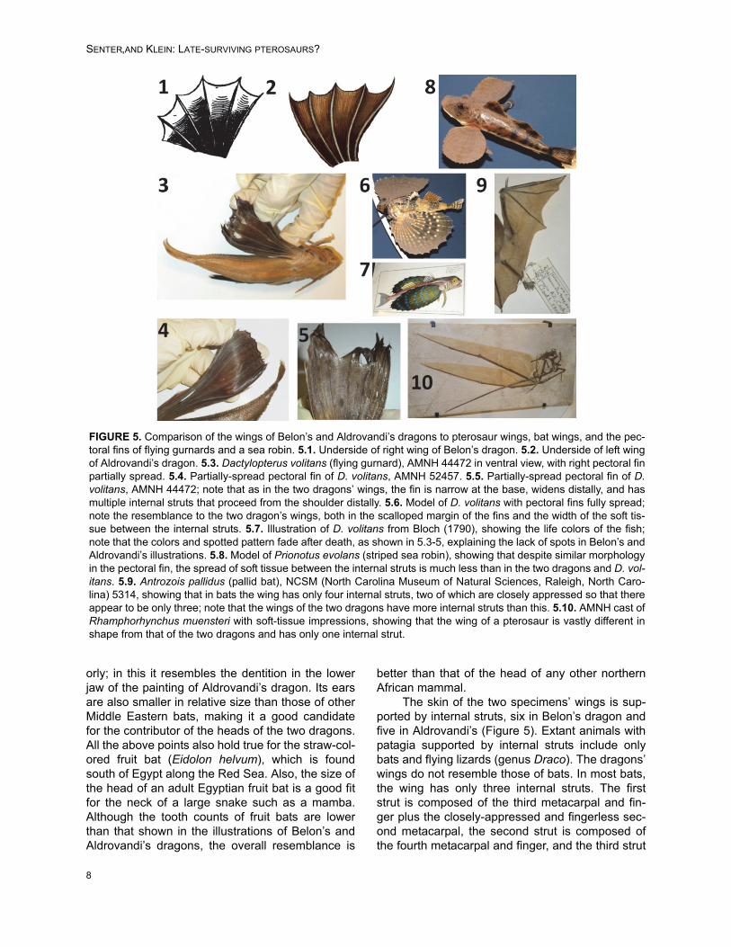
The skin of the two specimens’ wings is sup-ported by internal struts, six in Belon’s dragon andfive in Aldrovandi’s (Figure 5). Extant animals withpatagia supported by internal struts include onlybats and flying lizards (genus Draco). The dragons’wings do not resemble those of bats. In most bats,the wing has only three internal struts. The firststrut is composed of the third metacarpal and fin-ger plus the closely-appressed and fingerless sec-ond metacarpal, the second strut is composed ofthe fourth metacarpal and finger, and the third strut
FIGURE 5. Comparison of the wings of Belon’s and Aldrovandi’s dragons to pterosaur wings, bat wings, and the pec-toral fins of flying gurnards and a sea robin. 5.1. Underside of right wing of Belon’s dragon. 5.2. Underside of left wingof Aldrovandi’s dragon. 5.3. Dactylopterus volitans (flying gurnard), AMNH 44472 in ventral view, with right pectoral finpartially spread. 5.4. Partially-spread pectoral fin of D. volitans, AMNH 52457. 5.5. Partially-spread pectoral fin of D.volitans, AMNH 44472; note that as in the two dragons’ wings, the fin is narrow at the base, widens distally, and hasmultiple internal struts that proceed from the shoulder distally. 5.6. Model of D. volitans with pectoral fins fully spread;note the resemblance to the two dragon’s wings, both in the scalloped margin of the fins and the width of the soft tis-sue between the internal struts. 5.7. Illustration of D. volitans from Bloch (1790), showing the life colors of the fish;note that the colors and spotted pattern fade after death, as shown in 5.3-5, explaining the lack of spots in Belon’s andAldrovandi’s illustrations. 5.8. Model of Prionotus evolans (striped sea robin), showing that despite similar morphologyin the pectoral fin, the spread of soft tissue between the internal struts is much less than in the two dragons and D. vol-itans. 5.9. Antrozois pallidus (pallid bat), NCSM (North Carolina Museum of Natural Sciences, Raleigh, North Caro-lina) 5314, showing that in bats the wing has only four internal struts, two of which are closely appressed so that thereappear to be only three; note that the wings of the two dragons have more internal struts than this. 5.10. AMNH cast ofRhamphorhynchus muensteri with soft-tissue impressions, showing that the wing of a pterosaur is vastly different inshape from that of the two dragons and has only one internal strut.
8

PALAEO-ELECTRONICA.ORG
is composed of the fifth metacarpal and finger (Fig-ure 5.9). In some bats the second finger is retainedand is separated from the third so that it formsanother strut, but even in such cases there are onlyfour struts. Furthermore, in bats the first finger pro-trudes from the leading edge of the wing, a featureabsent in the two dragons.
The dragons’ wings are also unlike the pata-gia of flying lizards. In the two dragons, the wing isnarrow at the base and wide distally, whereas thepatagium of a flying lizard is wider at the base thanit is distally.
The Mediterranean fish known as the flyinggurnard (Dactylopterus volitans) has pectoral finswith morphology like that of these specimens’wings (Figure 5.3-5). There are multiple internalstruts, the skin can spread as wide between thestruts as in the wings of the two dragons, and thefins can be cut into the shapes shown in Belon’sand Aldrovandi’s illustrations. The pectoral fins of alive flying gurnard have brightly colored spots (Fig-ure 5.7), which are absent in Belon’s and Aldro-vandi’s dragons, but the spots fade after death,leaving the entire fin a dull brown (Figure 5.3-5).
Sea robins (Triglidae) have spreadable pecto-ral fins of similar shape (Figure 5.8), but they aretoo small to have been the source of these twospecimens’ wings, and the fins cannot spread aswidely between the struts as in Belon’s and Aldro-vandi’s dragons (Figure 5.8). The pectoral fins ofother bony fishes lack the wide membranesbetween internal struts that are found in the flyinggurnard and sea robins. We therefore concludethat the “wings” of Belon’s and Aldrovandi’s drag-ons were made from the pectoral fins of flying gur-nards.
The limbs of the two specimens are robust,and each terminates in four thick fingers with prom-inent, curved claws (Figure 1.1-4). Such forelimbsare not found in any circum-Mediterranean reptiles.Local lizards and crocodiles with robust forelimbshave five-fingered hands, and amphibians lackmanual claws. Among mammals, robust forelimbsthat terminate in four thick fingers are found inmembers of the Leporidae (the rabbit family), Cani-dae (the dog family), and Felidae (the cat family).The former two have fingers that are straighterthan those of felids and therefore better resemblethe fingers of the two dragons.
The scaly covering of the limbs of the twospecimens indicates that they were covered in asleeve of reptile skin. Rabbits, canids, and felidshave a dewclaw (a short thumb that does not reachthe tip of the forelimb), but its absence in the illus-
trations can be explained by its having beenclipped off, with the reptile-skin sleeve covering thespot where it was removed. It is noteworthy thatthe illustrations do not show scales or fur coveringthe fingers. This suggests that the original mammalskin had been removed and that the reptile-skinsleeve did not extend onto the fingers.
We conclude that Belon’s and Aldrovandi’swinged dragons were taxidermic composites. Eachwas composed of a decapitated snake to whichwas added a fruit bat’s head, the pectoral fins of aflying gurnard, the forelimbs of a rabbit or canid,and an internal insert that distended the torso.
COMPARISON OF CARDINAL BARBERINI’S DRAGON WITH EXTANT ANIMALS
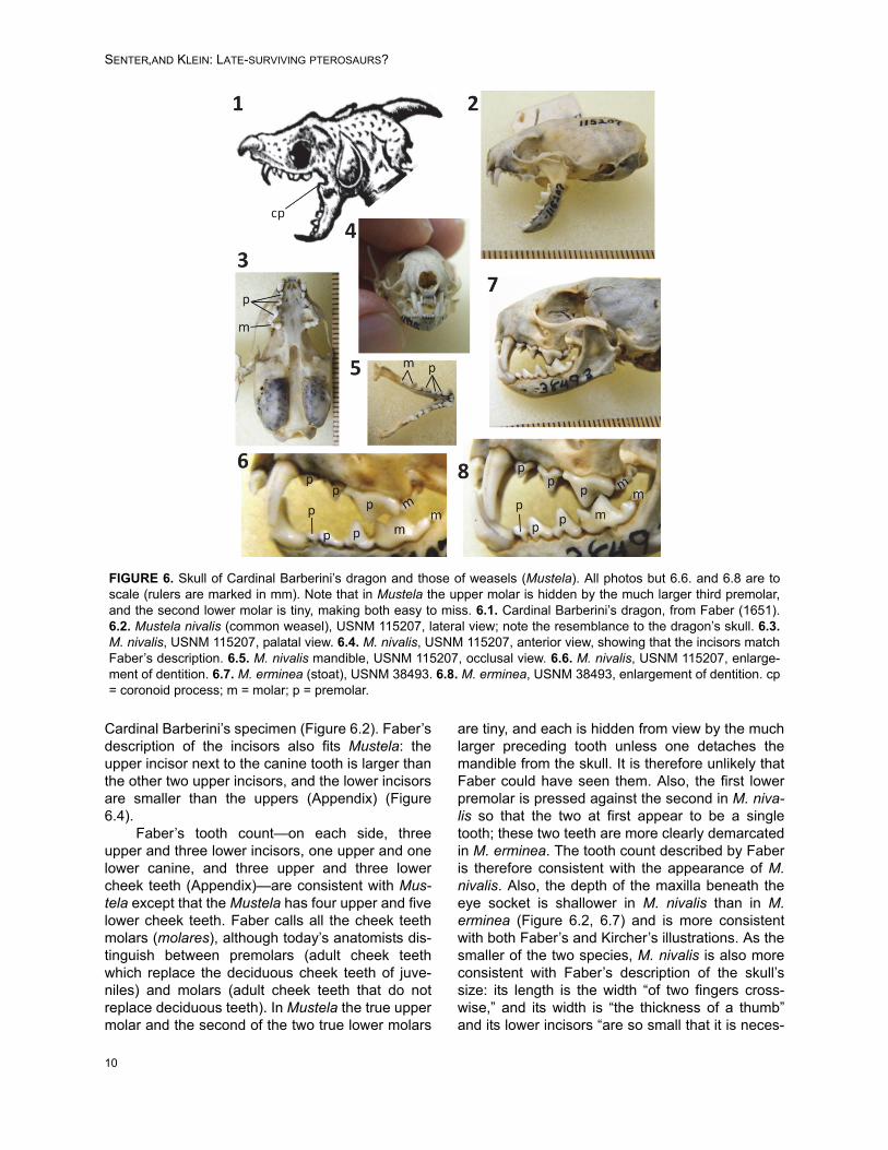
Faber’s and Kircher’s illustrations of CardinalBarberini’s dragon show a clear distinctionbetween canine teeth and molariform cheek teeth,a mammalian trait. They also show a coronoid pro-cess on the mandible, another mammalian trait(Figure 1). The dragon’s single, posteriorly curvinghorn and hooklike, beaked proboscis are absent inall mammals but are known to have been added toa mammalian skull on at least one other fakedragon from the seventeenth century (Senter andWilkins, 2013). It is therefore safe to dismiss theseas fake additions to the skull of Cardinal Barberini’sdragon and not part of the original skull.
Details of the skull and dentition differbetween Faber’s and Kircher’s drawings. The for-mer is detailed and precise in its depiction of theskull and neck, whereas the skull and neck of thelatter are more sloppily and less realistically ren-dered. For example, it includes teeth on the coro-noid process (Figure 1.7), a feature present in noknown extant or extinct tetrapod. We therefore con-sider the skull and neck in Faber’s drawing morereliable than those in Kircher’s. In Faber’s drawing,the prominence of the canine teeth, the generalsize and shape of the cheek teeth, the bulbousshape of the rear of the skull, and the fact that themandible is only about half the total length of theskull, are consistent with only one mammaliangenus: Mustela (weasels) (Aulagnier et al., 2009)(Figure 6). The lower incisors are hidden by thecanines in lateral view, which is consistent with theMustelidae (Aulagnier et al., 2009). The two small-est circum-Mediterranean species of Mustela areM. erminea (stoat) and M. nivalis (common wea-sel). No other western Eurasian species of Mustelais small enough to match the size of the specimenas described by Faber (Appendix). In both cases,the maximum gape of the jaw matches the gape of
9
SENTER,AND KLEIN: LATE-SURVIVING PTEROSAURS?
Cardinal Barberini’s specimen (Figure 6.2). Faber’sdescription of the incisors also fits Mustela: theupper incisor next to the canine tooth is larger thanthe other two upper incisors, and the lower incisorsare smaller than the uppers (Appendix) (Figure6.4).
Faber’s tooth count—on each side, threeupper and three lower incisors, one upper and onelower canine, and three upper and three lowercheek teeth (Appendix)—are consistent with Mus-tela except that the Mustela has four upper and fivelower cheek teeth. Faber calls all the cheek teethmolars (molares), although today’s anatomists dis-tinguish between premolars (adult cheek teethwhich replace the deciduous cheek teeth of juve-niles) and molars (adult cheek teeth that do notreplace deciduous teeth). In Mustela the true uppermolar and the second of the two true lower molars
are tiny, and each is hidden from view by the muchlarger preceding tooth unless one detaches themandible from the skull. It is therefore unlikely thatFaber could have seen them. Also, the first lowerpremolar is pressed against the second in M. niva-lis so that the two at first appear to be a singletooth; these two teeth are more clearly demarcatedin M. erminea. The tooth count described by Faberis therefore consistent with the appearance of M.nivalis. Also, the depth of the maxilla beneath theeye socket is shallower in M. nivalis than in M.erminea (Figure 6.2, 6.7) and is more consistentwith both Faber’s and Kircher’s illustrations. As thesmaller of the two species, M. nivalis is also moreconsistent with Faber’s description of the skull’ssize: its length is the width “of two fingers cross-wise,” and its width is “the thickness of a thumb”and its lower incisors “are so small that it is neces-
FIGURE 6. Skull of Cardinal Barberini’s dragon and those of weasels (Mustela). All photos but 6.6. and 6.8 are toscale (rulers are marked in mm). Note that in Mustela the upper molar is hidden by the much larger third premolar,and the second lower molar is tiny, making both easy to miss. 6.1. Cardinal Barberini’s dragon, from Faber (1651).6.2. Mustela nivalis (common weasel), USNM 115207, lateral view; note the resemblance to the dragon’s skull. 6.3.M. nivalis, USNM 115207, palatal view. 6.4. M. nivalis, USNM 115207, anterior view, showing that the incisors matchFaber’s description. 6.5. M. nivalis mandible, USNM 115207, occlusal view. 6.6. M. nivalis, USNM 115207, enlarge-ment of dentition. 6.7. M. erminea (stoat), USNM 38493. 6.8. M. erminea, USNM 38493, enlargement of dentition. cp= coronoid process; m = molar; p = premolar.
10

PALAEO-ELECTRONICA.ORG
sary to view them with a magnifying lens, if onewishes to discern them” (Appendix). We thereforeconclude that the skull of Cardinal Barberini’sdragon is that of M. nivalis.
The skin of Cardinal Barberini’s dragon hastransversely elongated ventral scutes, as insnakes. However, Kircher’s illustration shows thatthe rest of its scales are in transverse rows that arevertical in lateral view (as in some lizards) ratherthan in diagonal rows (as in snakes). It is thereforelikely that the specimen’s belly skin is that of asnake, while its dorsal and lateral skin is that of alizard. A similar situation was present in anothercomposite “dragon” hoax from RenaissanceEurope, in which the torso had the belly skin of asnake but the dorsal and lateral skin of a fish(Senter et al., 2013).
The color of Cardinal Barberini’s dragon’sskin, according to Faber (Appendix), is a mixture ofaquamarine, yellowish, and blackish, with moregreen dorsally and more yellow ventrally. Severallizard species of the appropriate color are presentin the circum-Mediterranean area, several of whichare in the genus Lacerta (Boulos, 1995; Schleich etal., 1996; Arnold, 2002; Baran et al., 2005; Baha elDin, 2006; Bar and Haimovitch, 2011). The skincovering the neck may be from the same individualas that covering the torso or it may be from a sec-ond individual. The range of sizes among appropri-ately colored, circum-Mediterranean species ofLacerta is consistent with the use of skin from asingle individual or two.
According to Faber, the wings of Cardinal Bar-berini’s dragon each have three internal struts, andare spotted with a background color of blue on oneside and a dark wheatlike color on the other. BothFaber’s and Kircher’s illustrations show a coveringof scales (Figure 7.1-2). Two small points thatFaber calls claws project anteriorly from each wing.The scaly covering eliminates bony fish fins andbat wings from consideration, because neither iscovered in scales. The patagia of flying lizards(Draco) are covered in scales, have ribs as internalstruts, can be cut into the shape of the wings of thedragon, and in several species of Draco are spot-ted (Das, 2010) (Figure 7.3-7). To test whether thetwo anterior “claws” could be the projecting tips ofsliced ribs of a Draco patagium, P.S. cut the rightpatagium off a dried specimen in his personal col-lection and found that even at the base of the pata-gium no two ribs converge closely enough to matchthe proximity of the claws on the dragon’s wing(Figure 7.4). To test whether the claws could bepieces of the pectoral girdle, P.S. cut the left pata-
gium of the specimen close enough to include partof the body wall (Figure 7.5), and found that no partof the pectoral girdle extended into the cut-offpiece. The pectoral girdle is slightly anterior to theanteriormost edge of the patagium where it meetsthe torso (Figure 7.5). No projecting pieces werepresent that could be identified as the “claws” ofthe dragon (Figure 7.6). We therefore could notconfirm that the wings of Cardinal Barberini’sdragon were made from cut Draco patagia.
Trade with Australasia had been establishedby Faber’s day, so P.S. examined specimens ofChlamydosaurus kingii (frilled lizard) to determinewhether the dragon’s wings could have been madeof frilled lizard frills (Figure 7.8-11). Some speci-mens have spots on the frill (Figure 7.9), as in Car-dinal Barberini’s dragon. The edge of the dorsalhalf of the frill has small, triangular projections thatare similar to the dragon’s “claws” (Figure 7.8-10),so the possibility that all but two of those projec-tions had been cut off a frill to form each of thedragon’s wings was deemed worthy of exploration.However, the frill has no internal struts. It doeshave three strong creases in its ventral half (Figure7.8, 7.11), but when the frill is spread the lack ofstruts in the creases is apparent. Furthermore, theventral half of the frill, which has the creases, lacksthe triangular projections around the edge that oth-erwise could hypothetically have been the “claws”.We therefore conclude that the wings of CardinalBarberini’s dragon were not made from frilled lizardfrills. We remain baffled as to the identity of thewings.
The limbs of Cardinal Barberini’s dragon ter-minate in five slender, well-defined fingers (Figure8.1). According to Faber’s description, each fingerhas a sharp claw, and the finger in the position ofthe thumb is shorter than the others. The presenceof five fingers with sharp claws eliminates manyanimals from consideration, but it is difficult to elim-inate any of the remaining candidates. Theyinclude weasels (Figure 8.2-4) and numerous spe-cies of shrews and lizards. It is possible that thehoaxer used the forelimbs of the same weasel fromwhich the skull came. The size of the hands ofMustela nivalis relative to its head (Figure 8.2) issimilar to that of the dragon (Figure 1.6-7). If aweasel’s hands were used, they were skinned;there is no indication of fur on the hands in theillustrations of the dragon. It is also possible thatthe hoaxer used the forelimbs of the same lizardfrom which the skin came. Digital proportions in thehands of Lacerta (Figure 8.5) are similar to those ofCardinal Barberini’s dragon.
11

SENTER,AND KLEIN: LATE-SURVIVING PTEROSAURS?
12
FIGURE 7. Wings of Cardinal Barberini’s dragon compared with patagium of Draco (flying lizards) and frill of Chlam-ydosaurus kingii (frilled lizard). 7.1. Wings of Cardinal Barberini’s dragon, from Faber (1651). 7.2. Wings of CardinalBarberini’s dragon, from Kircher (1664). 7.3. Draco haematopogon (personal collection of P.S.), showing spots onpatagium. 7.4. Detached right patagium of specimen in 7.3, with yellow arrows showing proximal ends of ribs andblack arrows showing distal tips of ribs. 7.5. Same specimen after detachment of left patagium plus part of the bodywall. 7.6. Internal view of detached left patagium, showing that no clawlike structures are present. 7.7. Draco maxi-mus, AMNH 29976, ventral view, showing spots on patagium. 7.8-11. Chlamydosaurus kingii, showing projections atedge of frill that resemble the claws on Cardinal Barberini’s dragon, showing the three strong creases in the ventralhalf of the frill (7.8, 7.10-11: AMNH 86512), and showing spots on the frill of one specimen (7.9: AMNH 99843).

PALAEO-ELECTRONICA.ORG
The bodies (centra) of the tail vertebrae ofCardinal Barberini’s dragon are of similar lengthand height. In contrast, those of small mammals,reptiles, and amphibians are much longer than tall.The vertebrae of fish tails tend to be of similarlength and height, but the tail vertebrae of sharksand kin (Chondrichthyes) lack the dorsal and ven-tral spines (neural spines and hemal spines,respectively) seen on this specimen’s tail verte-brae. Such spines are the norm in bony fishes. Wetherefore conclude that the tail skeleton is that of abony fish. In Faber’s illustration the neural andhemal spines point anteriorly, whereas in Kircher’sthey point posteriorly. The coiling of the tail evi-dently confused the eye of one of the two illustra-tors. The more realistic of the two is the tail inKircher’s illustration, because posteriorly pointingneural and hemal arches are the norm in verte-brates generally, including bony fishes (Figure 9).
In most bony fishes there is an abruptdecrease in the size of the vertebrae at the base ofthe tail, unlike the gradual decline in vertebral sizedown the length of the tail in Cardinal Barberini’sdragon. Eels are exceptional among bony fishes inthat their vertebrae exhibit a gradual decline in sizedown the length of the tail, as in Cardinal Barber-ini’s dragon. Of the three Mediterranean and Euro-pean species of eel, Anguilla anguilla (Europeaneel) is the best match to the tail of this dragon (Fig-
ure 9.4). The neural and hemal spines of the tailvertebrae of Muraena helena (Mediterraneanmoray) are shaped differently from those of thedragon (Figure 9.8). The neural and hemal spinesof Conger conger (European conger) are relativelymuch longer than in the dragon (Figure 9.6). Wetherefore conclude that the tail skeleton of CardinalBarberini’s dragon is that of Anguilla anguilla.
We conclude that Cardinal Barberini’s dragonwas a taxidermic composite. It includes the skull ofa common weasel; the belly skin of a snake; thedorsal and lateral skin of one or two individual liz-ards, possibly of the genus Lacerta; the tail skele-ton of an eel; and “wings” that remain a mystery.
DISCUSSION
To date, no “evidence” for pterosaur-humancoexistence has survived scrutiny. Claims of sight-ings of live pterosaurs in the United States, Cuba,Africa, and Papua New Guinea (e.g., Gibbons andHovind, 1999; Woetzel, 2006; Stuckwish, 2009;Whitcomb, 2010) are thus far unaccompanied byphotographic evidence or physical specimens. Analleged prehistoric painting of a pterosaur in BlackDragon Canyon, Utah, is actually a composite pic-ture of two humanoid figures, two quadrupedal ani-mals, and a horned serpent (Senter, 2012). Twoalleged rock paintings of pterosaurs near Alton, Illi-
FIGURE 8. Hands of Cardinal Barberini’s dragon compared with those of a common weasel (Mustela nivalis) (USNM115211) and an ocellated lizard (Lacerta lepida) (MCZ [Museum of Comparative Zoology, Cambridge, Massachusetts]R-29871). Thumbs are circled in yellow. 8.1. Cardinal Barberini’s dragon, from Faber (1651). 8.2. M. nivalis, showingthat the size of the hand relative to the head resembles that in Cardinal Barberini’s dragon (see Figure 1.5-6). 8.3. M.nivalis, showing that the thumb is shorter than the other fingers. 8.4. M. nivalis, showing curvature of the finger claws.8.5. L. lepida, showing that the thumb is shorter than the other fingers.
13

SENTER,AND KLEIN: LATE-SURVIVING PTEROSAURS?
nois, are fictions created by a nineteenth-centuryAmerican author, based on now-destroyed rockpaintings of wingless creatures (Senter, 2012).Alleged pterosaurs on Greco-Roman coinage arewinged snakes (Senter, 2013). Herodotus’ descrip-tion of the anatomy of Egyptian winged serpents—alleged by some to be pterosaurs—is inconsistentwith pterosaur anatomy (Senter, 2013). The“dragon” specimen that Cornelius Meyer exhibitedin 1691 is a taxidermic hoax (Senter and Wilkins,2013). The three specimens investigated here arealso taxidermic hoaxes. These three specimens
now join the ranks of investigated and falsified “evi-dence” for human-pterosaur coexistence andagainst evolutionary theory.
Cardinal Barberini’s dragon was a relativelyfamous specimen in its day, and it may have beenthe inspiration for the subsequent hoax perpetratedby Cornelius Meyer in 1691. The latter specimenwas adorned with a backward-curving horn and afake hook on the snout, rare attributes in fake drag-ons that are both present in Cardinal Barberini’sdragon. As in Cardinal Barberini’s dragon, its skull,ribcage, and tail skeleton were exposed, while its
FIGURE 9. Tail of Cardinal Barberini’s dragon compared with the tails of eels. In the photos and x-ray images of eels,including the close-ups, the anteriormost complete vertebra shown is the thirtieth vertebra from the tip of the tail, tofacilitate comparison with Cardinal Barberini’s dragon, on which Faber counted 30 tail vertebrae (Appendix). 9.1. Car-dinal Barberini’s dragon, from Kircher (1664). 9.2. Tail skeleton of Anguilla rostrata (American eel) (NCSM 69208),showing the shortness of the neural and hemal spines in Anguilla, as in Cardinal Barberini’s dragon. 9.3. X-ray imageof Anguilla anguilla (European eel) (MCZ 9244), the one Mediterranean species of Anguilla. 9.4. Close-up of 9.3 withone vertebra outlined, showing that the neural and hemal spines are short relative to centrum height. 9.5. Tail skeletonof Conger oceanicus (American conger) (NCSM 69209), showing the great length of the neural and hemal spines.9.6. Conger conger (European conger) (MCZ 9293), showing that in the one Mediterranean species of Conger, theneural and hemal spines are longer relative to the centrum than they are in Anguilla, and in fact are longer than theheight of the centrum. 9.7. Muraena helena (Mediterranean moray) (MCZ 9293). 9.8. Close-up of 9.7, with one verte-bra outlined, showing that the shapes of the neural and hemal arches are not simple spikes and therefore do notmatch Cardinal Barberini’s dragon. c = centrum; hs = hemal spine; ns = neural spine.
14

PALAEO-ELECTRONICA.ORG
neck was covered in skin, and its tail was coiled.We therefore suggest that it was modeled afterCardinal Barberini’s dragon. Meyer’s dragon wassaid to have been killed in the marshes nearRome. However, it is but another taxidermic com-posite hoax, in this case one with the skull of adomestic dog and the forelimbs of a bear (Senterand Wilkins, 2013).
It is noteworthy that Meyer’s hoax and thethree hoaxes investigated here continue to foolpeople over 300 years later. In all three casesauthors writing in the 1990s or the twenty-first cen-tury have insisted that the specimens were genu-ine, recently-killed pterosaurs (Goertzen, 1993,1998; Woetzel, 2006, 2012) or dinosaurs (Cooper,1992; Gilmer 2011). These authors are in goodcompany. A plethora of other dragons created bytaxidermic hoaxers in Renaissance Europe hood-winked many prominent citizens and, in somecases, scientists—although not all scientists werefooled (Dance, 1975; Senter et al., 2013). Thatpresent-day authors continue to be fooled byhoaxes that were perpetrated centuries ago is atestament to the hoaxers’ skill.
It is also noteworthy that some authors iden-tify these specimens as pterosaurs despite theirlack of resemblance to pterosaurs in all parts oftheir anatomy. The tendency to see nonexistentprehistoric animals in artifacts from centuries pastis widespread among authors who contend thatpterosaurs and dinosaurs coexisted with humans(Senter, 2013). As shown here, such inaccurateinterpretations of old artifacts could be corrected byone’s simply taking the time to compare an allegedprehistoric animal with actual examples, e.g., com-paring an alleged pterosaur with actual pterosaurs.It could also be alleviated by the study of compara-tive anatomy, so that parts of extant animals aremore readily recognized in taxidermic composites.It is our hope that in future publications, others willheed the lesson of this study and employ these twosolutions before publicly jumping to conclusionsthat are so easily shown to be false.
ACKNOWLEDGMENTS
We would like to thank the following people fortheir contributions to this project. R. Arrindell pro-vided access to fish specimens at the AmericanMuseum of Natural History. M. Arnold and D.Kizirian provided access to reptile specimens atthe American Museum of Natural History. C. Potterprovided access to mammal specimens at theUnited States National Museum. D. Lunde, M. Krol,and N. Edmison provided a loan of bat skulls from
the United States National Museum. L. Gatens andB. Hess provided access to mammal specimens atthe North Carolina Museum of Natural Sciences. G.Hogue provided a loan of eel specimens from theNorth Carolina Museum of Natural Sciences. J.Woodward provided images of lizard specimensfrom the Museum of Comparative Zoology. K. Har-tel provided x-ray images of eel specimens fromthe Museum of Comparative Zoology. C. Bennettand an anonymous reviewer provided helpfulreviews of this article.
REFERENCES
Aldrovandi, U. 1640. Serpentum et Draconum Historiae.M. Antony Berm, Bologna.
Anthon, C. 1878. A New Classical Dictionary of Greekand Roman Biography, Mythology, and Geography.Harper and Brothers, New York.
Arnold, E.N. 2002. Reptiles and Amphibians of Europe.Princeton University Press, Princeton.
Aulagnier, S., Haffner, P., Mitchell-Jones, A.J., Moutou,F., and Zima, J. 2009. Mammals of Europe, NorthAfrica and the Middle East. A&C Black, London.
Baha el Din, S. 2006. A Guide to the Reptiles andAmphibians of Egypt. The American University inCairo Press, Cairo.
Bar, A. and Haimovitch, G. 2011. A Field Guide to Rep-tiles and Amphibians of Israel. Pazbar, Herzliya,Israel.
Baran, I., Ilgaz, Ç., Avci, A., Kumlutaş, Y., and Olgun, K.2005. Türkiye Amfibi ve Sürüngenleri. Tubitak,Ankara.
Bartholin, T. 1678. De Unicornu. Observationes Novae.Henry Wetstenium, Amsterdam.
Belon, P. 1557. Portraits d’Oiseaux, Animaux, Serpents,Herbes, Arbres, Hommes et Femmes, d’Arabie &Egypte, Observés par P. Belon du Mans. GuillaumeCavellat, Paris.
Belon, P. 1588. Les Observations de Plusieurs Singular-ités et Choses Mémorables, Trouvées en Grèce,Asie, Judée, Egypte, Arabie & Autres Pays Etrangés.Guillaume Cavellat, Paris.
Biblioteca Universitaria di Bologna. 2013. Le TavoleAcquerellate di Ulisse Aldrovandi. www.filoso-fia.unibo.it/Aldrovandi/pinakesweb/main.asp (lastaccessed December 16, 2013).
Bloch, M.E. 1790. Allgemeine Naturgeschichte desFische. J. Morino, Berlin.
Boulos, I. 1995. Les Amphibiens, les Reptiles et lesOiseaux du Liban. Wizarat al-Zira ‘ah, Beirut.
Cooper, B. 1992. The early history of man—part 4. Livingdinosaurs from Anglo-Saxon and other early records.Creation Ex Nihilo Technical Journal, 6:49-66.
Dalla Vecchia, F.M. 2003. An Eudimorphodon (Diapsida,Pterosauria) specimen from the Norian (Late Trias-sic) of north-eastern Italy. Gortania, 25:47-72.
Dance, P. 1975. Animal Fakes and Frauds. SampsonLow, Maidenhead, United Kingdom.
15
SENTER,AND KLEIN: LATE-SURVIVING PTEROSAURS?
Das, I. 2010. A Field Guide to the Reptiles of South-eastAsia. New Holland, London.
Faber, G. 1651. Dracunculus Monoceros Illustris. Card.Barberini, pp. 816-822. In Lyncean Academy (ed.),Rerum Medicarum Novae Hispaniae Thesaurus.Uncio Vascardi, Rome.
Freedberg, D. 2002. The Eye of the Lynx. University ofChicago Press, Chicago.
Gessner, C. 1589. Schlangenbuch. Froschauer, Zurich.Gibbons, W.J. and Hovind, K. 1999. Claws, Jaws &
Dinosaurs. CSE, Pensacola.Gilmer, J.E. 2011. 100 Year Cover-up Revealed. We
Lived with Dinosaurs! AuthorHouse, Bloomington.Goertzen, J. 1993. Living dinosaurs. Creation Ex Nihilo
Technical Journal, 7:200-201.Goertzen, J. 1998. The rhamphorhynchoid pterosaur
Scaphognathus crassirostris: a “living fossil” until the17th century?, pp. 253-269. In Walsh, R.E. (ed.), Pro-ceedings of the Fourth International Conference onCreationism. Creation Science Fellowship, Pitts-burgh.
Hare, J.B. 2010. Internet Sacred Text Archive.www.sacred-texts.com (last accessed May 20,2013).
Kircher, A. 1664. Mundus Subterraneus. Johan Johan-sen and Elias Weyerstraet, Amsterdam.
Lyncean Academy. 1651. Rerum Medicarum Novae His-paniae Thesaurus. Uncio Vascardi, Rome.
Olmi, G. 2007. Ulisse Aldrovandi, pp. 59-62. In Huxley,R. (ed.), The Great Naturalists. Thames and Hudson,London.
Schleich, H.H., Kästle, W., and Kabisch, K. 1996.Amphibians and Reptiles of North Africa. Koeltz Sci-entific, Koenigstein.
Senter, P. 2012. More “dinosaur” and “pterosaur” rock artthat isn’t. Palaeontologia Electronica, 15(2.22A):1-14.
Senter, P. 2013. Dinosaurs and pterosaurs in Greek andRoman art and literature? An investigation of young-Earth creationist claims. Palaeontologia Electronica,16(3.25A):1-16.
Senter, P. and Wilkins, P.D. 2013. Investigation of a claimof a late-surviving pterosaur and exposure of a taxi-dermic hoax: the case of Cornelius Meyer’s dragon.Palaeontologia Electronica, 16(1.6A):1-11.
Senter, P., Hill, L.C., and Moton, B.J. 2013. Solution to a440-year-old zoological mystery: the case of Aldro-vandi’s dragon. Annals of Science, 70:531-537.
Spawls, S. and Branch, B. 1995. Dangerous Snakes ofAfrica. Ralph Curtis, San Sanibel.
Stuckwish, D. 2009. Biblical Cryptozoology: RevealedCryptids of the Bible. Xlibris, Bloomington.
Topsell, E. 1608. The History of Serpents. William Tag-gard, London.
Unwin, D.M. 2006. The Pterosaurs. From Deep Time. PiPress, New York.
Wellnhofer, P. 1975. Die Rhamphorhynchoidea (Ptero-sauria) der Oberjura-Plattenkalke Süddeutschlands.Palaeontographica Abteilung A, 148:1-33.
Wellnhofer, P. 1991. The Illustrated Encyclopedia ofPterosaurs. Crescent, New York.
Whitcomb, J.D. 2010. Live Pterosaurs in America. Self-published.
Woetzel, D. 2006. The fiery flying serpent. CreationResearch Society Quarterly, 42:241-251.
Woetzel, D. 2012. Chronicles of Dinosauria. MasterBooks, Green Forest, Arkansas.
16
PALAEO-ELECTRONICA.ORG
APPENDIX
English translation of Giovanni Faber’s latin description of Cardinal Barberini’s dragon. The following translation is byD.K. Notes in square brackets are by D.K. and are not in the original document.
The Miniature One-Horned Dragon of the Most Illustrious Cardinal Barberini
If one extends this animal lengthwise and measures it from head to tail, its lengthis one span and four fingers crosswise [A span is the width of a human hand, mea-sured from the tip of the thumb to the tip of digit V (approximately eight and seven/twenty-fourths of an inch, or 211 millimeters by the ancient Roman standard, but withlater regional variations). By “crosswise”, what is meant is across the fingers in a hori-zontal direction as they are held together. The “exact” measurements associated withthese terms varied from country to country, region to region; the Roman measurementsare cited here.]. The head is oblong and beaked, and its tip is composed entirely ofhorn. The mouth of the specimen is larger than one would expect for an animal of thissize. Three molars apiece can be found in each jaw, along with certain projections andserrations of a horny material which prominently protrude; there are twelve molars alto-gether. There is also a pair of canine teeth in each jaw, fearsome in appearance but notprotruding; the upper ones are a little larger than the lower. Along with these teeth, sixother incisors can be seen, with the upper ones being larger than the lower by far. Theupper incisors are themselves of unequal length: the two incisors next to the caninesare a little more elongated than the four middle ones, which are plainly all of the samesize. Those six incisors which can be found in the lower jaw are so small that it is nec-essary to view them with a magnifying lens, if one wishes to discern them. For this rea-son, I employed my microscope and was thus able to detect twenty-eight teeth all in all.
It is possible to see the eye-sockets, now empty and quite large, as well as earswhich still bear skin, but are sunken and deep, nor small in size. At the crown of thehead a small horn protrudes, which one would marvel to see. It is jointed at the tip likean index finger, long, and having a curvature which extends in the opposite direction ofthe curvature of the neck. It is protected by a scaly skin, an integument made attractiveby its little, variegated nodules, which, when slightly torn, exhibit a horny substanceunderneath themselves which shines very prettily. The entire head is the length of twofingers crosswise, and has the thickness of a thumb. The neck, when extended fromthe head, is the length of the Lesser Palm, or four fingers [approximately three inches/seventy-four millimeters in length, in Rome], until it reaches the first vertebrae of thethorax. The neck is the thickness of an index finger where it adjoins the upper end ofthe thorax; it extends horizontally over the breast, which is the thickness of a thumb.The site and position of the neck is the same as what is found in birds; and this animal,moreover, carries its neck so that it extends upward, and not extended in a straightline. The thorax follows after the neck, where one can find closest to the neck two clav-icles, and then six ribs which hold it together. I was unable to perceive any other struc-ture containing the thorax besides the six ribs and the two clavicles: in my experiencethis is only very rarely described in animals, the most noteworthy exceptions beingapes and hedgehogs. It seems, moreover, that the edge of the sternum is closed offbetween these six ribs. I shall have to omit any discussion as to whether the eight ribspresent on each side adhere to the cartilage in the middle: for these have collapseddue to the age of the specimen, and nothing can therefore be determined. The bellybegins as one moves down the thorax, and this region is surrounded by eight ribswhich extend in the direction of the tail; thus, there are altogether fourteen bones foundin the thoracic region, if the clavicles are excluded. The belly is three times as long as
17

SENTER,AND KLEIN: LATE-SURVIVING PTEROSAURS?
the thorax, and twice as wide in the middle, while becoming narrower as it nears thetail. Since the tail appears to have been truncated, it was impossible to measure theexact length of the specimen; but whatever was missing could not have amounted tomuch.
But by beginning from the end of the tail, I was able to enumerate thirty vertebraeall the way to the mouth of the sacrum, which finishes at the final rib of the belly. Thesevertebrae are indeed very polished and thoroughly denuded of flesh, and being verytightly and firmly connected they adhere to one another as if held by some sort ofextremely tenacious glue, or silver thread that appeared as if it had come into being inthe skeleton, causing the vertebrae to become compact and elegantly twisted, more-over, into a spiral.
The jointed bones of the femur, which are the length of two fingers crosswise,adhere to the clavicles, while the tibiae are slightly smaller. Very small feet are joined tothe tibiae, divided into four digits armed with rather sharp talons, of almost equallength, and not connected, as far as I could tell, by any membrane. Next to the four dig-its is the fifth, which is like a thumb in appearance—that is, shorter than the other digits,and located in the internal part of the foot.
This specimen bears two more or less quadrangular wings the width of two fin-gers, and a little longer than three fingers; the quadrangular shape is mitigated by thefact that the edges of the wings end in three sharply curved points, between which aretwo indentations of a half-moon shape. At the apex of each wing, directly above theplace where the wings arise from the body, two small claws are visibly displayed. Thelower border of the wings is close to the belly [It is difficult to see how this is so from theillustration, unless “belly” means “abdomen”], and is shorter in length than the upperborder. The wings are joined to the body between the seventh and eighth ribs – that is,right in the middle of the fourteen ribs. When the wings are extended, they do not raisethe borders of their most extreme points up high, but extend it toward the tail—in themanner of true birds, as I (and anyone else) would diligently note here. It appears,moreover, that the wings are not composed of feathers, but rather consist of a certainthin skin, with three nerves [note by P.S.: this is a term for internal struts such as thosein the wing of an insect or the fingers in the wings of a bat] in each wing; these nerves,composed of rather tough fibers, run along the length of the wings and strengthen theskin. No other small claws, besides the insignificant and scarcely conspicuous onesmentioned above, are visible along the edges of the wings. The wings are accordinglymore or less identical to those of bats, which use their wings to cling to walls, ramparts,and trees.
This cutaneous membrane of the wings, moreover, is transparent by candlelight.Their color on the internal side is a dark, wheaten hue, while that of the external part isblue, with a slightly red and black tint when reflecting light. These are distinct from anumber of small orbs, both oblong and ovoid in shape, and similar in appearance topeacocks’ eyes [i.e., the “eyes” on a peacock’s tail], which in us instilled more of asense of delight than terror.
The bones of the jaws, femurs, thorax, and vertebrae of the tail, as well as themany ribs, are entirely bereft of flesh; accordingly, they can be described as similar notto the spines of fish and snakes, but to those of birds and mammals. The skin or hide,by which the entire animal is covered [The specimen is apparently mummified, in thatthere is no flesh, while the skin remains], seems reptilian [serpentina] rather than thatof any other animal. Its color is varied, a mixture of an aquamarine tone, a yellowishhue, and a blackish hue. On the back and upper part of the creature’s body it is more ofa green color, while the underside, including the neck and belly, is more yellow.
And this concludes a concise description, and indeed genuine and most accurate,of this animal. It has been composed not artificially by some itinerant peddler, but trulybrought forth into the light of day by God and by Nature. We wish to show it as an illus-tration to the eyes of the curious reader that he may contemplate it, just as if it were
18
PALAEO-ELECTRONICA.ORG
expressed like words in a document or on a tablet. Thus with perfect liberty we haveset before you that which we know with complete certainty, not from a zoologicaldescription alone; and thus we have exactly described and elegantly depicted a dragonof this kind, just as it existed.
19


![[Domeniconi] Koyunbaba (guitare, 19p).pdf](https://img.dokumen.tips/doc/110x75/55cf8ed5550346703b9617fe/domeniconi-koyunbaba-guitare-19ppdf.jpg)