Embed Size (px)
Citation preview

Palaeontologia Electronica http://palaeo-electronica.org
FINITE ELEMENT ANALYSIS OF UNGULATE JAWS: CAN MODE OF DIGESTIVE PHYSIOLOGY BE DETERMINED?
Thomas M. Fletcher, Christine M. Janis, and Emily J. Rayfield
ABSTRACT
In order to efficiently deal with cellulose-rich vegetation, different ungulate(hoofed) mammals utilize either foregut (e.g., ruminant artiodactyls) or hindgut fermen-tation (e.g., perissodactyls, proboscideans and hyraxes). Hindgut fermenters areknown to have a greater food intake than ruminants (of similar size and diet), andhorses may chew their food more thoroughly on initial ingestion. These facts have ledto the prediction that jaws of hindgut fermenters should be more ‘robust’ than those ofruminants, and on this basis extinct hindgut or foregut fermenters may be identified inthe fossil record. This hypothesis was tested by creating 2D finite element (FE) modelsof the mandible of six pairings of extant foregut and hindgut fermenters matched forbody mass. All models were scaled to the same size, constrained at the jaw condyleand first molar, and loaded with 100 N of muscle force, divided between the temporalisand masseter muscles in proportion to the size of their relative insertion areas. MeanVon Mises stress through the mandible at a mid-point transect of the tooth row wasrecorded and the two groups compared with a paired t-test. The mandibles of extanthindgut and foregut fermenters differed significantly in robustness (p = 0.023) with verylittle overlap in mean stress values.
Thomas M. Fletcher. Department of Earth Sciences, Wills Memorial Building, University of Bristol, Bristol, BS8 1RJ, United Kingdom. [email protected] M. Janis. Department of Ecology and Evolutionary Biology, Brown University, Providence, Rhode Island 02912. [email protected] J. Rayfield. Department of Earth Sciences, Wills Memorial Building, University of Bristol, Bristol, BS8 1RJ, United Kingdom. [email protected]
KEY WORDS: Digestive physiology; finite element analysis; functional morphology; herbivory; ungulate
INTRODUCTION
A challenge common to all herbivores is theprocessing of tough plant tissues, notably high in
lignin and cellulose. Cellulose (a polysaccharide) isthe major structural component of the plant cellwall, and it cannot be hydrolysed by the endoge-nous enzymes of vertebrates (Stevens and Hume
PE Article Number: 13.3.21ACopyright: Society for Vertebrate Paleontology November 2010Submission: 17 February 2010. Acceptance: 15 September 2010
Fletcher, Thomas M., Janis, Christine M., and Rayfield, Emily J. 2010. Finite Element Analysis of Ungulate Jaws: Can mode of digestive physiology be determined? Palaeontologia Electronica Vol. 13, Issue 3; 21A:15p; http://palaeo-electronica.org/2010_3/234/index.html

FLETCHER, JANIS, & RAYFIELD: UNGULATE FEA
1995). It is only after symbiosis with cellulase-pro-ducing micro-organisms that sufficient nutritionalcontent can be gained from this food source. Her-bivorous mammals all utilise fermentation cham-bers in some portion of the gastrointestinal tract tomaximise exposure of fibrous foods to these diges-tive bacterial agents. The fermentation chambermay be situated in the foregut area of the stomach(e.g., ruminating artiodactyls, hippos, colobinemonkeys, sloths, muroid rodents, kangaroos, koa-las) or in the hindgut area of the caecum and/orcolon (e.g., perissodactyls, hyraxes, proboscide-ans, ateline monkeys, caviomorph rodents, rabbits,wombats). This study is concerned primarily withthe differences between ruminating artiodactyls(antelope, cattle, deer, giraffe, camels, etc.), whichare the only foregut fermenters to regurgitate theirfood (i.e., “chew the cud”), and perissodactyls(horses, rhinos, and tapirs) (Figure 1).
Note: the opportunity for a confusion of termi-nology: the term “ruminant” can either refer tophysiology, that of foregut fermentation combinedwith cud-chewing, or to a phylogenetic grouping ofartiodactyls [subfamily Ruminantia] that excludescamelids (camels and lamas, in the artiodactyl sub-
family Tylopoda). We will use the term “ruminant” inthe physiological sense here, to include camelids.
Ruminating artiodactyls possess three non-absorptive chambers of the stomach (two in cam-elids) where food is stored and processed, fol-lowed by a digestive chamber called theabomasum, the equivalent of the true stomach inother mammals (Figure 1.1). The first (and largest)chamber is the rumen, which serves as the mainfermentation “vat”, where the huge numbers ofbacteria and protozoans that break down celluloseare cultured. The products of fermentation (volatilefatty acids) account for the majority of the animal’snutritional requirement (up to 70% in cattle)(Schmidt-Nielson 1997). The acids are buffered bylarge volumes of saliva, containing dilute sodiumbicarbonate (100-190 litres a day in cattle), and thisalso aids in maintaining an appropriate growingmedium for the digestive microorganisms(Schmidt-Nielson 1997). This system is extremelyefficient as the cellulose is fermented prior to thesite of absorption in the small intestine, and addi-tionally this breakdown of the plant cell wall meansthat the cell contents are released prior to the siteof absorption. Ruminants also engage in a processtermed “nitrogen cycling” whereby the ammonia
Figure 1.1. Details of the digestive physiology of a typical ruminant (a cow) (modified from Stevens and Hume1995). 1.2. Details of the digestive physiology of a typical hindgut fermenting species (a horse) (modified from Ste-vens and Hume 1995).
2

PALAEO-ELECTRONICA.ORG
produced by protein fermentation in the rumen istransported via the blood system to the liver,returned to the rumen as urea and then used forfurther bacterial growth. The overspill of bacteriainto the abomasum then provides the animal withmicrobial protein as its protein source. As a result,ruminants can afford to be specialist feeders, giventhat all essential amino acids and many vitaminsare synthesised by the bacteria (Schmidt-Nielson1997). The process of thorough fermentation in therumen, although allowing for a high degree of cellu-lose digestion, entails a long retention time of thedigesta, and as a result food intake may be morelimited than in nonruminants (Clauss et al. 2003).
Hindgut fermentation takes place mainly in thecolon in perissodactyls and additionally in theenlarged caecum (Figure 1.2), which act like fer-mentation chambers in much the same way as therumen does in ruminants. This arrangement pres-ents the problem that the cellulose is not fermenteduntil this point, and the volatile fatty acids must beabsorbed in the colon, rather than in the smallintestine. Hindgut fermenters have a shorter pas-sage time than ruminants, and hence are less effi-cient in cellulose digestion, for which theycompensate with a higher intake of food (Clauss etal. 2003, 2007, 2009b). Note that an additionalproblem for hindgut fermenters is that they mustaccess the cell contents of the herbage prior to thefermentation of the cellulose in the hindgut.Although the products of cellulose fermentationcan be absorbed in the colon, the enzyme-produc-ing glands for the digestion of the sugars, fats andproteins of the cell contents are located in the smallintestine. Some non-ungulate hindgut fermenters,such as rabbits and certain rodents, circumventthis problem by refection (eating the initially-pro-duced faeces): however, however, refection is notpracticed by any ungulate (nor by hyraxes and ele-phants). Thus it must be the case, for hindgut fer-menting ungulates, that the initial mastication ofthe food is sufficient to fracture the plant cell wallsto release the cell contents prior to the site of cellu-lose fermentation (Janis et al. 2010).
Thus, hindgut fermenters face two functionalproblems with food comminution in which they dif-fer from ruminants. Not only must they consumemore food per day than a ruminant of similar sizeand diet, but they must also ensure that the cellwalls are ruptured on initial food ingestion (while aruminant can rely on fermentation to break downthe cell walls). One would therefore predict that ini-tial food mastication would be more prolonged andintensive in hindgut fermenters than in ruminants.
Even though ruminants later regurgitate their foodand chew it as cud, at this point the food has beensoftened by fermentation processes and may pres-ent a reduced load on the masticatory system(Fortelius 1985). Morphological studies do appearto show that hindgut fermenters have deeper jaws,larger areas for the insertion of masticatory mus-cles and greater cheek tooth occlusal area thanruminants (e.g., Turnbull 1970; Janis 1990a; Men-doza et al. 2002), which would accord with thehypothesis that hindgut fermenters experience agreater load on the masticatory system than rumi-nants. However, these observations have not beensubjected to rigorous biomechanical analysis.
With regards to food ingestion, and digestivephysiology in general, the usual comparisonbetween hindgut fermenters and ruminants isbetween horses and cattle both medium to large-sized ungulates (~300 kg) with a similar diet ofgrass. Both animals have been the subject of manyagricultural studies, and while other ruminantshave also been studied in this fashion (sheep,deer, llamas, etc.) studies of other extant hindgutfermenting ungulates (rhinos, tapirs, hyraxes, ele-phants) are few in number. Direct comparisons offood intake behaviour between horses and cowsare rare, although horses do seem to show longergrazing times and/or higher food intake than cattle(Arnold 1984; Duncan et al. 1990; Menard et al.2002). In addition, it has been shown that whileruminants initially swallow large particles, whichare later reduced in size via rumination (Clauss etal. 2009a), the faecal particle size in horses is rela-tively small, despite the fact that they only chewtheir food once (Fritz et al. 2009). One pilot studyhas directly compared the ingestion behaviour ofhorses and cows (Janis et al. 2010). This studysuggests, although it cannot statistically be demon-strated (because of small sample sizes), thathorses chew their food longer than cows on initialingestion, and that this difference is more pro-nounced with forage of increasing fibre content.
Here we test the hypothesis that, as hindgutfermenters most likely chew their food more on ini-tial ingestion than ruminants, and also must pro-cess more food per day, then hindgut fermentersshould possess features that increase mandibularrobustness relative to ruminants to deal with pro-longed, cyclical stress and strain produced duringmastication. As mentioned above, morphologicalobservations and measurements suggest that thiscase holds true, but has not been tested within abiomechanical framework. For this study it isassumed that a robust jaw would exhibit less defor-
3

FLETCHER, JANIS, & RAYFIELD: UNGULATE FEA
mation under a set load than a more gracile one,and thus robustness is defined here by the stressobserved in the jaw when experiencing quasi-feed-ing loads. More robust jaws would be expected toexperience lower stresses. In this study we esti-mate Von Mises stress, which is a function of thethree principle stress directions formed under load-ing conditions that distort a material.
MATERIALS AND METHODS
Study specimens - 25 specimens of 23 spe-cies were included (of which 8 species wereextinct) (Table 1) from the University Museum ofZoology Cambridge (UMZC), the AmericanMuseum of Natural History, New York (AMNH), thecollections of the University of Bristol School ofBiological Sciences (UBBS), and from the Digi-morph collection of the University of Texas at Aus-tin (http://digimorph.org) (see Appendix for details).Lateral aspect photographs comprised the majorityof raw data, with the exception of a CT scan of askull of Tapiris terrestris (lowland tapir), from whicha lateral aspect image was created. This specimenwas originally obtained from collections of theTexas Memorial Museum, University of Texas,United States of America (TMM).
In general, grazers tend to have jaws thatappear to be more robustly built than those ofbrowsers (Janis 1990a, 1990b, 1995; Clauss et al.
2007). To reduce the likely influence of diet, oncraniodental morphological features, and also pos-sibly confounding effects of body size (allometricscaling, etc.), ruminant and hindgut-fermentingspecies were paired according to feeding strategy(i.e., browser, mixed feeder or grazer) and bodymass (Table 1). Comparisons were made betweeneach matched pair, and then compared overall.
There are substantially fewer extant hindgutfermenters than ruminants in all dietary types andsize groups. This imbalance is problematic as itmeans we do not have a full representative rangeof feeding strategies and body masses for the twogroups. To partially redress this balance andexplore the hypothesis in a broader range of taxa(i.e., removing the influence of phylogenetic affilia-tion), we included a species of hyrax (Dendrohyraxdorsalis), and we also examined some extinctequids to increase the range of size and dietarycomparisons. (Other hyraxes [such as species ofthe mixed-feeding Heterohyrax or the grazing spe-cies of Procavia] were not included because thereare no extant [or extinct] ruminants of this small ofa body size [< 5 kg] that have this type of morefibrous diet [all are browsers or frugivores].) Theextinct equids were assumed to be hindgut fer-menters, like all other perissodactyls, and theirdiets were estimated from their dental morphology,from the degree of hypsodonty (see Janis 1995)and also from microwear studies (see Solounias
Table 1. Study species pairings based on body mass and feeding strategy. Emboldened pairs used in extant rumi-nant and hindgut paired t-test comparison.
Approximate Mass*
Feeding Strategy Foregut Fermenters Hindgut Fermenters
~3kg Browser Tragulus javanicus (lesser mouse deer) Dendrohyrax dorsalis (treehyrax)
~6kg Browser Cephalophus monticolor (blue duiker) Eurohippus parvalus †Hyracotherium sp.†
~25kg Browser Cephalophus ogilbly (Ogilbly’s duiker) Mesohippus sp.†Mesohippus sp.†
~60kg Mixed Feeder Dama dama (fallow deer) Merychippus sp.†Merychippus insignis †
~75kg Browser Odocoileus virginianus (white- tailed deer) Kalobatippus sp.†
~120kg Grazer Damaliscus lunatus(tsessabe)
Calippus martini†
~250kg Grazer Tragelaphus strepsiceros(greater kudu)Okapia johnstoni (okapi)
Tapiris bairdii (Baird’s tapir)
Tapiris terrestris (lowlandtapir)
~250kg Browser Connochaetes taurinus(blue wildebeest)
Equus burchelli (commonzebra)
~450kg Grazer Bos taurus (domestic cow) Equus caballus (domestic horse)
~800kg Browser Giraffa camelopardalis(giraffe)
Dicerorhinus sumatrensis(Sumatran rhino)
4

PALAEO-ELECTRONICA.ORG
and Semprebon 2002). Ontogenetic variation in alltaxa was accounted for by using only specimens ofadult animals (as determined by a fully eruptedthird molar).
Modelling Teeth
To test the effect of tooth row inclusion, 2Djaw FE-models of 16 species were created withand without the tooth row (simplified to a quadrilat-eral block consisting of all premolars and molars).Scaling was 1:1 with 10 mm model surfaces usedfor all species below ~100 kg and 20 mm for thoseabove. Constraints were added at the dentary con-dyle above the mandibular notch and the backedge of the coronoid process and downward forceof 100 N applied to a distal node of the tooth rowand results recorded at five point intervals along avertical transect originating from the mid-point ofthe tooth row. Although mean Von Mises stressacross the transect differed, similar patterns ofstress distribution were recovered.
It was clear from this study, that the uniquedental morphology of individual taxa would heavilyinfluence perceived robustness. It appeared alsothat the treatment of the tooth row as an immobilestrip was wholly inaccurate, and each tooth unitwould require individual modelling if they were tobe included. Besides time constraints, this createsa problem generally in that it would also require theinclusion of periodontal ligament (a fibrous soft tis-sue attached to the cementum) around the base ofeach tooth. Unlike bone and dentine (where stressand strain increase in proportion under normalloading conditions) soft tissue in general is knownto act with nonlinear elasticity with some studiessuggesting transmission of load from teeth to themandible is affected as a result (Kober et al 2008).Finally, it is important to consider that the directionof force transmitted through the tooth acts princi-pally in the vertical plane; having little impact onresulting mandibular stress patterns. These addi-tional intrinsic variables and the time involved inmodelling realistic tooth units was therefore notjustified for the remit of this study.
2D FE-Models
To create 2D FEmodels, basic line outlines ofthe jaw were generated from lateral aspect photo-graphs of all 25 mandibles. Finite element analysis(FEA) is an engineering analysis tool that calcu-lates stress and strain in a digital structure after theapplication of user-defined loads. It is usedincreasingly to determine functional mechanicalbehaviour of zoological and palaeontological speci-
mens (see Rayfield 2007; Richmond et al. 2005 forreviews). The digital structure of interest is dividedinto a finite number of element blocks of regulargeometry, linked at apices by nodal points. Thestress-strain behaviour of each discrete region iscomputed, dependent upon user-defined loads andmaterial properties, to provide a composite pictureof the mechanical behaviour of the structure. Two-dimensional FE models have a standard thickness(20 mm), and so the models used in this study cap-ture the outline geometry of the mandible but donot account for any differences in mandibular thick-ness. Jaw images were digitised using a polyhe-dral line tool of the java-based imaging softwareImageJ (http://rsb.info.nih.gov/ij/).
Planar (x,y) co-ordinates of the image outlinewere then plotted in the Geostar component ofCosmosM Finite Element Analysis (FEA) package(v. 2.8, SRAC, Ca. USA and Cenit Ltd, UK). Splinecurves were generated to connect imported co-ordinates to recreate 2D FE model geometry withappropriately scaled surfaces (Pierce et al. 2008,2009; Rayfield 2005).
A user error study was conducted by digitisingthe outline of the jaw of a plains zebra (Equusburchelli) 20 times. Error involved while capturinggeometry was negligible with 0.98% difference inarea between attempts, with areas of complex cur-vature appearing to produce the greatest variation.It is worth noting that these and more subtle devia-tions from true geometry are largely negated byrecreation of object margins with the spline curvetool of the FE processing software.
The model was meshed using triangular 3-noded finite elements, which were then attributedthe material properties of bovine Haversian bone(Young’s modulus = 10 GPa; Poisson ratio = 0.4:Reilly and Burstein 1975).
The 2D FE models were linearly transformedto the scale of Equus burchelli), a mid-sized spe-cies (~250 kg) from the study set (Table 1). Thescaling allowed analysis of pure geometric proper-ties, reducing the influence that skull size mayhave had on the results. Average element size andmodel thickness was 20 mm, with number of ele-ments ranging from 807 to 1561 depending largelyon shape.
Loading and Constraint. Areas of attachment forboth the temporalis and masseter muscles werestandardized (Figure 2, Figure 3) and the ratio forforce allocation calculated. The masseter attach-ment was limited to an area below the mandibularnotch between the condyle and coronoid process,and behind a vertical line drawn at the posterior
5
FLETCHER, JANIS, & RAYFIELD: UNGULATE FEA
6
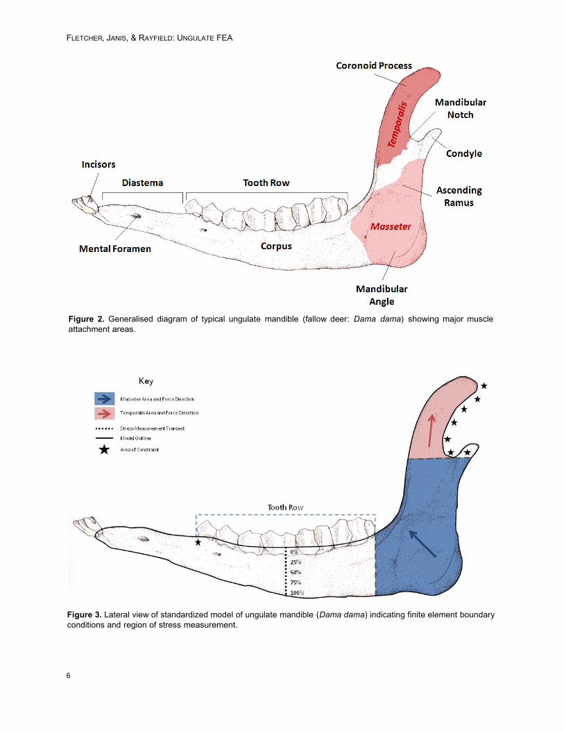
Figure 2. Generalised diagram of typical ungulate mandible (fallow deer: Dama dama) showing major muscleattachment areas.
Figure 3. Lateral view of standardized model of ungulate mandible (Dama dama) indicating finite element boundaryconditions and region of stress measurement.

PALAEO-ELECTRONICA.ORG
border of the lower molar row (i.e., behind the thirdmolar). The temporalis attachment consisted of thecoronoid process area immediately above themandibular notch. The models were constrained atboth the distal anterior tooth of the tooth row in theY direction to simulate a bite at this point, and anarea extending from the posterior edge of the coro-noid process and the condyle above the level ofthe mandibular notch: in all comprising six degreesof freedom to represent immobilisation of the man-dible at the temporomandibular joint. An arbitrarymuscle force value of 100 N was used for compar-ative purposes, but was distributed in proportion tosurface area across the finite element nodes of themasseter and temporalis in directions appropriatefor the relative direction of force during mastication(Figure 3).
Measurement. Stress measurements were takenfrom five evenly distributed nodes along a verticaltransect of the model at the mid-line of the toothrow (Figure 3). These nodes were measured at0%, 25%, 50%, 75% and 100% from the upper-most to lowermost point, respectively. Von Misesstress values were recorded for each of thesepoints, and whole jaw colour plots were created tovisualize the distribution of stress. Differences instress patterns and magnitudes between ruminantsand hindgut fermenters were analysed for all jawmodels pooled together; pairs of extant ruminants
and hindgut fermenters matched for dietary habit(grazer, browser, mixed feeder) and body size;pairs of extant ruminants and extinct hindgut fer-menters also matched for dietary habit and bodysize.
RESULTS
Stress Distribution
Figure 4 documents that large stressesappear in the jaws when subject to a quasi-func-tional feeding load. Warm colours (red, orange andyellow) indicate regions of high stress; blue indi-cates little or no stress. All models are shown to thesame stress scale. Areas of unusually high stresswere always observed at the condyle where themodel was constrained from movement, an issuefamiliar to engineers as Saint-Venant’s Principle(Cook 1995).
These stresses are artificially inflated by theconstraints, but occur at a reasonable distancefrom our region of interest (mid-point of the toothrow) to not significantly influence the outcome ofour analysis. Similarly high stresses were com-monly observed around the base of the coronoidprocess (Figure 4). This area is the attachmentpoint of two muscle groups with differently orien-tated force vectors, which may be at least partly
Figure 4. Typical jaw FE-models of Connochaetes (wildebeest, ruminant) with colour plot of Von Mises stress distri-bution and position of mid-toothrow transect (black line).
7
FLETCHER, JANIS, & RAYFIELD: UNGULATE FEA
responsible for this observed pattern. Again, how-ever, the region of interest appeared unaffected.
Within the ramus of the jaw, a typical bendingpattern was observed in which the highest stressvalues were recorded from the dorsal and ventraledges of the jaws, whilst the central jaw experi-enced little stress (akin to the neutral axis of abeam). As the load is applied, material of the uppermargin undergoes tension, and the lowermost mar-gin is compressed. As the model is two dimen-sional, this represents purely parasaggital bending.On average the dorsal and ventral jaw marginsexhibited 2.84 MPa greater Von Mises stress thanthe centre.
The distal portions of the mandible, includingmost of the diastema, tended to exhibit very little ifany stress. Such low stresses were also observedin the mandibular angle, where two patterns ofstress could be differentiated. In species where thisarea was discordantly bulbous in relation to thethickness of the mandible corpus, stress tended toconcentrate in exterior borders immediately sur-rounding it. In mandibular angles with wider curvesand less protuberance, stress was generally dis-tributed further from this area (Figure 4).
Comparative Stress Magnitudes
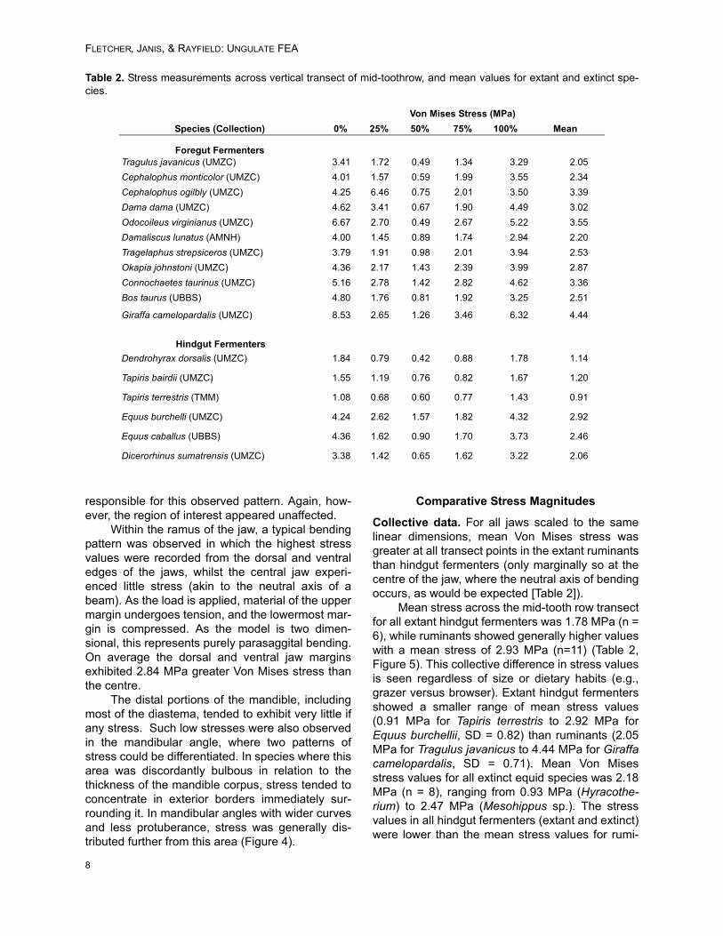
Collective data. For all jaws scaled to the samelinear dimensions, mean Von Mises stress wasgreater at all transect points in the extant ruminantsthan hindgut fermenters (only marginally so at thecentre of the jaw, where the neutral axis of bendingoccurs, as would be expected [Table 2]).
Mean stress across the mid-tooth row transectfor all extant hindgut fermenters was 1.78 MPa (n =6), while ruminants showed generally higher valueswith a mean stress of 2.93 MPa (n=11) (Table 2,Figure 5). This collective difference in stress valuesis seen regardless of size or dietary habits (e.g.,grazer versus browser). Extant hindgut fermentersshowed a smaller range of mean stress values(0.91 MPa for Tapiris terrestris to 2.92 MPa forEquus burchellii, SD = 0.82) than ruminants (2.05MPa for Tragulus javanicus to 4.44 MPa for Giraffacamelopardalis, SD = 0.71). Mean Von Misesstress values for all extinct equid species was 2.18MPa (n = 8), ranging from 0.93 MPa (Hyracothe-rium) to 2.47 MPa (Mesohippus sp.). The stressvalues in all hindgut fermenters (extant and extinct)were lower than the mean stress values for rumi-
Table 2. Stress measurements across vertical transect of mid-toothrow, and mean values for extant and extinct spe-cies.
Von Mises Stress (MPa)
Species (Collection) 0% 25% 50% 75% 100% Mean
Foregut FermentersTragulus javanicus (UMZC) 3.41 1.72 0.49 1.34 3.29 2.05
Cephalophus monticolor (UMZC) 4.01 1.57 0.59 1.99 3.55 2.34
Cephalophus ogilbly (UMZC) 4.25 6.46 0.75 2.01 3.50 3.39
Dama dama (UMZC) 4.62 3.41 0.67 1.90 4.49 3.02
Odocoileus virginianus (UMZC) 6.67 2.70 0.49 2.67 5.22 3.55
Damaliscus lunatus (AMNH) 4.00 1.45 0.89 1.74 2.94 2.20
Tragelaphus strepsiceros (UMZC) 3.79 1.91 0.98 2.01 3.94 2.53
Okapia johnstoni (UMZC) 4.36 2.17 1.43 2.39 3.99 2.87
Connochaetes taurinus (UMZC) 5.16 2.78 1.42 2.82 4.62 3.36
Bos taurus (UBBS) 4.80 1.76 0.81 1.92 3.25 2.51
Giraffa camelopardalis (UMZC) 8.53 2.65 1.26 3.46 6.32 4.44
Hindgut Fermenters
Dendrohyrax dorsalis (UMZC) 1.84 0.79 0.42 0.88 1.78 1.14
Tapiris bairdii (UMZC) 1.55 1.19 0.76 0.82 1.67 1.20
Tapiris terrestris (TMM) 1.08 0.68 0.60 0.77 1.43 0.91
Equus burchelli (UMZC) 4.24 2.62 1.57 1.82 4.32 2.92
Equus caballus (UBBS) 4.36 1.62 0.90 1.70 3.73 2.46
Dicerorhinus sumatrensis (UMZC) 3.38 1.42 0.65 1.62 3.22 2.06
8

PALAEO-ELECTRONICA.ORG
nants. Our results show that there is little overlapbetween mean stress for ruminants and hindgutfermenters (Figure 5), but this observation may beinfluenced by sample size.
Paired data. Data were paired according to similarbody size and feeding behaviour (grazer, mixedfeeder or browser; see Table 1) to remove dietaryhabit and allometric effects from consideration.Paired t-tests comparing six extant pairings ofruminants and hindgut fermenters (Table 1)revealed a statistically significant difference inmean transect values (p = 0.023).
The extinct species of hindgut fermenters (allequids) were paired with extant ruminants of simi-lar body size and likely similar diet (Table 1). Thereason for including these forms was to “fill in thegaps” that no extant hindgut fermenting ungulateoccupies today (i.e., small to medium-sized brows-ers and mixed feeders) to see if the pattern heldover the entire range of diets and body sizes. Asdiet was obviously conjectural in these extinct spe-cies, estimated from dental features (hypsodontyindex and microwear, as previously discussed),statistical differences in stress between these pairswas analysed separately from the pairings thatcontained only extant taxa.
Pairings of extant ruminants and extinctequids (Table 1) were compared on an individualbasis (Figure 6.2) and showed that the jaws ofmost extinct equids were more robust than those ofthe extant ruminants. The exception here was thepairing of Damaliscus lunatus (tsessabe) with theequid Calippus martini: here the equid showed amean stress of 2.67, greater than that of the rumi-nant, with a mean stress of 2.20. Without furthersampling it is impossible to know if this figure is sig-nificant.
The jaws of the extinct equids showed anaverage of 24.68% less stress across the transectthan their paired extant ruminant, with values rang-ing from 18.11% less stress (Merychippus sp.) to60.39% less stress (Hyracotherium sp.). With theexclusion of Damaliscus and Calippus, the averagefor all pairs increased to 31.23% (n = 7) lowerstress in extinct equids than their extant ruminantpairing. Kruskal-Wallis nonparametric analysisshowed borderline insignificance between meanVon Mises stress values between ruminants andextinct hindgut fermenters (p = 0.058). If Damalis-cus and Calippus are excluded, the groups are sig-nificantly different (p = 0.019).
Figure 5. Box plot showing mean Von Mises stresses for all ruminants, hindgut fermenters and extinct equid spe-cies.
9

FLETCHER, JANIS, & RAYFIELD: UNGULATE FEA
10
Figure 6.1. Jaw FE-models of extant pairs (ruminant and hindgut fermenting ungulates) with colour plot of VonMises stress distribution.

PALAEO-ELECTRONICA.ORG
11
Figure 6.2. Jaw FE-models of mixed extant (ruminants) and extinct (equids) pairs with colour plot of Von Misesstress.
FLETCHER, JANIS, & RAYFIELD: UNGULATE FEA
DISCUSSION
Extant hindgut fermenters have more robustjaws than ruminants, when size is removed fromcomparison, and regardless of whether dietaryhabit (grazer versus browser) is considered.“Robustness” is defined by our criteria of a jawpossessing lower mandibular stress than otherjaws subject to the same loading regime. A similartrend is seen in the pairings that include extinctequids, which can be assumed on phylogeneticgrounds to be hindgut fermenters, with extant rumi-nants. A prediction from this result is that this meth-odology could be used to identify the probabledigestive physiology of extinct ungulates with noextant relatives, such as the endemic South Ameri-can litopterns and notoungulates.
Feeding Strategy and Body Size
As previously discussed, feeding strategy hasa significant effect on jaw morphology. For exam-ple, grazers have larger masseter muscles thanbrowsers (Clauss et al. 2009b), which will producelarger forces during mastication, and the jaws ofgrazers appear to be more robustly built in terms ofgeneral morphology. However, no clear pattern ofdifferences between stresses in browsers andgrazers within digestive physiology groupsemerges from this study. Perhaps surprisingly,some of the lowest stress levels (and hence higherrobustness) were recorded in the smaller browsingforms, in the tree hyrax (Dendrohyrax) among thehindgut fermenters, and the mouse deer (Tragulus)among the ruminants and also in the larger brows-ing tapirs (hindgut fermenters). It appears thatdepth of the jaw at the point in front of the mandib-ular angle generally has the most effect on robust-ness here, but further work would be required toelucidate the true nature of the biomechanicsinvolved. It might be the case that smaller animalscan “afford” to have jaws that are more robust thanlarger ones, but that with increasing body size theabsolute weight of the jaw becomes an increasingconsideration in craniodental design, given that it ismanipulated with muscles placed only at the poste-rior end, with a relatively weak mechanical advan-tage. Of course, this remains to be tested.
These observations on extant browsers canalso be seen in some extinct ones. Hyracotheriumis the earliest known (early Eocene) and mostprimitive fossil equid, usually considered to have afolivorous/frugivorous type of diet (like that of amodern mouse deer), due to its brachydont andbunolophodont cheek teeth (MacFadden 2005).However, despite the apparent gracile nature of the
jaw morphology (see Figure 6.1), the FEA analysisrevealed the jaw of this species shows low levels ofstress (0.93 MPa), and thus possess a high level ofrobusticity, much more so than in other, slightlyyounger brachydont equids (Mesohippus), or eventhe more closely related, contemporaneous Euro-hippus. One morphological feature that may berelated to this is the unusual (for an equid) deepen-ing of the mandibular corpus beneath the premo-lars. This morphology could suggest feedingadaptations such as cracking seeds or nuts, anotion possibly supported by dental microwear(Solounias and Semprebon 2002 p. 30). Alterna-tively, at least one species of Hyracotherium (H.tapirinum) has been shown to be sexually dimor-phic in canine size, with (presumed) males havinglarger canines (Gingerich 1981). If this mandibularrobusticity was sexually dimorphic it might relate tomale fighting behaviour. Further study would beneeded to elucidate this idea.
Model Limitations
The finite element models presented here aretwo-dimensional and can only measure in-planestress and strain generated by behaviour such asparasaggital bending. In vivo strain gauge, dataand kinematic analysis of feeding in two ruminatingartiodactyls (goat and alpaca) (Lieberman andCrompton, 2000; Williams et al. 2007, 2009) dem-onstrate that the working side mandible also under-goes torsion about its longitudinal axis, andtransverse and/or parasagittal bending in the bal-ancing side mandible. Feeding loads are thereforeexperienced in three dimensions in these artiodac-tyls, and probably also in other ungulates, due tothe laterally directed adductor muscle resultantseen in these taxa. Our 2D FE models restrict theextent to which the full effect of ungulate feedingloads on jaw morphology can be assessed. Furtherwork using three dimensional FE-models is desir-able, but there are practical and time-constraintissues to this process. Computed tomography (CT)scanning would be required to capture the internaland external 3D geometry of specimens, and forsome taxa that are poorly preserved or of a largesize, scanning would be required to capture theinternal and external 3D geometry of specimens,which would be difficult for taxa that are poorly pre-served or of a large size. This paper offers a firststep towards these future research directions.
CONCLUSION
The mandibular robustness of ungulate jawsappears to be reliably correlated with digestive
12

PALAEO-ELECTRONICA.ORG
physiology in a range of extant species and poten-tially also in extinct species. In general hindgutfeeders have jaws which show lower levels ofstress when equal muscle forces are applied inFEA analysis (i.e., are more “robust”), and thesejaws can be significantly differentiated from the rel-atively more gracile jaws of ruminants, especiallywhen the species are matched for body mass anddietary type. This observation accords with thegreater amount of stress predicted for hindgut fer-menters due to their relatively greater levels of foodingestion and mastication.
ACKNOWLEDGMENTS
We would like to thank R. Asher (University ofCambridge), S. Holwell (University of Bristol), J.Hooker (Natural History Museum, London) and C.Norris (American Museum of Natural History) foraccess to various specimens. We are also gratefulto T. Rowe and J. Maisano (University of Texas) for3D data produced with funding from NSF grant IIS-0208675 awarded to T. Rowe, and DigiMorph.org.Lastly, we are indebted to J. Bright for her adviceand assistance with FEA and imaging software,and the anonymous reviewers for their helpful con-tributions to the final script.
REFERENCES
Arnold, G.W. 1984. Comparison of the time budgets andcircadian patterns of maintenance activities in sheep,cattle and horses grouped together. Applied AnimalBehaviour Science, 13:19-30.
Clauss, M., Frey, R., Kiefer, B., Lechner-Doll, M., Loeh-lein, W., Polster, C., Rössner, G., and Streich, W.J.2003. The maximum attainable body size of herbivo-rous mammals: morphophysiological constraints onforegut, and adaptations of hindgut fermenters.Oecologia, 136:14–27.
Clauss, M., Kaiser, T., and Hummel, J. 2007. The mor-phophysiological adaptations of browsing and graz-ing mammals, p. 47-88. In Gordon, I.J. and Prins,H.H.T (eds.), The Ecology of Browsing and Grazing.Springer, New York.
Clauss, M., Nunn, C., Fritz, J., and Hummel J. 2009a.Evidence for a tradeoff between retention time andchewing efficiency in large mammalian herbivores.Comparative Biochemistry and Physiology A,154:376-382.
Clauss, M., Fritz, J., Bayer, D., Nyrgyn, K., Hammer, S.,Hüatt, J.-M., Südekum, K.-H., and Hummel, J.2009b. Physical characteristics of rumen contents infour large ruminants of different feeding type, theaddax (Addax nasomaculatus), bison (Bison bison),red deer (Cervus elaphus) and moose (Alces alces).Comparative Biochemistry and Physiology A,152:398-406.
Cook, R.D. 1995. Finite Element Modelling for StressAnalysis, 1st Ed. Wiley, New York.
Duncan, P., Foose, T.J., Gordon, I.J., Gakahu, C.G., andLloyd, M. 1990. Comparative nutrient extraction fromforages by grazing bovids and equids: a test of thenutritional model of equid/bovid competition andcoexistence, Oecologia, 84:411-418.
Fortelius, M. 1985. Ungulate cheek teeth: developmen-tal, functional, and evolutionary interrelations. ActaZoologica Fennica, 180:1-76.
Fritz, J., Hummel, J., Kienzle, E., Arnold C., Nunn, C.,and Clauss, M. 2009. Comparative chewing effi-ciency in mammalian herbivores. Oikos 118:1623-1632.
Gingerich, P.D. 1981. Variation, sexual dimorphism, andsocial structure in the early Eocene horse Hyracothe-rium (Mammalia, Perissodactyla). Paleobiology,7:443-455.
Janis, C.M. 1990a. Correlation of cranial and dental vari-ables with dietary preferences: a comparison ofmacropodoid and ungulate mammals. Memoirs ofthe Queensland Museum, 28: 349-366.
Janis, C.M. 1990b. Correlation of cranial and dental vari-ables with body size in ungulates and macropodoids,p. 255-299. In Damuth, J. and MacFadden, B.J.(eds.), Body Size in Mammalian Paleobiology: Esti-mation and Biological Implications, Cambridge Uni-versity Press, Cambridge.
Janis, C.M. 1995. Correlations between craniodentalmorphology and feeding behavior in ungulates: recip-rocal illumination between living and fossil taxa, p.76-98. In Thomason, J.J. (ed.), Functional Morphol-ogy in Vertebrate Paleontology, Cambridge Univer-sity Press, Cambridge.
Janis, C.M., Constable, E.C., Houpt, K.A., Jurgun Stre-ich, W., and Clauss, M. 2010. Comparative ingestivemastication in domestic horses and cattle: a pilotinvestigation. Journal of Animal Physiology and Ani-mal Nutrition. DOI: 10.1111/j.1439-0396.2010.01030
Kober, C., Stubinger, S., Hellmich, C., Sader, R., andZeilhofer, H.F. 2008. Finite element simulation of thehuman mandible: the role of (natural) teeth. Interna-tional Journal of Computational Dentistry, 11:169-174.
Lieberman, D.E. and Crompton, A.W. 2000. Why fusethe mandibular symphysis? A comparative analysis.American Journal of Physical Anthropology, 112:517-540.
MacFadden, B.J. 2005. Fossil horses - evidence for evo-lution. Science, 307:1728-1730.
Menard, C., Duncan, P., Fleurance, G., Georges, J.Y.,and Lila, M. 2002. Comparative foraging and nutritionof horses and cattle in European wetlands. Journal ofApplied Ecology, 39:120-133.
Mendoza, M., Janis, C.M., and Palmqvist, P. 2002. Char-acterizing complex craniodental patterns related tofeeding behaviour in ungulates: a multivariateapproach. Journal of Zoology, London, 258:223-246.
13

FLETCHER, JANIS, & RAYFIELD: UNGULATE FEA
Pierce, S.E., Angielczyk, K.D., and Rayfield, E.J. 2008.Patterns of morphospace occupation and mechanicalperformance in extant crocodilian skulls: a combinedgeometric morphometric and finite element modellingapproach. Journal of Morphology, 269:840-864.
Pierce, S.E., Angielczyk, K.D., and Rayfield, E.J. 2009.Shape and mechanics in thalattosuchian (Crocodylo-morpha) skulls: implications for feeding behavioursand niche partitioning. Journal of Anatomy, 215:555-576.
Rayfield, E.J. 2005. Aspects of comparative cranialmechanics in the theropod dinosaurs Coelophysis,Allosaurus and Tyrannosaurus. Zoological Journal ofthe Linnean Society, 144:309-316.
Rayfield, E.J. 2007. Finite element analysis and under-standing the biomechanics and evolution of livingand fossil organisms. Annual Review of Earth andPlanetary Sciences, 35:541-576.
Reilly, D.T. and Burstein, A.H. 1975. The elastic and ulti-mate properties of compact bone tissue. Journal ofBiomechanics, 8:393-405.
Richmond, B.G., Wright, B.W., Grosse, I., Dechow, P.C.,Ross, C.F., Spencer, M.A., and Strait, D.S. 2005.Finite element analysis in functional morphology. TheAnatomical Record, 283A, 259-274.
Schmidt-Nielson, K. 1997. Animal Physiology: Adapta-tion and Environment (5th Edition). Cambridge Uni-versity Press, Cambridge.
Solounias, N. and Semprebon, G. 2002. Advances in thereconstruction of ungulate ecomorphology with appli-cations to early fossil equids. American MuseumNovitates, 366:1-49.
Stevens, C.E. and Hume, I.D. 1995. Comparative Physi-ology of the Vertebrate Digestive system. 2nd Edi-tion. Cambridge University Press, Cambridge.
Turnbull, W.D. 1970. Mammalian masticatory apparatus.Fieldiana: Geology, 18:149-356.
Williams, S.H., Vinyard, C.J., Wall, C.E., and Hylander,W.L. 2007. Masticatory motor patterns in ungulates:a quantitative assessment of jaw-muscle coordina-tion in goats, alpacas and horses. Journal of Experi-mental Zoology, 307A:226-240.
Williams, S.H., Vinyard, C.J., Wall, C.E., and Hylander,W.L. 2009. Mandibuluar corpus bone strain in goatsand alpacas: Implications for understanding the bio-mechanics of mandibular form in selenodont artio-dactyls. Journal of Anatomy, 214:65-78.
14

PALAEO-ELECTRONICA.ORG
APPENDIX – MUSEUM SPECIMEN CATALOGUE NUMBERS
Specimen Reference Code CollectionCephalophus monticolor H21621 University Museum of Zoology, Cambridge, UK
Cephalophus ogilbly H21461
Connochaetes taurinus H21242
Dama dama H17179
Dendrohyrax dorsalis H5281
Dicerorhinus sumatrensis H6387
Equus burchelli H9357
Giraffa camelopardalis H23090
Odocoileus virginianus H189902
Okapia johnstoni H20302
Tapirus bairdii H7451
Tragulus javanicus H15013
Tragelaphus strepsicerus H25161
Eurohippus parvulus Uncatalogued Cast of Skull
Natural History Museum, UK (original in Senkenberg Musuem, Germany)
Kalobatippus Uncatalogued Cast of Skull
Natural History Museum, UK (original in AMNH, New York)
Merychippus sp. Uncatalogued Cast of Skull
Natural History Museum, UK
Mesohippus sp. MT00033
Calippus martini FAM114159 American Museum of Natural History, New York, USA
Damaliscus lunatus AMNH150150
Hyracotherium sp. AMNH55986
Merychippus insignis FAM87003
Mesohippus sp. AMNH 39001
Tapiris bairdii AMNH 80076
Bos taurus S2J3.6 School of Biological Sciences, University of Bristol, Bristol, UK
Equus caballus R8a.3
Tapiris terrestris TMM M-16 Texas Memorial Museum, University of Texas, Austin, Texas, USA
15


![Pool - stetten.com.br · 3 234-05 Yankauer 234-06 Yankauer Pool 234-01 234-02 234-03 234-04 229-07 229-08 230-24 Universal 230-25 230-26 Pierce 3LXV /DQJ 0HGL]LQWHFKQLN *PE+ 0DGH](https://img.dokumen.tips/doc/110x75/5fb63cd83b5e3f1b5525f393/pool-3-234-05-yankauer-234-06-yankauer-pool-234-01-234-02-234-03-234-04-229-07.jpg)
![ВЕСТНИК - vestnik-cspu.ruvestnik-cspu.ru/upload/pdf/issues/2010/2010_3.pdfШибкова Д.З. – доктор биологических наук, ... [2]. Сам Урсул](https://img.dokumen.tips/doc/110x75/60048d2ded0f763c4b31561a/-vestnik-cspuruvestnik-cspuruuploadpdfissues201020103pdf.jpg)