Embed Size (px)
Citation preview

PAKISTAN JOURNAL OF CHEST MEDICINE
Pak J Chest Medwww.pjcm.net
October - December 2014Vol. 20 No. 4
ISSN: 2224-9710ISSNE: 2309-9844
EDITOR IN CHIEFProf. Arshad Javaid
President PCSProf. Kamran Chima
BIBLIOGRAPHERMr. Mazhar Kamal
EPIDEMIOLOGIST & STATISTICIANMr. Mazhar Ali Khan
ASSOCIATE EDITORS
Prof. Martyn R. Partridge, UK
Prof. Sohail Akhtar, Karachi
Dr. Wajid Ali, Islamabad
Dr. Ali Musani, USA
Dr. Muhammad Irfan, Karachi
Dr. Saifullah Baig, Karachi
Maj Gen Jawad K Ansari, Islamabad
Dr. Ali Zubairi, Karachi
Dr. Shereen Khan, Quetta
Dr. Talha Mahmud, Lahore
Dr. Ashraf Jamal, Lahore
Dr. M. Irfan Malik, Lahore
Dr. Zia Ullah, Peshawar
Dr. Sana Ullah Tareen, Quetta
EDITORProf. Mukhtiar Zaman
MANAGING EDITORDr. Zafar Iqbal
EDITORProf. Aamir Bilal
DEPUTY EDITORDr. Saadia Ashraf

Printer: Khyber Mail, Office No. 3, 3rd Floor, Syed’s Tower Opposite Customs House,
University Road, Peshawar - Pakistan.
Publication Cell: Pakistan Journal of Chest Medicine, Lady Reading Hospital, Peshawar, Pakistan.
Visit us at: www.pjcm.net
Indexed & abstracted in Directory of Research Journal Indexation (DRJI)
Approved by Pakistan Medical & Dental Council
Covered by Pakmedinet, EBESCO
E-mail: [email protected]
Pakistan Journal of Chest Medicine is published on controlled circulation basis and distributed among the chest Physicians & members of PCS, faculty of all medical colleges and tertiary referral centres,
main libraries and private clinics throughout Pakistan.
All rights are reserved. No part of this publication may be reproduced, stored in a retrieval system, or transmitted in any form or by any means, electronic, mechanical, photocopying, except for internal or
personal use, without the prior permission of the publisher.
The publisher and the members of the editorial board cannot be held responsible for errors or for any consequences arising from the use of the information contained in this journal.
Pakistan Journal of Chest Medicine is published quarterly, composed and printed at Khyber Mail Peshawar.

EDITORIAL
ASPERGILLUS IN THE LUNG: THE SPECTRUM OF DISEASES. 131
Muhammad Irfan
ORIGINAL ARTICLES
MODS ASSAY FOR RAPID DIAGNOSIS OF TUBERCULOSIS AMONG HIV TB CO INFECTED INDIVIDUALS IN A TERTIARY CARE HOSPITAL, ANDHRA PRADESH. 133
T. Jaya Chandra, Ramesh Redddy Alan, R. Selvaraj, YV Sharma
FACTORS DETERMINING THE DIAGNOSTIC YIELD OF CT-GUIDED CORE NEEDLE BIOPSY OF LUNG NODULES 139
Mohammad Iqbal, Zafar Iqbal, Farooq Ahmed
12 YEARS EXPERIENCE OF SURGICAL MANAGEMENT OF PULMONARY ASPERGILLOMA 142
Abdul Baseer, Aamir Bilal, Muhammad Imran
TREATMENT OUTCOME OF MULTI-DRUG RESISTANT TUBERCULOSIS (MDR-TB). 147
Naveed Inayat, Riaz Hussain Shah, Qurban Rahoo
CASE REPORT
INCIDENTAL FINDING OF COLOPLEURAL FISTULA DURING PLEUROCUTANEOUS WINDOW SURGERY FOR EMPYEMA THORACIS 151
Niaz Hussain, Aneeqa Ahsan Zafar
ABSTRACTS 154
INSTRUCTIONS TO AUTHORS 160
ContentsVol. 20 No.4
PJCM


131PJCM 2014; 20 (4)
Aspergillus in the Lung: The Spectrum of Diseases
Dr. Muhammad Irfan
The infection caused by fungi associated with high mortality and morbidity rates. The frequency of
pulmonary fungal infection is increasing over the past decades with the development of immuno-suppressed therapy, solid organ transplantation, steroid applica-tion and HIV-infection. Fungi causes the pulmonary infection include Candida, Cryptococcus, Aspergillus and others relatively uncommon fungus.1 Although the treatment is difficult, but the results are encouraging. Hence, this is a need of today to know these diseases well so that we are able to manage them scientifically.
Aspergillosis is a mycotic disease caused by As-pergillus species, a genus of ubiquitous soil fungi. Although exposure to Aspergillus conidia through in-halation is common, only a minority of those exposed develop lung disease. The clinical features, course and prognosis of Aspergillus infections are largely depend on the host immune response and the number and vir-ulence of the organisms.2, 3
Pulmonary aspergillosis can be subdivided into five categories: (a) saprophytic aspergillosis (aspergilloma), (b) hypersensitivity reaction (allergic bronchopulmona-ry aspergillosis), (c) tracheobronchial aspergillosis (d) chronic pulmonary aspergillosis, and (e) angioinvasive aspergillosis.
Saprophytic aspergillosis (aspergilloma) is an As-pergillus infection without tissue invasion. It consists of conglomeration of intertwined fungal hyphae admixed with mucus and cellular debris within a preexistent pulmonary cavity.4 The common underlying causes are tuberculosis, cystic fibrosis and sarcoidosis. The most common clinical presentation of aspergilloma is hemoptysis, although patients may remain asymptom-atic. On imaging, aspergilloma are characterized by the presence of a solid, round or oval mass with soft-tissue opacity within a lung cavity. Usually the mass is sep-arated from the wall of the cavity by airspace of vari-able size and shape, and present as “air crescent” sign.
Surgical resection is indicated for patients with severe life-threatening hemoptysis, and selective bronchial ar-tery embolization can be performed in those with poor lung function.
In this issue of Journal Baseer and colleagues pres-ent their 12 years’ experience of surgical management of aspergilloma with excellent results with different sur-gical techniques. They also nicely review literature on this disease modality.
Allergic bronchopulmonary aspergillosis (ABPA) is caused by a complex hypersensitivity reaction to As-pergillus. ABPA is seen most commonly in patients with long-standing bronchial asthma. ABPA is char-acterized by the presence of plugs of inspissated mu-cus containing Aspergillus organisms and eosinophils. This results in bronchial dilatation and bronchiectasis in segmental and sub segmental bronchi. Patients usually cough up thick mucus plugs in which hyphal fragments can be demonstrated at culture or histologic analysis. Common clinical presentations include recur-rent wheezing, malaise with low-grade fever, cough, sputum production, and a history of recurrent pneumo-nia. Radiologic manifestations include homogeneous, tubular, finger-in-glove areas of increased opacity in a bronchial distribution, usually predominantly involving the upper lobe and can migrate from one region to an-other. Treatment of ABPA aims to prevent progressive bronchiectasis. Corticosteroids are the main stay of treatment for several weeks or months. Iatraconaazole is used in patients with frequent exacerbations and to reduce the fungal burden and steroid dependence.5
Chronic Pulmonary Aspergillosis (CPA) has various patterns of presentation. Semi-invasive aspergillosis (SIA), also known as chronic necrotizing aspergillosis, is one of the commonest forms and characterized by the presence of tissue necrosis and granulomatous inflammation similar to that seen in reactivation of tu-berculosis. Factors associated with the development
EDITORIAL
This Article may be cited as: Irfan M. Aspergillus in the Lung: The Spectrum of Diseases. Pak J Chest Med 2014; 20(4): 131-32
Address for Correspondence:Dr. Muhammad IrfanAssociate Professor
Section of Pulmonary and Critical Care Medicine
Department of MedicineAga Khan University, Karachi
Email: [email protected]

132PJCM 2014; 20 (4)
ASPERGILLUS IN THE LUNG: THE SPECTRUM OF DISEASES
of this form of aspergillosis include diabetes mellitus, malnutrition, alcoholism, prolonged corticosteroid therapy, and chronic obstructive pulmonary disease.3, 6 Another, more common pattern is chronic cavitary pul-monary aspergillosis (CCPA), characterized by slowly evolving, single or multiple lung cavities, usually with thick walls and with pleural fibrosis. In some cases of CCPA extensive pulmonary fibrosis may develop. These patients are classified as chronic fibrosing pul-monary aspergillosis.3
Clinical presentations of CPA are often insidious and include chronic cough, sputum production, fever, and constitutional symptoms. Management of patients with CPA is complicated and Azoles are the initial choice of treatment. Itraconazole, voriconazole and posacon-azole can be used. The duration of treatment is usually prolong and associated with side effects of drugs. The relapse rate is also high in this form of aspergillosis.3, 7
Angioinvasive aspergillosis usually occurs in immu-nocompromised patients with severe neutropenia. The clinical diagnosis is difficult, and the mortality rate is high. Angioinvasive aspergillosis is characterized by the invasion and occlusion of small to medium-sized pulmonary arteries by fungal hyphae. This leads to the formation of necrotic hemorrhagic nodules or pleu-ra-based, wedge-shaped hemorrhagic infarcts. Char-acteristic CT findings consist of nodules surrounded by a halo of ground-glass attenuation known as “halo sign” or pleura-based, wedge-shaped areas of consol-idation.8 Definite diagnosis is usually based on fungal culture, galactomannan, or PCR in blood and respira-tory samples and on histopathology. Respiratory sam-ples are better than blood for all tests except β-D-glu-can. Voriconazole is the treatment of choice and has a significant mortality benefit. Duration of treatment in non-neutropenic patient is minimum of 12 weeks.9
Tracheobronchial aspergillosis or Aspergillus bron-chitis is a less common form of aspergillosis and usu-ally present in immunocompetent patients. These pa-tients usually present with recurrent chest infections unsuccessfully managed with antibiotics and repeated isolation of Aspergillus from sputum or BAL and pos-itive PCR but without pulmonary parenchymal dis-ease.3,10 They respond well to antifungals, but relapses are common.
In summary, the spectrum of disease caused by As-pergillus in the lung is wide, ranging from aspergilloma to invasive aspergillosis and can be viewed as a con-tinuous spectrum of disease. The manifestations are depending on interaction between fungus and host. A broad knowledge of clinical presentation and high sus-picion are required for timely diagnosis and treatment of aspergillus related lung diseases.
REFERENCES:
1. Liao Wanqing. Pulmonary Fungal Infection. Current Respiratory Medicine Reviews, 2012, 8, 345
2. Gefter WB. The spectrum of pulmonary aspergillosis. J Thorac Imaging 1992; 7:56-74.
3. Kosmidis C, Denning DW. The clinical spectrum of pulmonary aspergillosis. Thorax. 2015;70(3):270-277.
4. Aquino SL, Lee ST, Warnock ML, Gamsu G. Pulmo-nary aspergillosis: imaging findings with pathologic correlation. AJR Am J Roentgenol 1994; 163:811-815
5. Moreira AS, Silva D, Ferreira AR, et al. Antifungal treatment in allergic bronchopulmonary aspergillosis with and without cystic fibrosis: a systematic review. Clin Exp Allergy 2014;44:1210–27.
6. Smith NL, Denning DW. Underlying conditions in chronic pulmonary aspergillosis including simple as-pergilloma. Eur Respir J 2011;37:865–72.
7. Al-Shair K, Atherton GT, Harris C, et al. Long-term an-tifungal treatment improves health status in patients with chronic pulmonary aspergillosis: a longitudinal analysis. Clin Infect Dis 2013;57:828–35.
8. Del Bono V, Mikulska M, Viscoli C. Invasive aspergil-losis: diagnosis, prophylaxis and treatment. Curr Opin Hematol 2008;15:586–93.
9. Mousset S, Buchheidt D, Heinz W, et al. Treatment of invasive fungal infections in cancer patients-updated recommendations of the Infectious Diseases Working Party (AGIHO) of the German Society of Hematology and Oncology (DGHO). Ann Hematol 2014;93:13–32.
10. Chrdle A, Mustakim S, Bright-Thomas RJ, et al. As-pergillus bronchitis without significant immunocom-promise. Ann N Y Acad Sci 2012; 1272:73–85.

133PJCM 2014; 20 (4)
MODS ASSAY FOR RAPID DIAGNOSIS OF TUBERCULOSIS AMONG HIV TB CO INFECTED INDIVIDUALS IN A TERTIARY CARE HOSPITAL, ANDHRA PRADESH.
T. Jaya Chandra*, Ramesh Redddy Alan**, R. Selvaraj***, YV Sharma*
ABSTRACT
BACKGROUND: Rapid, reliable, economical methods are required for diagno-sis of tuberculosis. The Microscopic Observation of Drug Susceptibility (MODS) assay is a relatively low-cost and simple liquid culture method. The objective of this study is to determine the sensitivity and specificity of MODS test in com-parison to the Lowenstein- Jensen medium to diagnose tuberculosis among HIV seropositive individuals in GSL Medical College.
METHODS: Sputum specimens were evaluated using smear microscopy, cul-ture on Lowenstein-Jensen medium and MODS assay. A study subject is con-sidered to have tuberculosis if at least 1 culture on Lowenstein- Jensen medium or MODS technique showed growth for M. tuberculosis.
RESULTS: Spot Morning sputum samples were obtained from 873 HIV sero-positive individuals. Two hundred and ninety seven (34%) [95% CI=30.8 – 37.2] patients were culture positive by MODS and 277 (32%) [95% CI=28.7 – 34.9] were culture positive on LJ slopes (P < 0.001). MODS sensitivity was 99.3% and specificity was 96.3%. Mean times for TB detection were 21 days (range 15 – 25 days) and 12 days (range 7- 15 days) for culture on Lowenstein-Jensen medium and MODS (including drug susceptible testing) respectively (P<0.001). Culture contamination was low in MODS assay than culture on Lowenstein-Jensen me-dium (1.35 vs. 15.6%; P<0.001). Drug resistance was 12.6% for both RIF and INH, 12.6 % for RIF and 15% for INH.
CONCLUSIONS: The MODS assay is a relatively simple test whose good per-formance for detection of pulmonary tuberculosis in HIV patients may make it suitable for resource-limited environments.
KEY WORDS: Tuberculosis, Sputum smear, HIV, MOD
* GSL Medical College Rajah-mundry, South India.** Share India.*** Sathyabama University
Adress for correspondence:T. Jaya ChandraDepartment of Microbiology, GSL Medical College Rajah-mundry, South India.E-mail: [email protected]
Introduction:
In India, ~ 5% of tuberculosis (TB) patients regis-tered under Revised National tuberculosis Control
Program (RNTCP) are co-infected with HIV. 1 The exis-tence of HIV and TB together, greatly amplifies harmful effects of each other at individual level and contribute substantially to mortality among patients living with HIV (PLHIV). 2 The risk of developing TB is estimated to be between 20-37 times greater in PLHIV than among those without HIV infection.3 Inadequate treatment, de-fault behavior further result in Drug Resistance (DR) TB, HIV is one of the main predisposing factors.
Well equipped clinical laboratories can detect Myco-
bacterium tuberculosis (MTB) within 7-14 days, using sophisticated liquid culture systems such as BACTEC and Mycobacterium Growth Indicator Tubes (MGITs).3,4 In most of the developing countries TB laboratories lack sophisticated, costly equipment and skilled tech-nicians. Though ZN staining is a rapid test, sensitivity is relatively low, require about ten thousand bacilli per ml of the specimen.5, 6 In half of the HIV TB patients spu-tum smears are negative for Acid Fast Bacilli (AFB) by ZN staining.7, 8 This is the major limitation of ZN staining in PLHIV.
Most of the laboratories in developing countries use solid media such as Lowenstein Jensen (LJ). Under op-timum conditions TB diagnosis takes 4 weeks and drug
ORIGINAL ARTICLE
This Article may be cited as: Chandra TJ, Alan RR, Selvaraj R, Sharma YV. MODS assay for rapid diagnosis of Tuberculosis among HIV TB co infected individuals in a tertiary care hospital, Andhra Pradesh. Pak J Chest Med 2014; 20(4): 133-38

134PJCM 2014; 20 (4)
MODS ASSAY FOR RAPID DIAGNOSIS OF TUBERCULOSIS AMONG HIV TB CO INFECTED INDIVIDUALS IN A
susceptibility test (DST) takes additional 3 to 4 weeks using LJ medium.9 To detect the TB rapid molecular tests like line probe assay have been developed.10, 11 These molecular diagnostic tests are rapid and highly accurate. But cost, expertise and infrastructure are the major obstacles to offer these tests. Mycobacterium growth is more rapid in liquid medium as strings and tangles.12 Based on this a new, rapid, reliable, inexpen-sive method13-16 namely Microscopic Observation of Drug Susceptibility (MODS) is devised, which permits MTB detection and drug susceptibility in less than 2 weeks.
Hence in the current study the diagnosis and DST of TB in HIV patients is done by MODS and culture re-sults are compared with gold standard LJ.
MATERIAL AND METHODS
The study was conducted from March 2009 to De-cember 2013, in the department of Microbiology, GSL Medical College, Rajahmundry. Study was approved by the Institutional Research and Ethics committee. Study included PLHIV with clinical suspicion sugges-tive of TB. Children with HIV aged below 14 years, in-dividuals who refused to give two consecutive samples of sputum and HIV sero negative individuals were ex-cluded from the study. An informed written consent in the presence of witness was taken from all the volun-teers who participated in the study. Sputum samples were collected from HIV sero-positive individuals by Spot Morning (SM) scheme.18
All the volunteers were explained regarding the im-portance of submission of good quality sputum sam-ple. The visual difference between the sputum and sa-liva and the procedure for production of good quality sputum sample was demonstrated.17 After collection of spot sample, the individuals were provided with pre labeled sample containers for collection of morning samples. Immediately after collection, sputum smears were prepared on new glass slide and were stained by ZN technique as per RNTCP guidelines.18
After smear preparation, decontamination and con-centration of the sputum samples was done by stan-dard N-acetyl -L-cysteine- Sodium hydroxide method.19 Specimen with equal parts of N-acetyl-L-cysteine- So-dium hydroxide solution, mixed for 15 seconds on ver-tex mixture. Then enough phosphate buffer saline was added to reach within 1cm of the top of the test tube, cap was closed tightly and inverted to mix the solu-tion. Then it was centrifuged at 3600xg for 15minutes. The supernatant was decanted and sediment was sus-pended in to 2ml of Middlebrook 7H9 broth.
New, sterile flat bottom 24 well microtitre plates
were used to test three sputum samples (eight wells per sample) by MODS technique. Five hundred and fourty ml of sputum medium solution was placed into each of four micro wells. Sixty ml of each drug solu-tions were added. The final drug concentrations are as follows:
a. Isoniazid (INH): 0.4& 0.1 mg/ml
b. Rifampcin (RIF): 1 & 0.5 mg/ml
In the remaining four wells first two were filled with sputum media mixture, acts as drug free control. In the other two wells, one was filled with media, this act as sterilization control. The last well was filled with known drug sensitive strain of MTB, by standardizing the turbidity with 0.5 Mc Farlands standard. Plates were sealed with scotch polyethylene tape, incubated at 37°C. Every day (except on Sundays and public hol-idays) wells were examined for the presence of MTB under an inverted light microscope under 40X objec-tive.
A drop of processed sputum sample was also inoc-ulated in blood agar and another drop was inoculated in Sabourauds Dextrose Agar. These were incubated at 37°C. If any bacteria or fungal growth was observed, another sample was collected from patient. Simultane-ously samples were inoculated in LJ slopes, incubated at 37°C for 3 – 4 weeks. Regular decontamination of incubators was followed to avoid contamination of cul-tures.
Presence of pink colored bacilli in the ZN staining indicates sputum sample is positive for AFB. Presence of growth in microtiter well containing sputum media mixture was considered as TB positive. Individual was considered as non TB, if growth is absent in media sample containing well. Presence of growth in all the four drug containing wells (two RIF wells and two INH wells) along with media sample wells indicates DR. Presence of growth in drug free wells and absence in drug containing wells indicates that the clinical sample is TB positive and drug sensitive. LJ Media was home made.
Statistical methods:
Data were analyzed using of SPSS v. 16 (SPSS Inc., Chicago, IL, USA), with the patient as the unit of analysis. The Wilcoxon signed-rank test was used to compare the times to each end point among the two methods. A P value of less than 0.05 was used to indi-cate statistical significance. The concordance of sus-ceptibility results was determined with the use of the sensitivity, specificity, and positive and negative pre-dictive values for the detection of resistance (with 95% confidence intervals [CIs]). For sensitivity and speci-
135PJCM 2014; 20 (4)
MODS ASSAY FOR RAPID DIAGNOSIS OF TUBERCULOSIS AMONG HIV TB CO INFECTED INDIVIDUALS IN A
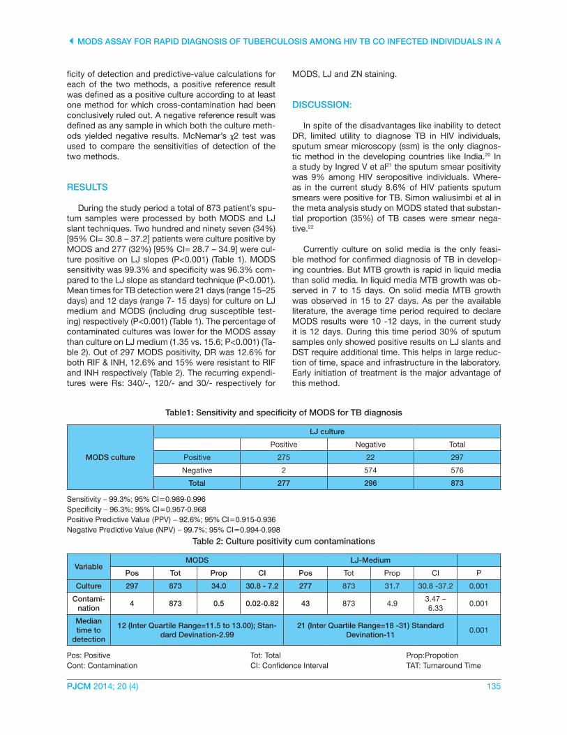
ficity of detection and predictive-value calculations for each of the two methods, a positive reference result was defined as a positive culture according to at least one method for which cross-contamination had been conclusively ruled out. A negative reference result was defined as any sample in which both the culture meth-ods yielded negative results. McNemar’s χ2 test was used to compare the sensitivities of detection of the two methods.
RESULTS
During the study period a total of 873 patient’s spu-tum samples were processed by both MODS and LJ slant techniques. Two hundred and ninety seven (34%) [95% CI= 30.8 – 37.2] patients were culture positive by MODS and 277 (32%) [95% CI= 28.7 – 34.9] were cul-ture positive on LJ slopes (P<0.001) (Table 1). MODS sensitivity was 99.3% and specificity was 96.3% com-pared to the LJ slope as standard technique (P<0.001). Mean times for TB detection were 21 days (range 15–25 days) and 12 days (range 7- 15 days) for culture on LJ medium and MODS (including drug susceptible test-ing) respectively (P<0.001) (Table 1). The percentage of contaminated cultures was lower for the MODS assay than culture on LJ medium (1.35 vs. 15.6; P<0.001) (Ta-ble 2). Out of 297 MODS positivity, DR was 12.6% for both RIF & INH, 12.6% and 15% were resistant to RIF and INH respectively (Table 2). The recurring expendi-tures were Rs: 340/-, 120/- and 30/- respectively for
MODS, LJ and ZN staining.
DISCUSSION:
In spite of the disadvantages like inability to detect DR, limited utility to diagnose TB in HIV individuals, sputum smear microscopy (ssm) is the only diagnos-tic method in the developing countries like India.20 In a study by Ingred V et al21 the sputum smear positivity was 9% among HIV seropositive individuals. Where-as in the current study 8.6% of HIV patients sputum smears were positive for TB. Simon waliusimbi et al in the meta analysis study on MODS stated that substan-tial proportion (35%) of TB cases were smear nega-tive.22
Currently culture on solid media is the only feasi-ble method for confirmed diagnosis of TB in develop-ing countries. But MTB growth is rapid in liquid media than solid media. In liquid media MTB growth was ob-served in 7 to 15 days. On solid media MTB growth was observed in 15 to 27 days. As per the available literature, the average time period required to declare MODS results were 10 -12 days, in the current study it is 12 days. During this time period 30% of sputum samples only showed positive results on LJ slants and DST require additional time. This helps in large reduc-tion of time, space and infrastructure in the laboratory. Early initiation of treatment is the major advantage of this method.
Table1: Sensitivity and specificity of MODS for TB diagnosis
MODS culture
LJ culture
Positive Negative Total
Positive 275 22 297
Negative 2 574 576
Total 277 296 873
Sensitivity – 99.3%; 95% CI=0.989-0.996Specificity – 96.3%; 95% CI=0.957-0.968Positive Predictive Value (PPV) – 92.6%; 95% CI=0.915-0.936Negative Predictive Value (NPV) – 99.7%; 95% CI=0.994-0.998
Table 2: Culture positivity cum contaminations
VariableMODS LJ-Medium
Pos Tot Prop CI Pos Tot Prop CI P
Culture 297 873 34.0 30.8 - 7.2 277 873 31.7 30.8 -37.2 0.001
Contami-nation
4 873 0.5 0.02-0.82 43 873 4.93.47 – 6.33
0.001
Median time to
detection
12 (Inter Quartile Range=11.5 to 13.00); Stan-dard Devination-2.99
21 (Inter Quartile Range=18 -31) Standard Devination-11
0.001
Pos: Positive Tot: Total Prop:Propotion Cont: Contamination CI: Confidence Interval TAT: Turnaround Time

136PJCM 2014; 20 (4)
MODS ASSAY FOR RAPID DIAGNOSIS OF TUBERCULOSIS AMONG HIV TB CO INFECTED INDIVIDUALS IN A
As per the Moore et al study,15 the diagnostic yield of single sputum sample among the patients suspect-ed with TB was 37%, 80% and 89% respectively for smear microscopy, LJ & MODS and with second sam-ple the additional use is 11.6%, 7.5% and 8.2% re-spectively. In HIV patients, with single sputum sample diagnostic yield of PT was 42.9%, 78.6% and 92.9% and the incremental yield was 3.6%, 3.6% and 3.6% with second sputum sample respectively for sputum smear, LJ and MODS. In the current study the diagnos-tic yield of ssm was 7.9%, 8.6% respectively for spot and morning samples. Due to limited resources single sputum sample was only processed for culture.
In a meta analysis study by Jessica Minion et al,23 DST of MODS has a sensitivity of 98% (95% CI 94·5 - 99·3), specificity 99·4% (95·7 - 99·9) for RIF resistance. For INH resistance, pooled sensitivity was 97·7% (94·4 - 99·1) and pooled specificity was 95·8% (88·1 - 98·6). In the current study, due to limited resources DST was not performed on LJ media. This could be the limitation of the current study.
In a study by Lazarnu24 the authors reported that MODS is 94.12% sensitive and 89.39% specific when compared with LJ media and the concordance with DST by the proportion method on LJ media to RIF & INH was 91.5% & 90.8% respectively. In one of the south Indian studies on MODS assay for detecting TB among HIV individuals, the overall sensitivity and specificity was 89.1% & 99.1%. In the diagnosis of DR TB, MODS was 84.2% sensitive and the authors also reported that MODS has 87% and 100% sensitivity for INH and RIF mono resistant.25
In another south Indian study MODS was report-ed to be 78.9% sensitive and 96.7% specific and the authors also coated that the true positivity in 4/6 ref-erence culture negative MODS positives.26 Kashmira Limaye et al studied MODS on sputum smear positive TB cases and the investigators declared that culture positivity was 100% for both MODS assay as well as LJ medium.27 In this study among sputum smear posi-tive cases the sensitivity is 100% for MODS as well as LJ media.
In a study by Reddy etal 28 MODS test had 100% sensitivity to detect TB among HIV patients. In the cur-rent study MODS sensitivity is 99%. So MODS could be used as a diagnostic test to detect TB in HIV sero positive patients.
Culture contamination was low in MODS assay, compared to LJ. In the current study contamination rate was 1.35 vs 15.6% for MODS and LJ media re-spectively. In a study by Reddy et al28 culture contam-ination was 7.3% vs 22% respectively for MODS and LJ.
Cord formation in MODS can be recognized more easily and rapidly than a ZN smear. With 2 weeks train-ing, one can read MODS cultures easily. But DST on LJ may take several months of training. The other avail-able rapid culture systems require computer attached incubators, in addition to the standard equipment. But MODS culture requires just an inverted microscope.
The recurring expenditure for MODS technique (both culture & DST) is 3 times more compared to LJ culture and 10 times high compared to ZN smears. So MODS is relatively expensive than the present routine techniques under the RNTCP conditions; Early detec-tion and treatment would prevent spread of infection which is estimated to be 10 - 15 individuals per year per open case.29 Due to misdiagnosis spread of PT can occur, for which national TB control programmes (NTPs) have to spend significant amount of money for anti TB treatment in the form of DOTS / DOTS plus. When compared to this, the expenditure on MODS is negligible.
Limitation of this study is that due to limited resourc-es only one set of LJ media was used which deviation from the RNTCP rule. Another point is high contami-nation rate of LJ media but this is possibly due to long incubation period of this media.
CONCLUSION:
To conclude MODS test is rapid, economical, re-quire minimum infrastructure, less contamination and the cord formation is read very easily than ZN smear. Like molecular techniques, MODS do not require any sophisticated equipment or skilled person, except bio-hazard safety cabinet and an inverted microscope. So, MODS is suggested as alternative method for the diag-nosis of TB and DST in HIV patients.
REFERENCES:
1. Central TB Division and National AIDS Control Orga-nization. Risk of various diseases infection in India: annual report 2011. New Delhi: Ministry of Health and Family Welfare; 2011.
2. National AIDS Control Organization (NACO). Develop state regional resources strengthening of state folk art based communication on HIV & AIDS: annual re-port 2011. New Delhi: Ministry of Health and Family Welfare; 2011.
3. Walters SB, Hanna BA. Testing of susceptibility of mycobacterium tuberculosis to isoniazid and rifamp-in by mycobacterium growth indicator tube method. J Clin Microbiol 1996;34:1565-7.

137PJCM 2014; 20 (4)
MODS ASSAY FOR RAPID DIAGNOSIS OF TUBERCULOSIS AMONG HIV TB CO INFECTED INDIVIDUALS IN A
4. Middlebrook G, Reggiardo Z, Tigertt WD. Automat-able radiometric detection of growth of mycobacte-rium tuberculosis in selective media. Am Rev Respir Dis 1977;115:1066-9.
5. Lawn SD, Wood R. Tuberculosis in antiretroviral treat-ment services in resource-limited settings: address-ing the challenges of screening and diagnosis. J In-fect Dis 2011;204:S1159–67.
6. Chandra TJ, Dash S, Srinivas G, Prabhakara Rao PV. A study on rapid confirmation of pulmonary tu-berculosis in smear-negative acid fast bacilli cases by using fiberoptic bronchoscopy, done through a trans oro pharyngeal spacer. J Fam Community Med 2012;19:43-46.
7. Getahun H, Harrington M, O’Brien R, Nunn P. Diag-nosis of smear negative pulmonary tuberculosis in people with HIV infection or AIDS in resource-con-strained settings: informing urgent policy changes. Lancet 2007;369:2042-9.
8. Reid MJ, Shah NS. Approaches to tuberculosis screening and diagnosis in people with HIV in re-source-limited settings. Lancet Infect Dis 2009;9:173-84.
9. Kent PT, Kubica GP. Public health mycobacteriology: a guide for the level III laboratory. Atlanta, Ga: U.S. Department of Health and Human Services, Centers for Disease Control; 1985.
10. Ling DI, Zwerling AA, Pai M. Genotype MTBDR assays for the diagnosis of multidrug resistance tuberculosis: a meta analysis. Eur Respir J 2008;32:1165-74.
11. Morgan M, Kalantri S, Flores L, Pai M. A commercial line probe assay for the rapid detection of rifampi-cin resistance in Mycobacterium tuberculosis: a sys-tematic review and meta analyses. BMC Infect Dis 2005;5:62.
12. Cheng AF, Li MS, Chan CY, Lyon D, Wise R, Lee JC. Evaluation of three culture media and their combina-tions for the isolation of mycobacterium tuberculosis from pleural aspirates of patients with tuberculosis pleurisy. J Trop Med Hyg 1994;97:249-53.
13. Moore DA, Evans CA, Gilman RH, Caviedes L, Coronel J, Vivar A, et al. Microscopic-observation drug-susceptibility assay for the diagnosis of TB. N Engl J Med 2006;355:1539-50.
14. Caviedes L, Lee TS, Gilman RH, Sheen P, Spellman E, Lee EH, et al. Rapid efficient detection and drug susceptibility testing of mycobacterium tuberculosis in sputum by microscopic observation of broth cul-
tures. J Clin Microbiol 2000;38:1203-8.
15. Moore DA, Mendoza D, Gilman RH, Evans CA, Hol-lm Delgado MG, Guerra J, et al. Microscopic ob-servation drug susceptibility assay, a rapid, reliable diagnostic test for multidrug-resistant tuberculosis suitable for use in resource-poor settings. J Clin Mi-crobiol 2004;42:4432-7.
16. Park WG, Bishai WR, Chaisson RE, Dorman SE. Performance of the microscopic observation drug susceptibility assay in drug susceptibility testing for mycobacterium tuberculosis. J Clin Microbl 2002;40:4750-2.
17. Chandra TJ. Same day sputum smear microscopy approach for the diagnosis of pulmonary tuberculosis in a microscopy center at Rajahmundry. Indian J Tu-berc 2012;59:141-4.
18. Revised National Tuberculosis Control Programme: DOTS-plus guidelines. New Delhi: Central TB Divi-sion, Directorate General of Health Services, Ministry of Health and Family Welfare; 2010.
19. Chandra TJ, Rao RO, Srinivas G, Moorthy, NVM, Rao PVP. Role of fiberoptic bronchoscopy in smear neg-ative and suspect cases of pulmonary tuberculosis. Natl Tuberc Inst Bull 2006;42:12-4.
20. Chandra TJ, Raj RS, Sharma YV. Same day sputum smear microscopy with modified ZN staining for the diagnosis of pulmonary tuberculosis in a micros-copy center at Rajahmundry. Indian J Med Microb 2014;32:153-6.
21. Bassett IV, Wang B, Chetty S, Giddy J, Losina E, Mazibuko M, et al. Intensive tuberculosis screening for HIV-infected patients starting antiretroviral therapy in Durban, South Africa. Clin Infect Dis 2010;51:823-9.
22. Walusimbi S, Bwanga F, De Costa A, Haile M, Joloba M, Hoffner S. Meta-analysis to compare the accuracy of GeneXpert, MODS and the WHO 2007 algorithm for diagnosis of smear negative pulmonary tuberculo-sis. BMC Infect Dis 2013;13:507.
23. Minion J, Leung E, Menzies D, Pai M. Microscop-ic-observation drug susceptibility and thin layer agar assays for the detection of drug resistant tuberculo-sis: a systematic review and meta-analysis. Lancet 2010;10:688-98.
24. Lazarus RP, Kalaiselvan S, John KR, Michael JS. Evaluation of the microscopic observation of drug susceptibility assay for rapid and efficient diagnosis of multi drug resistant tuberculosis. Indian J Med Mi-

138PJCM 2014; 20 (4)
MODS ASSAY FOR RAPID DIAGNOSIS OF TUBERCULOSIS AMONG HIV TB CO INFECTED INDIVIDUALS IN A
crobiol 2012;30:64-8.
25. Solomon S, Balakrishnan P, Vignesh R, Waldrop G, Solomon SS, Murugavel KG, et al. A rapid and low cost microscopic observation drug susceptibility as-say for detecting TB and MDR-TB among individuals infected by HIV in South India. Indian J Med Microbiol 2013;31:130-7.
26. Michael JS, Daley P, Kalaiselvan S, Latha A, Vijay-akumar J, Mathai D, et al. Diagnostic accuracy of the microscopic observation drug suscepti-bility assay: a pilot study from India. Int J Tuber Lung Dis 2010;14:482-8.
27. Limaye K, Kanade S, Nataraj G, Mehta P. Utility of Mi-croscopic observation drug susceptibility (MODS) as-
say for Mycobacterium tuberculosis in resource con-strained settings. Inidan J Tuberc 2010;57:207-12.
28. Reddy KP, Brady MF, Gilman RH, Coronel J, Navinco-pa M, Ticona E, et al. Microscopic observation drug susceptibility assay for tuberculosis screening prior to isoniazid preventive therapy in HIV-infected per-sons. Clin Infect Dis 2010;50:988-96.
29. World Health Organization. WHO 2007 annual re-port [Online]. 2007 [cited on 2013 Dec 16th]. Avail-able From URL: http://www.who.int/entity/whr/2007/whr07en.pdf.

139PJCM 2014; 20 (4)
FACTORS DETERMINING THE DIAGNOSTIC YIELD OF CT-GUIDED CORE NEEDLE BIOPSY OF LUNG NODULES
Mohammad Iqbal*, Zafar Iqbal**, Farooq Ahmed***
ABSTRACT
OBJECTIVE: The purpose of the study is to identify the factors determining the diagnostic yield of CT guided core needle biopsy of lung nodules.
MATERIALS AND METHODS: This study was conducted on 46 patients (from January to October 2013), who underwent CT guided core needle biopsy in the department of Radiology. All the patients were referred from Pulmonology, Medicine, and Cardiothoracic units of Lady Reading Hospital Peshawar that is a 1400 bedded tertiary care hospital of the province.
RESULTS: Amongst 46 patients, final diagnoses were twenty-three malignant lesions and fifteen benign lesions. The size of the mass was a significant factor contributing to diagnostic yield. Greater the size of the mass, the higher was the chances of yield. Lesions with a size between 1-2 cm., the yield were 87.9%, for lesions with a size between 2.1-3 cm., the yield was 86.7%, and beyond 3 cm., the yield was 100%. Ten patients developed small pneumothorax after the procedure.
CONCLUSION: Lesion size was a determining factor in diagnostic yield of CT guided core needle biopsy. Diagnostic yield increases with the increase in the lesion’s size.
KEY WORDS: Lung nodules, CT guided core needle biopsy
* Department of Radiology, Postgraduate medical insti-tute, Lady Reading Hospital, Peshawar** Department of Pulmonol-ogy, Postgraduate medical institute, Lady Reading Hos-pital, Peshawar*** Medical unit, Lady Read-ing Hospital, Peshawar
Address of correspondence:
Dr. Mohammad IqbalAssistant Professor of Ra-diology, Postgraduate med-ical institute, Lady Reading Hospital, Peshawar, Pakistan
INTRODUCTION
Transthoracic CT-guided percutaneous fine-nee-dle biopsy has been a reliable means of differ-
entiating benign and malignant pulmonary lesions. Success rates have been well documented, with diag-nostic accuracy rates in excess of 93%and sensitivity rates in excess of 95%.1, 2 Aside from pneumothorax (16.0%–44.6%), reported complications are uncom-mon for image-guided fine needle aspiration biopsy3. Successful biopsy of lesions as small as 3 mm in diam-eter has been reported.4
As imaging techniques and technology advance our ability to detect smaller lesions, our definition of small pulmonary nodules continues to change. This results in increased demand for sampling lesions 1.0 cm or smaller. These lesions are usually difficult to de-tect with fluoroscopy and typically require computed tomography (CT) to guide any biopsy attempt. On rare occasions, pleural-based lesions can be identified and biopsy performed with ultrasonographic guidance.5 In-vestigators in several studies have reported a decline in the accuracy of percutaneous biopsy to less than 75% for lesions 1.0 cm or smaller.6 Newer techniques
with respiratory gating and CT fluoroscopy have been used to improve success rates.7 This study is intended to identify the factors determining the diagnostic yield of such lesions in the department of Radiology of Lady Reading Hospital Peshawar with special emphasis over the size of lesion whether benign or malignant.
MATERIAL & METHODS
Forty-six CT guided core needle biopsies were per-formed in patients referred from Pulmonology, Medical and Cardiothoracic units of Lady Reading Hospital Pe-shawar from January to October 2013.
Toshiba Astion CT scanner scanned all the lesions and measurements were taken with the help of pul-monary windows. Depth of the lesion from the near-est skin surface was also measured. All biopsies were performed by consultant radiologists assisted by se-nior resident or another radiologist. The patient was placed in such a position to allow penetration of the le-sion from the position closest to the skin surface. Laser lights were used for localization of lesions on the skin surface. All biopsies were done using 18 gauge core needle biopsy systems. The position of the needle tip
ORIGINAL ARTICLE
This Article may be cited as: Iqbal M, Iqbal Z, Ahmed F. Factors Determining The Diagnostic Yield Of CT-Guid-ed Core Needle Biopsy Of Lung Nodules. Pak J Chest Med 2014; 20(4): 139-141

140PJCM 2014; 20 (4)
FACTORS DETERMINING THE DIAGNOSTIC YIELD OF CT-GUIDED CORE NEEDLE BIOPSY OF LUNG NOD-
in the lesion was checked again on CT. All 46 biopsies were sent for histological examination.Upright postero-anterior expiratory chest radiographs were obtained immediately after biopsy in all patients.
RESULTS
The study included 28 men and 18 women. Mean age was 66 years (Ranging from 28-83 years and SD+_8.3). More than 50% of patients were in the age range of 46-65 years (Table-1). Regarding the size of lesions on CT scan, there were 7 lesions of 1-2 cm, 15 lesions 2.1-3cm, 17 lesions of 3.1-5cm, 07 lesions of 5.1-7cm. Final diagnoses were twenty-three malignant lesions and fifteen benign lesions. The size of the mass was a significant factor contributing to diagnostic yield. Greater the size of the mass, the higher was the chanc-es of yield. Lesions with a size between 1-2 cm., the yield were 87.9%, for lesions with a size between 2.1-3 cm., the yield was 86.7%, and beyond 3 cm., the yield was 100% (Table-2). Post biopsy small pneumothorax occurred in 10 cases (22%) and post biopsy hemopty-sis occurred in 01 case (2.2 %).
DISCUSSION
Computed Tomography is better than plan X-ray for detecting small pulmonary nodules. Once a nodule is detected the most important is to determine whether the lesion is malignant or benign. In this regards CT guided core needle biopsy is the best modality for conforming the diagnosis of pulmonary nodules. There are various factors determining the diagnostic yield of CT guided core needle biopsies of the lung nodules whether these are benign or malignant. In this study, nodule size was significant criteria (factor) for diagnos-tic yield in CT guided core needle biopsy of the lung. Diagnostic yield is increased with an increase in size8. In a series of CT guided aspiration biopsies, Van Son-
nesbergreported diagnostic yield of 90% for lesion 3-4cm in size, 89.3% for lesion 2.1-3cm in size, 83.9% for lesions 1.1-2cm in size and 73.9% for lesion 0.3-1cm in diameter recognizing a decrease in diagnostic accuracy with decrease in size.9
Kaziroonireported that the presence of pneumo-thorax before the biopsy decreases diagnostic yield. It is due to the fact that pneumothorax with partial lung collapse displaces the lesion from the point of initial localization.10
Pulmonary lesion changes position with respiration. Thus patient`s cooperation is very crucial for core nee-dle biopsy. Minimal movement or unstable respiration during biopsy causes the initial localization of the le-sion inaccurate. If the lesion is under a rib, then patient cooperation is very important as reported by Moore.11
The experience of the physician performing the pro-cedure must also be included when success rates are compared. Similarly, sub-pleural pulmonary nodules are often more challenging than deeper lesions. In the literature, factors discussed in relation to increased risk of pneumothorax include smaller lesion size, increas-ing lesion depth, number of passes, pleural surfaces crossed, and underlying lung disease.12 Rizo et al. de-scribed a higher incidence of pneumothorax in smaller and deeper lesions on which biopsies were performed in 121 procedures, with a mean lesion diameter of 1.7 cm.13 Our study revealed pneumothorax in 22% of cases, and are possiblythe result of these factors de-scribed.
CONCLUSION
The most important factors in predicting the di-agnostic yield in pulmonary nodules are tumor size, pre-existing pneumothorax, preprocedural pulmonary
Table 1: (Age-wise distribution of participants in the study)
S. No Ages (in years)Number of
participantsPercentage
1 28-45 6 13
2 46-65 25 54
3 66-83 15 33
Total 46 100
Table 2: (size of lesion and accuracy rate in the CT guided core needle biopsy)
Size of lesion Success group Failure group Yield rate
1-2 cm 15 4 78.9 %
2.1-3 cm 13 2 86.7%
3.1-5 cm 5 0 100 %
5.1-7 cm 2 0 100 %

141PJCM 2014; 20 (4)
FACTORS DETERMINING THE DIAGNOSTIC YIELD OF CT-GUIDED CORE NEEDLE BIOPSY OF LUNG NOD-
function tests and location of lesions. Our study is a small one but the first of its kind in the country to iden-tify the factors, which will predict the success of the procedure. Similar studies are needed in other centers to increase the reliability of this procedure and identify some other yet unknown factors predicting the diag-nostic yield of pulmonary lesions.
REFERENCES
1. Klein JS, Salomon G, Stewart EA. Transthoracic nee-dle biopsy with a coaxially placed 20-gauge automat-ed cutting needle: results in 122 patients. Radiology 1996;198:715-20.
2. Haramati LB. CT-guided automated needle biopsy of the chest. AJR Am J Roentgenol 1995;165:53-5.
3. Naidich DP. Recommendations for the management of subsolid pulmonary nodules detected at CT: a statement from the Fleischner Society. Radiology 2013;266:304-17.
4. Nour-Eldin NE, Alsubhi M, Naguib NN, Lehnert T, Emam A, Beeres M. Risk factor analysis of pulmonary hemorrhage complicating CT-guided lung biopsy in coaxial and non-coaxial core biopsy techniques in 650 patients. Eur J Radiol 2014;83:1945-52.
5. Inoue D, Gobara H, Hiraki T, Mimura H, Kato K, Shibamoto K, et al. CT fluoroscopy-guided cutting needle biopsy of focal pure ground-glass opacity lung lesions: diagnostic yield in 83 lesions. Eur J Radiol 2012;81:354-9.
6. Choi SH, Chae EJ, Kim JE, Kim EY, Oh SY, Hwang HJ, et al. Percutaneous CT-guided aspiration and core bi-opsy of pulmonary nodules smaller than 1 cm: analy-
sis of outcomes of 305 procedures from a tertiary re-ferral center. AJR Am J Roentgenol 2013;201:964-70.
7. Li Y, Du Y, Yang HF, Yu JH, Xu XX. CT-guided percuta-neous core needle biopsy for small (≤ 20 mm) pulmo-nary lesions. Clin Radiol 2013;68:e354.
8. Takeshita J, Masago K, Kato R, Hata A, Kaji R, Fuji-ta S, et al. CT-guided fine-needle aspiration and core needle biopsies of pulmonary lesions: a single-center experience with 750 biopsies in Japan. AJR Am J Roentgenol 2015;204:29-34.
9. vanSonnenberg E, Casola G, Ho M, Neff CC, Var-ney RR, Wittich GR, et al. Difficult thoracic lesions: CT-guided biopsy experience in 150 cases. Radiolo-gy 1988;167:457-61.
10. Kazerooni EA, Lim FT, Mikhail A, Martinez FJ. Risk of pneumothorax in CT-guided transthoracic needle as-piration biopsy of the lung. Radiology 1996;198:371-5.
11. Wang Y, Li W, He X, Li G, Xu L. Computed tomog-raphy-guided core needle biopsy of lung lesions: di-agnostic yield and correlation between factors and complications. Oncol Lett 2014;7:288-94.
12. Wu RH, Tzeng WS, Lee WJ, Chang SC, Chen CH, Fung JL, et al. CT-guided transthoracic cutting needle biopsy of intrathoracic lesions: comparison between coaxial and single needle technique. Eur J Radiol 2012;81:e712-6.
13. Rizzo S, Preda L, Raimondi S, Meroni S, Belmonte M, Monfardini L, et al. Risk factors for complications of CT-guided lung biopsies. Radiol Med 2011;116:548-63.

142PJCM 2014; 20 (4)
12 YEARS EXPERIENCE OF SURGICAL MANAGEMENT OF PULMONARY ASPERGILLOMA
Abdul Baseer*, Aamir Bilal*, Muhammad Imran*
ABSTRACT
OBJECTIVE: To analyze the results of surgery in the management of Pulmonary Aspergilloma.
METHODOLOGY: Computerized records of 450 cases of diagnosed Pulmo-nary Aspergilloma were retrospectively analyzed from Jan 2003 to May 2014. Patients of all ages, both sexes, medically fit and unilateral Pulmonary Asper-gilloma were included in the study. Medically unfit and bilateral pulmonary as-pergilloma were excluded from the study. Routine investigations, serology for aspergillus, sputum culture, Computed Tomography, Pulmonary Function Tests and Bronchoscopy were performed in all cases. Type of pulmonary resection done according to extent of the disease. All patients underwent preoperative anesthetic evaluation by anesthetist and one lung ventilation during surgery and specimen sent for histopathology in all cases.
RESULTS: Out of 450 patients, 255 patients were male and 195 were female, age ranges from 16 years to 70 years, mean age was 35.6 years. The most common symptom was hemoptysis (92%) followed by persistent chest pain (30.7%) and recurrent cough with sputum (23%). The most common under-lying lung disease was tuberculosis in 407 (90.44%), whereas lung abscess was present in 42 (9.33%) and lung cancer in 1(.22%) case. Simple Myceto-ma was observed in 22 (4.88%) cases whereas complex Mycetoma was di-agnosed in 428 (95.11%) cases. The procedures performed were Lobectomy in 380 (84.44%) cases, Bilobectomy 36 (8%), wedge resection 22 (4.8%) and Pneumonectomy in 12 (2.66%) cases. Postoperative complications occurred in 32 (7.11%) patients, of which 15 (3.33%) had prolonged air leak, 4 (.88%) had significant postop bleeding out of which two required re-exploration, 2 (0.44%) patients developed Empyema and wound infection occurred in 11 (2.44%) pa-tients. Mortality was 10 (2.2%) of which 09 patients died due to respiratory failure and one patient due to pulmonary embolism.
CONCLUSION: Even surgical resection for complex aspergilloma can be done with low morbidity and mortality rate in a high volume center with harmonic and intercostal muscle flap utilization.
KEYWORDS: Pulmonary Aspergilloma, Tuberculosis, Surgery.
* Cardiothoracic Surgery Unit, Postgraduate Medical Institute, Lady Reading Hos-pital, Peshawar, Pakistan
Address of correspondence:
Dr. Abdul BaseerCardiothoracic Surgery Unit, Postgraduate Medical Insti-tute, Lady Reading Hospital, Peshawar, Pakistan
INTRODUCTION
Pulmonary aspergilloma, the so-called fungus ball or mycetoma, refers to colonization of pre-ex-
isting lung cavities with the Aspergillus fungus, most commonly the fumigatus species, and the lesion itself consists of a tangled mass of fungal hyphae, fibrin, epithelial cells, mucus, debris and blood cells.1,2 Tu-bercular lesions are the most common cause of such cavities although aspergillomas may occur within cav-ities of diverse etiologies including lung abscesses, bronchiectasis, cysts and bullae, necrotic malignant
cavities, pleural spaces.3,4 Other rare etiologies include ankylosing spondylitis, Wegener’s granulomatosis, and pulmonary infarction.5-8
Clinical spectrum of this pathology ranges from an incidental radiologic finding to life threatening hemop-tysis. Several mechanisms for hemoptysis have been proposed including erosion of vascular cyst wall, elab-oration of endotoxin, Fibrinolytic substance produced by aspergillus micelles induces caseous necrosis of tissues, and this is the cause of haemoptysis by the fungus and the patient’s underlying disease.3-8 As pul-
ORIGINAL ARTICLE
This Article may be cited as: Baeer A, Bilal A, Imran M. 12 Years experience of surgical management of pulmo-nary aspergilloma. Pak J Chest Med 2014; 20(4): 142-146

143PJCM 2014; 20 (4)
12 YEARS EXPERIENCE OF SURGICAL MANAGEMENT OF PULMONARY ASPERGILLOMA
monary Aspergilloma may cause life-threatening he-moptysis, the disease has been brought to the atten-tion of chest physicians and thoracic surgeons.
Medical treatment has little role in the management and surgery offers significant benefit for the patient di-agnosed with pulmonary aspergilloma. However, high mortality and morbidity rates of the operation have been reported in literature.5,9,10 Surgery of aspergillo-ma has been known to be a techni cal challenge be-cause of its high intra- and postoperative complication rate.11,12 Patients with the so-called simple aspergil-loma (thin-walled cavity; no parenchymal/pleural dis-ease) are offered surgical treatment liberally because operation carries low risk. However, surgical resection for complex aspergilloma (thick-walled cavity; associ-ated parenchymal/pleural disease) carries significant morbidity and mortality rates 13 which must be weighed against the clinical benefits. In particular, notoriously high morbidity rates have been reported when patients with complex aspergilloma undergo a Pneumonecto-my.
The purpose of the present study is to analyze the results of open surgery in the management of Pulmo-nary Aspergilloma.
METHODOLOGY:
Computerized records of 450 cases of diagnosed Pulmonary Aspergilloma were retrospectively analyzed from Jan 2003 to May 2014. Patients of all ages, both sexes, medically fit and unilateral Pulmonary Aspergil-loma were included in the study. The most common symptom was hemoptysis (92%) followed by persistent chest pain (30.7%) and recurrent cough with sputum (23%).Routine investigations, serology for aspergillus, sputum culture, Computed Tomography, Pulmonary Function Tests and Bronchoscopy were performed in all cases. Specimen sent for histopathology in all cases.
Procedure: The surgery was performed under gen-eral anesthesia e one lung ventilation via the use of a double-lumen endobronchial tube. The chest was opened via a posterolateral thoracotomy. Pleural space was often obliterated with fibrous and vascular adhe-sions. Lung was mobilized by extra pleural dissection; avoiding entry to the infected cavity Harmonic scalpel was used to dissect adhesions to minimize the blood loss. Bleeding from the chest wall was checked and stopped .Lung was visualized for the diseased areas and surgical resections done in the form of wedge re-section.lobectomy, bilobectomy or pneumonectomy with the aim to preserve healthy lung as much as pos-sible with complete resection of the mycetoma cavity. The extent of lung resection was determined by the amount of involvement by the aspergilloma and the de-gree of lung function. At the end of surgery, bronchial
stump was checked for any air leak. Pleural flap /inter-costals muscle flaps were used to prevent Bronchop-leural fistulas .Two Chest Drains (apical and basal) were kept in the pleural cavity in cases of wedge resection, lobectomy and bilobectomy while single basal drain were kept in cases of pnemonectomies. Chest cavity was closed in two layer with Vicryl 2. All patients were put on low pressure suction to avoid air space problem post operatively. Patients were encouraged to have in-centive spirometry, physio therapy postoperatively with judious use of analgesia.
RESULTS:
Out of 450 patients, 255 patients were male and 195 were female, age ranges from 16 years to 70 years, mean age was 35.6 years. The most common symptom was hemoptysis (92%) followed by persistent chest pain (30.7%) and recurrent cough with sputum (23%). The most common underlying lung disease was tuber-culosis in 407 (90.44%), whereas lung abscess was present in 42 (9.33%) and lung cancer in 1(.22%) case. Simple Mycetoma was observed in 22 (4.88%) where-as complex Mycetoma was diagnosed in 428 (95.11%) cases. The procedures performed were Lobectomy in 380 (84.44%) cases, Bilobectomy 36 (8%),wedge re-section 22 (4.8%) and Pneumonectomy in 12 (2.66%) cases.Postoperative complications occurred in 32 (7.11%) patients, of which 15 (3.33%) had prolonged air leak, 4 (.88%) had significant postop bleeding out of which two required re-exploration, 2 (0.44%) patients developed Empyema and wound infection occurred in 11 (2.44%) patients. Mortality was 10 (2.2%) of which 09 patients died due to respiratory failure and one pa-tient due to pulmonary embolism.
DISCUSSION:
Pulmonary aspergilloma is an opportunist infection of the lung complicating necrotic cavitary lesions.Sou-bani, Regnard, and Lin et al pointed out that fungi grow in a pre-existing cavity, either in the lung or a dilated bronchus. Patients with aspergilloma are usually non typical and have chronic underlying lung diseases in-cluding advanced tuberculosis, bronchiectasis, inter-stitial fibrosis or emphysema, solid or cavitating neo-plasm, abscess cavity containing necrotic tissue.17-19
Tuberculosis is the most common underly-ing disease which ranges from 50% to 90% of the patients.1-3, 8, 13 In our series, we found that open healed tuberculosis cavity contributed 90.44% of the patients. The British Thoracic and Tuberculosis Association re-ported 6% of patients with open healed tuberculous cavity developing an aspergilloma within three years.20
The high mortality from aspergilloma is related to the underlying disease and to the frequent occurrence

144PJCM 2014; 20 (4)
12 YEARS EXPERIENCE OF SURGICAL MANAGEMENT OF PULMONARY ASPERGILLOMA
of hemoptysis.As pulmonary aspergilloma may cause life-threatening hemoptysis, symptomatic patients with aspergilloma are deemed candidates for therapy.10 Ef-ficacy of medical treatment for aspergilloma is still lim-ited, and definitive treatment is surgical removal of the affected lung.14
Belcher and Plummer15 divided aspergilloma into two groups: simple aspergilloma and complex asper-gilloma, according to the nature and extent of the un-derlying disease of the lung. Simple aspergilloma de-velops in isolated thin walled cysts of bronchial origin with little or no abnormality in the surrounding lung. On the other hand, complex aspergilloma develops in cav-ities with gross disease in the surrounding lung tissue. Patients with simple Aspergilloma are considered good candidates for pulmonary resection, because surgery carries little risk. However, surgical removal of complex aspergilloma is associated with a high incidence of complications following operation.4-16
Hemoptysis is the most common presenting symp-tom, occurring in 48% to 100% of patients,1-3,8,10 which may be mild, severe, or even exsanguinating; especial-ly in the intracavitary type. Ninety two percent (92%) of patients presented with recurrent hemoptysis in our study. Bronchial artery embolization rarely results in control of hemoptysis because of the massive collater-al blood vessels.2, 8, 14 However, it should be considered as a temporary treatment in patients with life-threaten-ing hemoptysis.14
Serological diagnosis has reasonably good sensitiv-ity and specificity but has limited clinical importance in a typical scenario. Serum precipitating antibodies (Ig G) are almost always present, initially in high concen-tration, but become weaker and even negative, if the fungus ball is taken out.11 In our series, as is usually the case, radiology formed the basis of diagnosis. Chest X-ray shows the typical ‘air-crescent’ sign in patients with Aspergilloma and CT gives you the extent of the disease.
In patients with pulmonary aspergilloma, surgical resection is generally performed through a standard posterolateral thoracotomy because of severe adhe-sions and the risk for massive hemorrhage.21 In the past decade, video-assisted thoracic surgery (VATS) has undergone significant evolution and refinement, and continues to change the way thoracic conditions are managed.22 However, the safety and feasibility of a thoracoscopic approach to lung resection for pulmo-nary aspergilloma have not been well evaluated. Gos-sot D et al.23
Use of the harmonic scalpel for the control of ves-sels during open thoracic surgical procedures is safe, shortens operative time by almost 30 minutes and min-
imize blood loss by almost 200mls compared with the conventional technique. This represents a refinement of technique, with decreased anesthesia and operating time, minimal blood loss, less post-operative compli-cation and significant cost savings.24 In our study we have used harmonic scalpel for dissection during sur-gical resection of complex Mycetoma and find it very effective in hemostasis and better surgical outcome.
Surgery offers three potential benefits: control of symptoms; prevention of hemoptysis; and prolonga-tion of life.2, 3 The ideal operative procedure should be a formal pulmonary resection. However, the technique involved ranks among the most complex in thoracic surgery due to severe intra pleural adhesion and many patients already have a poor pulmonary reserve that is a contraindication of pulmonary resection. When surgical resection is performed, lobectomy is the most common procedure.2-8, 25 Pneumonectomy is preferred over less aggressive procedures for patients with mul-tiple lobes affected by aspergilloma or with a totally destroyed underlying lung. However, previous studies have reported that pneumonectomy for complex as-pergilloma is associated with extremely high compli-cation rates.26 During the Pneumonectomy procedure for complex aspergilloma, surgeons encounter dense fibrosis with obliteration of the pleural space, exten-sion beyond the extra pleural plane of dissection, and distortion of hilar structures.25-27 These structural alter-ations due to the inflammatory disease process make dissection extremely difficult. Many investigators expe-rience excessive blood loss in patients undergoing a pneumonectomy for complex aspergilloma.1,3,5 In our series Simple Mycetoma was observed in 22 (4.88%) whereas complex Mycetoma was diagnosed in 428 (95.11%) cases, the procedures performed were Lo-bectomy in 380 (84.44%) cases, Bilobectomy 36 (8%), and Pneumonectomy in 12 (2.66%) cases for complex mycetoma whereas wedge resection was done in 22 (4.8%) cases for simple Mycetoma cases.
The overall complication rate in our study was 7.11% which is comparable to recent reports.1–3,8 The most common complications included prolonged air leak in 15 cases (3.33%),which was conservatively treated with low pressure suction .Wound infection oc-curred in 11 (2.44%) patients was treated according to culture sensitivity. Four patients (0.88%) had significant postop bleeding out of which two required re-explo-ration and empyema occurred in two (0.44%) patients whom operated again.
Recent reports show mortality rates of 1% to 9.5 %.2,28 In our study mortality was 10 (2.2%) of which 09 patients died due to due to respiratory failure and one patient due to pulmonary embolism. Though surgical resection for complex Mycetoma carry high morbidity and mortality, in our series the results of surgical resec-

145PJCM 2014; 20 (4)
12 YEARS EXPERIENCE OF SURGICAL MANAGEMENT OF PULMONARY ASPERGILLOMA
tion for complex Mycetoma are good and low compare to other series mainly because:
1) high volume center in which all the team mem-bers are gear up for pre op, per op and post-operative management.
2) use of harmonic for dissection reduces blood loss and post-operative complications.
3) use of intercostal muscle flap to prevent Broncho pleural fistulas.
CONCLUSION:
Even surgical resection for complex aspergilloma can be done with low morbidity and mortality rate in a high volume center with harmonic and intercostal mus-cle flap utilization.
REFERENCES:
1. Babatasi G, Massetti M, Chapelier A, Fadel E, Mac-chiarini P, Khayat A, et al. Surgical treatment of pul-monary aspergilloma: current outcome. J Thorac Car-diovasc Surg 2000;119:906-12.
2. Park CK, Jheon S. Results of surgical treatment for pulmonary aspergilloma. Eur J Cardiothorac Surg 2002;21:918-23.
3. Passera E, Rizzi A, Robustellini M, Rossi G, Della Pona C, Massera F, et al. Pulmonary aspergilloma: clinical aspects and surgical treatment outcome. Thorac Surg Clin 2012;22:345-61.
4. Yilmaz B, Onen A, Kececi Y, Mermut G, Selek E. A Case report: lung adenocarcinoma with pulmonary aspergilloma. Turk Respir J 2004;5:43-5.
5. Kim YT, Kang MC, Sung SW, Kim JH. Good long-term outcomes after surgical treatment of simple and complex pulmonary aspergilloma. Ann Thorac Surg 2005;79:294-8.
6. Akbari JG, Varma PK, Neema PK, Menon MU, Neelakandhan KS. Clinical profile and surgical out-come for pulmonary aspergilloma: a single centre ex-perience. Ann Thorac Surg 2005;80:1067-72.
7. Ahmad T, Ahmed SW, Hussain N, Rais K. Clinical profile and postoperative outcome in patients with simple and complex aspergilloma of lung. J Coll Phy-sicians Surg Pak 2010;20:190-3.
8. Shah R, Vaideeswar P, Pandit SP. Pathology of pul-monary aspergillomas. Indian J Pathol Microbiol 2008;51:342-5.
9. Ichinose J, Kohno T, Fujimori S. Video assisted tho-racic surgery for pulmonary aspergilloma. Interact
Cardiovasc Thorac Surg 2010;10:927-30.
10. Camuset J, Nunes H, Dombret MC, Bergeron A, Hen-no P, Philippe B, et al. Treatment of chronic pulmonary aspergillosis by voriconazole in nonimmunocompro-mised patients. Chest 2007;131:1435-41.
11. Vencevicius V, Cicenas S. Surgical treatment of pul-monary aspergilloma. Acta Med Litu 2008;15:125-9.
12. Lejay A, Falcoz PE, Santelmo N, Helms O, Kochetko-va E, Jeung M, et al. Surgery for aspergilloma: time trend towards improved results? Interact Cardiovasc Thorac Surg 2011;13:392-5.
13. Nam HS, Jeon K, Um SW, Suh GY, Chung MP, Kim H, et al. Clinical characteristics and treatment outcomes of chronic necrotizing pulmonary aspergillosis: a re-view of 43 cases. Int J Infect Dis 2010;14:e479-82.
14. Pratap H, Dewan RK, Singh L, Gill S, Vaddadi S. Sur-gical treatment of pulmonary aspergilloma: a series of 72 cases. Indian J Chest Dis Allied Sci 2007;49:23-7.
15. Belcher JR, Plummer NS. Surgery in broncho-pulmo-nary aspergillosis. Br J Dis Chest 1960;54:335-41.
16. Muniappan A, Tapias LF, Butala P, Wain JC, Wright CD, Donahue DM, et al. Surgical therapy of pulmo-nary aspergillomas: a 30-year North American experi-ence. Ann Thorac Surg 2014;97:432-8.
17. Soubani AO, Chandrasekar PH. The clinical spectrum of pulmonary aspergillosis. Chest 2002;121:1988-99.
18. lin SJ, Schranz J, Teutsch SM. Aspergillosis case fatality rate: systematic review of the literature. Clin Infect Dis 2001;32:358-66.
19. Regnard JF, Icord P, Nicolosi M. Aspergilloma: a series of 89 surgical cases. Ann Thorac Surg 2000;69:898-903.
20. Aspergilloma and residual tuberculous cavities--the results of a resurvey. Tubercle 1970;51:227-45.
21. Chen QK, Jiang GN, Ding JA. Surgical treatment for pulmonary aspergilloma: a 35-year experience in the Chinese population. Interact Cardiovasc Thorac Surg 2012;15:77-80.
22. Weber A, Stammberger U, Inci I, Schmid RA, Dutly A, Weder W. Thoracoscopic lobectomy for benign dis-ease-a single centre study on 64 cases. Eur J Cardio-thorac Surg 2001;20:443-8.
23. Gossot D, Validire P, Vaillancourt R, Socie´ G, Esperou H, Devergie A, et al. Full thoracoscopic approach for

146PJCM 2014; 20 (4)
12 YEARS EXPERIENCE OF SURGICAL MANAGEMENT OF PULMONARY ASPERGILLOMA
surgical management of invasive pulmonary aspergil-losis. Ann Thorac Surg 2002;73:240-4.
24. Baseer A, Bilal A, Imran M. Use of harmonic scalpel in open thoracic surgery. Pak J Chest Med 2011;17:12-7.
25. Citak N, Sayar A, Metin M, Pekçolaklar A, Kok A, Akanıl Fener N, et al. Results of surgical treatment for pulmonary aspergilloma with 26 cases in six years: a single center experience. Tuberk Toraks 2011;59:62-9.
26. Shiraishi Y, Katsuragi N, Nakajima Y, Hashizume M, Takahashi N, Miyasaka Y. Pneumonectomy for com-
plex aspergilloma: is it still dangerous? Eur J Cardio-thorac Surg 2006;29:9-13.
27. Andrejak C, Lescure FX, Pukenyte E, Douadi Y, Yazdanpanah Y, Laurans G, et al. Mycobacterium xe-nopi pulmonary infections: a multicentric retrospec-tive study of 136 cases in north-east France. Thorax 2009;64:291-6.
28. Video assisted thoracic surgery for pulmonary asper-gilloma: a safe and effective procedure. Ann Thorac Surg 2014;97:218-23.

147PJCM 2014; 20 (4)
TREATMENT OUTCOME OF MULTI-DRUG RESISTANT TUBERCULOSIS (MDR-TB).
Naveed Inayat*, Riaz Hussain Shah**, Qurban Rahoo***
ABSTRACT
OBJECTIVE: To assess the treatment outcome of multi-drug resistant tubercu-losis (MDR-TB).
PLACE AND DURATION: People’s University and Medical Health Sciences, Nawabshah during the period of four years from January 2007 to December 2010.
MATERIAL AND METHOD: Patients were selected from Pulmonology outpa-tient department and ward, after having confirmed MDR-TB by Laboratory AFB culture and DST (Drug Sensitivity Test). Patients were admitted in Pulmonology ward till sputum conversion was achieved. The details of demographic data, chemotherapy, adverse reactions to drugs, follow-up assessment as well as regular sputum bacteriology and chest radiograph result were recorded.
Medical records were reviewed of patients treated for MDR-TB from January 2007 to 2010 and monitored three years after initiation of treatment. Initial treat-ment outcomes and survival rates were analyzed.
RESULTS: 14 out of 36 patients (38.44%) treatment success rate was found at the end of treatment and 8 patients (22.24%) were failed to achieved sputum conversion by smear and culture at the end of MDR-TB treatment. Where as eight patients (22.24%) were defaulter and 4 patients (11.22%) were died and 2 patients (5.56%) were relapsed after completing their treatment.
CONCLUSION: Adequate TB control polices should be implemented to prevent the further spread of drug resistance.
KEY WORDS: MDR-TB, XDR-TB, AFB Sputum Smear, AFB Culture, Treatment Failure, DST.
* Associate Professor Pulm-onology, Liaquat University of Medical & Health sciences,Jamshoro, Pakistan.** Associate Professor Pul-monology, Chandka Medical College, Larkana, Pakistan.*** Associate Professor Car-diology, People’s University of Medical & Health sciences, Nawabshah, Pakistan.
Address of correspondence:
Dr. Naveed InayatAssociat Professor Pulmon-ology, Liaquat University of Medical & Health sciences,Jamshoro, Pakistan.
INTRODUCTION
Multi-drug resistant tuberculosis is a growing hazard to human health worldwide and a threat
to tuberculosis control, the management of MDR-TB is difficult, more expensive challenging and quite often leads to treatment failure.1 Drug resistance is the end result of poor TB control, drug resistant form of tuber-culosis is poised to kill tens of millions people across the world.2
MDR-TB is an iatrogenic problem (Man made error). MDR-TB is one of the earth’s deadliest infection with resistant strain of mycobacterium tuberculosis. MDR-TB is defined as resistance to Rifampicin and Isoniazid with or without resistance to other anti-tuberculosis drug.3 Increased prevalence of MDR-TB is due to in-correct regimens and poor patient’s adherence.4 Pa-kistan is one of the most adversely affected country
by MDR-TB it is caused by incorrect prescription, poor compliance, poor drug quality, irregular drug supply, inadequate TB control program, lack of DOTS results in MDR-TB. MDR-TB is more difficult and more expen-sive to treat; treatment of this disease can take up to 24 months with a combination of toxic, less potent drugs.
According to a recent W.H.O report, approximately 490,000 MDR-TB cases occur globally every year, cor-responding to approximately 4.8% of the world’s TB cases. MDR-TB is an increasing health problem in Pa-kistan. W.H.O report 2008 Geneva, the estimated cases of MDR-TB in Pakistan are 3.4% and 36% in new and previously treated cases of tuberculosis respectively. Globally Pakistan is ranked 8th in terms of estimated number of tuberculosis (TB) cases with an incidence of 181/100,000 persons.5 Patients infected with MDR strains are not only difficult to cure but also more likely to remain sources of infection for a longer period of
ORIGINAL ARTICLE
This Article may be cited as: Inayat N, Shah R H, Rahoo Q. Treatment outcome of multi-drug resistant tuber-culosis (MDR-TB). Pak J Chest Med 2014; 20(4): 147-150

148PJCM 2014; 20 (4)
TREATMENT OUTCOME OF MULTI-DRUG RESISTANT TUBERCULOSIS (MDR-TB).
time than those drug susceptible organism.6 Uprising problem of treatment failure in pulmonary tuberculo-sis cases needs to find out the treatment outcome of MDR-TB patients with second line anti-tuberculosis drugs, whether the MDR-TB patients are treatable or not with second line anti-TB drugs.
XDR-TB is the occurrence of TB in person whose M. Tuberculosis isolates are resistance to Isoniazid and Rifampin Plus resistant to any Fluoroquinolone and at least one of three Kanamycin, Amikacin or Capreo-mycin. XDR is an extremely serious, emerging threat to public health and TB control. XDR-TB (Extensively Drug Resistant Tuberculosis) which cannot be cure by either first line or second line drugs.7
MATERIAL AND METHODS
PLACE OF STUDY:
This prospective study was conducted on thirty six MDT-TB patients between January 2007 to 2010 at People’s University of Medical & Health Sciences Nawabshah.
SELECTION OF PATIENTS:
Patients were selected from out patients depart-ment and Pulmonology ward after having confirmed MDR-TB by Laboratory identification and susceptibility test were enrolled.
INCLUSION CRITERIA:
Adult patients of TB with sputum for AFB culture and sensitivity report showing resistant to at least Ri-fampicin and Isoniazid.
EXCLUSION CRITERIA:
Pregnancy, Mental illness.
All Patients who were included in this study were ad-mitted and treated with ofloxacin and at least three oth-er second line anti-tuberculosis drugs based on drug susceptibility test. Patients were admitted in TB Ward till sputum conversion was achieved. Radiological ex-amination was done by serial chest x-rays at least once every three months. Radiological severity was estimat-ed by using the recommendation of the National Tu-berculosis Association of the United States.8 We used the modified six treatment outcome categories recom-mended by WHO (Cure, Default Death, Treatment Fail-ure, Relapse and transfer out).9
Guidelines for the Programmatic Management of Drug- Resistant Tuberculosis, Geneva, Switzerland Publication No: WHO/HTM/TB/2006. 361. The duration of adequate treatment was defined as 18 months or more and 12 months or more after culture conversion.
Treatment was given daily and directly observed during the intensive phase (3-5 months) patients were closely observed for any side effects of second line drugs. During this period bacteriological monitoring was done by smear and culture monthly for 6 month then quarterly till the end of treatment (18-24 months). LFT, Urea, creatinine were also done at a monthly in-terval.
DEFINITION OF TREATMENT OUTCOME
Cured: A patient who has completed treatment for at least for 18 months and has been culture negative for the final 12 consecutive months of treatment.
Death: A patient who dies during the course of treat-ment.
Failure: A patient who remains culture positive at least 6 months or those who become consistently pos-itive subsequently during treatment and require change treatment.
Default: A patient who had interrupted treatment for two or more consecutive months.
Relapse: A patient previously treated for TB or treat-ment completed and is diagnosed with bacteriological-ly positive (smear or culture) tuberculosis.
RESULTS
Demographic data of MDR-TB patients are present-ed in Table- 1
There were twenty three (63.88%) males and thir-teen (36.12%) females with mean age of 37.42 + 7.72 years (Range 16-75 years) and mean weight 40.65 + 7.70 kg (Range 25-58 kg) nine (25%) out of thirty six patients were smoker and all of them were males. All the patients were having a definite history of anti-tu-berculosis therapy varying from 5 months to 3 years. In this study all patients were acquired drug resistance MDR-TB.
Associated medical problem were seen in four pa-tients, two of them had diabetes mellitus where as one was hypertensive and other one had pericardial effu-sion.
Resistant pattern of MDR-TB listed in Table-2.
Two drugs resistance were found in two patients (5.56%), three drugs seen in ten patients (27.78%), four drugs resistance were seen in ten patients (27.78%), five drugs resistance were found in ten patients (27.78%) and six drugs seen in four patients (11.12%) and remaining four patients were resistance to six drugs.

149PJCM 2014; 20 (4)
TREATMENT OUTCOME OF MULTI-DRUG RESISTANT TUBERCULOSIS (MDR-TB).
28 patients (77.8%) out of 36 sputum smear and conversion rate was found, where as 8 (22.2%) pa-tients were failed to achieved sputum smear and cul-ture at the end of 5 months therapy. (P-value= 0.001, Chi Square = 44.44).
Finally the treatment outcome after two year of an-ti-tuberculosis treatment for MDR-TB was obtained in 36 patients, cure rate favourable clinical and bacterio-logical response was recorded in 14 (38.4%) out of 36 patients and treatment failure was found in 8 patients (22.24%) where as 8 patients (22.24%) defaulted and
treatment restarted, after 2 months they lost to follow up and failed to keep their OPD appointment, and 4 pa-tients (11.12%) defaulted and 4 patients (11.12%) died before treatment completion and 2 patients (5.56%) were relapsed after completing their treatment.
DISCUSSION
MDR-TB has a high mortality rate even with treat-ment. Treatment of MDR-TB is both difficult and ex-pensive even in industrialized countries. The treatment success rate in this study was 38.89% (14/36) LOCK-
Table 1: Demographic Data of MDR-TB
Patients Characteristics
Number of Cases (n=36) Percentage
Age
Mean 37.42 + 7.72
Range (16-75 years)
Sex
Male 23 63.88 %
Female 13 36.12 %
Body weight 40.65 + 7.70
(Kg) 25-58 Kg
Smoker 9 25 %
History of contact with TB Patients
27 75 %
Previous History 5 month to 3 years
Table 3: Outcomes of MDR-TB n=36
CureTreatment
FailureDefault Died Relapse
MDR-TB 14 (38.89%) 8 (22.24%) 6 (16.68%) 2 (5.56%) 2 (5.56%)
XDR-TB - - 2 (5.56%) 2 (5.56%) -
Table 2:
Resistance to DrugsNo. of Patients
n=36Total percent of Patients
n=36
Two drugs RH 2 2 (5.56%)
Three drugs
RHZ 3
10 (27.78%)RHE 3
RHS 4
Four drugsRHEZ 4
10 (27.78%)SRHE 6
Five drugsSREHZ 6
10 (27.78%)KSRH OFX 4
Six drugsKSRH OFX Z 2
4 (11.12%)KSRH OFX ETO 2
R= Rifampcin, H= Isoniazid, E= Ethambutol, S=Streptomycin, K= Kanamycin, Z= Pyrazinamide, ETO= Ethionamide, OFX=Ofloxacin.
150PJCM 2014; 20 (4)
TREATMENT OUTCOME OF MULTI-DRUG RESISTANT TUBERCULOSIS (MDR-TB).
MAN reported cure rate (defined as completed > 6 months of therapy and had negative AFB smear result at the end of treatment of 37% (17/46), which is com-patible to our study.10 Default rate 22.24% (8/36) was high in our study. Authors have reported high default rates 28.9%.11, 12 In resource limited countries, where default during treatment is high. We believe that the reason for default in our study was because of side effects of second line drugs and economic constraint i.e poverty. It has been reported patients who received initial therapy in Hospital had significantly higher treat-ment completion rate (79%) than those treated as out-patient alone (48%). Our treatment completion rate was low in spite of all patients being admitted in the initial phase. The relapse rate is quite low in this study. This could be because of the patients who benefitted from their own purchased good quality drugs. Relapse rate during the study period 2 patients (5.6%) reported with positive sputum among those who were successfully completed the treatment. The gap between completion of treatment and relapse was between 4-6 months (at-tempts were made to contact but failed to traced them) due to non availability of facilities for follow up.
The limitation of this study was that the patients were followed up with sputum smear and chest X-ray only as the facility for AFB culture on a large scale was not possible. The other limitation was lack of follow up after completion of treatment.
CONCLUSION:
MDR-TB is a treatable disease with proper man-agement and strategy. Adequate TB polices should be implemented to prevent the further development and spread of drug resistance.
RECOMMENDATION
The top priority is not the management, but the pre-vention of MDR-TB by implementation of good Nation-al TB Program (NTP) using shout course chemothera-py (SCC) by directly observed treatment shout course (DOTS).
REFERENCES
1. Prasad R. Management of multi-drug resistant tu-berculosis: ractitioners view point, Indian J Tube 2007:54;3-11.
2. VN stepanshina. Drug resistant strains of mycobacte-rium tuberculosis isolated in Russia. Int J Tuber Lung Dis 1999;3:149-52.
3. Iseman MD. Treatment of multi-drug resistant tuber-culosis. N Engl J Med 1993;329:784-91.
4. Anuradha B, Aparna S, Hari Sai Priya V, Vijaya Lakhshmi V, Akbar Y, Suman Latha G, et al. Preva-lence of drug resistance under the DOTS strategy in Hyderabad South India 2001-2008. Int J Tuber Lung Dis 2006;10:58-62.
5. World Health Organization. Global tuberculosis con-trol surveillance planning, financing. Geneva: WHO; 2008.
6. Centers for Disease Control and Prevention (CDC). Nosocomial transmission of multi-drug resistant tu-berculosis to health care workers and HIV infected patients in a urban Florida. MMWR Morb Mortal Wkly Rep 1990;39:718-22.
7. Centers for Disease Control and Prevention (CDC). Revised definition of extensively drug resistant tuber-culosis. MMWR Morb Mortal Wkly Rep 2006;55:1176.
8. National Tuberculosis Association Diagnostics Stan-dards and Classification of Tuberculosis. New York: The Association: 1961.
9. World Health Organization. Guidelines for the pro-grammatic management of drug-resistant tuberculo-sis. Geneva, Switzerland: WHO; 2006.
10. Lockman S, Kruuner A, Binkin NJ, Levina K, Wang Y, Danilovitsh M, et al. Clinical outcome of Estonian patients with primary multi-drug resistant versus drug susceptible tuberculosis. Clin Infect Dis 2001:32;373-80.
11. Chiang CY, Enarson DA, Yu MC, Bai KJ, Huang RM, Hsu CJ, et al. Outcome of pulmonary multi-drug resistant a 6 year follow up study. Eur Respir J 2006;28:980-5.
12. Park SK, Lee WC, Lee DH, Mitnick CD, Han L, Seung KJ. Self administered standardized regimens for multi-drug resistant tuberculosis in South Korea. Int J Tuber Lung Dis 2004:8;361-8.

151PJCM 2014; 20 (4)
INCIDENTAL FINDING OF COLOPLEURAL FISTULA DURING PLEUROCUTANEOUS WINDOW SURGERY FOR
EMPYEMA THORACIS
Niaz Hussain*, Aneeqa Ahsan Zafar*
ABSTRACT
We report a 35 year old male patient with a 43 day history of left sided chest pain, shortness of breath and fever. Before presenting to us, he was admitted at a local hospital, where left sided tube thoracostomy was performed for empy-ema thoracis. After failure of pus to resolve, he was referred to the Department of Thoracic Surgery at Dow University of Health Sciences, Karachi, Pakistan. Pus for gram stain and culture grew multiple organisms and antibiotics were started promptly. The patient’s condition did not improve, hence surgery was planned. During surgery, undigested food particles were found in the pleural cavity along with foul smell, suggesting a diagnosis of enteropleural fistula. Bar-ium studies confirmed the diagnosis of colo-pleural fistula. Adopting a staged approached, laprotomy and fistulectomy with primary closure of bowel was attempted. Patient’s recovery was unremarkable and he was discharged after eight days.
KEY WORDS: Colopleural fistula, diaphragmatic hernia, empyema
*Department of Thoracic Surgery, DOW University of Health Sciences, Karachi, Pakistan.
Adress for correspondence:Aneeqa Ahsan ZafarDepartment of Thoracic Surgery, DOW University of Health Sciences, Karachi, Pakistan.
E-mail: [email protected]
INTRODUCTION
Fistulas are abnormal communications between two epithelial-lined surfaces. Gasterointestinal
fistulas include all such connections that involve the al-imentary tract. Gasterointestinal fistulas can be exter-nal if they communicate with the skin surface i.e. enter-cutaneous fistula & internal if they connect to another internal organ, system or space. Colopleural fistula is a rare entity. Possible cause include hepato gastrointes-tinal surgery, fistula secondary to diaphragmatic her-nia1 malignancy, complication of pneumonectomy2 and diverticular disease.3
CASE REPORT
We report a case of 35 year old male patient referred from a local hospital in Sindh. He was received in a cachexic, septic condition with a left sided chest tube in place discharging foul smelling pus. According to the patient, around 2 months back, he developed abdom-inal pain with constipation and vomiting. He received conservative management at a local hospital and was discharged after a week. About 12 days later, he start-ed spiking a fever and developed left sided chest pain. He was diagnosed with left hydropneumothorax with possible empyema formation and a tube thoracostomy was performed.Chest tube bottle collected more than a litre of frank pus, so antibiotics were started and the
patient was referred to our department.
We sent Culture and sensitivity of pus which report-ed growth of Ecoli, Proteus mirabilis and Streptococci. Despite appropriate antibiotics, the patient’s condition continued to deteriorate rapidly. As his condition did not permit any major surgical interventions, we decid-ed to create a pleurocutaneous window to drain the foul smelling pus. During the procedure, the lung was found to be collapsed and there were no pleural adhe-sions. We were surprised to find undigested food parti-cles in the pleural cavity at the diaphragmatic site and a small opening was seen at the diaphragm.
Suspecting a possible enteropleural fistula ,patient was kept NPO and total parenteral nutrition started. It was observed that while patient was NPO his pus dis-charge decreased significantly which was suggestive of Sister Leena’s Sign.2 The fistulous communication was confirmed on barium enema which showed contrast entering the pleural cavity. After confirmation of fistu-lous communication, elective laparotomy was planned. During surgery, the splenic flexure of colon was found entrapped within the left dome of diaphragm with un-derlying diaphragmatic hernia. The colon appeared healthy on assessment, so the fistula tract was ex-cised and the intestine was repaired in two layers. The defect in diaphragm was also closed. Colonic tissue specimen was sent for histopathology which revealed
CASE REPORT
This Article may be cited as: Hussain N, Zafar AA. Incidental finding of colopleural fistula during pleurocutane-ous window surgery for empyema thoracis. Pak J Chest Med 2014; 20(4): 151-53

152PJCM 2014; 20 (4)
INCIDENTAL FINDING OF COLOPLEURAL FISTULA DURING PLEUROCUTANEOUS WINDOW SURGERY FOR
a nonspecific inflammatory infiltrate without any obvi-ous pathology. Patient’s post-op recovery was smooth and he was orally allowed on the 6th postoperative day.
Figure 1: Barium enema showing splenic flexure communi-cating with left thoracic cavity
Figure 3: Large bowel repair being done
Figure 2: Fistulectomy being performed
DISCUSSION
Feculent empyma thoracis is a very uncommon pre-sentation of diaphragmatic hernia. Our literature review revealed similar cases in which colopleural fistula was diagnosed as an incidental finding secondary to dia-phragmatic hernia repair,1 due to sigmoid perforation4 or resulting as a complication post pneumonectomy.2,5 While traumatic diaphragmatic hernia is a well-recog-nized complication of blunt and penetrating injuries to the abdomen and thorax,6 strangulation of the large bowel that migrates through that hernia into the thorax with subsequent rupture and the development of fecal pneumothorax is most unusual.6 The clinical presenta-tion of traumatic diaphragmatic hernia maybe acute or delayed in its appearance. Late presentations may be dramatic if they occur months or years after the initial

153PJCM 2014; 20 (4)
INCIDENTAL FINDING OF COLOPLEURAL FISTULA DURING PLEUROCUTANEOUS WINDOW SURGERY FOR
Figure 4: Diaphragmatic repair
injury.4,6 During history taking, our patient recalled two injuries from adolescence, one inflicted with a knife on the left side of his chest, managed by primary closure and another injury that resulted after a fall from the back seat of a loader vehicle but he did not recieve any treatment for it.
Patients with colopleural fistula usally present like empyema thoracis, with fever, chest discomfort and dyspnea.1 Diagnosis requires a high degree of suspi-cion. Presence of a foul smelling thickened discharged should raise the possibility of fistula formation.3 Ob-serving sister leena’s sign may aid in diagnosis.2 In our case, the detection of coliforms in culture, polymicrobi-al flora on gram stain6 and presence of fecal pyopneu-omothorax along with the correlation of amount of pus with food intake was highly suggestive of colopleural fistula. Definite diagnosis requires barium imaging. Recommended method of treatment of this pathology is a laparotomy or thoracotomy either as urgent surgery once diagnosed3 or a staged procedure after stabiliza-tion of the patient with TPN and antibiotics and defin-itive surgery.
CONCULSION
Diagnosis of colopleural fistula is a challenge. It is important to diagnose it early to avoid patient morbid-ity. Presence of foul smelling discharge with pyopneu-mothorax, growth of polymicrobial flora on gram stain with E coli detection and a decrease in discharge fol-lowing reduction in food intake should raise the suspi-cion of a colopleural fistula.
REFERENCES
1. Komatsu T, Henteleff H. Colopleural fistula with atyp-ical presentation as a complication of diaphrag-matic hernia repair. Ann Thorac Surg 2010;90:662-3.
2. Ibrahim WH, Thomas L. Sister Leena’s sign: a sign that may be useful in differentiating colopleural fis-tula (fecal empyema) from usual empyema. Chest 2007;131:1616-7./
3. Barisiae G, Krivokapiae Z, Adziae T, Pavloviae A, Popoviae M, Gojniae M. Fecopneumothorax and col-opleural fistula - uncommon complications of Crohn’s disease. BMC Gastroenterol 2006;6:17.
4. Papagiannopoulos K, Gialvalis D, Dodo I, Darby MJ. Empyema resulting from a true colopleural fistu-la complicating a perforated sigmoid diverticulum. Ann Tthorac Surg 2004;77:324-6.
5. Olubaniyi BO, Fontaine EJ, Page RD. Colo-pleural fis-tula following pneumonectomy. Eur J Cardiothorac Surg 2006;30:950-1.
6. Lacayo L, Taveras JM, Sosa N, Ratzan KR. Ten-sion fecal pneumothorax in a postpartum patient. Chest 1993;103:950-1.

ABSTRACTS
154PJCM 2014; 20 (3)
SIX- VS. EIGHT-MONTH ANTI-TUBERCULOSIS REGIMEN FOR PULMONARY TUBERCULOSIS UNDER PROGRAMME
CONDITIONSSETTING: One urban tertiary care and one rural sec-ondary care hospital in Nigeria.
OBJECTIVE: To compare the epidemiological charac-teristics and treatment outcomes of tuberculosis (TB) patients treated with an 8-month or 6-month anti-tu-berculosis regimen in a low-resource setting.
DESIGN: Retrospective cohort study.
RESULTS: A total of 928 newly diagnosed smear-pos-itive TB patients were treated with either daily etham-butol (EMB), isoniazid (INH), rifampicin (RMP) and pyr-azinamide (PZA) for 2 months followed by EMB and INH for 6 months (2RHZE/6EH), or the same inten-sive phase as the first regimen followed by 4 months of daily RMP and INH (2RHZE/4RH). The proportion of successful outcomes was 381/490 (77.8%) with
2RHZE/6EH and 373/438 (85.2%) with 2RHZE/4RH (P = 0.004). Defaulting was significantly more frequent in patients who received 2RHZE/6EH (14.3% vs. 5.5%; P < 0.001). Treatment failure was not significantly higher in patients who received 2RHZE/6EH (2.9% vs. 1.6%; P = 0.15). After adjusting for confounders, older age (adjusted odds ratio [aOR] 1.7), 2RHZE/6EH treatment (aOR 1.6) and male sex (aOR 1.5) independently pre-dicted unsuccessful outcomes in human immunodefi-ciency virus negative TB patients.
CONCLUSIONS: Newly diagnosed TB patients on 2RHZE/4RH have a higher treatment success rate than those treated with 2RHZE/6EH under programme con-ditions in a low-resource, high-burden setting. Current World Health Organization recommendations should be maintained.
BRONCHOSCOPE INSERTION ROUTE AND PATIENT COMFORT DURING FLEXIBLE BRONCHOSCOPY
González Aguirre, J. E.; Chavarría Martínez, U.; Rodríguez Mier, D.; Acosta Moreno, M.; Mercado Longoria, R.
SETTING: Diagnostic flexible bronchoscopy performed in hospitalised and ambulatory patients in a tertiary care academic hospital in Monterrey, Mexico.
OBJECTIVE: To determine the effect of the route of in-sertion of the bronchoscope (oral or nasal) on patient comfort, vocal cord visualisation, local anaesthetic and sedation requirements and possible complications.
DESIGN: Prospective study carried out in patients aged >18 years with an indication for flexible bronchoscopy. The route of insertion was randomly assigned. Symp-toms related to the procedure were evaluated using a questionnaire.
RESULTS: Sixty-three patients were included: 32 in the
oral insertion group and 31 in the nasal insertion group. There was no statistically significant difference in pa-tient discomfort (1.91 ± 2.95 vs. 2.39 ± 3.56 points on a scale of 1 to 10, P = 0.74) or procedural complications (4 vs. 0 events, P = 0.12) between study groups. Oral insertion was associated with less time to vocal cord visualisation (25.5 ± 156 s vs. 56 ± 61 s, P < 0.01), lower requirement for lidocaine (15 ± 7.50 vs. 16 ± 4 ml, P = 0.01) and fewer insertion failures (0 vs. 6 cases, P < 0.01).
CONCLUSIONS: With intravenous sedoanalgesia, route of insertion did not affect patient comfort. How-ever, the oral route was associated with faster vocal cord visualisation, less use of lidocaine and no inser-tion failure.

ABSTRACTS
155PJCM 2014; 20 (3)
SUBSTITUTING OR ADDING FLUOROQUINOLONES TO ESTABLISHED FIRST-LINE ANTITUBERCULOUS DRUG
REGIMENS GIVES NO ADDITIONAL BENEFIT OR RISKSBACKGROUND: Currently the World Health Organiza-tion only recommend fluoroquinolones for people with presumed drug-sensitive tuberculosis (TB) who cannot take standard first-line drugs. However, use of fluoro-quinolones could shorten the length of treatment and improve other outcomes in these people. This review summarises the effects of fluoroquinolones in first-line regimens in people with presumed drug-sensitive TB.
OBJECTIVES: To assess fluoroquinolones as substi-tute or additional components in antituberculous drug regimens for drug-sensitive TB.
Search strategy: We searched the Cochrane Infectious Diseases Group Specialized Register; CENTRAL (The Cochrane Library 2013, Issue 1); MEDLINE; EMBASE; LILACS; Science Citation Index; Databases of Russian Publications; and metaRegister of Controlled Trials up to 6 March 2013.
SELECTION CRITERIA: Randomized controlled trials (RCTs) of antituberculous regimens based on rifampi-cin and pyrazinamide and containing fluoroquinolones in people with presumed drug-sensitive pulmonary TB.
DATA COLLECTION AND ANALYSIS: Two authors independently applied inclusion criteria, assessed the risk of bias in the trials, and extracted data. We used the risk ratio (RR) for dichotomous data and the fixed-effect model when it was appropriate to combine data and no heterogeneity was present. We assessed the quality of evidence using the GRADE approach.
MAIN RESULTS: We identified five RCTs (1330 par-ticipants) that met the inclusion criteria. None of the included trials examined regimens of less than six months duration.
Fluoroquinolones added to standard regimens
A single trial (174 participants) added levofloxacin to the standard first-line regimen. Relapse and treatment failure were not reported. For death, sputum conver-sion, and adverse events we are uncertain if there is an effect (one trial, 174 participants, very low quality evidence for all three outcomes).
Fluoroquinolones substituted for ethambutol in stan-dard regimens
Three trials (723 participants) substituted ethambutol with moxifloxacin, gatifloxacin, and ofloxacin into the standard first-line regimen. For relapse, we are uncer-tain if there is an effect (one trial, 170 participants, very low quality evidence). No trials reported on treatment failure. For death, sputum culture conversion at eight weeks, or serious adverse events we do not know if there was an effect (three trials, 723 participants, very low quality evidence for all three outcomes).
Fluoroquinolones substituted for isoniazid in standard regimens
A single trial (433 participants) substituted moxiflox-acin for isoniazid. Treatment failure and relapse were not reported. For death, sputum culture conversion, or serious adverse events the substitution may have little or no difference (one trial, 433 participants, low quality evidence for all three outcomes).
Fluoroquinolines in four month regimens
Six trials are currently in progress testing shorter regi-mens with fluoroquinolones.

ABSTRACTS
156PJCM 2014; 20 (3)
NOT ENOUGH EVIDENCE ON THE ROUTINE USE OF SURGERY IN ADDITION TO DRUG TREATMENT FOR
PEOPLE WITH TUBERCULOSIS OF THE SPINEBACKGROUND: Tuberculosis is generally curable with chemotherapy, but there is controversy in the literature about the need for surgical intervention in the one to two per cent of people with tuberculosis of the spine.
OBJECTIVES: To compare chemotherapy plus surgery with chemotherapy alone for treating people diagnosed with active tuberculosis of the spine.
SEARCH STRATEGY: We searched the Cochrane In-fectious Diseases Group Specialized Register (Febru-ary 2010), CENTRAL (The Cochrane Library 2010, Issue 1), MEDLINE (1966 to February 2010), EMBASE (1974 to February 2010), LILACS (1982 to February 2010), conference proceedings, and reference lists. A search update in November 2012 revealed no new studies.
SELECTION CRITERIA: Randomized controlled trials with at least one year follow up that compared che-motherapy plus surgery with chemotherapy alone for treating active tuberculosis of the thoracic and/or lum-bar spine.
DATA COLLECTION AND ANALYSIS: Two authors in-dependently assessed trial eligibility, methodological
quality, and extracted data. We analysed data using odds ratio with 95% confidence intervals.
MAIN RESULTS: Two randomized controlled trials (331 participants) met the inclusion criteria. They were con-ducted in the 1970s and 1980s with follow-up reports available after 18 months, three years, and five years; one trial also reported 10 years follow up. Complete-ness of follow up varied at the different time points, with less than 80% of participants available for anal-ysis at several time points. There was no statistically significant difference for any of the outcome measures: kyphosis angle, neurological deficit (none went on to develop this), bony fusion, absence of spinal tuber-culosis, death from any cause, activity level regained, change of allocated treatment, or bone loss. Neither trial reported on pain. Of the 130 participants allocated to chemotherapy only, 12 had a neurological deficit and five needed a decompression operation. One trial sug-gested that an initial kyphosis angle greater than 30° is likely to deteriorate, especially in children.

ABSTRACTS
157PJCM 2014; 20 (3)
CHEST PHYSIOTHERAPY FOR PNEUMONIA IN ADULTS
BACKGROUND: Despite conflicting evidence, chest physiotherapy has been widely used as an adjunctive treatment for adults with pneumonia.
OBJECTIVES: To assess the effectiveness and safety of chest physiotherapy for pneumonia in adults.
SEARCH STRATEGY: We searched CENTRAL 2012, Issue 11, MEDLINE (1966 to November week 2, 2012), EMBASE (1974 to November 2012), Physiotherapy Ev-idence Database (PEDro) (1929 to November 2012), CINAHL (2009 to November 2012) and CBM (1978 to November 2012).
SELECTION CRITERIA: Randomised controlled trials (RCTs) assessing the efficacy of chest physiotherapy for treating pneumonia in adults.
DATA COLLECTION AND ANALYSIS: Two authors in-dependently assessed trial eligibility, extracted data and appraised trial quality. Primary outcomes were mortality and cure rate. We used risk ratios (RR) and mean difference (MD) for individual trial results in the data analysis. We performed meta-analysis and mea-sured all outcomes with 95% confidence intervals (CI).
MAIN RESULTS: Six RCTs (434 participants) appraised four types of chest physiotherapy (conventional chest physiotherapy; osteopathic manipulative treatment (which includes paraspinal inhibition, rib raising and myofascial release); active cycle of breathing tech-niques (which include active breathing control, thoracic expansion exercises and forced expiration techniques); and positive expiratory pressure).
None of the physiotherapies (versus no physiothera-py or placebo) improved mortality rates of adults with pneumonia.
Conventional chest physiotherapy (versus no physio-therapy), active cycle of breathing techniques (versus no physiotherapy) and osteopathic manipulative treat-ment (versus placebo) did not increase the cure rate or chest X-ray improvement rate.
Osteopathic manipulative treatment (versus placebo) and positive expiratory pressure (versus no physiother-apy) reduced the mean duration of hospital stay by 2.0 days (mean difference (MD) -2.0 days, 95% CI -3.5 to -0.6) and 1.4 days (MD -1.4 days, 95% CI -2.8 to -0.0), respectively. Conventional chest physiotherapy and active cycle of breathing techniques did not.
Positive expiratory pressure (versus no physiotherapy) reduced fever duration (MD -0.7 day, 95% CI -1.4 to -0.0). Osteopathic manipulative treatment did not.
Osteopathic manipulative treatment (versus placebo) reduced the duration of intravenous (MD -2.1 days, 95% CI -3.4 to -0.9) and total antibiotic treatment (MD -1.9 days, 95% CI -3.1 to -0.7).
Limitations of this review are that the studies address-ing osteopathic manipulative treatment were small, and that six published studies which appear to meet the inclusion criteria are awaiting classification.

ABSTRACTS
158PJCM 2014; 20 (3)
EARLY (LESS THAN 30 DAYS AFTER THE START OF CHEMOTHERAPY) OR LATE (MORE THAN 30 DAYS AFTER THE START OF CHEMOTHERAPY) CHEST RADIOTHERAPY FOR PATIENTS SUFFERING FROM LIMITED SMALL CELL
LUNG CANCERBACKGROUND: This is an update of the original re-view published in Issue 1, 2005. It is standard clinical practice to combine chemotherapy and chest radio-therapy in treating patients with limited-stage small cell lung cancer. However, the best way to integrate both modalities is unclear.
OBJECTIVES: To establish the best timing of chest radiotherapy with chemotherapy for patients with lim-ited-stage small cell lung cancer in order to improve long-term survival.
SEARCH STRATEGY: We ran a new search in January 2009. We searched MEDLINE (through PubMed), EM-BASE (through Ovid), CINAHL (through EBSCO), the Cochrane Central Register of Controlled Trials (CEN-TRAL) (The Cochrane Library 2009, Issue 1) and refer-ence lists, handsearched journals and conference pro-ceedings, and contacted experts to identify potentially eligible trials, published and unpublished.
SELECTION CRITERIA: Randomised controlled clini-cal trials comparing different timing of chest radiother-apy in patients with limited-stage small cell lung cancer.
DATA COLLECTION AND ANALYSIS: Seven ran-
domised trials were included. There were differences in the timing and overall treatment time of chest radio-therapy, and the type of chemotherapy used.
MAIN RESULTS: We found no significant differences in overall survival, whether chest radiotherapy was de-livered within 30 days after the start of chemotherapy or later, even after exclusion of the only study that de-livered chest radiotherapy during cycles of non-plat-inum chemotherapy (HR 0.86 in favour of early radi-ation, P = 0.11). The same was observed for studies having early chest radiotherapy delivered in an overall treatment time of less than 30 days compared to a lon-ger treatment time (HR 0.82, P = 0.13). These results should be interpreted with caution because the largest trial has follow-up data up to three years only. The out-come of longer follow up for overall survival remains to be seen. Local tumour control was not significant-ly different between early and late chest radiotherapy, nor the incidence of severe pneumonitis or severe oe-sophagitis. However, we observed a trend towards a higher chance of developing oesophagitis and pneu-monitis when early chest radiotherapy was delivered during chemotherapy, which remained for oesophagi-tis, but not pneumonitis, after exclusion of studies with non-platinum based chemotherapy.

ABSTRACTS
159PJCM 2014; 20 (3)
EFFECTIVENESS AND SAFETY OF INHALERS CONTAINING THE DRUG ACLIDINIUM BROMIDE FOR MANAGING PA-
TIENTS WITH STABLE COPDBACKGROUND: Bronchodilators are the mainstay for symptom relief in the management of stable chronic obstructive pulmonary disease (COPD). Aclidinium bromide is a new long-acting muscarinic antagonist (LAMA) that differs from tiotropium by its higher selec-tivity for M3 muscarinic receptors with a faster onset of action. However, the duration of action of aclidinium is shorter than for tiotropium. It has been approved as maintenance therapy for stable, moderate to severe COPD, but its efficacy and safety in the management of COPD is uncertain compared to other bronchodila-tors.
OBJECTIVES: To assess the efficacy and safety of acli-dinium bromide in stable COPD.
SEARCH STRATEGY: We identified randomised con-trolled trials (RCT) from the Cochrane Airways Group Specialised Register of trials (CAGR), as well as www.clinicaltrials.gov, World Health Organization (WHO) International Clinical Trials Registry Platform (ICTRP), US Food and Drug Administration (FDA) website and Almirall Clinical Trials Registry and Results. We con-tacted Forest Laboratories for any unpublished trials and checked the reference lists of identified articles for additional information. The last search was performed on 7 April 2014 for CAGR and 11 April 2014 for other sources.
SELECTION CRITERIA: Parallel-group RCTs of acli-dinium bromide compared with placebo, long-acting beta2-agonists (LABA) or LAMA in adults with stable COPD.
DATA COLLECTION AND ANALYSIS: Two review au-thors independently selected studies, assessed the risk of bias, and extracted data. We sought missing data from the trial authors as well as manufacturers of aclidinium. We used odds ratios (OR) for dichotomous data and mean difference (MD) for continuous data, and reported both with their 95% confidence intervals (CI). We used standard methodological procedures ex-pected by The Cochrane Collaboration. We applied the GRADE approach to summarise results and to assess the overall quality of evidence.
MAIN RESULTS: This review included 12 multicentre RCTs randomly assigning 9547 participants with sta-ble COPD. All the studies were industry-sponsored and
had similar inclusion criteria with relatively good meth-odological quality. All but one study included in the meta-analysis were double-blind and scored low risk of bias. The study duration ranged from four weeks to 52 weeks. Participants were more often males, mainly Caucasians, mean age ranging from 61.7 to 65.6 years, and with a smoking history of 10 or more pack years. They had moderate to severe symptoms at randomis-ation; the mean post-bronchodilator forced expiratory volume in one second (FEV1) was between 46% and 57.6% of the predicted normal value, and the mean St George’s Respiratory Questionnaire score (SGRQ) ranged from 45.1 to 50.4 when reported.
There was no difference between aclidinium and pla-cebo in all-cause mortality (low quality) and number of patients with exacerbations requiring a short course of oral steroids or antibiotics, or both (moderate qual-ity). Aclidinium improved quality of life by lowering the SGRQ total score with a mean difference of -2.34 (95% CI -3.18 to -1.51; I2 = 48%, 7 trials, 4442 participants) when compared to placebo. More patients on aclidin-ium achieved a clinically meaningful improvement of at least four units decrease in SGRQ total score (OR 1.49; 95% CI 1.31 to 1.70; I2 = 34%; number needed to treat (NNT) = 10, 95% CI 8 to 15, high quality evi-dence) over 12 to 52 weeks than on placebo. Aclidini-um also resulted in a significantly greater improvement in pre-dose FEV1 than placebo with a mean difference of 0.09 L (95% CI 0.08 to 0.10; I2 = 39%, 9 trials, 4963 participants). No trials assessed functional capacity. Aclidinium reduced the number of patients with exac-erbations requiring hospitalisation by 4 to 20 fewer per 1000 over 4 to 52 weeks (OR 0.64; 95% CI 0.46 to 0.88; I2 = 0%, 10 trials, 5624 people; NNT = 77, 95% CI 51 to 233, high quality evidence) compared to placebo. There was no difference in non-fatal serious adverse events (moderate quality evidence) between aclidinium and placebo.
Compared to tiotropium, aclidinium did not demon-strate significant differences for exacerbations re-quiring oral steroids or antibiotics, or both, exacer-bation-related hospitalisations and non-fatal serious adverse events (very low quality evidence). Inadequate data prevented the comparison of aclidinium to formo-terol or other LABAs.

INSTRUCTIONS
166PJCM 2014; 20 (3)
INSTRUCTIONS TO AUTHOR
The Pakistan journal of Chest Medicine (PJCM) is the official journal of Postgraduate Medical Institute, Peshawar, Pakistan. PJCM is a quarterly, peer reviewed medical journal and follows the uniform requirements for Man-uscripts (URM) submitted to Biomedical journals as approved by the International Committee of Medical Journal Editors (ICMJE) as revised in 1997 published in N Eng J Med 1997; 336:309-15. Detailed information about up-dated URM can be downloaded from www.icmje.org.
SUBMISSION OF ARTICLE1
PJCM shall soon be providing user friendly online submission of articles on its website.
Until then the anthors are request to submit their article via E-mail to the [email protected]
AUTHOR’S DECLARATION
All submitted manuscripts submitted online or via email should be accompanied by a covering letter from the authors responsible for correspondence regarding the manuscript. The covering letter should contain the following copyright disclosure statement/undertaking, duly signed by ALL contributing authors. Please note that we cannot accept electronic signatures; all authors must sign by hand. Please complete multiple forms if necessary, and submit scanned copy with your sub-mission through our online submission system or email to:
We, the undersigned authors of the article
______________________________________________
Submitted for publication in Journal of Pakistan Journal of Chest Medicine (PJCM), have contributed significantly to and share in the resposibility for above.
The undersigned stipulate that the material submitted to PJCM is new, original and has not been submitted to another publication for concurrent consideration.
Upon acceptance by PJCM, all copyright ownership for the article is transferred to PJCM.
It is attested that all human and/or animal studies un-dertaken as a part of the research are in compliance with regu-lation of our institution(s) and with generally accepted guidelines governing such work.
It is hereby submitted that the manuscript has been
seen and approved by all authors.
Any conflict of Interest: (including employment, consul-tancies, stock ownership, honoraria, paid expert testimony, patent applications and travel grants etc)--- Yes / No
If Yes give details ______________________________________________________________________________
If there has been any prior publication of any part of the work, this should be acknowledged and appropriate written permission included.
Role of the funding source: (details of any funding sources and their role, if any, in the writing of the manuscript or the decision to submit it for publication)---Yes/No
If Yes, give details of funding source and its role in manuscript writing _______________________________________________________________________________
Note: Authors submitting their article online, are as-sumed to agree with the above mentioned statements and they have to check off the submission check list at step no 1 of the 5 steps of online submission of articles. However, they have to submit the scanned copy/hard copy of printed declaration, duly signed by all authors. Without author’s declaration, manuscripts will not be processed further.
FORMAT/REQUIREMENTS2
While submitting manuscripts, please carefully fol-low the instructions:
Summary of Technical Requirements
The journal will accept: (a) Original research articles (b) Review articles (c) Case reports (d) Editorials (f) Special communication (g) Short communications (h) Letter to the Editor.
It should be typed in double space with clear margins on both sides.
Begin each section or component on a new page.
Review the sequence: title page, abstract and key words, text, acknowledgments, references, tables (each on sep-arate page), legends.
Illustrations, unmounted prints, should not be larger than 203 × 254 mm (8 × 10 inches).
Manuscript should not exceed 20 pages excluding ta-
INSTRUCTIONS

INSTRUCTIONS
167PJCM 2014; 20 (3)
bles and references.
There should be no more than 40 references in original article, <10 references in case report and no more than 100 refer-ences in a review article.
Include permission to reproduce previously published material or to use illustrations that may identify human subjects.
Keep copies of everything submitted.
Approval certificate from Institutional review board (IRB)/ research ethical committees.
MATERIAL FOR PUBLICATION3
All manuscripts of original research should contain following sections:-
Title Pagea
The title page should carry
1) The title of the article, which should be concise, specific and informative. Authors should include all in-formation in the title that will make electronic retrieval of the article both sensitive and specific.
2) Full name of each author, with his or her highest academic degree(s) and institutional affiliation.
3) The name of the department(s) and institution(s) to which the work should be attributed.
4) Disclaimers, if any.
5) The name, email and postal address of the author responsible for correspondence about the manuscript.
6) The name and address of the author to whom re-quests for reprints should be addressed, source(s) of support in the form of grants, equipment, drugs, or all of these.
7) A short running title/head or footline of no more than 40 characters (count letters and spaces) at the foot of the title page.
Abstract and Key Wordsb
The second page should carry structured abstract of not more than 250 words.
The abstract should state the objective: purpose of the study or investigation; methodology: study design,
place and duration of study, basic procedures as se-lection of study subjects or laboratory animals, obser-vational and analytical methods; results: main findings givingspecific data and their statistical significance, if possible and conclusion: the principal conclusion. It should emphasize new and important aspects of the study or observations.
Below the abstract authors should provide, and identify as such, 3 to 10 key words or short phrases that will assist indexers in cross-indexing the article and may be published with the abstract. Terms from the Medical Subject Headings (MeSH) list of Index Medicus should be used. If suitable MeSH-terms are not yet available for recently introduced terms, present terms may be used.
* The main manuscript of original article is di-vided into subsections according to “IMRAD” structure, with the headings Introduction, Methodology, Results and Discussion.
Introductionc
State the purpose of the article and summarize the rationale for the study or observation. Give only strictly pertinent references and do not include data or conclu-sions from the work being reported.
Methodologyd
Describe your selection of the observational or ex-perimental subjects (patients or laboratory animals, in-cluding controls) clearly. Identify the age, sex, and oth-er important characteristics of the subjects. Because the relevance of such variables as age, sex, and ethnic-ity to the object of research is not always clear, authors should explicitly justify them when they are included in a study report. The guiding principle should be clarity about how and why a study was done in a particular way. For example, authors should explain why only subjects of certain ages were included or why wom-en were excluded. Authors should avoid terms such as “race,” which lacks precise biological meaning, and use alternative descriptors such as “ethnicity” or “ethnic group” instead. Authors should specify carefully what the descriptors mean, and tell exactly how the data were collected (for example, what terms were used in survey forms, whether the data were self-reported or assigned by others, etc.). Identify the methods, appa-ratus (give the manufacturer’s name and address in pa-rentheses), and procedures in sufficient detail to allow other workers to reproduce the results. Give references to established methods, including statistical methods (see below); provide references and brief descriptions for methods that have been published but are not well known; describe new or substantially modified meth-

INSTRUCTIONS
168PJCM 2014; 20 (3)
ods, give reasons for using them, and evaluate their limitations. Identify precisely all drugs and chemicals used, including generic name(s), dose(s), and route(s) of administration. Reports of randomized clinical trials should present information on all major study elements, including the protocol (study population, interventions or exposures, outcomes, and the rationale for statisti-cal analysis), assignment of interventions (methods of randomization, concealment of allocation to treatment groups), and the method of masking (blinding). Authors submitting review manuscripts should include a sec-tion describing the methods used for locating, select-ing, extracting, and synthesizing data. These methods should also be summarized in the abstract.
Ethicse
When reporting experiments on human subjects, indicate whether the procedures followed were in ac-cordance with the ethical standards of the responsible committee on human experimentation (institutional or regional) and with the Helsinki Declaration of 1975, as revised in 1983. Do not use patients’ names, initials, or hospital numbers, especially in illustrative materi-al. When reporting experiments on animals, indicate whether the institution’s or a national research council’s guide for, or any national law on, the care and use of laboratory animals was followed. Send the copy of ap-proval certificate from Institutional review board for bioethics/ research ethical committees.
Statisticsf
Describe statistical methods with enough detail to enable a knowledgeable reader with access to the orig-inal data to verify the reported results. When possible, quantify findings and present them with appropriate in-dicators of measurement error or uncertainty (such as confidence intervals). Avoid relying solely on statistical hypothesis testing, such as the use of P values, which fails to convey important quantitative information. Dis-cuss the eligibility of experimental subjects. Give de-tails about randomization. Describe the methods for and success of any blinding of observations. Report the complications of treatment, if any. Give numbers of observations and report losses to observation (such as dropouts from a clinical trial). References for the design of the study and statistical methods should be to standard works when possible (with pages stated) rather than to papers in which the designs or meth-ods were originally reported. Specify any general-use computer programs used. Put a general description of methods in the Methods section. When data are sum-marized in the Results section, specify the statistical methods used to analyze them. Restrict tables and fig-ures to those needed to explain the argument of the
paper and to assess its support. Use graphs as an al-ternative to tables with many entries; do not duplicate data in graphs and tables. Avoid nontechnical uses of technical terms in statistics, such as “random” (which implies a randomizing device), “normal,” “significant,” “correlations,” and “sample.” Define statistical terms, abbreviations, and most symbols.
Resultsg
Present your results in logical sequence in the text, tables, and illustrations. Do not repeat in the text all the data in the tables or illustrations; emphasize or summa-rize only important observations.
Discussionh
Emphasize the new and important aspects of the study and the conclusions that follow from them. Do not repeat in detail data or other material given in the Introduction or the Results section. Include in the Dis-cussion section the implications of the findings and their limitations, including implications for future re-search. Relate the observations to other relevant stud-ies. Link the conclusions with the goals of the study but avoid unqualified statements and conclusions not completely supported by the data. In particular, authors should avoid making statements on economic benefits and costs unless their manuscript includes economic data and analysis. Avoid claiming priority and allud-ing to work that has not been completed. State new hypothesis when warranted, but clearly label them as such. Recommendations, when appropriate, may be included.
Acknowledgmentsi
List all contributors who do not meet the criteria for authorship, such as a person who provided pure-ly technical help, writing assistance, or a department chair who provided only general support. Financial and material support should also be acknowledged. Groups of persons who have contributed materially to the paper but whose contributions do not justify au-thorship may be listed under a heading such as “clin-ical investigators” or “participating investigators,” and their function or contribution should be described for example, “served as scientific advisors,” “critically re-viewed the study proposal,” “collected data,” or “pro-vided and cared for study patients.” Because readers may infer their endorsement of the data and conclu-sions, all persons must have given written permission to be acknowledged.

INSTRUCTIONS
169PJCM 2014; 20 (3)
Referencesj
References should be numbered consecutively in the order in which they are first mentioned in the text. Identify references in text, tables, and legends by Ara-bic numerals in parentheses. References cited only in tables or figures legends should be numbered in ac-cordance with the sequence established by the first identification in the text of the particular table or figure. Use the style of the examples below, which are based on the formats used by the NLM in Index Medicus. The titles of journals should be abbreviated according to the style used in Index Medicus. Consult the List of Journals Indexed in Index Medicus, published annu-ally as a separate publication by the library and as a list in the January issue of Index Medicus. The list can also be obtained through the library’s web site. Avoid using abstracts as references. References to papers accepted but not yet published should be designat-ed as “in press” or “forthcoming”; authors should ob-tain written permission to cite such papers as well as verification that they have been accepted for publica-tion. Information from manuscripts submitted but not accepted should be cited in the text as “unpublished observations” with written permission from the source. Avoid citing a “personal communication” unless it pro-vides essential information not available from a pub-lic source, in which case the name of the person and date of communication should be cited in parentheses in the text. For scientific articles, authors should ob-tain written permission and confirmation of accuracy from the source of a personal communication. The ref-erences must be verified by the author(s) against the original documents. The Uniform Requirements style (the Vancouver style) is based largely on an ANSI stan-dard style adapted by the NLM for its databases. Notes have been added where Vancouver style differs from the style now used by NLM.
Articles in Journals
1. Standard journal article
Up to six authors: Zafar I, Ali S, Basit A, Ziaul-lah, Khan MY, Javaid A. Comparison of treat-ment outcome of tuberculosis before and after the introduction of daily DOTS in TB clinic at Lady Reading Hospital Peshawar. Pak J Chest Med 2014; 20(2): 54-60
More than six authors: List the first six authors followed by et al. Basit A, Khan MA, Dost M, Ahmad M, Ziaullah, Zafar I, et al. Need for establishing a linkage between tertiary care hospital and peripheral DOTS centres. Pak J Chest Med 2013;19(3): 54-60
2. Organization as author
The Cardiac Society of Australia and New Zea-land. Clinical exercise stress testing. Safety and performance guidelines. Med J Aust 1996; 164: 282-4.
3. No author given
Cancer in South Africa [editorial]. S Afr Med J 1994; 84:15.
4. Article not in English
(Note: NLM translates the title to English, encloses the translation in square brackets, and adds an abbre-viated language designator.) Ryder TE, Haukeland EA, Solhaug JH. Bilateral infrapatellar seneruptur hostid-ligere frisk kvinne. Tidsskr Nor Laegeforen 1996; 116: 41-2.
5. Volume with supplement:
Shen HM, Zhang QF. Risk assessment of nickel carcinogenicity and occupational lung cancer. Environ Health Perspect 1994;102 Suppl 1:275-82.
6. Issue with supplement
Payne DK, Sullivan MD, Massie MJ. Women’s psy-chological reactions to breast cancer. Semin Oncol 1996; 23 (1 Suppl 2):89-97.
7. Volume with part
Ozben T, Nacitarhan S, Tuncer N. Plasma and urine sialic acid in non-insulin dependent diabetes mellitus. Ann Clin Biochem 1995; 32(Pt 3): 303-6.
8. Issue with part
Poole GH, Mills SM. One hundred consecutive cas-es of flap lacerations of the leg in ageing patients. N Z Med J 1994; 107 (986 Pt 1): 377-8.
9. Issue with no volume
Turan I, Wredmark T, Fellander-Tsai L. Arthroscopic ankle arthrodesis in rheumatoid arthritis. Clin Orthop 1995; (320): 110-4.
10. No issue or volume
Browell DA, Lennard TW. Immunologic status of the cancer patient and the effects of blood transfusion on antitumor responses. Curr Opin Gen Surg 1993: 325-33.
11. Pagination in Roman numerals

INSTRUCTIONS
170PJCM 2014; 20 (3)
Fisher GA, Sikic BI. Drug resistance in clinical oncol-ogy and hematology. Introduction. Hematol Oncol Clin North Am 1995 Apr;9(2):xi-xii.
12. Type of article indicated as needed
Enzensberger W, Fischer PA. Metronome in Parkin-son’s disease [letter]. Lancet 1996;347:1337. Clement J, De Bock R. Hematological complications of hantavi-rus nephropathy (HVN) [abstract]. Kidney Int 1992; 42: 1285.
13. Article containing retraction
Garey CE, Schwarzman AL, Rise ML, Seyfried TN. Ceruloplasmin gene defect associated with epilepsy in EL mice [retraction of Garey CE, Schwarzman AL, Rise ML, Seyfried TN. In: Nat Genet 1994; 6: 426-31]. Nat Genet 1995; 11: 104.
14. Article retracted
Liou GI, Wang M, Matragoon S. Precocious IRBP gene expression during mouse development [retract-ed in Invest Ophthalmol Vis Sci 1994; 35: 3127]. Invest Ophthalmol Vis Sci 1994; 35: 1083-8.
15. Article with published erratum
Hamlin JA, Kahn AM. Herniography in symptomatic patients following inguinal hernia repair [published er-ratum appears in West J Med 1995;162:278]. West J Med 1995;162:28-31.
Books and Other Monographs
(Note: Previous Vancouver style incorrectly had a comma rather than a semicolon between the publisher and the date.)
16. Personal author(s)
Ringsven MK, Bond D. Gerontology and leadership skills for nurses. 2nd ed. Albany (NY): Delmar Publishers; 1996.
17. Editor(s), compiler(s) as author
Norman IJ, Redfern SJ, editors. Mental health care for elderly people. New York: Churchill Livingstone; 1996.
18. Organization as author and publisher
Institute of Medicine (US). Looking at the future of the Medicaid program. Washington: The Institute; 1992.
19. Chapter in a book
(Note: Previous Vancouver style had a colon rather than a p before pagination.) Phillips SJ, Whisnant JP. Hypertension and stroke. In: Laragh JH, Brenner BM, editors. Hypertension: pathophysiology, diagnosis, and management. 2nd ed. New York: Raven Press; 1995. p. 465-78.
20. Conference proceedings
Kimura J, Shibasaki H, editors. Recent advances in clinical neurophysiology. Proceedings of the 10th Inter-national Congress of EMG and Clinical Neurophysiolo-gy; 1995 Oct 15-19; Kyoto, Japan. Amsterdam: Elsevier; 1996.
21. Conference paper
Bengtsson S, Solheim BG. Enforcement of data pro-tection, privacy and security in medical informatics. In: Lun KC, Degoulet P, Piemme TE, Rienhoff O, editors. MEDINFO 92. Proceedings of the 7th World Congress on Medical Informatics; 1992 Sep 6-10; Geneva, Swit-zerland. Amsterdam: North-Holland; 1992. p. 1561-5.
22. Scientific or technical report
Issued by funding/sponsoring agency: Smith P, Gol-laday K. Payment for durable medical equipment billed during skilled nursing facility stays. Final report. Dallas (TX): Dept. of Health and Human Services (US), Office of Evaluation and Inspections; 1994 Oct. Report No.: HHSIGOEI69200860. Issued by performing agency: Field MJ, Tranquada RE, Feasley JC, editors. Health services research: work force and educational issues. Washington: National Academy Press; 1995. Contract No.: AHCPR282942008. Sponsored by the Agency for Health Care Policy and Research.
23. Dissertation
Kaplan SJ. Post-hospital home health care: the el-derly’s access and utilization [dissertation]. St. Louis (MO): Washington Univ.; 1995.
24. Patent
Larsen CE, Trip R, Johnson CR, inventors; Novoste Corporation, assignee. Methods for procedures relat-ed to the electrophysiology of the heart. US patent 5,529,067. 1995 Jun 25.
25. In press
(Note: NLM prefers “forthcoming” because not all items will be printed.) Leshner AI. Molecular mecha-nisms of cocaine addiction. N Engl J Med. In press

INSTRUCTIONS
171PJCM 2014; 20 (3)
1996.
26. Journal article in electronic format
Morse SS. Factors in the emergence of infectious diseases. Emerg Infect Dis [serial online] 1995 Jan-Mar [cited 1996 Jun 5];1(1):[24 screens]. Available from: URL: http://www.cdc.gov/ncidod/EID/eid.htm
27. Monograph in electronic format
CDI, clinical dermatology illustrated [monograph on CD-ROM]. Reeves JRT, Maibach H. CMEA Multime-dia Group, producers. 2nd ed. Version 2.0. San Diego: CMEA; 1995.
28. Computer file
Hemodynamics III: the ups and downs of hemody-namics [computer program]. Version 2.2. Orlando (FL): Computerized Educational Systems; 1993.
Illustrations and Legendsk
Submit 2 hard copies on high-quality laser printer paper or bond paper. For best possible reproduction, avoid using shading or dotted patterns; if unavoid-able, submit this type of illustration in the form of a glossy photograph for best results. Use thick, solid lines and bold, solid type. Place lettering on a white background; avoid reverse type (white lettering on a dark background). Illustrations (three complete sets of glossy prints) should be numbered in the order of their mention in the text and should be marked lightly on the back with the first author’s last name and an arrow to indicate the top edge. Special charges will be made by the publisher for publishing figures in color. Before publication the corresponding author will be sent a cost estimate; at that time he or she may decide to pay the costs or print the illustration in black and white. Only good photographic prints of original drawings should be supplied. All lettering must be done profes-sionally. Do not send original artwork, x-ray films, or ECG tracings. Glossy photographs are preferred; good black-and-white contrast is essential. Preferred size for submitted illustrations is 5 x 7 inches. Suitable figure legends should be typewritten double spaced on a separate sheet of paper and included at the end of the manuscript. If a figure has been taken from previously copyrighted material, the legend must give full credit to the original source and letters of permission must be submitted with the manuscript. Articles appear in both the print and online versions of the Journal, and word-ing of the letter should specify permission in all forms and media. Failure to get electronic permission rights may result in the images not appearing in the online version. Illustrations cannot be returned by the pub-
lisher. Figures may be submitted in electronic format. All images should be at least 5 inches wide. Graphics software such as Photoshop and Illustrator, not pre-sentation software such as PowerPoint, CorelDraw, or Harvard Graphics, should be used in the creation of the art. Color images need to be CMYK, at least 300 DPI, and be accompanied by a digital color proof, not a color laser print or color photocopy. Please include hardware and software information, in addition to the file names.
Tablesl
Tables should be self-explanatory and numbered in Roman numerals in the order of their mention in the text. Provide a brief title for each. Type each double-spaced on a separate page. Abbreviations should be defined in a double-spaced footnote at the end of the table. If any material in a table or a table itself has been taken from previously copyrighted material, a double paced foot-note must give full credit to the original source and per-mission of the author and publisher must be obtained. Send letters of permission to the Editor with the manu-script.
Conflict of Interest Notificationm
Authors should declare any potential conflict of in-terest and any financial support for the study may be disclosed as well.
Systematic Review Articlen
A systematic review paper should have a struc-tured Abstract of no more than 250 words using head-lines as Objective, Data Sources, Study Selection, Data Extraction, Data Synthesis and Conclusions and with 3-10 key words for indexing.
Objective: Give precise statement of the primary ob-jective for the review. Define if the review emphasises cause and diagnosis, prognosis, therapy and interven-tion, or prevention. Define if the review would be highly selective as including only randomized controlled trials (RCT) or have wider inclusion criteria.
Data Sources: Present data sources used, including any time restriction.
Study Selection: Describe criteria to select studies for detailed review. Specify methods used, as blinded re-view, consensus, multiple reviewers.
Data Extraction: Describe how extraction was made, including assessment of quality and validity.

INSTRUCTIONS
172PJCM 2014; 20 (3)
Data Synthesis: Present the main results of the review and state major identified sources of variation between studies.
Conclusion: Give a clear statement of the conclusions made, its generalisability and limitations.
The Introduction of the paper could be similar to an original report, but without any longer literature survey, only reviewing shortly previous structural reviews and stating the reason and aim of the present review.
The Methodology section may have subheadings cor-responding to the Abstract (Data Sources, Study Selection, Data Extraction) and should include clearly defined and reported inclusion and exclusion criteria, and specifica-tion of databases and other formal register, conference proceedings, reference lists and trial authors, which are used as sources. The full search strategy should be giv-en so that it is easy to reproduce. If it is considered too long to be published in the article, an electronic docu-ment as an Appendix may be alternative. The stages of selection usually include several steps, each undertak-en by at least two independent researchers (identified in the Methods). There will be an initial selection from titles/abstracts to select the articles to be examined in full. The full articles should be re-screened against the selection criteria. The articles fulfilling the criteria should be subjected to quality assessment. Summarize in a flow chart with the number of articles selected and reasons for rejection at each stage. The quality of the methodology should be assessed having an appropri-ate tool and also for outcome measures and blinding of outcome assessors. The tool that is most appropriate will depend on the extent and nature of the anticipated research evidence.
The Result section corresponds to Data synthesis in the Abstract and may present tables with long lists of selected articles. Extracted data from trials should, when available, include report of randomization meth-od, study population, intervention methods and de-livery, reasons to losses at follow-up, information related to treatment monitoring, post-intervention as-sessments and follow-up. Report the major outcomes, which were pooled, and include odds ratios or effects sizes. Use when applicable meta-analysis. Numerical values should, when possible, be accompanied with confidence intervals. State the major identified sources of variation between reported studies, as differences in treatment protocols, co-interventions, confounders, outcome measures, length of follow-up, and dropout rates. Tables and figures must be self-explanatory and have appropriate title or caption. The methods for syn-thesis of evidence should be pre-determined. Some-times it may not be possible to pool the data, but a synthesis of best evidence ought to be given.
The Discussion section should be structured similar to an original report. The findings should be discussed with respect to the degree of consistency, variation, and generalisability. New contribution to the literature based on the review conducted and where information is insufficient must be stated. Providing the limitations of the review would be helpful. Suggest the need for new studies and future research agenda.
Length of paper: The total length of the text should usually not be more than 5000 words (corresponding to 8-9 printed pages) and in addition tables and the reference list. The reference list should be comprehen-sive and will therefore often be rather long. However, in the printed version of a review paper normally not more than 100 references will be accepted. If needed and without an upper limit, additional references may be published only electronically with a link to such an Appendix given in the original version of the paper.
Narrative Review Articleo
A narrative (educational) review should have an un-structured Abstract which should not exceed 200 words, summarizing the current status of the knowledge about the topic reviewed followed by 3-10 key words for index-ing.
Introduction: This should provide a background to a review which focuses on relevant literature published over the last few years that has advanced our under-standing of the issue under consideration. The head-lines in the review have to be chosen according to the need of that particular review.
There is usually no Method section. However proper Research strategy should be given. Give in detail the strategy for inclusion of article in the review. Details of the database searched and the time period for which it was searched should be stated.
The Discussion section could be structured along the lines for an original report. At the end of discussion, limitations of the study and key message may be given.
Conclusions of the article also highlighting the prob-lems, or areas for future research may be included.
The word count should be between 2000 and 5000 words with up to 5 tables, up to 3 illustrations and up to 100 references.
Case Reportsp
Case Reports should be limited to three type writ-ten pages, including an unstructured abstract, a short

INSTRUCTIONS
173PJCM 2014; 20 (3)
introduction, details of the case report followed by dis-cussion and 6 to 10 references. Relevant documentary proof including pictures of the case (with the consent of the patient) or investigations like radiological or histo-pathological evidence should be submitted along with manuscript.
Letter to the Editorq
Letter to the Editor are considered for publication (subject to editing and abridgment) provided they do not contain material that has been submitted or pub-lished elsewhere. The letter must be typewritten and double-spaced. Its text, not including reference, must not exceed 250 words if it is in reference to a recent journal article, or 400 words in all other cases (please provide a word count). It must have no more than five references and one figure or table. Letters referring to a recent journal article must be received within four weeks of its publication. Please include your full ad-dress, telephone number, fax number and e-mail ad-dress.
Guidelinesr
Authors should take help from following guidelines in writing manuscripts
CHECKLIST FOR THE AUTHOR4
Covering letter (should include section for which manu-script is submitted)
Copyright transfer statement signed by all authors
Original and two photocopies of the article (dou-ble-spaced)
Title page
Section of Journal to be published in (or note if a review article)
Title of article & short title (40 characters or fewer)
Authors, academic degrees, and affiliations
Author to whom correspondence and reprint requests are to be sent, including address, business phone and fax num-bers, and e-mail address
Structured abstract, 250-words (maximum)
Text (including Introduction, Methodology, Results and Discussion)
References
Illustrations, properly labeled (3 glossy sets)
Legends
Tables (provide brief title for each), typed on separate sheets
Permission to reproduce published material in all forms and media
Informed consent to publish patient photographs
AUTHORSHIP5
All persons designated as authors should qualify for authorship. An “author” is generally considered to be someone who has made substantive intellectual contri-butions to a published study. To qualify as an author one should;
1) have made substantial contributions to the conception or design of the work; or the acquisition, analysis, or interpretation of data for the work; AND
2) have been involved in drafting the work or revising it critically for important intellectual content; AND
3) have given final approval of the version to be published; AND
4) have made agreement to be accountable for all aspects of the work in ensuring that questions relat-ed to the accuracy or integrity of any part of the work are appropriately investigated and resolved.
Each author should have participated sufficiently in the work to take public responsibility for appropriate portions of the content. Acquisition of funding, col-lection of data, or general supervision of the research group, alone, does not justify authorship.
CONFLICT OF INTEREST6
At the end of the text, under a subheading “Conflict of interest”, all authors must disclose any financial and personal re-lationships with other people or organizations that could inappro-priately influence (bias) their work. Examples of financial conflicts include employment, consultancies, stock ownership, honoraria, paid expert testimony, patents or patent applications, and travel grants, all within 3 years of beginning the work submitted. If there are no conflicts of interest, authors should state that.
All authors are required to provide a signed statement of

INSTRUCTIONS
174PJCM 2014; 20 (3)
their conflicts of interest as part of the author’s declaration.
ROLE OF THE FUNDING SOURCE7
All sources of funding should be declared as an ac-knowledgment at the end of the text.
At the end of the Methodology section, under a sub-heading “Role of the funding source”, authors must describe the role of the study sponsor(s), if any, in study design; in the collection, analysis, and interpretation of data; in the writing of the report; and in the decision to submit the paper for publication.
If there is no Methodology section, the role of the fund-ing source should be stated as an acknowledgment. If the funding source had no such involvement, the authors should state.
The corresponding author should confirm that he or she had full access to all the data in the study and had final responsi-bility for the decision to submit for publication.
PATIENTS’ CONSENT AND PERMISSION TO PUBLISH
8
Studies on patients or volunteers need approval from an ethical committee and informed consent from participants. These should be documented in the paper.
If there is an unavoidable risk of breach of privacy e.g., in a clinical photograph or in case details — the patient’s written consent for publication, or that of the next of kin, must be obtained.
To respect your patient’s privacy, please do not send the consent form to us. Instead, we require you to send a statement signed by yourself confirming that you have obtained consent from the patient using consent form.
Consent must be obtained for all Case Reports, Clinical Pictures, and Adverse Drug Reactions.
MANUSCRIPT EVALUATION9
Every new manuscript submitted to PJCM is immedi-ately assessed by an editor for an initial inspection (internal peer review).
An article with publication potential is sent to TWO ex-ternal peer reviewers to evaluate the suitability of the article for publication based on its quality, novelty, and relevance for publica-tion.
A time frame of minimum 4 weeks will be given for a
reviewer to go through a manuscript and send his suggestions to the editor. Failing which will generate a reminder from the editor with additional 4 weeks time for review to be completed.
If a reviewer is unable to meet the time frame agreed upon or he declines to review the manuscript, the manuscript will be sent to another reviewer.
The editor may establish a system for rapid review of especially important manuscripts. This may include review only by editors or asking reviewers to complete their evaluations within a shorter period of time than is allowed routinely. Authors who seek rapid review should explain why their manuscripts merit such re-view.
Reviewers are advisors to authors and editors. The ed-itor may ask reviewers to make recommendations regarding ac-ceptance or rejection of manuscripts, and should pay attention to the recommendations, but the editor must be the one who makes the decisions.
The editor may reject manuscripts without outside re-view, for example if the subject matter is outside the purview of the journal, a manuscript on the same topic is just about to be published, the quality of the manuscript is poor, or criteria for the submission of manuscripts are not met.
DECISION MAKING AND COMMUNICATION TO AUTHORS
10
The editor makes a decision about the manuscript (ac-cept, invite a revision, or reject) based on a consideration of all the reviewer comments, his own critique, and other external factors.
What considerations should enter into the decision? These may include the comments and recommendations of the reviewers, the availability of space, and the most important is the judgment of the editor(s) regarding the suitability of the manuscript for the journal and the value and interest of the manuscript to the journal’s readers.
The editor may always seek additional review and ad-vice if required.
Decisions are communicated to authors by the editor. This means that the editor may need to provide explanations for the decision independent of the comments of the reviewers that are to be sent to the authors.
Decisions to reject a manuscript may be based on sci-entific weakness (poor research design, inappropriate methods of study), lack of originality, lack of importance and interest to read-ers, or simply lack of space. The editor will explain to authors the reasons for decisions to reject manuscripts. This is particularly important when the editor rejects a manuscript but the tone of
INSTRUCTIONS
175PJCM 2014; 20 (3)
the comments of the reviewers that will be sent to the authors is favorable.
The editor should actively encourage revision of manu-scripts thought to be potentially acceptable. When an editor seeks revision of a manuscript, he should make clear which revisions are essential, and which are optional. If the comments of the reviewers are contradictory, the editor must decide and tell the authors which comments the authors should follow. Editors may add their own comments and suggestions for re-vision, and they (or some person in the editorial office designated by the editor) are responsible for ensuring that manuscripts meet the journal’s policies regarding length and style.
In general, manuscripts that are potentially acceptable but need very major revision or additional data should be rejected, but the editor can encourage resubmission. When this is done, the editor should explain precisely what is needed to make the man-uscript acceptable. It is a disservice to authors to request revision and then later reject the manuscript. As an alternative, the editor may choose to work closely with the authors to make the manu-script acceptable for publication.
The editor should not make decisions regarding manu-scripts about which he may have a conflict of interest, for example manuscripts submitted by members of the editor’s own institution or people who have been collaborators of the editor in the past. In this instance, the manuscript should be handled by an assistant editor or preferably a person outside of the editorial office who is given full power to select reviewers and make decisions regarding acceptance or rejection. The same policy should be followed if the editor himself submits a manuscript - other than an editorial - to his journal, which he should only rarely.
Revised manuscripts should be evaluated by editors, to determine if the revisions are satisfactory, and not returned to reviewers. An exception might be when the revised manuscript includes changes that may have introduced important new short-comings about which the editor needs advice from one or more of the original reviewers. Revised manuscripts should not be sent to new reviewers.
Editors should immediately reject a resubmitted manu-script that was previously rejected and has not been revised.
PLAGIARISM11
All articles submitted to PJCM are subjected to plagia-rism testing. PJCM follows the standard definition and description of plagiarism (http://facpub. stjohns.edu/~roigm/plagiarism/Index.html) and we endorse Committee of Publication Ethics (COPE), IC-MJE, Pakistan Association of Medical Editors (PAME), Higher Edu-cation Commission (HEC) policies regarding plagiarism available on www.cope.org, www.icmje.org and www.hec.gov. pk
Intellectual contribution and originality of every article is
to be defined by the authors and this is the responsibility of authors to be aware of various forms of plagiarism like plagiarism of ideas, text, paraphrasing, self plagiarism including redundant/duplicate publication, salami slicing (data fragmentation) and text recycling etc. ignorance regarding plagiarism and its various forms will not be considered as an excuse.
Any manuscript submitted for publication or a manu-script accepted for publication or even an article that has already been published in the journal is found to be plagiarized, the matter will be dealt with according to COPE guidelines.
Editorial Board will immediately stop the processing/publication of the article and will ask for an explanation from the authors. The corresponding author will be required to respond with an explanation within 30 days of receiving the letter from the edi-tor.
In case an acceptable explanation is provided by the author(s), the PJCM editorial board may recommend appropriate changes after which the review process for the submitted manu-script may commence.
In case of non response in the stipulated time or unsat-isfactory explanation, the PJCM editorial board will decide regard-ing the fate of the article and authors including
Rejection of the manuscript:
Withdrawal of already published article (as the case may be)
Debarment of the authors(s) from further publication in the PJCM for one year or permanent depending upon the nature of offence.
The author will be on watch.
HEC, PMDC, PAME and author’s institute will also be no-tified for information and possible action.
In case of multiple submissions, other editors will also be informed. The author(s) will have to provide documentary proof of retraction from publication, if such a defence is pleaded.
Those claiming intellectual/idea or data theft of an arti-cle must provide documentary proof in their claim.
PUBLICATION AND DISTRIBUTION12
PJCM is published on controlled circulation basis and distributed among the faculty of all medical colleges and tertiary referral centers, main libraries and private clinics throughout Paki-stan and abroad. All rights are reserved. No part of this publication may be reproduced, stored in a retrieval system, or transmitted in any form or by any means, electronic, mechanical, photocopying, except for internal or personal use, without the prior permission of

INSTRUCTIONS
176PJCM 2014; 20 (3)
the publisher. The publisher and the member of the editorial board cannot be held responsible for errors or for any consequences arising from the use of the information contained in this journal.
Write to us at:
Editor in Chief, Pakistan Journal of Chest Medicine
Publication Cell, Department of Pulmonology, Lady Reading Hospital, Peshawar - Pakistan.
E-mail: [email protected] Web: www.pjcm.net


![Public Policy by Aslam Tareen[1]](https://img.dokumen.tips/doc/110x75/5501c06d4a7959c0558b494c/public-policy-by-aslam-tareen1.jpg)