Embed Size (px)
Citation preview

Pages 141-145

http://www.pennmedicine.org/encyclopedia/em_DisplayAnimation.aspx?gcid=000112&ptid=17

There are two major phases of ossification in long bones
1. Osteoblasts (builder cells) osteoblasts multiply (through mitosis) cartilage calcifies- it is replaced with bone by the
osteoblasts
2. Cartilage inside the diaphysis is digested away This opens up the medullary cavity
© 2015 Pearson Education, Inc.

By birth, most cartilage is converted to bone except:1. Articular cartilages (the epiphyseal surfaces)2. Epiphyseal plates
New cartilage is continuously formed by chondrocytes
© 2015 Pearson Education, Inc.

Bones grow in two ways:◦ length (longitudinal)◦ width (appositional)
Growth in diameter
Controlled by growth hormones Epiphyseal plates are converted from
cartilage to bone during adolescence ◦ Fused by the age of 18 (W), 21 (M)
© 2015 Pearson Education, Inc.
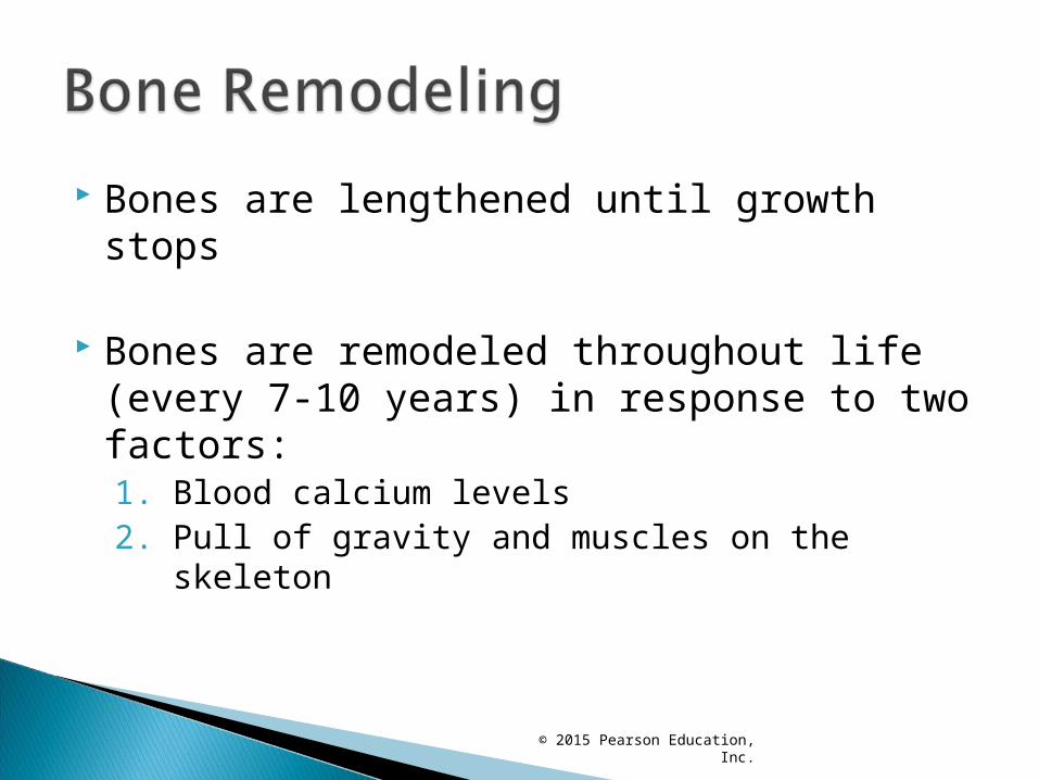
Bones are lengthened until growth stops
Bones are remodeled throughout life (every 7-10 years) in response to two factors:1. Blood calcium levels2. Pull of gravity and muscles on the skeleton
© 2015 Pearson Education, Inc.

Parathyroid hormone (PTH) ◦ Released when blood calcium levels are low◦ Activates osteoclasts (bone-destroying cells)
Osteoclasts break down bone and release calcium ions into the blood
Hypercalcemia (high blood calcium levels) prompts calcium storage to bones◦ Regulated by calcitonin (secreted by thyroid)
© 2015 Pearson Education, Inc.

http://highered.mheducation.com/sites/0072495855/student_view0/chapter6/animation__bone_growth_in_width.html

Fracture: break in a bone Types of fractures
© 2015 Pearson Education, Inc.

Bone fractures are treated by reduction and immobilization
◦ Closed reduction: bones are manually coaxed into position by physician’s hands
◦ Open reduction: bones are secured with pins, screws, or wires during surgery
© 2015 Pearson Education, Inc.

1. Hematoma (blood-filled swelling) is formed2. Fibrocartilage callus forms
1. A soft mixture of cartilage matrix, bony matrix, and collagen fibers splint the broken bone
3. Bony callus (hard) replaces the fibrocartilage callus1. Osteoblasts and osteoclasts migrate in
4. Bone remodeling- compact bone replaces cartilage
© 2015 Pearson Education, Inc.

Figure 5.7 Stages in the healing of a bone fracture.
Hematoma
1
Externalcallus
Internalcallus(fibroustissue andcartilage)
New bloodvessels
Spongybonetrabecula
Hematomaforms.
2 3 Fibrocartilagecallus forms.
Bony callusforms.
Boneremodelingoccurs.
Bonycallus ofspongybone
Healedfracture
4

Common Types of Fractures• Closed (simple) fracture: break that does not penetrate the skin
• Open (compound) fracture: broken bone penetrates through the skin
• Comminuted: bone breaks into many fragments
• Compression: bone is crushed
• Depressed: broken bone portion is pressed inward
• Impacted: broken bone ends are forced into each other
• Spiral: ragged break occurs when excessive twisting forces are applied to a bone
• Greenstick: bone breaks incompletely (common in children)
© 2015 Pearson Education, Inc.

Table 5.2 Common Types of Fractures.