Embed Size (px)
Citation preview

UNIT 3
How does experience affect behaviour and mental processes?
area oF StuDy 1
How does the nervous system enable psychological functioning?
cHaPter 2 Nervous system functioning
cHaPter 3 Stress as a psychobiological process
area oF StuDy 2
How do people learn and remember?
cHaPter 4 Neural basis of learning and memory
cHaPter 5 Models to explain learning
cHaPter 6 Process of memory
cHaPter 7 Reliability of memory
outcoMe 1
■ explain how the structure and function of the human nervous system enables a person to interact with the external world and analyse the different ways in which stress can affect nervous system functioning
■ apply biological and psychological explanations for how new information can be learned and stored in memory, and provide biological, psychological and social explanations of a person’s inability to remember information
c02NervousSystemFunctioning 1 12 July 2016 11:13 AMc02NervousSystemFunctioning 1 12 July 2016 11:13 AM
UNCORRECTED PAGE P
ROOFS

c02NervousSystemFunctioning 2 12 July 2016 11:13 AM
UNCORRECTED PAGE P
ROOFS

Key KnoWLeDge
■ the roles of different divisions of the nervous system (central and peripheral nervous systems and their associated subdivisions) in responding to, and integrating and coordinating with, sensory stimuli received by the body
■ the distinction between conscious and unconscious responses by the nervous system to sensory stimuli, including the role of the spinal refl ex
■ the role of the neuron (dendrites, axon, myelin and axon terminals) as the primary cell involved in the reception and transmission of information across the synapse (excluding details related to signal transduction)
■ the role of neurotransmitters in the transmission of neural information between neurons (lock-and-key process) to produce excitatory effects (as with
glutamate) or inhibitory effects (as with gamma amino butyric acid [GABA])
■ the effects of chronic changes to the functioning of the nervous system due to interference to neurotransmitter function, illustrated by the role of dopamine in Parkinson’s disease.
Roles of different divisions 00
Conscious and unconscious responses to sensory stimuli 00
Role of the neuron 00
Role of neurotransmitters 00
How interference to neurotransmitter function can affect nervous system functioning 00
cHaPter
2 Nervous system functioning
c02NervousSystemFunctioning 3 12 July 2016 11:13 AM
UNCORRECTED PAGE P
ROOFS

Unit 3 How does experience affect behaviour and mental processes?4
c02NervousSystemFunctioning 4 12 July 2016 11:13 AM
The human nervous system is a complex, highly organised network of specialised cells that enables the brain to receive information about what is going on from both inside and outside the body and to respond appropriately. Everything you sense, feel, think and do is controlled by your nervous system in some way. This includes not only your everyday sensing, perceiving, learning, remembering, thinking, imagining, speaking, moving and the vast array of other responses you voluntarily make, but also your involuntary responses such as breathing, heart rate, squinting when someone turns on a bright light in the middle of the night, and the ‘butterflies’ you may feel in your stomach when anxious or meeting someone special.
The nervous system achieves this by serving as a communication system between the body’s internal cells and organs and the external world. Through its vast network of nerves distributed throughout the body, the nervous system enables the brain to obtain information about what is going on inside and outside the body and to respond appropriately. Its three main functions are to:• receive information• process information, and• coordinate a response to information.
Although the nervous system is a single body system, it is made up of different sub-systems. These are commonly referred to as ‘divisions’ or ‘branches’. Although each division carries out identifiable functions, the nervous system functions as a coordinated whole.
As shown in figure 2.1, the two main divisions are the central nervous system and the peripheral nervous system. They are connected by the spinal cord and constantly work together maintaining communication throughout the body, thereby enabling us to not only think, feel and act as we do, but also to keep us alive.
The brain is kept continually informed of the ever-changing external and internal environments of the body through sensory information received by the many and varied receptor cells located at or near the surface of the body and also deep within the body. These sensory receptors specialise in detecting and responding to different types of information.
Sensory information from the external environment is received through sensory receptors that are sensitive to specific types of stimuli arising outside the body. For example, neurons that function as sensory receptors at the back of the eye respond only to light for vision, the inner ear contains receptors for hearing, balance and body position, and the skin has receptors that are responsive to touch, pressure, temperature and pain. The nervous system also receives information from within various parts of the body. For example, sensory receptors located in the muscles, joints and tendons provide information about muscle tension, position and movement, and receptors located in internal organs such as the heart, lungs, liver and intestines provide information about the body’s internal environment.
Central nervous system(CNS)
Carries messages toand from the PNS
BrainResponsible for
virtually everythingwe think, feel and do
Spinal cordConnects brain
and PNS initiatesspinal reflexes
Peripheral nervous system(PNS)
Carries messages toand from the CNS
Nervous system
Autonomicnervous systemConnects CNS to
internal organs andglands; self-regulating
Somaticnervous system
Carries messages fromsensory receptors in the
body to the CNS, andmotor messages from
the CNS to skeletalmuscles
Sympatheticnervous system
Prepares bodyfor action
Parasympatheticnervous system
Calms bodyafter actionFigure 2.1 The major divisions of the human nervous system
UNCORRECTED PAGE P
ROOFS

5CHAPtER 2 Nervous system functioning
c02NervousSystemFunctioning 5 12 July 2016 11:13 AM
When the sensory information is received at the brain it is processed. This enables perception — interpretation of the sensory information so meaning can be assigned. Processing often involves integrating (combining) incoming information with other information already in the brain. For example, incoming auditory and visual sensory information may be combined with information stored in memory in order to recognise what was seen and heard. If required, the brain will also coordinate a response by initiating appropriate action; for example, by sending neural messages to muscles, glands and internal organs. This, in turn, enables muscles to move, causes glands to secrete (release) hormones and initiates the responses of internal organs, thereby enabling our body systems to function effectively.
Neurons and glial cells (or glia) are the building blocks of the nervous system. Basically, neurons are responsible for communicating information and glia support their functions. For example, some glia surround neurons to provide a coating (i.e. myelin) that insulates them, whereas others clear up debris that could interfere with efficient neural transmission.
In this chapter we examine the roles of different divisions of the nervous system in responding to, and integrating and coordinating with, sensory stimuli received by the body. We also explore how the specialised structures and functioning of neurons allow the nervous system to transmit neural information throughout all points of the body.
RolEs of diffEREnt divisionsCentral nervous systemThe central nervous system (Cns) comprises the brain and its extension, the spinal cord. Its main function is to process information received from the body’s internal and external environments and to activate appropriate responses.
the brainThe brain is an intricate network of cells that plays a vital role in processing information received through neural pathways from the body and in directing actions within the body. It continuously receives and analyses sensory information, responding by controlling all bodily actions and functions. Because of its crucial role in almost everything we think, feel and do, it is sometimes called the ‘control centre’ or ‘master regulator’.
The brain is more than a mass of networked cells. Brain cells are organised into many identifiable areas (or ‘regions’) and structures that have specialised functions. For example, some parts are
dedicated to sensory or motor functions. Most parts, however, have integrating and overlapping functions. The apparently simple task of naming a familiar object, such as a car or mobile phone, will trigger activity in multiple structures and areas throughout the brain. These include areas at the back and side to process visual information received from the eyes, areas at the front, at the sides and near the centre to recover information from memory and to identify the object, and areas at the front involved in language and speech production to state the name of the object.
Many brain functions involve the activation of neural pathways that link different brain areas and structures. Neural pathways (also called tracts) comprise one or more circuits of interconnected neurons that form communication networks. Some span short distances and others extend from one side of the brain to the other. Neural pathways also connect the brain to other parts of the nervous system and the body. Although much is known about the brain’s neural circuitry, chemistry, structures and functioning, more remains unclear or unknown. For example, although it is known that different types of memory are associated with activity in distinctive parts of the brain, it is not fully understood how the brain goes about locating and retrieving specific memories when needed. Nor is it known exactly how different types of memories are actually stored.
Figure 2.2 The human brain has a complex structure and is responsible for virtually everything we think, feel and do.
UNCORRECTED PAGE P
ROOFS

Unit 3 How does experience affect behaviour and mental processes?6
c02NervousSystemFunctioning 6 12 July 2016 11:13 AM
processed by your brain such as the size, shape, texture, weight, distance and location of the bottle in relation to your eyes, mouth and hand, so that you can successfully execute a highly coordinated series of individual movements performed in one, well-timed, smooth action with just enough pressure to grasp the bottle and hold it without squeezing it too hard.The transmission of information along the
spinal cord, to and from the brain, occurs through interconnected neurons that form nerve pathways. When the spinal cord is injured, the brain loses both sensory input from and control over the body. The severity of feeling loss and paralysis depends on where the spinal cord is injured. The higher up on the spine the injury is, the greater the number of nerve connections between the brain and body that are severed.
The spinal cord has a relatively simple organisation but does more than provide pathways for messages to and from the brain. It can also initiate some simple motor reactions in the form of refl exes that occur extremely rapidly, independently of the brain. We consider the function of spinal refl exes and how they occur in the next section.
the spinal cordThe spinal cord is the long, thin bundle of nerve tissue that extends from the base of the brain to the lower back. As can be seen in fi gure 2.3, the spinal cord links the brain and the parts of the body below the neck.
Two major functions of the spinal cord are to:• receive sensory information from the body (via
the peripheral nervous system) and send these messages to the brain for processing. For example, an itch on your big toe, the sensation of heat as you step into a warm bath and the pain of a sprained wrist are all carried via the spinal cord to the brain area responsible for initially processing this type of sensory information
• receive motor information from the brain and send it to relevant parts of the body (via the peripheral nervous system) to control muscles, glands and internal organs so that appropriate actions can be taken. For example, to pick up a water bottle and bring it to your mouth for a drink, millions of neural messages are sent from the primary motor cortex to the muscles in your shoulder, upper arm, forearm, wrist and fi ngers. This is complemented by other relevant information that has been
Brain
(a) (b)
Brain
Spinal cord
Spinalcord
Figure 2.3 (a) The CNS consists of the brain and spinal cord. (b) Anatomically, the spinal cord links the brain and peripheral nervous system
UNCORRECTED PAGE P
ROOFS

7CHAPtER 2 Nervous system functioning
c02NervousSystemFunctioning 7 12 July 2016 11:13 AM
Peripheral nervous systemThe CNS does not have direct contact with the outside world. It relies on the peripheral nervous system to link it to the rest of the body so that messages can be carried to and from the brain via the spinal cord.
The peripheral nervous system (Pns) is the entire network of nerves located outside the CNS. It extends from the top of the head, throughout the body to the tips of the fi ngers and toes and to all parts of the skin. Its main function is to transmit information to and from the CNS. More specifi cally, the PNS:• carries information to the CNS from the body’s
muscles, organs and glands (about the internal environment) and from the sensory organs (about the external environment)
• carries information from the CNS to the body’s muscles, organs and glands.The peripheral nervous system does this through it
two subdivisions: the somatic nervous system and the autonomic nervous system.
3 Spinal cord carries motorinformation to the hand.
4 Motor neurons carry message toskeletal muscles of the hand andforearm.
5 Sensory receptors on the fingers send message to primarysomatosensory cortex that the bottle has been grasped.
8 Primary somatosensory cortexin parietal lobe receives messagethat the bottle has been grasped.
Sensorynerve
Motornerve
2 Premotor cortex in the frontal lobeplans the reach and adjacentprimary motor cortex commandsthe movement.
6 Spinal cord carries sensoryinformation to the brain.
7 Basal ganglia structures at thebase of the forebrain judge graspforces, and cerebellum at the rearcorrects movement errors toensure well-timed execution.
1 Prefrontal cortex in frontal lobesprocesses visual informationrequired to locate the target.Figure 2.4 Some of
the brain processes and information transmission via the spinal cord that occur to pick up a water bottle in one, well-timed, smooth action with just enough pressure to grasp the bottle and hold it without squeezing it too hard. Note that the right arm is picking up the bottle. This means that motor information will be sent from the brain’s left hemisphere (because it controls voluntary movements on the right side of the body) and somatosensory (‘body sense’) information will be sent to the brain’s left hemisphere.
Central nervous system (CNS)(blue)
Peripheral nervoussystem (PNS)
Brain
Spinal cord
Somatic (black)Sensory and motorfunctionControls voluntarymuscles
Autonomic (orange)Controls involuntarymuscles
SympatheticPrepares the bodyfor action
ParasympatheticCalms the body after action
Figure 2.5 The peripheral nervous system (PNS) consists of all nerves outside the CNS. It carries information to and from the CNS.
UNCORRECTED PAGE P
ROOFS

Unit 3 How does experience affect behaviour and mental processes?8
c02NervousSystemFunctioning 8 12 July 2016 11:13 AM
Box 2.1
Structure and function of brain areasNeuropsychologists often describe the brain using three main areas (or regions) — the hindbrain, midbrain and forebrain. This is based on how the brain develops
early in life. Each area is associated with identifiable mental processes and behaviour but these function in an integrated way to enable us to think, feel and behave as we do.
<insERt fig 2.6. imAgE mUst bE lARgE>
Figure 2.6
UNCORRECTED PAGE P
ROOFS

9CHAPtER 2 Nervous system functioning
c02NervousSystemFunctioning 9 12 July 2016 11:13 AM
Learning activity 2.1
review questions1 Describe three main functions of the human nervous
system, with reference to examples not used in the text.
2 Which part of the nervous system coordinates the activity of the entire nervous system?
3 (a) Describe the two main functions of the spinal cord in terms of the types of messages that travel up and down its length, and the branch of the nervous system to which it connects.
(b) What is a third function of the spinal cord?4 Explain why spinal cord damage can result in loss of
brain–body control.5 (a) What is the peripheral nervous system?
(b) What is its primary function?6 Describe the relationship between the central
nervous system and the peripheral nervous system, with reference to key functions of each division.
Learning activity 2.2
refl ectionThe term ‘peripheral’ means outlying or surrounding, suggesting lesser importance than the term ‘central’. With this in mind, suppose that your peripheral nervous system suddenly stopped ‘working’ for 30 seconds right now. Comment on your experience of the world during the 30 second period. What does this suggest about the importance of the PNS?
somatic nervous systemThe somatic nervous system (sns) is a network of nerves that carry sensory information to the CNS and motor information from the CNS. Sensory information is received at sensory receptor sites in the body (skin, muscles, joints and tendons) and carried along sensory neural pathways by sensory neurons. Motor information is carried along motor neural pathways by motor neurons to skeletal muscles to control their activity by causing them to contract or relax. Skeletal muscles are attached to our bones and respond to messages from the CNS to initiate, change or stop movement.
The sensory information is called afferent and the motor information efferent. These terms refer to the direction of the neural information fl ow. More specifi cally, afferent information is sensory information coming into the CNS (incoming information), whereas efferent information is motor information leaving the CNS (outgoing information).
The sensory function of the SNS is demonstrated when someone touches your hand. The SNS sends the sensory signals about touch from the skin to your brain, resulting in the sensation of touch (or pressure on the skin). The motor function of the SNS is demonstrated whenever voluntary actions are performed. For example, when you ‘text’, talk, chew, shower, surf or dance, your somatic nervous system is active.
Thus, the somatic nervous system is involved in all skeletal muscle activity that enables us to participate in our relationship with the external environment. Its nerves send information to the brain from the body’s various sensory receptors. These nerves also enable us to respond to these stimuli by moving through the environment.
Although motor pathways carry messages that initiate or stop movement, voluntary movement is controlled through the coordinated actions of both motor and sensory information. For example, when you use a pen to scratch your nose, your brain (primary motor cortex) sends messages via motor neurons to skeletal muscles in your arm, hands and fi ngers to move in specifi c ways. Sensory receptors in your skin and muscles send back messages through sensory neurons that help determine how much pressure is needed to hold the pen. However, your somatic nervous system does not make your heart beat faster when you’re suddenly threatened or regulate your internal environment. For these reactions, the other subdivision of the PNS is required—the autonomic nervous system.
Sensory neural pathwaysare incoming and afferent.
Motor neural pathwaysare outgoing and efferent.
Sensory receptors
Figure 2.7 Information fl ow in the somatic nervous system
UNCORRECTED PAGE P
ROOFS

Unit 3 How does experience affect behaviour and mental processes?10
c02NervousSystemFunctioning 10 12 July 2016 11:13 AM
Figure 2.8 Sensory nerves in the SNS carry information to the central nervous system when someone touches your hand. Your SNS is active when you voluntarily move, such as when walking up a set of stairs.
Learning activity 2.3
review questions1 (a) Briefly describe the two main functions of the
somatic nervous system. (b) Give an example of each of these functions, using
examples not referred to in the text.2 Distinguish between the afferent and efferent
information with reference to the type of information and the direction in which it is transmitted.
3 Whenever you reach to pick up a glass of water on a table, both the sensory and motor functions of the somatic nervous system are involved. Explain both the sensory and motor roles in grasping the glass.
4 This athlete has restricted movement due to paraplegia caused by spinal cord damage.
Explain the athlete’s restricted movement with reference to the somatic nervous system.
the autonomic nervous systemThe autonomic nervous system (Ans) is a subdivision of the PNS that connects the CNS to the body’s internal organs (such as the heart, stomach and liver) and glands (such as sweat, salivary and adrenal glands), providing feedback to the brain about their activities. The ANS is called ‘autonomous’ because many of the organs, glands and processes under its control are self-regulating and therefore occur without conscious effort and are not usually under our voluntary control. For example, your heartbeat, breathing, digestion and perspiration occur without you consciously activating or controlling them.
While skeletal muscles are completely inactive in the absence of motor neuron messages from the brain, the muscles involved in the activity of internal organs and glands (called visceral muscles) have built-in mechanisms for generating activity and do not depend on voluntary control by the brain. This is an important feature of the ANS, as it functions continuously — whether we are awake, active, asleep, under an anaesthetic or even in a coma. Regardless of our level of awareness or alertness, the ANS keeps the vital organs and systems of our body functioning, thereby maintaining our survival.
Unlike the somatic nervous system, which is responsible for initiating skeletal muscle movement, the ANS regulates the activity of the visceral muscles, organs and glands. Thus the messages carried between the CNS and the visceral muscles, organs and glands either increase or decrease their respective activities in response to the varying demands placed on the body throughout each day.
You often become consciously aware of ANS functions when you experience emotions such as fear, anger and excitement at intense levels because this is when there is heightened ANS
UNCORRECTED PAGE P
ROOFS

11CHAPtER 2 Nervous system functioning
c02NervousSystemFunctioning 11 12 July 2016 11:13 AM
activity. For example, think about how you can feel your heart and breathing rates change when you suddenly become very frightened, or during exhilarating moments on a roller-coaster ride. Recall also the physiological changes you can instantly feel when the fear or exhilaration start to diminish. Your heart rate noticeably slows and your breathing becomes more regulated. Any goosebumps or feelings of butterflies in your stomach will also eventually disappear.
The ANS is not completely self-regulating. It is linked to the brain’s cerebral cortex so we can voluntarily control a few autonomic responses at certain times. For example, with conscious effort, you could control your breathing rate right now.
Some people are able to use techniques they have learned to exercise extraordinary control over specific autonomic responses. For example, it has been reported that some Hindu holy men in India who are highly skilled yoga practitioners have been able to increase their heartbeat from the
normal resting rate of 75 beats or so per minute to 300 per minute without undertaking any physical activity, or have slowed their heartbeat to less than 50 beats per minute. Some have also been reported as being able to control their body temperature to the extent that one side of their hand is warm while the other side is cold (Blanchard & Young, 1973; Pines, 1973).
People who aren’t yogis can also learn to control various specific autonomic responses using a technique called biofeedback training. Biofeedback is a process by which a person receives information (‘feedback’) about the state of an internal bodily activity that normally occurs automatically, and then uses thought processes to exert control over that activity. The person learns a strategy, such as relaxation and/or visualisation, in order to control a particular autonomic response. Feedback about the state of the autonomic response being controlled is usually provided by a monitoring device connected to the person.
Figure 2.9 In outer space, the temperature is extremely cold and there is no oxygen. Astronauts wear special space suits to restrict heat loss and to maintain adequate oxygen pressure for brain function. On Earth, these functions occur automatically through the activity of the autonomic nervous system.
Learning activity 2.4
review questions1 (a) Explain why the autonomic nervous system is
described as autonomous. (b) Is ‘autonomous’ a truly accurate term for describing
this nervous system? Explain with reference to an example.
2 Explain the relationship of the autonomic nervous system to the central nervous system with reference to a physiological response.
3 What is a key difference between skeletal muscles and visceral muscles?
4 Which is more important in maintaining our survival: the autonomic nervous system or the central nervous system? Explain with reference to an example.
UNCORRECTED PAGE P
ROOFS

Unit 3 How does experience affect behaviour and mental processes?12
c02NervousSystemFunctioning 12 12 July 2016 11:13 AM
Learning activity 2.5
Distinguishing between the somatic nervous system and the autonomic nervous systemComplete the following table to indicate which division of the peripheral nervous system is more likely to be involved
in each of the following responses: the somatic nervous system (S), the autonomic nervous system (A) or both (B)?• pressing a key to send an email• eating dinner• sweating before having to give an important speech• clenching your fists while watching a scary movie• crouching on the blocks awaiting the starting siren before swimming in a 50-metre freestyle final• washing the dog• blinking• talking on the phone• laughing at a joke• heart races when you are startled by a loud noise
Responsesomatic nervous system (S), autonomic nervous system (A), both (S & A)
divisions of the AnsThe ANS consists of two distinct divisions that complement (‘balance’) each other, but generally have opposite effects. These are:• the sympathetic nervous system, which is
responsible for increasing the activity of most visceral muscles, organs and glands in times of vigorous activity, stress or threat
• the parasympathetic nervous system, which is responsible for decreasing the activity of most visceral muscles, organs and glands, and restoring body functioning to its normal state.The complementary actions of the sympathetic
and parasympathetic nervous systems occur without conscious effort and are demonstrated when you engage in an activity requiring physical exertion over a period of time. For example, when playing tennis vigorously, your sympathetic nervous system speeds up your heart rate to pump more blood and oxygen to your muscles. It causes your liver to release sugar into your bloodstream for energy, and induces sweating to keep your skin cool and prevent you from overheating. Because the body is pumping more blood and oxygen to the muscles, these are diverted from non-essential functions such as digestion, so this is inhibited. After the game, your parasympathetic nervous system takes over. Your heart rate slows, constricting the blood vessels in your muscles so the blood flow is diverted to the internal organs. Your sweat glands gradually slow down the production of sweat as the body returns to its ‘normal’ state.
The sympathetic and parasympathetic nervous systems do not function in an ‘on/off’ or ‘either/or’ way. They are both active at the same time. However,
one system is usually dominant at any given time. For example, the sympathetic division dominates and is more active during emotional arousal, whereas the parasympathetic division is dominant and more active during rest and digestion.
the sympathetic nervous systemThe sympathetic nervous system activates internal muscles, organs and glands to prepare the body for vigorous activity or to deal with a stressful or threatening situation. It is activated by a stressor or fear stimulus and enhances survival by providing an immediate response, in a split second, to any kind of emergency.
When you perceive an emergency or experience a crisis, the sympathetic nervous system activates specific organs and glands to respond. Glands that are activated include the adrenal glands, which are located just above your kidneys and release hormones (such as adrenaline) into the bloodstream. These circulate throughout your body, enhancing the effects of the sympathetic nervous system by activating various muscles, organs and other glands in preparation for dealing with the stressor or potential threat.
The result is that your heart rate and blood pressure increase, and your breathing rate increases so more oxygen can be taken in. Sugar and fat are released from storage to provide instant energy to the skeletal muscles. Your pupils dilate (‘expand’) to allow more light to enter the eye and enhance vision. Your sweat glands increase production of sweat to cool the body. In addition, digestion is slowed down. The sympathetic nervous system is also involved when you blush or get goosebumps, making the hairs on your body stand on end (see box 2.2).
UNCORRECTED PAGE P
ROOFS

13CHAPtER 2 Nervous system functioning
c02NervousSystemFunctioning 13 12 July 2016 11:13 AM
Figure 2.10 The sympathetic nervous system is activated in both these animals.
Box 2.2
goosebumpsGoosebumps appear when the fine hairs on your skin stand on end. Their appearance is controlled by the sympathetic nervous system.
Human body hairs are so short that when they become erect, nothing much happens. The response of goosebumps has been described as an evolutionary response linked to our ancient ancestors, who had hairier bodies. Erecting the hairs helps non-human mammals conserve their body warmth in a cold environment by increasing insulation around their bodies. In several species it also serves as a defence against enemies in emergency situations. Consider, for example, a frightened cornered cat. By erecting its hairs, it looks larger and by doing so may deter its opponent.
The echidna’s quills, which are an effective defence against potential predators, are actually modified body hairs. In an emergency situation, sympathetic nervous system activity leads to erection of the quills, just as it leads to erection of hairs in other mammals. The
behaviour that makes the quills so useful (their erection in response to fear) is said to have evolved before the quills themselves did.
Figure 2.11
the parasympathetic nervous systemIn times of minimal stress and in the absence of threat, the parasympathetic nervous system helps to maintain the internal body environment in a steady, balanced state of normal functioning. The parasympathetic nervous system generally has the effect of counterbalancing the activities of the sympathetic nervous system. It restores the body to a state of calm, once the need for sympathetic nervous system activation has passed.
The parasympathetic nervous system dominates the sympathetic nervous system most of the time. It is involved in routine, everyday activities. For example, when you eat, the parasympathetic nervous system stimulates the stomach and intestines to digest food. It is also involved in the elimination of wastes and the protection of the visual system through the production of tears and through automatic pupil constriction in conditions
UNCORRECTED PAGE P
ROOFS

Unit 3 How does experience affect behaviour and mental processes?14
c02NervousSystemFunctioning 14 12 July 2016 11:13 AM
of bright light. In addition, when returning the body to a balanced state, the parasympathetic nervous system reduces heart and breathing rates, and minimises the release of sugar (glucose) and fats into the bloodstream.
If you had to jump out of the way of an oncoming car, your sympathetic nervous system would immediately be activated. Once the danger had passed, your parasympathetic nervous system would take over and the various bodily systems and functions activated by the sympathetic nervous system would gradually begin to return to normal. The parasympathetic nervous system takes longer to return the body to its normal state compared with the sympathetic nervous system’s immediate activation. This is because of the lingering presence of the hormones that are released when the sympathetic nervous system is activated. They remain in the bloodstream for some time after the threat has passed.
taBLe 2.1 The activities of the sympathetic and parasympathetic nervous systems
bodily organ bodily function sympathetic nervous system action
Parasympathetic nervous system action
Pupils Regulate the amount of light entering the eye
Dilate (expand) Contract
Salivary glands Digestion Decrease salivation Increase salivation
Heart Pumps blood Accelerates heart rate Slows heart rate
Bronchioles of lungs Breathing Dilate (expand) Contract
Stomach Digestion Decreases contractions Increases contractions
Liver Produces bile to aid digestion
Maintains blood sugar (glucose) level
Increases the release of glucose (sugar)
Decreases the release of glucose (sugar)
Gall bladder Stores bile Inhibits the release of bile Stimulates the release of bile
Adrenal glands Secrete the hormones adrenaline (epinephrine) and noradrenaline (norepinephrine) from the medulla
Stimulate hormone secretion resulting in increased heart rate, blood pressure and breathing rate, and relaxation of intestinal muscles
Inhibit hormone secretion
Bladder Stores urine Relaxes Increases contractions
Intestine Digestion Relaxes Increases contractions
Genitals Reproduction Excite Relax
Sweat glands Regulate temperature Increase production of perspiration
Decrease production of perspiration
Learning activity 2.6
review questions1 In what main way do the sympathetic nervous system
and the parasympathetic nervous system differ?2 (a) What is the role of the sympathetic nervous system
in enhancing survival? (b) Give three examples of bodily functions that
increase their activity as a result of sympathetic nervous system activation.
(c) Give three examples of bodily functions that decrease their activity as a result of sympathetic nervous system activation.
3 (a) Describe the main roles of the parasympathetic nervous system.
(b) Give three examples of bodily functions that are affected as a result of the action of the parasympathetic nervous system. Briefly explain the purpose of these changes if resulting from parasympathetic nervous system activation.
4 Explain why it can take longer for the parasympathetic nervous system to ‘slow down’ bodily functions than it does for the sympathetic nervous system to ‘speed up’ bodily functions.
Figure 2.12 Riding on a roller-coaster activates the sympathetic nervous system. After the ride is over, the parasympathetic nervous system restores the body to a state of calm.
UNCORRECTED PAGE P
ROOFS

15CHAPtER 2 Nervous system functioning
c02NervousSystemFunctioning 15 12 July 2016 11:13 AM
Learning activity 2.7
Sympathetic versus parasympathetic nervous systems1 Which division of the autonomic nervous system is likely to
be dominant if you are in each of the following situations? (a) lying on the beach reading a book (b) waiting for the delivery of your VCE results (c) feeling anxious about a blind date (d) hearing an unexpected loud knock on the window at
2 am while watching TV alone (e) eating dinner
(f) watching a terrifying scene in a movie (g) sitting in class listening with interest to a teacher’s
explanation.2 Which division of the autonomic nervous system
is likely to be dominant when each of the following physiological responses is observed?
(a) increased rate of digestion (b) decreased salivation (c) increased pulse rate (d) decreased pupil size (e) increased perspiration.
Learning activity 2.8
Summarising the activities of the sympathetic and parasympathetic nervous systems
Copy the diagram below, then summarise the activities of the sympathetic and parasympathetic nervous
systems. Insert your answers on the lines connecting the various organs and glands, as shown in the example for the pupil.
Central nervous system
Sympathetic
Pupil
Salivary glands
Lungs
Heart
Intestine Stomach
Pancreas Gall bladder
Liver
Kidney Adrenal gland
Bladder
Parasympathetic Central nervous systemDilates Contracts
UNCORRECTED PAGE P
ROOFS

Unit 3 How does experience affect behaviour and mental processes?16
c02NervousSystemFunctioning 16 12 July 2016 11:13 AM
ConsCioUs And UnConsCioUs REsPonsEs to sEnsoRy stimUliSome people believe that we use only 10% of our brain and the rest is a huge reservoir of untapped potential for some kind of remarkable ‘power’. The reality is that we ordinarily use virtually every part of the brain, and that the brain is active almost all the time. In neurosurgery, where it is possible to observe the functions of a patient’s brain under local anaesthetic while the patient is awake, electrical stimulations in virtually all parts show activity at the neuronal level, even when no sensory experience, movement or any other reaction is being observed. Moreover, no areas of the brain are completely inactive, even during sleep. If they were, it would indicate a serious functional disorder (CERI, 2007; Horstman, 2009; Kolb & Whishaw, 2014).
Our brain and nervous system are constantly processing sensory stimuli detected by sensory receptors and organs that respond to the different types of information received from both our internal and external environments. Our responses to these internally and externally sourced stimuli may be conscious or unconscious. Psychologists distinguish between these reactions primarily in terms of whether or not there is awareness.
A conscious response to a sensory stimulus is a reaction that involves awareness. You will have paid attention to the stimulus and therefore know about it. The response will usually be a voluntary, ‘intentional’ reaction. The reaction, even if momentary, is also likely to be goal directed (‘purposeful’) and you will be able to exercise some degree of control over it.
In the course of a typical day we make numerous conscious responses of varying complexity to all kinds of external sensory stimuli that bombard our senses. For example, when you step outside and feel the air temperature you will make a conscious response when you decide whether to put on a jacket. Similarly, if the sun is shining brightly, you may choose to wear sunglasses, a hat or both.
A conscious response may also be made to an internally sourced stimulus, as might occur if you feel a stomach ache in class at school. Depending on the severity of the ache, you may decide to ignore it, stroke your stomach, tell someone about it, excuse yourself and leave the room, or react in some other way that you believe is best.
An unconscious response to a sensory stimulus is a reaction that does not involve awareness. It is involuntary, unintentional, automatic and we cannot ordinarily control its occurrence. Bodily responses regulated by the ANS occur automatically without conscious effort. For example, in response to stimuli about the state of different bodily systems, your ANS is unconsciously regulating their functioning, pumping blood from your heart, digesting your food and so on. You don’t consciously have to think about making
your heart beat, your eyes blink or your lungs fill with oxygen. Many of these ANS functions are actually reflexive responses (called autonomic reflexes).
Other reflexive responses also serve to help us avoid danger and minimise harm. Sometimes, we need to react so quickly that there is no time for conscious thought. These unconscious, automatically occurring responses are reflexes involving contraction of skeletal muscles. Most are very simple responses. They occur in the same way each time and do not require learning. We consider the role of the spinal reflex.
Figure 2.13 Reflexes are unconscious, automatic involuntary behaviours that does not require prior experience and occur in the same way each time. We are born with a large number of reflexes, but most of these disappear or are incorporated as parts of other behaviours within several months after birth. a. The sucking reflex is an involuntary response that is important for survival as it enables the newborn infant to feed. b. This newborn infant demonstrates the grasping reflex. Although it is strong enough to allow infants to support their own weight, this reflex disappears within the first few months of life, like many other reflexive behaviours we are born with.
the spinal reflexThe spinal cord does more than provide pathways for messages to and from the brain. It can also initiate some simple responses on its own independently of the brain. These responses include spinal reflexes.
A spinal reflex is an unconscious, involuntary and automatically occurring response to certain stimuli without any involvement of the brain. It is often referred to as a reflex arc because the response to an incoming stimulus is automatically ‘reflected back’ from the spinal cord without any initial input from the brain and before the brain processes a conscious perception of the stimulus.
For example, if you were to touch the hot handle of a frying pan, you would automatically withdraw your hand to release the handle before the sensory information travels all the way to your brain and therefore before pain is actually experienced. The sensory receptors in your finger would send messages to your CNS, but the first point of contact in the CNS is the spinal cord. It responds with a message via motor
UNCORRECTED PAGE P
ROOFS

17CHAPtER 2 Nervous system functioning
c02NervousSystemFunctioning 17 12 July 2016 11:13 AM
neurons to move the appropriate muscles in your hand to release the hot object and withdraw the hand.
The immediate response at the spinal cord enables a faster reaction time, a fraction of a second before the sensory information reaches the brain. Consequently, a spinal refl ex involving a withdrawal reaction is believed to be an adaptive response. The spinal refl ex is adaptive in that it saves time in a situation that may be very harmful to the organism. When a spinal refl ex occurs, the spinal cord responds to the message directly, before the message is carried further on to the brain. While the transmission of information from the spinal cord to the brain only takes a fraction of a second, this saved time may be important in terms of minimising harm, or even saving the life of the organism. Other examples of this type of spinal refl ex are when you jerk your bare foot up from the hot pavement and when you withdraw your hand if you touch a sharp rose thorn or needle.
Because refl exes are normally so predictable they provide useful information about the functioning of
Spinal cord(cross section)
1 Sensory neurons carry the message along a sensory pathway to the spinal cord.
5 The message is received in the area of the brain that processes this type of sensory information and interprets it as pain in the hand.
4 While the spinal reflex occurs, sensory neurons are also carrying the message further up the spinal cord to the brain.
3 Motor neurons carry the message along a motor pathway to hand muscles, causing a withdrawal reflex. The frying pan is released before the brain perceives pain.
2 Interneurons in the spinal cord relay the message to motor neurons.
Figure 2.14 This sequence shows a spinal refl ex involving a withdrawal response. Sensory receptors respond to the stimulation and initiate a neural message that is carried by sensory neurons to interneurons in the spinal cord. Interneurons act as a link between sensory and motor neurons, relaying information from one to the other (because sensory and motor neurons rarely ever connect directly). The interneurons send the message to motor neurons that carry a message back to the appropriate muscles, causing them to contract and pull away from the stimulus. The spinal cord will also carry the message to the brain, including information about the action taken. The hot and potentially harmful saucepan handle is released before the brain processes the conscious perception of pain.
the nervous system, and greatly assist in the diagnosis of neural disorders. Damage or disease anywhere along its refl ex arc can cause a refl ex to be absent or abnormal. For example, when the knee is tapped on the patellar ligament, the sensory nerve that receives this stimulus carries the information to the spinal cord, where it is relayed to a motor nerve. This normally causes the quadriceps muscle at the front of the thigh to contract and jerk the leg up. The leg begins to jerk up while the brain is just becoming aware of the tap. Absence of this patellar refl ex could indicate damage within sensory or motor pathways, or a spinal cord injury in the lower back area (Jenkins, Kemniz & Tortora, 2010).
The spinal refl ex demonstrates that a response to a particular sensory stimulus can have both an unconscious and conscious component, one occurring before the other. For each refl ex action, a handful of neurons simply convert a sensori stimulus into action. These neurons are shown in fi gure 2.14. We consider their specifi c roles in the next section.
UNCORRECTED PAGE P
ROOFS
Unit 3 How does experience affect behaviour and mental processes?18
c02NervousSystemFunctioning 18 12 July 2016 11:13 AM
Learning activity 2.9
review questions1 (a) Distinguish between a conscious and unconscious
response by the nervous system to a sensori stimulus with reference to three key points.
(b) Give an example of when you may respond to an external stimulus before you know that you have responded.
2 Formulate a definition for a reflex.3 (a) Explain what a spinal reflex is. (b) Why is it also called a reflex arc? (c) Why is a spinal reflex considered to have an
‘adaptive’ or ‘survival’ role? (d) Give an example of a reflex response that you
believe may not be involved in a spinal reflex arc. Explain your choice of example.
4 Draw a simple flow chart to name the three types of neurons involved in a spinal reflex and show the sequence in which they contribute to the response.
5 How might damage to interneurons affect the spinal reflex?6 Sam is using a wet knife to remove a broken piece
of toasted bread that is jammed in the toaster. She experiences an electric shock and spontaneously releases the knife and pulls her hand away from the stimulus.
(a) Will Sam experience pain? Explain your answer. (b) List, in their correct order, the steps that enabled
Sam’s spinal reflex.
Learning activity 2.11
Main divisions and subdivisions of the human nervous systemComplete the following chart to show the main divisions and subdivisions of the human nervous system.
Nervous system
Learning activity 2.10
visual presentationBarefooted Jake steps on the sharp end of a drawing pin and immediately jerks up his foot before completing the step.
Create a flow chart which outlines the sequence of events in Jake’s spinal reflex response and refers to the relevant anatomical features.
You may present your flow chart in a digital format, for example, as a Powerpoint slide with SmartArt graphics. The flow chart may include a copy of the stimulus event shown above.
UNCORRECTED PAGE P
ROOFS
19CHAPtER 2 Nervous system functioning
c02NervousSystemFunctioning 19 12 July 2016 11:13 AM
RolE of tHE nEURonThe human nervous system consists of billions of neurons and glial cells. The neuron is the primary cell involved in the reception and transmission of information throughout the nervous system and different types of glial cells support neuronal function.
When even one part of the process breaks down, the results can be devastating. Many brain disorders and nervous system diseases, including schizophrenia, Alzheimer’s disease, Parkinson’s disease, epilepsy, multiple sclerosis and even botulism that causes paralysis, have been linked to problems with neurons and communication within and between neurons. Glial cells are also implicated in most neurological disorders and diseases given their numbers and crucial roles (Fields, 2011; Miller, Cookson & Dickson, 2004).
A neuron is an individual nerve cell that is specialised to receive, process and/or transmit information. Neurons not only communicate with each other, but also with muscles and glands. They are the building blocks of the brain and nervous system. The entire nervous system is comprised of neurons organised into networks that form neural circuits and pathways of varying complexity through which information is continuously transmitted. With support from glial cells, communication throughout the nervous system is an extremely fast and efficient process.
Figure 2.15 The entire nervous system is comprised of neurons which interconnect to form neural circuits and pathways, enabling the continuous communication of information around the entire body. Glial cells within the nervous systems support neural activity. This computer enhanced image highlights its complexity.
Neurons are also described as the ‘primary functional units’ of the nervous system because of their vital role in enabling the nervous system to function as it does. They carry information (‘neural
messages’) in the form of an action potential (or neural impulse) to the appropriate part of the nervous system, or interpret the message and enable a response.
Neurons have specialised functions and vary in shape and size depending on where they are located and on their specific function. Some neurons specialise in transmitting (sending) information from sensory receptors, sensory organs, tendons or muscles to the CNS. Other neurons specialise in sending information to cells in bodily organs, muscles or glands from the CNS. Some neurons simply serve as communication links and carry information between neurons (see box 2.3). However, most neurons typically have several structural features in common. These include dendrites, an axon, myelin and axon terminals.
dendritesA dendrite is an extension of a neuron that detects and receives information from other neurons. As shown in figure 2.16, dendrites separate out like the branches of a tree. Most dendrites have little protrusions, or ‘outgrowths’, called dendritic spines. Each spine provides a site with receptors where a neuron can connect with and receive information from a neighbouring neuron. The neuron’s capability to form (‘grow’) new dendritic spines is associated with and demonstrates neuroplasticity.
A neuron may have from one to 20 dendrites, each dendrite may have from one to many branches, and the total numbers of spines on the branches may be in the hundreds or thousands. This means that a single neuron can have many thousands of connections to other neurons through its dendritic branches and spines. Each spine may have multiple kinds of receptors to gather different types of chemical messages from other neurons. Consequently, a single neuron can have the capability to receive, virtually simultaneously, many and various kinds of information from dozens, hundreds, or even thousands of other neurons.
When the dendrites receive information from other neurons, they pass it on to the neuron’s soma (cell body) where it is integrated. The soma may collect and integrate information from thousands of other neurons. Once the incoming information from other neurons has been integrated in the soma, it is transmitted along the axon.
AxonAn axon is a single, tubelike, extension that transmits neural information to other neurons (or cells in muscles and glands). Most neurons have only one axon but many axons have branches that allow a message to be sent to multiple cells. Axons vary in length; for example, some axons extend over a metre from your spine to your big toe, others are as small as the width of a single hair. Nerves are actually cable-like bundles of multiple axons (see figure 2.16).
UNCORRECTED PAGE P
ROOFS

Unit 3 How does experience affect behaviour and mental processes?20
c02NervousSystemFunctioning 20 12 July 2016 11:13 AM
myelinThe axons of many, though not all, neurons are myelinated. myelin is a white, fatty substance (made up of certain types of glial cells) that surrounds and insulates the axon, much like the plastic tubing around copper wires in an electrical cord. Without the coating, called the myelin sheath, interference from the activity of other nearby axons may occur. In addition, the myelin sheath allows for the rapid movement of the message along the axon without being interrupted or distorted. Messages travel much faster through neurons wrapped in myelin than unmyelinated neurons.
The myelin sheath is not continuous along the full length of the axon. It occurs in segments that are separated by small unmyelinated gaps (called nodes of Ranvier). The neural message jumps from node to node and this is believed to speed up transmission.
The importance of the myelin sheath is evident in people with multiple sclerosis. In this disease, the body’s immune system attacks the myelin or the glial cells that produce and maintain it. Recurrent attacks by antibodies break down the myelin (called demyelination) and the axons themselves may also be damaged. The cause is still unknown. It may be due to something which causes the immune system to react in this way, or because there are issues with the myelin and that the immune system attempts to clean up the damage. Demyelination ultimately creates areas of scarring (‘sclerosis’) which disrupt the transmission of
Soma
Dendritic spineDendrites
Myelin sheath
Nodes of Ranvier
Nucleus
Axon
Terminal buttonsAxon terminals
Axon collateralsSoma
Dendrite
Axon
Figure 2.16 Incoming neural messages are received by the dendrites and transmitted to the soma (cell body) where the information is integrated. Outgoing information is transmitted from the soma to the axon terminals and on to dendrites of other neurons. Within the neuron, the message is in an electrical form called an action potential (or neural impulse).
neural messages within the nervous system. This can result in impairment of motor, sensory and cognitive functions to a greater or lesser extent, depending on which part of the nervous system is damaged and the severity of the damage (MS Australia, 2016).
Figure 2.17 Myelin surrounds and insulates the axon, as well as allowing for the rapid movement of the message along the axon. This photo, taken using a coloured scanning electron micrograph (SEM), shows myelinated nerve fi bres.
UNCORRECTED PAGE P
ROOFS

21CHAPtER 2 Nervous system functioning
c02NervousSystemFunctioning 21 12 July 2016 11:13 AM
Axon terminalsThere are small branches at the end of an axon called axon collaterals. At the end of the collaterals are axon terminals. Each axon terminal has a small knob-like swelling at its tip called a terminal button (sometimes called a synaptic vesicle, synaptic knob or synaptic button). The terminal button is a small structure like a sac that stores and secretes neurotransmitter that is manufactured by the neuron and carries its chemical message to other neurons or cells.
Information always travels in one direction through a neuron. It is received by dendrites, passes through the soma and exits from the axon. The neural message to be sent by a neuron originates at the soma. It is in the form of an electrical signal. When it reaches the axon terminals, it stimulates the release of neurotransmitter from the terminal buttons. The neurotransmitter will carry the message to the next neuron in a chemical form. Although each neuron has only one axon, the collaterals and axon terminals allow its message to be sent to many other neurons simultaneously.
Neuron 1 Neuron 2
Axon
Axon terminalTerminal button
A bundle of axons running together form a nerve.
Figure 2.18 The term ‘nerve’ is commonly used for a neural pathway or circuit outside the CNS and ‘tract’ for a pathway within the CNS.
Box 2.3
three type of neuronsNeurons can be classified in terms of their specific function and the direction that they send information. The three classes are sensory neurons, motor neurons and interneurons.
Sensory and motor neurons are found throughout the nervous system, whereas interneurons are found only in the CNS.
Sensoryreceptor
(a) (b) (c)
Actionpotential
Actionpotential
Actionpotential
Dendrite
Axon
Axon
Soma
Myelin sheath
Nucleus
Nucleus
Soma
Axon
Dendrites Dendrites
Soma
Nucleus
Axonterminals
Axonterminals
Motor neuron InterneuronSensory neuron
Axonterminals
Figure 2.19 (a) Sensory neuron receives and carries sensory information from both the external and internal environments and transmits to the CNS. Also called an afferent neuron or affector. (b) Motor neuron carries messages from the CNS to cells in skeletal muscles, organs and glands to stimulate activity. Also called an efferent neuron or effector or motoneuron. (c) Interneuron sends messages between sensory and motor neurons within the CNS, relaying information from one to the other (because sensory and motor neurons rarely ever connect directly). Also called a connecting or association neuron.
(continued)
UNCORRECTED PAGE P
ROOFS

Unit 3 How does experience affect behaviour and mental processes?22
c02NervousSystemFunctioning 22 12 July 2016 11:13 AM
(continued from previous page)
Figure 2.20 shows the interaction between the three types of neurons to enable a spinal refl ex.
Interneuron
Spinal cord
Sensory neuron
Motorneuron
Sensoryreceptors
Figure 2.20 Interaction between three types of neurons
Learning activity 2.12
review questions1 What is a neuron?2 Explain the meaning of the statement ‘Neurons are the
primary functional units of the entire nervous system’.3 In what way are the structure and functions of a
dendrite different from those of axon terminals?4 (a) Label the following diagram of a neuron. A copy of the
diagram can be accessed in your eBook. It may also be labelled within, then printed from your eBook.
(b) Draw each of the following on the diagram i dendrites of an adjacent neuron ii axon of an adjacent neuron
5 Match the part of the neuron in the list with its role in the transmission of a neural message.
a. myelin d. axon g. axon collateral b. terminal button e. axon terminal h. soma c. dendrite f. dendritic spine ___ integrates information received by dendrites ___ receives information from another neuron ___ provides a specifi c receptor site for neural
information ___ transmits neural information to another neuron ___ white, fatty substance surrounding an axon that
insulates the neuron and increases the speed of neural communication
___ small branch at the end of an axon ___ extension of an axon collateral ___ stores and secretes neurotransmitter The correct answers can be found at the end of the text.
4
5
3
1
2
Figure 2.21
UNCORRECTED PAGE P
ROOFS

23CHAPtER 2 Nervous system functioning
c02NervousSystemFunctioning 23 12 July 2016 11:13 AM
RolE of nEURotRAnsmittERsWhen neurons communicate with one another, they do so by sending neurotransmitter across the tiny space between the terminal buttons of one neuron, which release the neurotransmitter, and the dendrites of another, which receive the neurotransmitter. This tiny space is called the synaptic gap (or synaptic cleft). The synaptic gap is about 500 times thinner than a strand of your hair. It is one component of the synapse. The synapse is the site where communication occurs between adjacent neurons. The other two components of the synapse are the terminal buttons of the presynaptic (‘sending’) neuron and the dendrites of the postsynaptic (‘receiving’) neuron.
neurotransmitter is a chemical substance produced by a neuron that carries a message to other neurons or cells in muscles, organs or other tissue. When carrying a message to another neuron, neurotransmitter works by attaching itself (‘binding’) to receptor sites of postsynaptic neurons that are specialised to receive that specific neurotransmitter. Therefore, receptors on dendrites play a vital role in the communication process.
Neurotransmitter that does not bind to receptors in the postsynaptic neuron is absorbed back into the terminal buttons by the presynaptic neuron in a process called reuptake. Once the postsynaptic neuron has received the neurotransmitter, any additional neurotransmitter that is left in the synapse will also be reabsorbed by the presynaptic neuron. Many medications work by affecting the process of reuptake in order to increase or reduce the availability of a particular neurotransmitter(s) in the brain.
Generally, a specific type of neurotransmitter will have either of two effects. Some neurotransmitters have an excitatory effect and consequently stimulate or activate postsynaptic neurons to perform their functions. Other neurotransmitters have an inhibitory effect and block or prevent postsynaptic neurons from firing.
Some neurotransmitters also occur as hormones. For example, noradrenaline (also called norepinephrine) is a neurotransmitter and a hormone. It is secreted as a hormone by the adrenal glands into the blood, and as a neurotransmitter from neurons.
Glutamate and GABA are the most common neurotransmitters in the CNS. Neurons in virtually every brain area use these two chemical messengers to communicate with each other.
glutamate (glu) is the primary excitatory neurotransmitter in the CNS. This means that glutamate enhances information transmission by making postsynaptic neurons more likely to fire. It is the second most abundant neurotransmitter in the brain and involved in most aspects of normal brain function, including perception, learning, memory, thinking and movement. The release of glutamate is associated with enhanced learning and memory.
Figure 2.22 Neurons do not link together like a chain. The branches of an axon almost touch the dendrites of an adjacent neuron, leaving a tiny space called a synaptic gap.
Synapse
Terminalbuttons
Axon terminals
Dendritic spines
Axon
DendritesSoma
Myelin sheath
Terminal buttons
Axon terminal
Neural message frompresynaptic (sending)neuron (action pontential)
Synaptic gap
Dendrite (or dendritic spine)of postsynaptic receiving neuron
Synapse
Receptor site
Neurotransmitter
Figure 2.23 When the neural message reaches the axon terminal, neurotransmitter is released from the terminal buttons, which carry the message across the synaptic gap to the receiving neuron.
UNCORRECTED PAGE P
ROOFS
Unit 3 How does experience affect behaviour and mental processes?24
c02NervousSystemFunctioning 24 12 July 2016 11:13 AM
Despite its importance, too much or too little glutamate can actually be harmful to neurons and brain functioning as a whole. For example, abnormally high concentrations of glutamate can lead to overexcitation of receiving neurons. This overexcitation can lead to effects that can cause neuronal damage and/or death by overstimulating them.
gamma-amino butyric acid (gAbA) is the primary inhibitory neurotransmitter in the CNS. It works throughout the brain to make postsynaptic (‘receiving’) neurons less likely to fire (i.e. it ‘inhibits’ firing). One of its roles is to fine-tune neurotransmission in the brain and maintain neurotransmission at an optimal, or ‘best possible’, level.
Without the inhibitory effect of GABA, activation of postsynaptic neurons might get out of control. Their uncontrolled activation could spread throughout the brain, causing seizures similar to those of epilepsy and other problems. Anxiety symptoms such as those experienced by people with phobias have been connected to a low level of GABA in the brain, thereby impacting on the regulation of neuronal transmission in the brain. The link between anxiety and a dysfunctional GABA system is examined in chapter 13 on page 000.
In sum, the inhibitory action of GABA counterbalances the excitatory activity of glutamate and vice versa. Consequently, GABA and glutamate have important roles in regulating central nervous system arousal.
Glutamate GABA
Figure 2.24 GABA is an inhibitory neurotransmitter and makes receiving neurons less likely to fire. Glutamate is an excitatory neurotransmitter and makes receiving neurons more likely to fire. The inhibitory action of GABA counterbalances the excitatory activity of glutamate and vice versa.
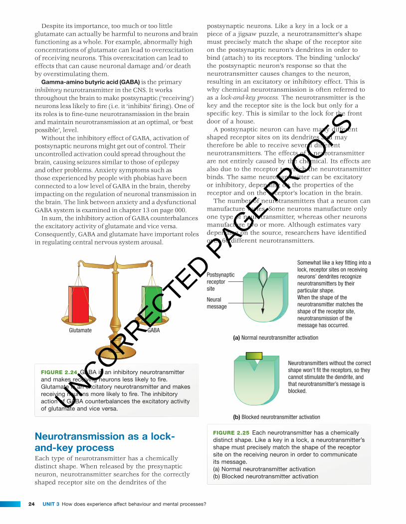
neurotransmission as a lock-and-key processEach type of neurotransmitter has a chemically distinct shape. When released by the presynaptic neuron, neurotransmitter searches for the correctly shaped receptor site on the dendrites of the
postsynaptic neurons. Like a key in a lock or a piece of a jigsaw puzzle, a neurotransmitter’s shape must precisely match the shape of the receptor site on the postsynaptic neuron’s dendrites in order to bind (attach) to its receptors. The binding ‘unlocks’ the postsynaptic neuron’s response so that the neurotransmitter causes changes to the neuron, resulting in an excitatory or inhibitory effect. This is why chemical neurotransmission is often referred to as a lock-and-key process. The neurotransmitter is the key and the receptor site is the lock but only for a specific key. This is similar to the lock for the front door of a house.
A postsynaptic neuron can have many different shaped receptor sites on its dendrites and may therefore be able to receive several different neurotransmitters. The effects of a neurotransmitter are not entirely caused by the chemical. Its effects are also due to the receptor to which the neurotransmitter binds. The same neurotransmitter can be excitatory or inhibitory, depending on the properties of the receptor and on the receptor’s location in the brain.
The number of neurotransmitters that a neuron can manufacture varies. Some neurons manufacture only one type of neurotransmitter, whereas other neurons manufacture two or more. Although estimates vary depending on the source, researchers have identified over 60 different neurotransmitters.
Postsynapticreceptorsite
Neuralmessage
(a) Normal neurotransmitter activation
(b) Blocked neurotransmitter activation
Neurotransmitters without the correctshape won’t fit the receptors, so theycannot stimulate the dendrite, andthat neurotransmitter’s message isblocked.
Somewhat like a key fitting into alock, receptor sites on receiving neurons’ dendrites recognizeneurotransmitters by theirparticular shape.When the shape of theneurotransmitter matches theshape of the receptor site,neurotransmission of themessage has occurred.
Figure 2.25 Each neurotransmitter has a chemically distinct shape. Like a key in a lock, a neurotransmitter’s shape must precisely match the shape of the receptor site on the receiving neuron in order to communicate its message.(a) Normal neurotransmitter activation(b) Blocked neurotransmitter activation
UNCORRECTED PAGE P
ROOFS

25CHAPtER 2 Nervous system functioning
c02NervousSystemFunctioning 25 12 July 2016 11:13 AM
While communication between one neuron and another is usually a chemical process involving neurotransmitters, communication between neurons also occurs in other ways. In some instances, communication between neurons is electrical; for
example, when axons transmit messages directly to other axons or directly to the cell body of other neurons and when dendrites of one neuron communicate directly with the dendrites of other neurons.
Figure 2.26 Acetylcholine (ACh) is the neurotransmitter that regulates the contraction of the muscles, including those required for breathing. In South America, indigenous Indian hunters dip their arrows and blowgun darts into a poison called curare, which blocks that action of ACh, paralysing their prey.
Learning activity 2.13
review questions1 (a) What is neurotransmitter? (b) What role does neurotransmitter play in neural
communication at a synapse?2 (a) What is a synapse? (b) Name the three components of a synapse and outline
their roles in communication between neurons.3 Mark each of the following on the following diagram. synaptic gap neurotransmitter synapse axon terminal terminal button receptor site postsynaptic neuron presynaptic neuron
Neural message
4 Distinguish between excitatory and inhibitory effects of a neurotransmitter with reference to glutamate and GABA.
5 Outline the chemical neurotransmission process with reference to the lock and key model.
6 Match each neurotransmission term with its correct description.
presynaptic neuron synaptic gap (cleft) reuptake binding receptor site excitatory effect glutamate synapse neurotransmitter terminal button inhibitory effect postsynaptic neuron gamma-amino butyric acid
(GABA) ___ tiny space between the terminal buttons
of a sending neuron and the dendrites of receiving neuron
___ receiving neuron ___ when terminal buttons ‘take back’ neurotransmitter ___ where neurotransmitter is received ___ an excitatory neurotransmitter in the CNS
sending neuron ___ neural message in a chemical form ___ point of communication occurs between
adjacent neurons ___ where neurotransmitter is released ___ block or prevent a postsynaptic neuron from firing ___ stimulate or activate a postsynaptic neuron ___ attachment of neurotransmitter to a receptor site ___ an inhibitory neurotransmitter in the CNS
UNCORRECTED PAGE P
ROOFS

Unit 3 How does experience affect behaviour and mental processes?26
c02NervousSystemFunctioning 26 12 July 2016 11:13 AM
HoW intERfEREnCE to nEURotRAnsmittER fUnCtion CAn AffECt nERvoUs systEm fUnCtioningThe vital role played by neurotransmitters in communication between neurons makes it clear that we are crucially dependent on them. More specifi cally, our ability to do virtually anything depends on the neurotransmitters in our nervous system functioning as they should, as well as having them in the biologically correct amounts. For example, there is compelling research evidence that too little or too much of a specifi c neurotransmitter can have a signifi cant impact on how we think, feel or behave because of its effect on nervous system functioning. Abnormal levels of specifi c neurotransmitters have been linked to various problems with mental processes and behaviour. This is illustrated by the role of dopamine in Parkinson’s disease.
Parkinson’s diseaseParkinson’s disease is a CNS neurodegenerative disorder characterised by both motor and non-motor symptoms. Motor symptoms result from the degeneration and loss of neurons in the substantia nigra. The substantia nigra, located in the midbrain, has a role in the control of voluntary muscle movements so they can be executed in a smooth and coordinated manner, such as the normal sequence of movements required for balance, walking, talking and writing.
Neurons in the substantia nigra produce the neurotransmitter called dopamine, so when the substantia nigra is diseased or damaged, the amount of dopamine available for motor activity reduces as neurons gradually die. The level of dopamine continues to fall over many years.
Dopamine from the substantia nigra carries messages on how to control body movements, in the fi rst instance to the nearby basal ganglia, and from there to motor cortex in the frontal lobes (Parkinson’s Australia, 2016a).
If there are fewer neurons in the substantia nigra, less dopamine will be produced. This means that the brain structures such as the basal ganglia and motor cortex that are involved in planning, coordinating and initiating voluntary movements receive slower, fewer and/or irregular dopamine messages about motor activity.
Ultimately, the primary motor cortex which executes voluntary movements receives inadequate information due to insuffi cient and impaired activation by dopamine. Movement commands are disrupted because essential messages about how and when to move have gaps or have not been received. The decrease in dopamine does not account for all symptoms experienced with the disorder (Parkinson’s Australia, 2016b).
Motor symptoms begin to appear only after extensive neuronal death. As we age, we all experience a loss of neurons in the substantia nigra, but only after we have lost about 60% of them would we start to show motor symptoms like those of Parkinson’s disease. Although Parkinson’s disease is progressive, the rate at which its symptoms worsen is variable, and only rarely is progression so rapid that a person becomes disabled within 5 years (Kolb & Whishaw, 2014; National Institute of Neurological Disorders and Stroke [NINDS], 2015).
Although Parkinson’s disease is linked to the degeneration of dopamine-producing neurons, it is not known what actually causes these neurons to become
Figure 2.27 a. Parkinson’s disease is a CNS neurological disorder primarily involving degeneration of dopamine producing neurons in the substantia nigra. These neurons are partly responsible for starting a circuit of messages that coordinate voluntary movement. b. This cross-section of the brain shows the substantia nigra, basal ganglia and motor cortex areas which interact with each other and other structures in planning, coordinating and initiating voluntary movements. The dopamine motor activity pathway from the substantia nigra is shown in red.
Primary motor cortex
Substantia nigra
Premotor cortexBasal ganglia
Substantianigra
(a)
(b)
UNCORRECTED PAGE P
ROOFS

27CHAPtER 2 Nervous system functioning
c02NervousSystemFunctioning 27 12 July 2016 11:13 AM
In a weakened substantia nigra,dopamine-producing neurons havedegenerated, eventually contributingto symptoms of Parkinson’s disease
View from above the substantianigra, a midbrain structureinvolved in initiating movement
A healthy substantia nigra, richin dopamine-producing neurons,shown in cross section
Figure 2.28 A variety of Parkinson’s disease motor symptoms appear after extensive loss of dopamine-producing neurons in the substantia nigra.
diseased and die. Therefore, it is described as idiopathic, which means ‘having no known cause’. Parkinson’s disease is not considered to be genetic though there is a family history of the disorder in about 15% of cases. The only real risk factor seems to be age (Parkinson’s Australia, 2016b).
symptoms of Parkinson’s diseaseThe symptoms of Parkinson’s disease develop slowly and gradually progress over years. They tend to vary greatly between individuals diagnosed with the disorder and no two people will be affected in the same way. In addition, both motor and non-motor symptoms also tend to vary in severity from day to day and at different times throughout the day.
According to Parkinson’s Australia (2016b), four key symptoms are used for diagnostic purposes. These are all motor symptoms.
Tremor involving continuous, involuntary shaking (trembling) of the body is the best-known symptom, but is not necessarily experienced in all cases. Some 30% of people with the disease will not experience tremor.
Most often, tremors are ‘resting tremors’ and occur when the affected limb is not in use. These tremors tend to be regular and rhythmic, occurring at the rate of about 4–6 times per second. ‘Restless legs’ is also common. This is when the person’s legs appear to move or feel as if they are moving constantly.
Muscle rigidity, or ‘stiff muscles’, whereby the muscles seem unable to relax and are tight, even when at rest, is another key symptom. Individuals report feeling that their muscles will not do what they want them to do. They may have diffi culty performing automatic movements, such as swinging their arms when walking or rolling over in bed. They may feel their muscles are so tight that they have frozen and won’t actually move.
Rigidity can also lead to lack of facial expression through loss of facial muscle tone. Muscle rigidity fi rst occurs on one side of the body, then eventually progresses to both sides. Over time, muscle rigidity can lead to the characteristic stooped or ‘forward bent’ posture apparent in many people with the disorder.
Slowness of voluntary movement (called bradykinesia), particularly when initiating and executing movement and in performing repetitive movements, presents in a variety of ways, including diffi culty starting new movements or stopping an ongoing movement. There is a decrease in fi ne motor coordination required for ‘delicate’ work with the hands, such as when doing up buttons, putting on make-up, shaving and hand writing.
Bradykinesia also affects the more critical aspects of daily living, such as walking, talking, chewing, swallowing and speaking, and also contributes to a lack of facial expression. Initially bradykinesia may be misinterpreted as slowing due to ageing; however, it is out of proportion to normal ageing.
Postural instability, balance problems and gait (walking) disturbances tend to occur later in the course of the disorder. Inability to maintain a steady, upright posture or to take a corrective action to prevent a fall often results in just that — falling. Gait disturbance is apparent in the short, shuffl ing steps taken by individuals and reduced arm swing. In advanced Parkinson’s disease, there may be episodes of freezing in which the feet appear to be glued to the fl oor.
Non-motor symptoms may include a decrease or loss of sense of smell (called anosmia), sweating and increased sensitivity to temperatures, fatigue which is not relieved by resting, and mental health problems such as confusion, panic attacks, anxiety disorder and depression. Problems with cognitive function such as slowness of thinking, impaired planning and decision making and memory loss may occur in up to 40–50% of people with Parkinson’s disease, especially late in the disease, especially late in the disease and in older people (Golbe, Mark & Sage, 2014; Parkinson’s Australia (2016b)).
Due to the very slow onset of Parkinson’s disease, it can often take a long time for people to realise their reduced ability to control movement and other motor symptoms. For some, a slight tremor of the hand when it is relaxed and not in use will be the fi rst sign that something is wrong. For others, diffi culty with walking, or falling due to disturbed balance control, may be the fi rst sign of the disorder.
UNCORRECTED PAGE P
ROOFS

Unit 3 How does experience affect behaviour and mental processes?28
c02NervousSystemFunctioning 28 12 July 2016 11:13 AM
Figure 2.29 The use of a balance aid often become necessary late in the course of Parkinson’s disease to assist with mobility and help prevent falls.
There is currently no known cure for Parkinson’s disease, but motor symptoms such as tremor, muscle rigidity and slowness of movement may be relieved by medications that restore the deficiency of dopamine by increasing the level of dopamine in the brain. Two types of medications can be used — those that can be converted into dopamine by neurons and those that mimic the role of dopamine and are able to effectively stimulate reception of dopamine by neurons within crucial motor areas by causing neurons to react as they would to dopamine. Not all symptoms respond equally to the medications (NINDS, 2015) (see box 2.4).
There is research evidence that midbrain dopamine neurons release not only dopamine but also another neurotransmitter called GABA, which has the effect of inhibiting or reducing neuronal activity (see pages 000–000). It is believed that overlooking the reduced level of GABA and adopting a single-focus approach of targeting only dopamine for treatment may explain why initial improvements in people with Parkinson’s disease associated with the use of dopamine medications tend not to persist and eventually disappear (Tritsch, Ding & Sabatini, 2012).
Box 2.4
Diagnosis and treatment of Parkinson’s diseaseThe average age of a person diagnosed with Parkinson’s disease is between 55 and 65 years, though it can affect anyone at any time, including adolescents. It is estimated that about 1 in 350 people in Australia have the disorder (Parkinson’s Australia, 2016b).
Diagnosis is based on an individual’s presenting symptoms, a neurological examination and a review of their past medical history. There are currently no really adequate or specific biological or radiological tests available for a diagnosis that would confirm the presence of the disorder.
At present, there is also no known cure for Parkinson’s disease. It is not contagious, life span is not necessarily shortened and medication can help treat symptoms and improve quality of life for a very long time. Because of the complex nature of the disease, for each individual the disease’s management requires a biopsychosocial approach (see p. 000 <ch 11>). This holistic approach takes account of how the disease is impacting on all aspects of the individual’s life, not just their motor problems.
Motor symptoms such as tremor, muscle rigidity and slowness of movement may be relieved by medications that can restore the brain’s dwindling supply of dopamine or mimic the effects of dopamine. One of the most commonly used and effective medications is L-dopa, made from levedopa, a chemical that is converted to dopamine by neurons and thereby replaces dopamine that is lost in Parkinson’s disease. Levodopa is usually taken with carbidopa. Carbidopa delays the conversion of levodopa into dopamine until it reaches the brain.
Tremor is also related to a low level of the neurotransmitter acetylcholine, which contributes to voluntary movement (see figure 2.26). For this reason, tremor is often the least responsive symptom to dopamine replacement therapy. Bradykinesia and rigidity respond best to levodopa. Problems with balance may not be alleviated at all. Other medications, such as bromocriptine, pramipexole, and ropinirole, mimic the role of dopamine in the brain, causing the neurons to react as they would to dopamine (NINDS, 2015; Parkinson’s Australia (2016b).
It is relatively common for people to require high doses of medication and therefore experience side effects as the disorder progresses and natural dopamine production is reduced. In many cases, medication has a maximum benefit for a period of 5–10 years. Neuron degeneration continues relentlessly and eventually too few dopamine-containing neurons remain in the substantia nigra, and levodopa stops being effective. Some non-motor symptoms may actually be aggravated by dopamine boosting medications and many people with Parkinson’s disease report that some of the side effects are as disabling as the disorder itself (Parkinson’s Victoria, 2016).
In some cases, the deep brain stimulation (DBS) procedure may be a treatment option. This is a surgical procedure to enable electrical stimulation of the brain. The patient is conscious during the surgery so that responses to stimulation can be assessed. Very fine wires with electrodes at their tips are implanted in the brain, usually in or near the basal ganglia. These are connected to a pulse generator (a device like a heart pacemaker), which is placed under the skin around the chest or
UNCORRECTED PAGE P
ROOFS

29CHAPtER 2 Nervous system functioning
c02NervousSystemFunctioning 29 12 July 2016 11:13 AM
stomach area. When the device is switched on, the electrodes stimulate the targeted area with tiny amounts of electric current, thereby blocking the abnormal neural messages (electrical signals) that cause motor symptoms.
As a result, some of the motor symptoms may be alleviated and the individual may be able to reduce the amount of medication previously required. However, not all Parkinson’s motor symptoms will necessarily respond to the electrical stimulation. At present, the procedure
is used only for individuals with debilitating motor symptoms that cannot be adequately controlled with medications. The treatment has been found to improve motor symptoms (particularly tremor) and reduce the need for levodopa, but it carries the risk of side effects such as impulsive behaviour and difficulties with decision making. In addition, some people experience increased depression and anxiety (Frank, et. al., 2007; Parkinson’s Australia (2016c)).
(a) (b)
Figure 2.30 a. One of the most commonly used and effective medications is L-dopa, made from levedopa, a chemical that is converted to dopamine by neurons. b. Deep brain stimulation may also be a treatment option to reduce the need for medications or when they are no longer effective, especially in the later stages of the disease when motor symptoms are very debilitating. It is a surgical procedure during which the patient is conscious so that responses to stimulation can be assessed.
DBS surgery and administration
Box 2.5
neuroimaging techniques used for brain researchA major problem confronting early researchers interested in studying the brain was the lack of technology to directly observe a normal intact human brain in action. Consequently, early researchers, most of whom were medical doctors, often studied the brains of dead animals and those of dead people who had donated their bodies to medical science.
Living people and animals were also studied. Studies with animals often included experiments. Many animal experiments involved the destruction or surgical removal of a specific brain structure or area to study the effects on behaviour. Studies with people were often case studies of individuals who had experienced brain damage due to an accident, crime or disease.
Although early research provided useful information about the brain, this was mainly limited to the structure of the brain. Relatively little was known about the function of the brain; that is, how the brain actually ‘works’ when we are thinking, feeling or behaving. The development of new brain recording and imaging technologies during the latter half of the 20th century helped advance understanding of the brain in significant ways.
Neuroimaging, commonly called ‘brain scanning’, can capture detailed images of the living intact brain as people engage in different mental processes or make behavioural responses. Importantly, neuroimaging techniques are ‘non-invasive’. This means that researchers can use them without entering the brain. Therefore, the risk of harm to participants is minimal, if not negligible. Table 2.2 summarises some of the better known neuroimaging techniques used for brain research conducted by psychologists.
(continued)
UNCORRECTED PAGE P
ROOFS

Unit 3 How does experience affect behaviour and mental processes?30
c02NervousSystemFunctioning 30 12 July 2016 11:13 AM
(continued from previous page)
taBLe 2.2 Examples of neuroimaging techniques for brain research
neuroimaging technique description Research applications
Computed tomography (Ct) or Computed axial tomography (CAt)
Figure 2.31a CAT scan showing brain damage (dark areas) in a stroke patient
Produces computer-enhanced cross-sectional images of the brain from X-rays taken at different angles
• Useful diagnostic tool that shows brain structures or areas affected by disease or injury
• Images are much more detailed than standard X-rays
• Cannot reveal brain function
magnetic resonance imaging (mRi)
Figure 2.31b MRI scan showing brain damage (colour areas) in a stroke patient
Harmless magnetic fields and radio waves are used to produce computer-enhanced colour images in 2D or 3D
• Useful diagnostic tool that shows brain structure more clearly and in greater detail than CT
• Cannot reveal brain function
functional magnetic resonance imaging (fmRi)
Figure 2.31c fMRI image (enhanced) showing areas of brain activity during the repetition of words
An enhanced version of MRI that detects brain activity during a given task through changes in oxygen levels in blood flowing through the brain Produces computer-generated colour-coded images showing areas of high and low activity
• Shows brain structure and brain activity as it happens in highly detailed images
• Like PET but more precise and detailed images of brain activityUNCORRECTED P
AGE PROOFS

31CHAPtER 2 Nervous system functioning
c02NervousSystemFunctioning 31 12 July 2016 11:13 AM
neuroimaging technique description Research applications
Positron emission tomography (PEt)
Figure 2.31d PET scan of brain activity during mathematical problem solving. At left is the brain of a person reciting multiplication tables, while at right the person is performing repeated subtractions. Active brain areas are shown in orange.
Radioactive tracer is injected into the bloodstream (or taken orally) and acts like glucose, which is used when the brain is active. When it reaches the brain, the amount used during a given task is recorded.
Computer-generated colour-coded images showing areas of high and low activity are produced.
• Widely used for experimental research on brain function
• Shows areas of brain activity (and inactivity) during a wide variety of tasks such as selective attention, daydreaming, limb movement, planning, problem solving, speech, learning and remembering
Learning activity 2.14
review questions1 Explain what Parkinson’s disease) is with reference
to three key motor symptoms and examples of non-motor symptoms.
2 Why can Parkinson’s disease be described as a neurological disorder? a neurodegenerative disorder?
3 (a) In which brain structure of people with Parkinson’s disease is dopamine found to be at a depleted level?
(b) Where is this structure located?
(c) Explain how a low level of dopamine in this specific structure is believed to impair control of voluntary movements.
4 (a) What two changes at the neurotransmitter level might be targeted by medications designed to treat Parkinson’s disease motor symptoms?
(b) What change to dopamine action is targeted by deep brain stimulation?
5 Explain how the psychological experience of motor symptoms can worsen or contribute to the development of non-motor symptoms.
Learning activity 2.15
reflectionParkinson’s disease is commonly described as a disorder that impairs the control of voluntary movements rather than the production of voluntary movements (as in paralysis). Comment on the accuracy of this description with reference to Parkinson’s disease motor symptoms.
UNCORRECTED PAGE P
ROOFS

Unit 3 How does experience affect behaviour and mental processes?32
c02NervousSystemFunctioning 32 13 July 2016 10:55 AM
Chapter 2 review
Chapter SUmmary
NERVOUS SYSTEMFUNCTIONING
Roles of differentdivisions
Conscious andunconscious responsesto sensory stimuli
Role of the neuron
Role ofneurotransmitters
How interference toneurotransmitter functioncan affect nervoussystem functioning
Peripheral nervoussystem
The spinal reflex
Dendrites
Axon
Myelin
Axon terminals
Neurotransmissionas a lock-and-keyprocess
Somatic nervoussystem
The autonomicnervous system
Divisions of theANS
Central nervoussystem
Symptoms ofParkinson’s disease
Parkinson’s disease
The brain
The spinal cord
The sympatheticnervous system
The parasympatheticnervous system
UNCORRECTED PAGE P
ROOFS

33CHAPtER 2 Nervous system functioning
c02NervousSystemFunctioning 33 12 July 2016 11:13 AM
KEy tERmsacid (gAbA) p. 000autonomic nervous system
(Ans) p. 000axon p. 000axon terminals p. 000brain p. 000central nervous system
(Cns) p. 000conscious response p. 000dendrite p. 000
gamma-amino butyric acid (gAbA) p. 000
glutamate (glu) p. 000myelin p. 000neuron p. 000neurotransmitter p. 000parasympathetic nervous
system p. 000Parkinson’s disease p. 000peripheral nervous system (Pns) p. 000
somatic nervous system (sns) p. 000
spinal cord p. 000spinal reflex p. 000sympathetic nervous
system p. 000synapse p. 000synaptic gap p. 000unconscious response p. 000
lEARning CHECKlistComplete the self-assessment checklist below, using ticks and crosses to indicate your understanding of this chapter’s key knowledge (a) before and (b) after you attempt the chapter test. Use the ‘Comments’ column to add notes about your understanding.
Key knowledge i need to know about mental health
self-assessment of key knowledge
i understand before chapter
test
self-assessment of key knowledge i need to revisit after chapter test Comments
UNCORRECTED PAGE P
ROOFS

Unit 3 How does experience affect behaviour and mental processes?34
c02NervousSystemFunctioning 34 12 July 2016 11:13 AM
CHAPtER tEst
Section A — Multiple-choice questionsChoose the response that is correct or that best answers the question.A correct answer scores 1, an incorrect answer scores 0.Marks will not be deducted for incorrect answers.No marks will be given if more than one answer is completed for any question.
Question 1A major function of the spinal cord is toA. protect the spinal column.B. initiate voluntary muscle movements.c. connect the brain and central nervous system.D. connect the brain and peripheral nervous system.
Question 2You are working quietly in the library when a friend sneaks up from behind and scares you, making your heart race. At this time, your _____ nervous system would be dominant.A. parasympatheticB. sympatheticc. somaticD. central
Question 3Sensory pathways carry information to the _____ and motor pathways carry information from the _____ .A. somatic nervous system; peripheral nervous systemB. central nervous system; somatic nervous systemc. central nervous system; central nervous systemD. peripheral nervous system; peripheral nervous system
Question 4A mosquito lands on your arm. You watch it carefully then move your hand to swat it. Your sensation and response are due to _____ activity.A. spinal reflexB. autonomic nervous systemc. somatic nervous systemD. parasympathetic nervous system
Question 5Parkinson’s disease motor symptoms are associated withA. an excessive amount of dopamine in motor pathways.B. loss of dopamine as it travels along motor pathways.c. overproduction of dopamine in the substantia nigra.D. a depleted amount of dopamine in the substantia nigra.
Question 6Sensory information is best described as _____ information.A. afferentB. efferentc. internalD. external
Question 7The ______ nervous system automatically restores bodily systems to their normal level of functioning after the need for heightened activity has passed.A. somaticB. centralc. sympatheticD. parasympathetic
Question 8Jana was diagnosed with paraplegia after a horse riding accident and can no longer walk. She is unable to do so because her _____ nervous system cannot communicate with her _____ nervous system.A. central; autonomicB. somatic; centralc. somatic; sympatheticD. autonomic; sympathetic
Question 9The autonomic nervous systemA. controls movements of skeletal muscles.B. initiates movements of skeletal muscles.c. controls the activities of visceral muscles, organs
and glands.D. controls biofeedback activities and responses.
Question 10A synapse isA. a neural connection.B. a type of neurotransmitter.c. the place where neurons chemically communicate.D. the part of the neuron on which small extensions
grow.
Question 11The ____nervous system initiates skeletal muscle movement, whereas the ____ nervous system regulates the activity of visceral muscles.A. somatic; autonomicB. parasympathetic; sympatheticc. autonomic; somaticD. peripheral; sympathetic
UNCORRECTED PAGE P
ROOFS

35CHAPtER 2 Nervous system functioning
c02NervousSystemFunctioning 35 12 July 2016 11:13 AM
Question 12The neurons in the spinal cord are part of the _____________ nervous system.A. centralB. peripheralc. somaticD. autonomic
Question 13The peripheral nervous system transmits information between the ____ and the _____.A. central nervous system; spinal cord.B. spinal cord; muscles, organs and glands.c. sensory receptors, muscles, organs and glands; central
nervous system.D. somatic nervous system; muscles, organs and glands.
Question 14The two major divisions of the central nervous system are the ______ and the two major divisions of the peripheral nervous are the ______.A. somatic and autonomic systems; brain and spinal cordB. brain and peripheral system; somatic and
sympathetic systemsc. somatic system and spinal cordD. brain and spinal cord; autonomic and somatic systems
Question 15The substantia nigra is located in the _____ region of the brain.A. midbrainB. forebrainc. hindbrainD. motor
Question 16An important role of an axon is toA. carry a neural message towards a neighbouring neuron.B. carry a neural message away from a
neighbouring neuron.c. integrate neural information and speed up its transmission.D. insulate a neuron to speed up its transmission.
Question 17A major function of the somatic nervous system is toA. carry neural messages between the CNS and internal
organs and glands.B. maintain the body’s internal states.c. carry motor messages to the CNS.D. transmit information from sensory receptors
to the CNS.
Question 18The division of the nervous system that is generally self-regulating is called the __________ nervous system.A. centralB. somaticc. autonomicD. peripheral
Question 19Which if the following bodily functions results from parasympathetic nervous system action?A. increased salivationB. increased perspirationc. increased respirationD. decreased stomach contractions
Question 20A neurotransmitter will have its effect when itB. reaches an axon terminal.c. binds to its receptor.c. contacts a dendrite.D. enters the synapse.
Section B — Short-answer questionsAnswer all questions in the spaces provided. Write using black or blue pen.
Question 1 (6 marks)
Label the following drawing of a neuron.
Include:(a) axon(b) axon terminal(c) myelin(d) dendrite(e) an arrow showing the direction of the neural message(f) a cross (x) showing where neurotransmitter would be secreted.
UNCORRECTED PAGE P
ROOFS

Unit 3 How does experience affect behaviour and mental processes?36
c02NervousSystemFunctioning 36 12 July 2016 11:13 AM
Question 2 (2 marks)(a) Give an example of an unconscious response to an internal sensori stimulus. 1 mark
(b) Name the source of the stimulus for (a) 1 mark
Question 3 (2 marks)
The synaptic gap is one component of a synapse. Name the other two components.
Question 4 (3 marks)
Describe the interrelationship of the sympathetic and parasympathetic nervous systems with reference to an example.
Question 5 (2 marks)
Distinguish between the roles of the autonomic and somatic nervous systems.
UNCORRECTED PAGE P
ROOFS

37CHAPtER 2 Nervous system functioning
c02NervousSystemFunctioning 37 12 July 2016 11:13 AM
Question 6 (2 marks)
Explain why someone in a comatose state with severe brain damage may still be able to remain alive for a prolonged period without artificial life support.
Question 7 (5 marks)(a) List the steps in the spinal reflex sequence of activity that enables a withdrawal response to occur before the brain
processes the conscious perception of pain. 3 marks
(b) Explain why a spinal reflex is considered to be an adaptive response with reference to conscious and unconscious responses to sensori stimuli. 2 marks
Question 8 (5 marks)
(a) What is Parkinson’s disease? 1 mark
(b) Explain how two different types of medications for Parkinson’s disease might be designed to work at the neurotransmitter level to reduce key motor symptoms. 4 marks
Question 9 (8 marks)
(a) Within the central nervous system, the neurotransmitter _____________________________________ has an inhibitory effect on neurons and the neurotransmitter _______________________________________ has an excitatory effect. 1 mark
(b) Explain the meaning of excitatory and inhibitory effects of neurotransmitters. 2 marks
UNCORRECTED PAGE P
ROOFS

Unit 3 How does experience affect behaviour and mental processes?38
c02NervousSystemFunctioning 38 12 July 2016 11:13 AM
(c) What primarily determines whether or not a neurotransmitter will have an excitatory or inhibitory effect? 2 marks
(d) Explain how chemical neurotransmission occurs as a lock-and-key process. 3 marks
Return to the checklist on page xxx and complete your self-assessment of areas of key knowledge where you need to do more work to improve your understanding.
the answers to the multiple-choice questions are in the answer section at the back of this book and in ebookPlUs.
the answers to the short-answer questions are in ebookPlUs.
Note that you can also complete Section A of the chapter test online through eBookPluS and get automatic feedback. int-0000
UNCORRECTED PAGE P
ROOFS