Embed Size (px)
Citation preview

EYE ON PACIFIC
INSIDE
-New options for dry eye patients: p. 6
-Special Needs Clinic in Forest Grove: p. 9
-First look at our new clinic lanes in Forest Gove: p. 10
pacificu.edu
As vision subspecialties continue to grow, we can ensure that patients
continue to get the care they deserve.
Winter | 2016
Pacific University College of Optometry
Treating Eyelid and Periocular Lesions LORNE YUDCOVITCH, OD, MS, FAAO | MEDICAL EYE CARE SERVICE CHIEF GERALD MELORE, OD, MPH | EYELID AND PERIOCULAR SERVICE CLINICAL
One of the specialty services at Pacific
University College of Optometry is the Eyelid
and Periocular Service, also known as the
“Lumps and Bumps Clinic.” This service is
currently available on Tuesday mornings
alternating between the Pacific EyeClinic in
Portland and Forest Grove. Patients are also
seen on select Tuesday afternoons at the
Pacific EyeClinic in Hillsboro. This service is
purposed for managing eyelid and adnexal
conditions.
Patients present to optometrists with a
variety of eyelid and periocular lesions. Most
lesions are benign, and are of no consequence
other than an unsightly appearance; however,
some can result in potentially serious
complications. Even if the lesion is benign,
Figure 1: Eyelid lesion as noted on general observation.
Note the pedunculated appearance and extension of
the lesion below the upper eyelid margin.

Eyelid Lesions (continued) the patient may be self-conscious about the
appearance and want it removed for cosmetic
reasons. In other situations, the lesion may
create physical discomfort, irritation/pain, or
even visual problems. In a worst-case
scenario, the condition may be cancerous in
nature, requiring prompt management. Often
these patients are extremely grateful and
appreciative once the lesion is removed.
Many eyelid lesions are a result of aging skin
and are unavoidable in susceptible individuals.
As our population continues to age, more of
these elderly patients will present with eyelid
anomalies. As primary eye care providers,
optometrists must understand these conditions
in order to best care for patients. Our Eyelid
and Periocular Service faculty is available to
provide consultations and second opinions for
your patients. In conjunction with professional
consultation, we offer management of several
conditions. Here we share an example of one
patient referred for management of his eyelid
condition.
Case Report
A 58-year-old Caucasian male presented as a
referral from a private practice optometrist for
further evaluation and management of a
longstanding bump on his right eyelid. He
reported that this bump has grown gradually
over the last year or two and that it was
physically irritating and cosmetically
bothersome. In the last year, the patient
felt that it interfered with his visual activities
on a daily basis. Medical history was positive
for hay fever and hypercholesteremia, for
which he was taking simvastatin. Ocular
history included mild pre-surgical nuclear
sclerotic cataracts, ocular rosacea, and
seasonal allergic conjunctivitis, for which he
was taking Bepreve ophthalmic eye drops
before and during the allergy season. Family
history was positive for a mother with type 2
diabetes mellitus. The patient reported no
medication allergies and did not smoke or
drink alcohol.
Entering visual acuities were 20/20 in each eye
with habitual spectacle correction. Pupils on
general observation appeared round and
reactive with no relative afferent pupil defect.
No photophobia was noted of either eye.
Ocular motilities were full and equal in both
eyes, with no pain or diplopia noted. Ocular
pressures were 14 OD, 13 OS @ 10:15 with
non-contact tonometry. Blood pressure was
112/82 mmHg.
General observation showed normal facial
symmetry, with mild rhinophyma and
telangiectatic vessels along the cheeks and
forehead - findings typical of rosacea. A
pedunculated (stalked) 8 mm x 10 mm lesion
was noted on the patient’s upper right eyelid
(Figure 1). This lesion was similar in
coloration to the surrounding skin and
extended below the eyelid margin into the area
of the palpebral fissure. Anterior segment
biomicroscopy revealed a lobulated and
‘cauliflower-like’ texture to the mass, with no
color irregularity, ulceration, or vascularization
(Figure 2). No eyelid ectropion nor entropion
Figure 2: Biomicroscopic appearance of the lesion.
Note the lobulated, ‘cauliflower-like’ appearance.

was noted, and no madarosis (lash loss) or
poliosis (lash whitening) was evident. All
other anterior segment findings were
unremarkable, save for the mild cataracts in
each eye. Dilated fundus examination
performed two weeks prior by the referring
doctor was reported to be remarkable only for
a small (1/2 disc-diameter) flat choroidal
nevus in the left eye that had remained stable
for several years.
Based on the history and appearance of the
lesion, the diagnosis of benign squamous
pedunculated papilloma was made. After
educating the patient regarding the diagnosis
and discussion of treatment options
(observation, removal/destruction) through
informed consent, the patient opted for
excision of the mass.
The patient was first screened for any allergies
to medication or anesthetic, or problems with
prior medical procedures. Due to the size of
the papilloma, a subcutaneous injection of 0.1
cc of 1% lidocaine with epinephrine was
placed around the lesion stalk to reduce pain
and limit bleeding. Using precision forceps
and scissors, the papilloma was removed with
mild bleeding (Figure 3). It was later
determined that the patient had forgotten to
mention having taken aspirin recently for joint
Eyelid Lesions (continued)
pain. After 1 minute with mild pressure
hemostasis, the bleeding subsided (Figure 4).
The patient was provided with prophylactic
antibiotic ointment and release instructions.
Although the patient missed his recommended
follow-up visit, several weeks later he
presented to the clinic reporting complete
relief of his ocular and visual symptoms, as well
as cosmetic satisfaction.
Discussion
Squamous cell papillomas are generally benign
growths arising from stratified squamous
epithelium. The human papilloma virus (HPV)
has been implicated in causing this growth,
although HPV is more closely associated with
conjunctival papilloma variant. Papillomas are
one of the most common benign eyelid lesions,
and there is no race or sex predilection.
Frequency of eyelid papillomas increases with
age, usually more often seen after age 30. (1)
Squamous papillomas are sessile or
pedunculated and often similar in color to the
surrounding skin. There can be more than one,
and they tend to develop at the eyelid margin.
With biomicroscopy, the lesions can be seen to
have fingerlike projections of tissue covered by
thickened, mildly keratinized epithelium. (2)
Figure 3: Biomicroscopic appearance of the stalk
location immediately after excision.
Figure 4: Eyelid appearance one minute post-excision.

Summary
The case above exemplifies a very common
eyelid condition and treatment. There are a
multitude of other eyelid lesions and anomalies
that can be treated with various in-office
procedures.
Examples of conditions treated with chemical
or thermal cautery include verrucae, dermatosa
papulosa nigra, xanthelasma, keratocanthoma,
solar keratosis, ectopic punctum with resulting
epiphora, spastic entropion with resulting
trichiasis, and punctal occlusion. Examples of
conditions requiring minor excisional and/or
drainage procedures include pedunculated
verruca and skin tags, sudoriferous cyst,
sebaceous cyst, chalazia, and abscess of the lid.
Additional treatments modalities include
fulguration (which utilizes electric current),
radiofrequency excision, steroid injection, and
pressure expression. In addition, consideration
and application of topical, injectable, and/or
oral medications is often part of the
management regimen.
Common referrals to our Eyelid and Periocular
Service include:
• Chalazion injection and/or excision
• Benign lesion excision/destruction
• Cyst removal/expression
• Lacrimal disorders/dilation &
irrigation/punctal closure
• Eyelid shape abnormalities/age-related
changes
• Eyelid or periocular
inflammations/infections
• Atypical lesion evaluation/triage
• Second opinions/consultations
We are happy to provide specialized evaluation
and, when indicated, treatment for your
patients. Please feel free to reach us at our
Forest Grove, Portland, and Hillsboro Pacific
EyeClinic locations.
Differential diagnoses of papilloma include, but
are not limited to, the following:
• acrochordon (skin tag)
• eyelid nevi
• molluscum contagiosum
• verruca (plantar wart)
• sebaceous cyst
• chalazion/hordeolum
• xanthelasma
• seborrheic keratosis
• pyogenic granuloma
• basal cell (Figure 5), squamous cell,
sebaceous cell carcinoma
• malignant melanoma (especially
amelanotic variant)
Treatment options for squamous papillomas
include lesion destruction (through
cryotherapy, thermal application, carbon
dioxide or argon laser ablation, photodynamic
therapy, or chemical cautery) or more
commonly, excision (3). Radiofrequency
excision has also been performed with some
success. (4) In cases where eyelid margin
papilloma excision may lead to unacceptable
cosmetic outcome, intralesional interferon
injection or similar medications such as
imiquimod may be an effective treatment
option. (5) Potential complications of
treatment include bleeding, scarring, lid
notching, infection, and lesion recurrence.
These complications are rare, and outcome
prognosis is usually excellent.
Eyelid Lesions (continued)
Figure 5: Basal cell carcinoma masquerading as a
pigmented papilloma in a different patient.

Advances in Medical Eye Care LORNE YUDCOVITCH, OD, MS, FAAO | MEDICAL EYE CARE SERVICE CHIEF
Green does not always mean good.
A patient presented for a repeat RNFL OCT. Two
years prior, the RNFL showed borderline thinning
(yellow) in the superior-nasal section (Figure 1).
The most recent RNFL OCT (Figure 2) shows
normal thickness (green) throughout indicating the
patient is within normal limits. Both scans show
excellent image acquisition quality with no
shadowing of the cross-section scans.
Did the patient’s RNFL improve? Note that the
scan circle position is slightly different between
scans (a baseline ‘lock’ was not established for
subsequent comparison). Additionally, disc
malinsertion and myopic atrophy influenced the
results, with one scan crossing the atrophy
inferior-temporally. The initial scan shows a large
peak inferiorly (rectangle), likely due to an
inferior-temporal retinal venule. The follow-up
scan shows a significant drop (20 micrometers) in
that inferior-temporal section (oval), yet the scan
analysis is normal, or green, in that quadrant.
Be careful trusting ‘green.’ Look at the details to
find the truth. We are happy to consult with you
on test data.
Figure 1. Initial OCT RNFL scan. Note the ‘borderline’
analysis and the inferior RNFL peak (rectangle).
Figure 2. Subsequent RNFL scan. Note the ‘within normal
limits’ analysis and the inferior RNFL drop (oval).
Eyelid Lesions (continued)
Conjunctival, and Orbital Tumors: An Atlas and
Textbook (3rd Ed). Lippincott Williams &
Wilkins. 2015.
4. Eshraghi B, Torabi HR, Kasaie A, Rajabi MT.
The use of a radiofrequency unit for excisional
biopsy of eyelid papillomas. Ophthal Plast
Reconstr Surg. 2010 Nov-Dec;26(6):448-9.
5. Lee BJ; Nelson CC. Intralesional interferon for
extensive squamous papilloma of the eyelid
margin. Ophthal Plast Reconstr Surg. 2012;
28(2):e47-8.
Selected References
1. Bashour, M. Eyelid Papilloma. Medscape:
Drugs & Diseases website article. Updated:
Mar 21, 2016.
http://emedicine.medscape.com/article/121
1855-overview#a4
2. Eagle R. The eyelid and lacrimal drainage
system. In: Eye Pathology: An Atlas and Text.
Philadelphia: LWW, 2011:241-242.
3. Shields JA, Shields CL. Chapter 1: Benign
tumors of the eyelid epidermis. In Eyelid,

Figure 1: Restasis offers a unidirectional valve.
Advances in Dry Eye Disease TRACY DOLL, OD, FAAO | PACIFIC DRY EYE SOLUTIONS COORDINATOR
For patients with dry eye disease we now have
two new prescribing options for inflammatory-
based ocular surface dryness: Restasis
Multidose™ and Xiidra™.
Restasis (cyclosporine ophthalmic emulsion
0.05%) is now available with a new delivery
system. The bottle offers a unidirectional valve
and venting system (Figure 1). The tip prevents
backflow and allows for sterility of the
medication. There are 5.5 mL of drops in a large,
10 mL bottle, making it easier to squeeze. There
is no cost difference between the vial and bottle
prescription, and all cost savings programs apply
to both delivery systems.
Xiidra (lifitegrast ophthalmic solution 5%) is an
entirely new class of topical medication that has
been FDA approved to treat the signs and
symptoms of dry eye disease (Figure 2). While the
exact mechanism of Xiidra is not known, we do
know that Xiidra prevents the interaction of ICAM-
1 on the surface of the ocular tissues from binding
to LFA-1, a receptor on the surface of T cells. Since
T cells signal the inflammatory cascade, the
inhibition of the binding of I-CAM-1 to LFA-1
results in an anti-inflammatory effect in ocular
surface dryness. Xiidra is supplied in single use
vials with twice per day dosing. The main side
effects include ocular irritation, dysgeusia (taste
alteration), and transient blurred vision. Patients
feel symptom relief and there is reduced corneal
staining between 2-12 weeks.
With the use of advanced diagnostics to aid in
the identification of proper candidates for anti-
inflammatory prescription therapy, Pacific Dry
Eye Solutions is seeing excellent results with
both Restasis and Xiidra.
If we can be of help to your dry eye patients,
don’t hesitate to contact us at the Pacific
EyeClinic Beaverton (503-352-1699).
Figure 2: Xiidra is a new topical medication
approved for dry eye disease.

Advances in Vision Rehabilitation Vision Rehabilitation QUIZ: What vision level constitutes LOW VISION?
A. 20/100; B. 20/80; C. 20/200; D. 20/40; E. 20/400
Low vision is in fact defined as 20/40 or worse in the
better eye!
If you guessed incorrectly, you’re not alone. Most of
us don’t even begin put low vision on our radar until
someone is 20/80 to 20/100, or worse. However,
patients with 20/40 are painfully aware how their
vision causes difficulty with even the simplest of
tasks.
Let’s look at the facts. According to the NEI, the
number of cases of low vision was almost 3 million in
2010. By 2030, the prediction is 5 million cases, and
by 2050 there will be almost 9 million cases.
The bottom line is that the number of low
vision patients is steadily increasing. We are
in a unique position to refer our patients to
low vision services.
So, whenever you see a patient with best
corrected vision of 20/40 or worse, consider
referring for low vision care.
Advances in Contact Lenses MATT LAMPA, OD, FAAO | CORNEA AND CONTACT LENS SERVICE CHIEF
Modern contact lenses have seen many advancements. As it
relates to the irregular cornea, few contact lens options have
rivaled the modern scleral lens. With the increasing utilization
of scleral lenses there are several questions that remain about
the effects on the ocular surface. One of the most fundamental
questions is how much do the lenses settle on the eye.
Scleral lenses contact the eye first on the bulbar conjunctiva.
Because the bulbar conjunctiva is comprised of fluid filled,
non-keratinized, stratified columnar epithelium, it can
compress when force is applied. How much the conjunctiva
compresses is of significant clinical importance.
Our studies at Pacific University have demonstrated that over
an eight hour period, scleral lenses settle approximately 130
microns (Figure 1). Over 30 days they settle 150 microns.
This indicates that most of the settling of the lens takes place
in the first several hours to days.
We don’t want the lens to settle to the point that it is in
contact with the central cornea, so it is imperative that
settling of the lenses is accounted for
during fitting. Generally, we look for
300 microns of clearance at dispensing.
The Cornea and Contact Lens Specialty
Service is available to aid you and your
patients in fitting these specialty contact
lenses.
CHRISTI CLOSSON, OD, FAAO | LOW VISION SERVICE CHIEF
Figure 1: Thinning of the tear layer as the
scleral lens settles.

Advances in Neuro-Ophthalmic Disease
Right tonic pupil that did not respond to
light but responded to a near stimulus.
DENISE GOODWIN, OD, FAAO | NEURO-OPHTHALMIC DISEASE CLINIC COORDINATOR
Often when we think about pupils that don’t respond
to light but constrict with accommodation (light-near
dissociation) we think of Argyll Robertson pupil, and
syphilis testing is appropriate. While this is true,
there are several other causes of light-near
dissociation.
Tonic pupils respond sluggishly to accommodation
despite being non-reactive to light. This is often
idiopathic but can be caused by systemic conditions
that affect the autonomic system such as diabetes or
by intraorbital/intraocular lesions or surgery.
Another reason for light-near dissociation is a lesion
at the dorsal midbrain. Here, mid-dilated pupils do
not respond to light but respond briskly to
accommodation. These patients may also exhibit
bilateral eyelid retraction, vertical gaze palsy, or
convergence-retraction nystagmus.
One other cause of light-near dissociation is
aberrant regeneration. This can be caused by
a slow growing mass (e.g. aneurysm). If fibers
intended for the medial rectus are misdirected
to the pupil, light-near dissociation occurs.
At the Neuro-ophthalmic Disease Clinic we
are happy to help with any questions you
may have regarding pupil abnormalities.
Advances in Binocular Vision HANNU LAUKKANEN, OD, MEd, FAAO, FCOVD-A | VISION THERAPY SERVICE CHIEF
in the patient’s quality of life.
While Dr. Luke’s clinical interests are in the
areas of vision therapy and pediatrics, she also
enjoys spending time with her husband and dog.
Dr. Luke is excited to be returning to Pacific
University College of Optometry as a faculty
member and looks forward to serving your
vision therapy and pediatric care needs.
vision care to
those with
Parkinson Disease.
This was a very
rewarding time
because she was
able to see that the
smallest changes
in vision
correction made a
significant change
On behalf of our VT Service, I am very pleased to
introduce Dr. Paula Luke, our newest vision
therapy/pediatric attending doctor! This
semester you can find Dr. Luke in Forest Gove on
Monday afternoons. In the spring she will also be
at our downtown Portland clinic. We are all very
happy to have Dr. Luke with us again!
Dr. Luke received her Bachelor of Science degree
in Microbiology from Montana State University
and completed her Doctor of Optometry and
post-doctoral residency in pediatrics, vision
therapy, and visual rehabilitation at PUCO.
Following her residency, Dr. Luke served as an
Assistant Professor at both Southern California
College of Optometry and Midwestern University
Arizona College of Optometry. There, Dr. Luke
worked in the areas of vision therapy and
acquired brain injury and became a referral
source for many local neurologists by delivering
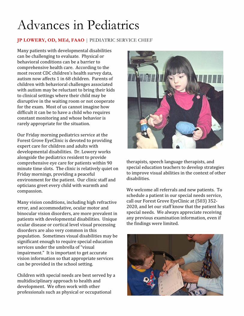
JP LOWERY, OD, MEd, FAAO | PEDIATRIC SERVICE CHIEF
Many patients with developmental disabilities
can be challenging to evaluate. Physical or
behavioral conditions can be a barrier to
comprehensive health care. According to the
most recent CDC children’s health survey data,
autism now affects 1 in 68 children. Parents of
children with behavioral challenges associated
with autism may be reluctant to bring their kids
to clinical settings where their child may be
disruptive in the waiting room or not cooperate
for the exam. Most of us cannot imagine how
difficult it can be to have a child who requires
constant monitoring and whose behavior is
rarely appropriate for the situation.
Our Friday morning pediatrics service at the
Forest Grove EyeClinic is devoted to providing
expert care for children and adults with
developmental disabilities. Dr. Lowery works
alongside the pediatrics resident to provide
comprehensive eye care for patients within 90
minute time slots. The clinic is relatively quiet on
Friday mornings, providing a peaceful
environment for the patient. Our clinic staff and
opticians greet every child with warmth and
compassion.
Many vision conditions, including high refractive
error, and accommodative, ocular motor and
binocular vision disorders, are more prevalent in
patients with developmental disabilities. Unique
ocular disease or cortical level visual processing
disorders are also very common in this
population. Sometimes visual disabilities may be
significant enough to require special education
services under the umbrella of “visual
impairment.” It is important to get accurate
vision information so that appropriate services
can be provided in the school setting.
Children with special needs are best served by a
multidisciplinary approach to health and
development. We often work with other
professionals such as physical or occupational
therapists, speech language therapists, and
special education teachers to develop strategies
to improve visual abilities in the context of other
disabilities.
We welcome all referrals and new patients. To
schedule a patient in our special needs service,
call our Forest Grove EyeClinic at (503) 352-
2020, and let our staff know that the patient has
special needs. We always appreciate receiving
any previous examination information, even if
the findings were limited.
Advances in Pediatrics

CAROLE TIMPONE, OD, FAAO, FNAP | ASSOCIATE DEAN OF CLINICAL PROGRAMS
Pacific EyeClinic, Forest Grove has a new look!
On November 21st, the first phase of our
EyeClinic remodel was completed. Gone are the
cinder block walls and the exposed plumbing.
New furnishings, equipment, instruments, and
lighting upgrades have been made possible
through the generous support of alumni, friends,
and industry, complementing significant
University funding. Additional attending faculty
offices, new restrooms and lockers with ADA
upgrades, as well as improved heating,
ventilation and air conditioning throughout,
complete the project.
The clinic is remaining open as we continue to
refurbish all 18 west wing exam rooms and
clinic attending offices in four phases,
anticipated to be completed in spring 2017.
Patients, students, and faculty have been thrilled
with the transformation. Please come by the
clinic to take a look!
Fundraising is ongoing, with opportunities for
naming of exam rooms in recognition of donors.
If you would like to support this endeavor,
please contact our Office of University
Development at https//community.pacificu.edu.
We are also pleased to present Pacific’s brand
new, 33 foot state-of-the-art mobile EyeVan.
Equipped with two complete exam lanes,
optometry students, under the supervision of Dr.
Sarah Martin, Director of Community Outreach,
are able to comfortably perform complete
comprehensive examinations, including
photography and visual field testing, for
underserved populations of all ages, in addition
to ongoing vision and eye health screenings.
This further expands Pacific’s opportunities for
community service throughout the state.
The College thanks the many donors and the
University for their contributions that made this
wonderful upgrade possible.
Pacific EyeClinic Updates
Original typical Forest Grove clinic exam room. Recently completed Phase 1 clinic exam room.

reR
Practice Management Tips CINDI RAPP, RDH | DIRECTOR OF CLINICAL OPERATIONS
Test Your HIPAA Knowledge
What is HIPAA? The Health Insurance
Portability and Accountability Act of 1996. It
was put in place to protect privacy and
security of health information.
What is PHI? Protected Health Information or
individually identifiable health information
includes demographic data that can be used to
identify an individual.
Can you name all 18 HIPAA identifiers? In
addition to the demographic information, the
list includes medical record number, social
security number, account number, license
number, photo image, finger or voice print,
etc. For a complete list, Google “18 HIPAA
Identifiers.” What you find may surprise you.
Did you know that PHI should not reside on any
personally owned mobile device, which includes, but
is not limited to, cell phones, tablets, or thumb drives?
If PHI is inadvertently downloaded to a personally
owned mobile device, it must be deleted from the
device as soon as possible.
When was your last HIPAA training? This is to be
completed annually by all staff who have access to
identifiable health information that is kept or
transmitted by a covered entity, i.e. your practice.
In addition to HIPAA, as the year end approaches, it is
always a good time to encourage your team to share
those things for which they are thankful. One way, is
to share appreciations during your monthly meetings.
“We often take for granted the very things that most
deserve our gratitude.” ~ Cynthia Ozick
January 2017: -PUCO Glaucoma Symposium; Woodinville, WA; Jan. 14. -Island Eyes Conference; Kauai Marriott Resort, Lihue, HI; Jan. 22-28. -PMOS Proliferative Diabetic Retinopathy in the Age of Anti-VEGF Therapy; BridgePort Brew Pub, Portland, OR; Jan. 23, 6:30.
February 2017: -OOPA Practice Management Seminar; Embassy Suites Hotel, Portland, OR; Feb. 23.
March 2017: -PMOS Spring CE Event; Aquariva, Portland, OR; March 4, 8:30 am -3:00 pm.
April 2017: -PUCO Coeur d’Alene CE; Coeur d’Alene Resort, Coeur d’Alene, ID; April 21-22.
CE Opportunities Research Opportunities
We are recruiting children 7-13 years old as research subjects for a research project: Effects of Alternate Occlusion on Children’s Fixation Disparity in Reading. Subjects need to make two visits to VPI, two hours each, with the task of reading text on a computer screen.
Subjects should have one of the following: � Exophoria > 8 prism diopters, � Esophoria > 3 prism diopters, � Stereoacuity > 60 arc seconds, or � Near point of convergence > 8 cm
Compensation: At the end of participation, subjects will be paid $20 per hour.
If interested, please schedule an appointment through our online scheduler at www.pacificu.edu/vpi and select "Display Resolution Study." For details or any questions, please email [email protected].
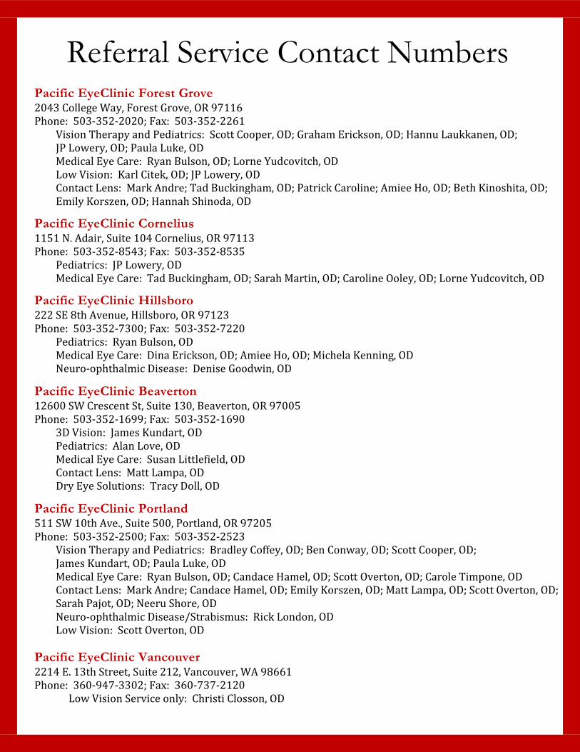
Referral Service Contact Numbers Pacific EyeClinic Forest Grove 2043 College Way, Forest Grove, OR 97116
Phone: 503-352-2020; Fax: 503-352-2261
Vision Therapy and Pediatrics: Scott Cooper, OD; Graham Erickson, OD; Hannu Laukkanen, OD;
JP Lowery, OD; Paula Luke, OD
Medical Eye Care: Ryan Bulson, OD; Lorne Yudcovitch, OD
Low Vision: Karl Citek, OD; JP Lowery, OD
Contact Lens: Mark Andre; Tad Buckingham, OD; Patrick Caroline; Amiee Ho, OD; Beth Kinoshita, OD;
Emily Korszen, OD; Hannah Shinoda, OD
Pacific EyeClinic Cornelius 1151 N. Adair, Suite 104 Cornelius, OR 97113
Phone: 503-352-8543; Fax: 503-352-8535
Pediatrics: JP Lowery, OD
Medical Eye Care: Tad Buckingham, OD; Sarah Martin, OD; Caroline Ooley, OD; Lorne Yudcovitch, OD
Pacific EyeClinic Hillsboro 222 SE 8th Avenue, Hillsboro, OR 97123
Phone: 503-352-7300; Fax: 503-352-7220
Pediatrics: Ryan Bulson, OD
Medical Eye Care: Dina Erickson, OD; Amiee Ho, OD; Michela Kenning, OD
Neuro-ophthalmic Disease: Denise Goodwin, OD
Pacific EyeClinic Beaverton 12600 SW Crescent St, Suite 130, Beaverton, OR 97005
Phone: 503-352-1699; Fax: 503-352-1690
3D Vision: James Kundart, OD
Pediatrics: Alan Love, OD
Medical Eye Care: Susan Littlefield, OD
Contact Lens: Matt Lampa, OD
Dry Eye Solutions: Tracy Doll, OD
Pacific EyeClinic Portland 511 SW 10th Ave., Suite 500, Portland, OR 97205
Phone: 503-352-2500; Fax: 503-352-2523
Vision Therapy and Pediatrics: Bradley Coffey, OD; Ben Conway, OD; Scott Cooper, OD;
James Kundart, OD; Paula Luke, OD
Medical Eye Care: Ryan Bulson, OD; Candace Hamel, OD; Scott Overton, OD; Carole Timpone, OD
Contact Lens: Mark Andre; Candace Hamel, OD; Emily Korszen, OD; Matt Lampa, OD; Scott Overton, OD;
Sarah Pajot, OD; Neeru Shore, OD
Neuro-ophthalmic Disease/Strabismus: Rick London, OD
Low Vision: Scott Overton, OD
Pacific EyeClinic Vancouver 2214 E. 13th Street, Suite 212, Vancouver, WA 98661
Phone: 360-947-3302; Fax: 360-737-2120
Low Vision Service only: Christi Closson, OD