Embed Size (px)
Citation preview

8/6/2019 Pa Tho Genesis of Ovca
http://slidepdf.com/reader/full/pa-tho-genesis-of-ovca 1/15
Pathogenesis of Ovarian Cancer. Lessons from Morphology and
Molecular Biology and their Clinical Implications
Robert J. Kurman, MD and Ie-Ming Shih, MD, PhD
From the Departments of Pathology, Gynecology and Obstetrics, and Medical Oncology, The JohnsHopkins Medical Institutions, Baltimore, Maryland, USA
Abstract
The received view of ovarian carcinogenesis is that carcinoma begins in the ovary, undergoes
progressive “dedifferentiation” from a well to a poorly differentiated tumor and then spreads to the
pelvic and abdominal cavities before metastasizing to distant sites. It has therefore been reasoned
that survival for this highly lethal disease could be improved by developing screening methods that
detect disease when it is confined to the ovary. To date, however, no prospective, randomized trial
of any ovarian cancer screening test(s) has demonstrated a decrease in mortality. We believe that oneof the main reasons for this is that the dogma underlying ovarian carcinogenesis is flawed. Based on
studies performed in our laboratory over the last decade we have proposed a model of ovarian
carcinogenesis that takes into account the diverse nature of “ovarian cancer” and correlates the
clinical, pathologic and molecular features of the disease. In this model, ovarian tumors are divided
into two groups designated Type I and Type II. Type I tumors are slow growing, generally confined
to the ovary at diagnosis and develop from well established precursor lesions that are termed
“borderline” tumors. Type I tumors include low-grade micropapillary serous carcinoma, mucinous,
endometrioid, and clear cell carcinomas. They are genetically stable and are characterized by
mutations in a number of different genes including KRAS, BRAF, PTEN , and beta-catenin. Type II
tumors are rapidly growing, highly aggressive neoplasms for which well defined precursor lesions
have not been described. Type II tumors include high-grade serous carcinoma, malignant mixed
mesodermal tumors (carcinosarcomas) and undifferentiated carcinomas. This group of tumors has a
high level of genetic instability and is characterized by mutation of TP53. The model helps to explainwhy current screening techniques, aimed at detecting stage I disease, have not been effective. Tumors
that remain confined to the ovary for a long period of time belong to the Type I group but they account
for only 25% of the malignant tumors. The vast majority of what is considered “ovarian cancer”
belongs to the Type II category and these are only rarely confined to the ovary. Although the reasons
for this are not entirely clear, it appears that some tumors evolve rapidly and spread to extra-ovarian
sites early in their development. In addition, a significant number of Type II “ovarian carcinomas”
develop outside the ovary, specifically, the peritoneum and fallopian tube, and involve the ovary
secondarily. These tumors are advanced stage at their inception. Therefore, a more realistic endpoint
for the early detection of ovarian carcinoma may be volume and not stage of disease. Thus, the model
which correlates the clinical and pathologic features of “ovarian cancer” with the specific molecular
genetic events that play a role in tumor progression can lead to a more rational approach to early
detection and to more targeted therapeutic intervention.
The received view of ovarian carcinogenesis is that carcinoma begins in the ovary, undergoes
progressive “dedifferentiation” from a well to a poorly differentiated tumor and then spreads
to the pelvic and abdominal cavities before metastasizing to distant sites. It is also believed
that since serous borderline tumors (SBTs) are rarely associated with invasive serous carcinoma
they are a distinct entity unrelated to invasive carcinoma. However, SBTs sometimes progress
to carcinoma and therefore some relationship must exist. In contrast, mucinous borderline
tumors (MBTs) are often associated with invasive mucinous carcinoma. These disparate
NIH Public AccessAuthor Manuscript Int J Gynecol Pathol. Author manuscript; available in PMC 2009 December 16.
Published in final edited form as:
Int J Gynecol Pathol. 2008 April ; 27(2): 151–160. doi:10.1097/PGP.0b013e318161e4f5.
N I H -P A A u
t h or Manus c r i pt
N I H -P A A ut h or Manus c r i pt
N I H -P A A ut h or M
anus c r i pt

8/6/2019 Pa Tho Genesis of Ovca
http://slidepdf.com/reader/full/pa-tho-genesis-of-ovca 2/15
findings suggest that some borderline tumors are precursors of invasive carcinoma and others
are not. In the past several years, clinicopathologic and molecular genetic analyses performed
on a large number of borderline ovarian tumors and invasive carcinomas (1–4) by our research
group have elucidated the relationship of borderline ovarian tumors to invasive cancer. Based
on these studies a model of ovarian carcinogenesis for all the surface epithelial tumors has been
proposed which reconciles the difference in the relationship of SBTs and MBTs to their
respective carcinomas. The model divides surface epithelial tumors into two groups designated
Type I and Type II (1,5). Type I tumors tend to be low grade, indolent neoplasms that arisefrom well characterized precursor lesions, specifically borderline tumors (6). The Type I group
includes low-grade micropapillary serous carcinoma (MPSC), mucinous carcinoma,
endometrioid carcinoma and clear cell carcinoma. In contrast, Type II tumors are aggressive
and high grade from the outset and since precursor lesions have not been identified, have been
said to arise de novo (7). Included in this group are high grade serous carcinoma, malignant
mixed mesodermal tumors (MMMTs) and undifferentiated carcinomas. In addition to the
clinical and pathologic differences, molecular genetic studies have shown that Type I tumors
are relatively genetically stable and are characterized by a number of different mutations
whereas the Type II tumors show considerable genetic instability. The mutations that
characterize Type I tumors are not detected in Type II tumors whereas the vast majority of
Type II tumors contain mutant p53 which is only rarely found in Type I tumors (1,8). This
model does not replace the standard histopathologic classification of ovarian carcinoma, its
purpose is to provide a framework for the study of ovarian carcinogenesis. The model, bydrawing attention to molecular genetic pathways that play a role in tumor progression, can shed
light on new approaches to early detection and novel methods of treatment.
Serous Tumors
The relationship of SBTs to invasive carcinoma has puzzled investigators since the category
was created by FIGO and the WHO over 30 years ago. In the past, it was not clear whether
SBTs were a distinct entity unrelated to serous carcinoma or whether they represented part of
a continuum of tumor progression that culminates in serous carcinoma (9–13). Until recently,
the molecular genetic studies have suggested that SBTs are unrelated to serous carcinoma.
(1,7,11,12,14,15)
Our initial clinicopathologic studies revealed that noninvasive serous tumors display twodistinctly different morphologic phenotypes. One is characterized by a hierarchical branching
pattern that we classify as an “atypical proliferative serous tumor (APST)”. This tumor behaves
in a benign fashion. The other morphologic variant is characterized by a nonhierarchical,
micropapillary pattern that we designate “micropapillary serous carcinoma (MPSC)”, a
noninvasive carcinoma. Foci of invasion can develop in these tumors and when they overgrow
the noninvasive component the tumor is classified as a low-grade micropapillary serous
carcinoma.” Invasion can be extensive or focal. Focal invasion can display a variety of patterns
and has traditionally been classified as “microinvasion”. The pattern that resembles
micropapillary serous carcinoma is the one associated with a poor outcome (16,17).
Accordingly, we classify tumors with small foci displaying a micropapillary architecture, as
“microcarcinoma” to distinguish them from the other patterns of “microinvasion” which do
not have an adverse effect on outcome. Data on the behavior of “microcarcinoma” are limited
(16) so there are no established criteria for separating microcarcinoma from bona fide invasivelow-grade micropapillary serous carcinoma. We have seen cases measuring less than 3 mm
associated with a poor outcome. This is an area that clearly warrants further investigation.
Invasive low-grade MPSC nearly always displays a micropapillary architecture although on
rare occasion other patterns can be observed. These include tumors with a macropapillary
pattern (18,19) and therefore there is merit to the view that all these low-grade tumors should
Kurman and Shih Page 2
Int J Gynecol Pathol. Author manuscript; available in PMC 2009 December 16.
N I H -P A A
ut h or Manus c r i pt
N I H -P A A ut h or Manus c r i pt
N I H -P A A ut h or
Manus c r i pt

8/6/2019 Pa Tho Genesis of Ovca
http://slidepdf.com/reader/full/pa-tho-genesis-of-ovca 3/15
be classified as low-grade serous carcinoma (6). We believe the qualifier “micropapillary” is
helpful to draw attention to the distinctive morphology, behavior and nature of this neoplasm
that distinguishes it from the usual type of serous carcinoma which is high-grade. Invasive low-
grade micropapillary serous carcinoma is characterized by small solid nests and micropapillae
haphazardly infiltrating the stroma (2) (Fig 1). Psammoma bodies are often present and there
is no evidence of necrosis. Cytologically, invasive low-grade MPSC is composed of a relatively
uniform population of cells with small, rounded nuclei often containing a small but conspicuous
nucleolus. On a scale of 1 to 3 this degree of nuclear atypia qualifies as grade 1. Mitotic activityis low and there is no evidence of abnormal mitotic figures. Multinucleation is almost absent.
It has been estimated that low-grade micropapillary serous carcinomas account for
approximately 10% of all serous carcinomas (6).
Compared to atypical proliferative serous tumors (APSTs) which are synonymous with the
usual type of SBT, noninvasive MPSCs are more often associated with invasive as opposed to
noninvasive implants. Invasive implants generally have a micropapillary architecture and
resemble low-grade invasive serous carcinoma (18). Accordingly, in our opinion invasive
implants are low-grade serous carcinomas and should be classified as such. Recurrences have
a similar appearance. The median survival of patients following recurrence of an SBT in which
the recurrent tumor looks like micropapillary serous carcinoma is the same as for women who
have a low-grade serous carcinoma at presentation (20) providing further support to the view
that the invasive low-grade tumors develop from atypical proliferative serous tumors andnoninvasive MPSCs.
Low-grade MPSC, the prototypic Type I tumor, and its precursor lesions, APST is
characterized by sequence mutations in KRAS, BRAF and ERBB2 oncogenes (8,21–24).
Oncogenic mutations in BRAF, KRAS and ERBB2 result in constitutive activation of the
mitogen activated protein kinase (MAPK) signal transduction pathway which plays a critical
role in the transmission of growth signals into the nucleus and contributes to neoplastic
transformation. Previous studies (21,23) have demonstrated that KRAS mutations at codons 12
and 13 occur in one third of invasive low-grade MPSCs and another one third of SBTs.
Similarly, BRAF mutations at codon 600 occur in 30% of low-grade serous carcinomas and
28% of SBTs (21). These findings have been confirmed by other investigators (24,25).
Mutations in KRAS, BRAF and ERBB2 are mutually exclusive. Therefore mutations in any of
the genes are detected in about two thirds of MPSCs and APSTs. In contrast, these genes arenot mutated in high-grade serous carcinomas (21,22). Mutations of KRAS and BRAF appear
to occur very early in the development of low-grade MPSC as evidenced by the demonstration
that the same KRAS and BRAF mutations detected in SBTs are detected in the cystadenoma
epithelium adjacent to SBTs (26). Mutations in TP53 are very rare.
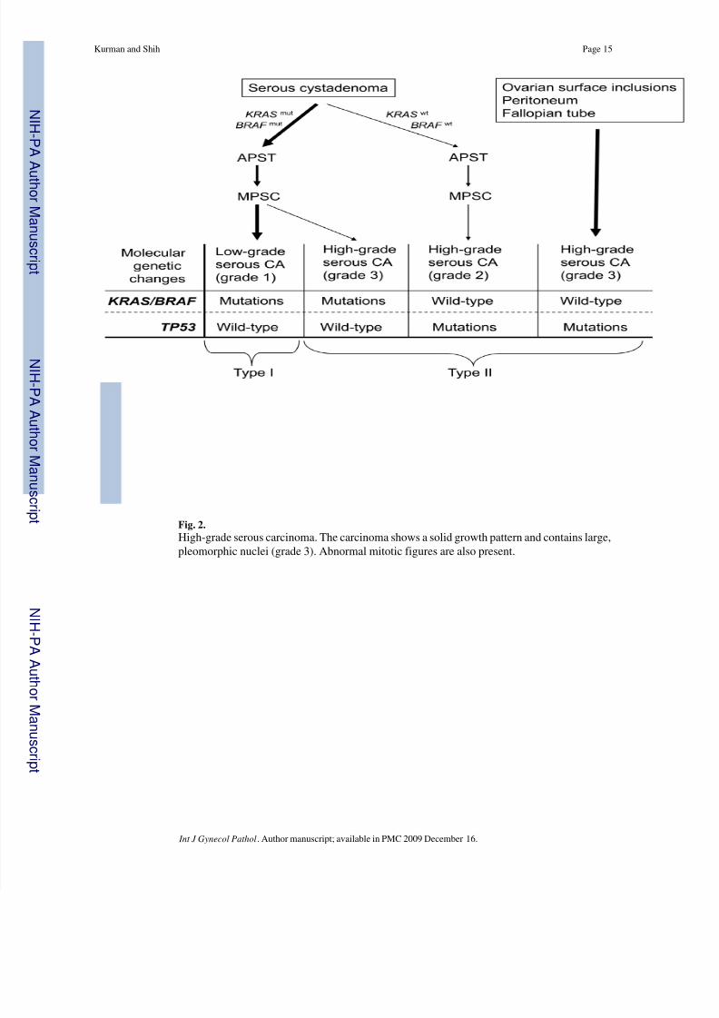
High-grade serous carcinoma, the prototypic Type II tumor, corresponds to the usual type of
ovarian serous carcinoma and accounts for approximately 90% of serous carcinomas. These
tumors are composed of large masses of cells that frequently display a papillary architecture.
Necrosis is a common feature. The tumor cells have large, pleomorphic nuclei, many of which
are multinucleated. There is a high level of mitotic activity and abnormal mitotic figures are
frequent. These tumors are considered poorly differentiated and the nuclear atypia is grade 3
(Fig 2). Studies have shown that the majority of advanced stage, high-grade serous carcinomas
have mutant TP53 (27–33) and the mutation frequency is over 80% when purified tumorsamples are used for analysis (33). It has also been reported that mutant TP53 is present in 37%
of stage I and II high-grade serous carcinomas (34). In a study of very early, microscopic stage
I high-grade serous carcinomas in ovaries removed prophylatically from women who were
BRCA heterozygotes, over expression of p53 and mutation of TP53 were found in these very
early invasive high-grade serous carcinomas as well as in the adjacent “dysplastic” surface
epithelium (35). These microscopic serous carcinomas had nuclear atypia resembling that
Kurman and Shih Page 3
Int J Gynecol Pathol. Author manuscript; available in PMC 2009 December 16.
N I H -P A A
ut h or Manus c r i pt
N I H -P A A ut h or Manus c r i pt
N I H -P A A ut h or
Manus c r i pt

8/6/2019 Pa Tho Genesis of Ovca
http://slidepdf.com/reader/full/pa-tho-genesis-of-ovca 4/15
found in advanced stage, high-grade serous carcinomas. It is plausible that inherited mutations
in BRCA genes compromise DNA repair and predispose to genetic instability that may
contribute to the “dysplastic changes”. Although sporadic ovarian carcinomas were not
analyzed in this study, the clinical and pathologic features of BRCA-linked ovarian carcinomas
and their sporadic counterparts are indistinguishable, suggesting that their histogenesis is
similar. Thus, although the findings are preliminary, they suggest that conventional high-grade
serous carcinoma, in its very earliest stage, resembles advanced stage serous carcinoma at a
molecular as well as at a morphologic level.
Correlation of the morphologic and molecular genetic studies elucidates some of the puzzling
features of serous tumors. First, they explain the lack of association of SBTs with the usual
type of serous carcinoma on the one hand and their occasional malignant behavior on the other.
Thus, recognizing that the vast majority of serous tumors fall into two distinct groups is of
considerable importance as it highlights the fact that the molecular genetic pathways leading
to the development of low-grade micropapillary serous carcinoma and the usual type of serous
carcinoma are entirely different (1). In one pathway, invasive low-grade micropapillary serous
carcinoma develops from a noninvasive, i.e., in situ tumor which has traditionally been termed
“serous borderline tumor (SBT)” (2). The progression of APST to MPSC, a noninvasive low-
grade serous carcinoma, (1,23) and then to invasive low-grade carcinoma (invasive MPSC)
mimics the adenoma-carcinoma sequence in colorectal carcinoma in which the carcinoma
evolves through a continuum of histologically recognizable precursor lesions. (36) Progressionof an APST to a noninvasive MPSC is supported by the finding of a greater level of allelic
imbalance in MPSCs as compared to APSTs (23). Atypical proliferative serous tumor and
noninvasive MPSC can be thought of as analogous to dysplasia and carcinoma in situ of the
cervix. That is to say, the APST is a benign proliferative tumor that can progress to noninvasive
MPSC, which is the immediate precursor of invasive low-grade micropapillary serous
carcinoma. Compared to women with high-grade serous carcinoma the low-grade tumors occur
at a younger age (median age 43 years compared to 61 years for the high-grade tumors) and
behave in a more indolent fashion (median survival 81 months compared to 24 months for
women with high-grade serous carcinoma) (6). In the second pathway, high-grade serous
carcinoma is thought to develop from ovarian surface epithelium or from surface inclusion
cysts (37). It is possible that the immediate precursor of high-grade serous carcinoma is an
intraepithelial high-grade serous carcinoma developing in inclusion cysts but this putative in
situ lesion has not been well characterized and therefore the origin of these tumors has beendescribed as de novo (1,7). It has also been recently reported that these tumors may develop
from an intraepithelial carcinoma in the fallopian tube (38,39). High-grade serous carcinomas
are very aggressive and spread rapidly, accounting for their advanced stage at presentation.
It is important to emphasize that low-grade micropapillary serous carcinoma and high-grade
serous carcinoma are distinctly different tumor types analogous to low-grade endometrial
stromal sarcoma and “undifferentiated stromal sarcoma”. This, concept of ovarian
carcinogenesis is very different from the traditional view in which high-grade serous
carcinomas are thought to develop progressively from well differentiated serous carcinoma.
In the proposed model, high-grade serous carcinomas are high-grade from their inception and
do not evolve from low-grade serous carcinomas. In fact, the low-grade tumors nearly always
retain the same morphologic phenotype during the entire course of the disease which in some
instances can be over 20 years.
Like high-grade serous carcinoma, other Type II tumors, specifically malignant mixed
mesodermal tumors (carcinosarcomas) also demonstrate TP53 mutations in almost all cases
analyzed (40–42). Moreover, when the carcinomatous and sarcomatous components of
MMMTs are analyzed for TP53 mutation the identical mutation has been detected in both the
epithelial and stromal component leading investigators to suggest that malignant mixed
Kurman and Shih Page 4
Int J Gynecol Pathol. Author manuscript; available in PMC 2009 December 16.
N I H -P A A
ut h or Manus c r i pt
N I H -P A A ut h or Manus c r i pt
N I H -P A A ut h or
Manus c r i pt

8/6/2019 Pa Tho Genesis of Ovca
http://slidepdf.com/reader/full/pa-tho-genesis-of-ovca 5/15
mesodermal tumors are in essence variants of carcinoma. The finding that MMMts have a very
high rate of TP53 mutations similar to that of high-grade serous carcinoma has additional
implications. Thus, when high grade tumors that show clear-cut evidence of endometrioid,
mucinous, or clear cell carcinoma differentiation are excluded, the remaining group of tumors,
namely high-grade serous carcinomas, adenocarcinomas NOS, and MMMTs have similar
molecular genetic profiles (Vang et al unpublished data). Also the group of tumors that
pathologists routinely classify as high-grade serous carcinoma is quite different in their
appearance from what is classified as “uterine serous carcinoma”. Uterine serous carcinomahas a relatively limited morphologic phenotype restricted to tumors with a clear-cut “low-grade
architecture (papillary or glandular) combined with high grade (grade 3) nuclei. In contrast,
the morphologic phenotype of ovarian high-grade serous carcinoma is much broader. Some
resemble uterine serous carcinoma but the majority does not. Instead these tumors display large
masses and nests of cells with slit-like spaces. Papillary and glandular patterns are frequently
not evident. Extensive areas of necrosis and fibrosis are frequently encountered. In short, many
of these high-grade carcinomas are poorly differentiated adenocarcinomas or undifferentiated
carcinomas without the slightest evidence of “serous” differentiation. As presently used
“moderate to poorly differentiated “or “high-grade serous carcinoma” is a “waste basket”
category for high grade carcinomas that show no clear-cut evidence of endometrioid, mucinous
or clear cell differentiation. The tumors in the Type II group appear to have a similar behavior.
Thus, despite the morphologic diversity, from a molecular genetic standpoint they are similar.
In the future it may be more appropriate to include all of these poorly differentiated/ undifferentiated carcinomas into a single category of “anaplastic carcinoma”.
This proposed model of carcinogenesis is the first step in trying to unravel what is a highly
complex process. For example, studies have shown that on rare occasion low-grade serous
tumors are associated with high-grade serous carcinomas (Fig 3) (43,44). In a recent analysis
of tumors in which a high-grade serous carcinoma was immediately adjacent to an APST we
found the identical KRAS mutation in both components of the tumor indicating that they had
a shared lineage (44). These findings suggest that on rare occasion APSTs and low-grade
micropapillary serous carcinomas may progress to high-grade serous carcinomas.
Interestingly, none of these high-grade tumors had a TP53 mutation. Only six cases were
studied but since over 80% of high-grade serous carcinomas contain TP53 mutations, the
absence of a TP53 mutation in the high-grade serous carcinomas associated with APSTs raises
the possibility that these high grade serous carcinomas develop along a different pathway thanthe usual high-grade serous carcinoma that is not associated with an APST (44).
We have also recently studied a group of moderately differentiated serous carcinomas,
characterized by the presence of grade 2 nuclei that were not associated with an APST or
noninvasive MPSC (Fig 4). Architecturally, several displayed a micropapillary pattern
simulating low-grade serous carcinoma but to date all have had TP53 mutations and lacked
mutations of KRAS, BRAF, or ERBB2 (unpublished data). These findings suggest that high-
grade serous carcinomas with grade 2 nuclei may develop along yet another pathway that is
different from the one described for the more common high-grade serous carcinoma with grade
3 nuclei. Since many of these tumors displayed a micropapillary architecture it is conceivable
that they developed from a subset of low-grade serous carcinomas that lacked mutations of
KRAS, BRAF and ERBB2. Subsequent acquisition of a TP53 mutation could lead to genetic
instability which in turn contributed to the increased level of nuclear atypia (grade 2).Accordingly, our preliminary findings suggest that although there is a dominant pathway for
the development of most high-grade serous carcinomas, there are other pathways that account
for the morphologic diversity displayed by this group of neoplasms. A schema depicting these
different pathways is shown in Figure 5.
Kurman and Shih Page 5
Int J Gynecol Pathol. Author manuscript; available in PMC 2009 December 16.
N I H -P A A
ut h or Manus c r i pt
N I H -P A A ut h or Manus c r i pt
N I H -P A A ut h or
Manus c r i pt
8/6/2019 Pa Tho Genesis of Ovca
http://slidepdf.com/reader/full/pa-tho-genesis-of-ovca 6/15
Finally, given the utility of the two-tier grading system in separating low- and high-grade serous
carcinomas with different clinical behaviors (6) and presumably would require different
treatment, establishment of a cut-point between low- and high tumors is imperative. The
distinction at either end of the spectrum is straight forward. Thus, low-grade tumors have small,
uniform (grade 1) nuclei whereas high-grade tumors have highly pleomorphic, often
multinucleated (grade 3) nuclei. Mitotic activity is low in the low-grade tumors and high in the
high-grade tumors. However, tumors with nuclei and mitotic levels that are intermediate
between low- and high-grade, i.e. moderately differentiated tumors with grade 2 nuclei dooccur. Are these tumors low- or high grade using a two tier grading system? As has been
discussed these grade 2 tumors have TP53 mutations and lack mutant KRAS and BRAF which
provides strong molecular genetic evidence that they are high-grade. This has important clinical
implications since at the present time some oncologists consider stage I, grade 2 tumors low-
grade and do no administer adjuvant chemotherapy whereas patients with grade 3 tumors
receive adjuvant chemotherapy.
Mucinous Tumors
Mucinous tumors share some similarities with serous tumors but also have profound
differences. Like SBTs, survival for women with stage I MBTs is 100% but based on the
literature, survival with advanced stage tumors is only 50% compared to 70% for advanced
stage SBTs. In addition, it has been reported that 80% of advanced stage MBTs are associatedwith pseudomyxoma peritonei (PMP). It is now well established that PMP results from a
ruptured mucinous appendiceal adenoma and that the ovarian involvement is secondary (45–
47). It has also been shown that metastatic mucinous carcinomas from the upper gastrointestinal
tract including the biliary tract, pancreas and cervix can metastasize to the ovary and simulate
a primary ovarian mucinous tumor. When mucinous tumors associated with PMP and
metastatic mucinous carcinomas that masquerade as primary ovarian tumors are excluded from
consideration, it becomes apparent that MBTs unlike SBTs never spread beyond the ovary.
A number of clinical and pathological observations link primary mucinous carcinomas of the
ovary to mucinous borderline tumors (MBTs). The mean size of MBTs and mucinous
carcinomas is the same at diagnosis (18 cm). The large size of mucinous carcinomas and their
unilateral presentation underscores the fact that they grow slowly. Mucinous carcinomas at the
time of their diagnosis are nearly always well differentiated and merge with areas of borderlinetumor and mucinous cystadenoma. In fact, it is not unusual to find a focus of carcinoma growing
within a tumor that is for the most part a MBT. Cytologic atypia within the noninvasive
component can range from minimal to marked, leading investigators to subclassify borderline
tumors into atypical proliferative and intraepithelial carcinoma based on the degree of cytologic
atypia. These findings strongly suggest that mucinous carcinoma develops slowly, in a stepwise
fashion from benign precursors. This conclusion is supported by molecular genetic studies
which have shown that the most common molecular genetic alteration in MBTs and mucinous
carcinomas is a point mutation of KRAS (25,48,49). An increasing frequency of KRAS
mutations at codons 12 and 13 has been described in cystadenomas, MBTs and mucinous
carcinomas, respectively (12,48–51). In addition, mucinous carcinoma and the adjacent
mucinous cystadenoma and borderline tumor share the same KRAS mutation (50). Besides
KRAS, other genetic alterations in ovarian mucinous tumors have not been reported.
Endometrioid and Clear Cell Tumors
There has not been a single well documented case of a borderline endometrioid or clear cell
tumor associated with malignant behavior since the category was first introduced in the early
1970s supporting the view that these are benign proliferative tumors. Nonetheless, they are
frequently associated with their malignant counterparts. As with the mucinous tumors, the
Kurman and Shih Page 6
Int J Gynecol Pathol. Author manuscript; available in PMC 2009 December 16.
N I H -P A A
ut h or Manus c r i pt
N I H -P A A ut h or Manus c r i pt
N I H -P A A ut h or
Manus c r i pt

8/6/2019 Pa Tho Genesis of Ovca
http://slidepdf.com/reader/full/pa-tho-genesis-of-ovca 7/15
endometrioid and clear cell carcinomas tend to be large (mean diameter 15 cm), unilateral and
frequent transitions between the proliferative tumor and the carcinoma are observed. Only a
few molecular genetic studies of these tumors have been performed. These are discussed in
greater detail in the accompanying article in this Symposium by Dr Kathleen Cho. Briefly,
mutations of KRAS and BRAF have been reported in approximately 10% of endometrioid
carcinomas (21,25,49,52–54) and mutation of the tumor suppressor, PTEN , in 20% of
endometrioid carcinomas, which rises to 46% in the tumors with 10q23 loss of heterozygosity
(55). Similar molecular genetic alterations including loss of heterozygosity at 10q23 andmutations in PTEN have been reported in endometriosis, atypical endometriosis and ovarian
endometrioid carcinoma in the same specimen (55–60). Furthermore, mutations of beta-
catenin have been detected in more than 60% of ovarian endometerioid carcinomas (grade I)
and their precursor lesions, endometrioid borderline tumors (61). These molecular genetic
findings together with the morphological data demonstrating a frequent association of
endometriosis with endometrioid adenofibromas and atypical proliferative endometrioid
tumorss adjacent to invasive well-differentiated endometrioid carcinoma provide evidence of
stepwise tumor progression in the development of endometrioid carcinoma (62. The critical
role of the genetic changes in PTEN and KRAS is highlighted by a recent report showing that
inactivation of PTEN and an activating mutation of KRAS are sufficient to induce the
development of ovarian endometrioid carcinoma in a mouse model (63). More recently,
inactivation of the Wnt/beta-catenin and the PI3K/Pten pathways has been reported to be
sufficient to induce endometrioid carcinoma in a engineered mouse model (64).
Clear cell carcinomas display clinical, morphologic and molecular genetic changes that are
shared with, but also differ from, tumors in the Type I and II groups suggesting that clear cell
carcinomas develop along an independent pathway. For example, although they tend to be
diagnosed in stage I, clear cell carcinomas present more often as advanced stage tumors than
the other neoplasms in the Type I group. Also, although they tend to be large at diagnosis, they
typically are high grade whereas other Type I tumors are low grade. As compared to other
types of ovarian epithelial tumors, the main molecular genetic changes associated with ovarian
clear cell borderline tumors and clear cell carcinomas remain to be identified. Although several
molecular genetic changes have been reported in clear cell tumors, most studies have analyzed
a limited number of cases and therefore the true prevalence of those changes is not known.
Microsatellite instability is present in endometrioid and clear cell carcinoma but is only rarely
detected in serous and mucinous tumors (62,65,66). This finding supports the view thatendometriosis is the common precursor for both endometrioid and clear cell carcinoma (62).
Mutations in KRAS, BRAF and TP53 are present in some clear cell carcinomas but their
frequency is low (25). Thus, although mutations have been identified in clear cell carcinomas
they differ from those found in the other Type I tumors. Finally, unlike Type II tumors but
similar the Type I tumors, they are relatively genetically stable.
Implications of the Model for Early Detection and Treatment
An appreciation of ovarian carcinogenesis based on this model sheds light on potential new
approaches to early detection and treatment. First, the model, by dividing ovarian cancer into
two broad groups, Type I and Type II, draws attention to the fact that ovarian cancer is a
heterogeneous group of diseases that not only behave differently but also develop differently.
Screening with pelvic examination and transvaginal ultrasound is reasonable for Type Icarcinomas because they develop slowly from well characterized precursors (borderline
tumors) and remain confined to the ovary while growing to a large size. However, Type I
carcinomas constitute only 25% of ovarian cancers so these approaches are inadequate for large
scale screening of “ovarian cancer”. The vast majority of ovarian cancers are Type II tumors
which are high-grade, advanced stage at presentation, rapidly growing, and highly aggressive.
Current approaches to screening, namely serum CA125 assays and transvaginal ultrasound
Kurman and Shih Page 7
Int J Gynecol Pathol. Author manuscript; available in PMC 2009 December 16.
N I H -P A A
ut h or Manus c r i pt
N I H -P A A ut h or Manus c r i pt
N I H -P A A ut h or
Manus c r i pt

8/6/2019 Pa Tho Genesis of Ovca
http://slidepdf.com/reader/full/pa-tho-genesis-of-ovca 8/15
have not assisted in the detection of these tumors at an early stage. The likely explanation is
that these are genetically unstable tumors that transit rapidly from the ovary to extra-ovarian
sites and therefore the time for detecting these tumors while still confined to the ovary (stage
I) is very brief. Furthermore, a substantial number of Type II carcinomas appear to develop in
the fallopian tube (39) and peritoneum and involve the ovary secondarily. It is well recognized
that serous carcinomas identical to ovarian serous carcinomas can develop following bilateral
salpingo oophorectomy. Also many cases of serous carcinoma extensively involving the pelvic
and abdominal cavities have only minimal ovarian involvement but are classified as ovarianif the ovarian tumor is greater than 5 mm. This is clearly an arbitrary decision in assigning
origin to the ovary. In any event, these tumors are advanced stage at their inception. It is
therefore apparent that early detection of these tumors is extraordinarily difficult since there
is no morphologically characterized precursor lesion. Despite these formidable difficulties,
strategies can be developed to enhance early detection and improve survival. Clues can be
gleaned from advances in the surgical treatment of ovarian cancer. It is well recognized that
the most important prognostic indicator is not stage but the volume of residual disease
following cytoreductive surgery. As surgical techniques have evolved, what constitutes
optimal cytoreduction has shifted from <2cm to <1.5cm to <1cm. With each reduction in the
amount of residual disease that is considered optimal, survival has improved (67). The smaller
the tumor volume the more effective chemotherapy will be. Therefore the current approach to
evaluating screening tests should be shifted from detection of stage I tumors to detection of
“minimal ovarian carcinoma” irrespective of stage. “Minimal ovarian carcinoma” can bedefined as microscopic to 1 cm. As technology advances and the sensitivity of assays is
improved, the definition of what constitutes “minimal” can be changed.
The ultimate goal of early detection, given the lack of morphologically recognizable precursor
lesions for the Type II tumors, is the identification of biomarkers that precede the development
of these precursors. It has been shown that mutations of TP53 are currently the most common
molecular genetic change in Type II tumors (33). Moreover, mutation of TP53 occurs very
early in the genesis of these neoplasms. In fact they have been observed in intraepithelial
neoplasia in the fallopian tube fimbria of BRCA patients (38,39). Importantly, TP53 mutations
are inherited during cancer evolution and contribute to the transformed state. As a result, the
initiating genetic changes are retained in both the primary and recurrent tumors. Furthermore,
it is likely that the tumor DNA containing mutant TP53 DNA or polypeptides released from
these tumors can be detected in body fluids. Accordingly, a test that detects mutant TP53 inthe blood could be very useful in early detection.
The proposed model for ovarian carcinogenesis also has important implications for targeted
treatment. With the characterization of specific genetic changes that occur early in the
development of Type II tumors, treatment could be administered using drugs that target the
pathways affected by the mutations, for example mutant TP53. Therapeutic options would be
offered based on the presence of these biomarkers alone. A precedent for this approach
currently exists for women who are identified as having BRCA mutations, many of whom
choose to undergo prophylactic bilateral salpingo oophorectomy and hysterectomy.
Type I tumors present different challenges as compared to Type II tumors because they tend
to be localized and indolent. Since Type I tumors are slow growing and therefore therapeutic
agents that are effective against Type II tumors are not as effective against Type I tumors. Forexample, Type I carcinomas harbor several mutations in protein kinases and therefore the
pathways that they control could be amenable to inhibitor treatment or targeted by
immunotherapy. In many Type I carcinomas, there is constitutive activation of the MAPK
signaling pathway due to mutations in either KRAS or BRAF genes, the upstream regulators of
MAPK . Accordingly, BRAF inhibitors and other MAPK inhibitors should be evaluated to
determine whether they could prolong disease-free interval and overall survival in patients with
Kurman and Shih Page 8
Int J Gynecol Pathol. Author manuscript; available in PMC 2009 December 16.
N I H -P A A
ut h or Manus c r i pt
N I H -P A A ut h or Manus c r i pt
N I H -P A A ut h or
Manus c r i pt

8/6/2019 Pa Tho Genesis of Ovca
http://slidepdf.com/reader/full/pa-tho-genesis-of-ovca 9/15
advanced-stage Type I tumors. The mutated biomarker sequences might also be specifically
targeted by immunotherapy since the mutated sequence is non-self and its expression is
restricted to the tumor cells.
Conclusions
A new model for the pathogenesis of ovarian cancer based on clinical, pathological, and
molecular genetic studies is proposed. In this model ovarian tumors are divided into two broadgroups designated Type I and Type II. Type I tumors are slow growing, generally confined to
the ovary at diagnosis and develop from well established precursor lesions that are termed
“borderline” tumors. Type I tumors included low-grade micropapillary serous carcinoma,
mucinous, endometrioid, and clear cell carcinomas. They are genetically stable tumors and are
characterized by mutations in a number of different genes including KRAS, BRAF, PTEN , and
beta-catenin. In contrast, Type II tumors are rapidly growing, highly aggressive, neoplasms
for which well defined precursor lesions have not been described. The vast majority of what
is considered “ovarian cancer” belongs to the Type II category. These tumors include high-
grade serous carcinoma, malignant mixed mesodermal tumors (carcinosarcomas) and
undifferentiated carcinomas. This group of tumors has a high level of genetic instability and
is characterized by mutation of TP53. The model has important implications for the early
detection and treatment of ovarian cancer. Specifically, it indicates that the current approach
to screening, aimed at detecting stage I ovarian carcinoma, is not likely to be of benefit forType II tumors, the vast majority of what constitutes “ovarian cancer”. Type II carcinomas,
are only rarely detected when the disease is confined to the ovary because those that begin in
the ovary appear to spread rapidly to extraovarian sites while a substantial number of them
appear to develop outside the ovary, specifically, the peritoneum and fallopian tube and involve
the ovary secondarily. Therefore a more realistic endpoint for the early detection of high-grade
serous ovarian carcinoma may be volume and not stage of disease. This has already been
observed in the treatment setting. Knowledge of the pathogenesis of various types of ovarian
cancer could also potentially lead to more targeted therapeutic interventions. In summary, this
model is an initial attempt to organize our thinking about what is undoubtedly a highly complex
process. Clearly, the morphologic diversity displayed by ovarian tumors indicates that a
number of different molecular pathways must be operative. As our knowledge of ovarian
carcinogenesis deepens, additional molecular genetic pathways will be discovered. The
challenge for the future will be to elucidate and characterize them in order to customizeapproaches to early detection and treatment.
Acknowledgments
Supported by NIH/NCI CA116184 and CA103937
References
1. Shih I-M, Kurman RJ. Ovarian tumorigenesis- a proposed model based on morphological and
molecular genetic analysis. Am J Pathol 2004;164:1511–1518. [PubMed: 15111296]
2. Burks RT, Sherman ME, Kurman RJ. Micropapillary serous carcinoma of the ovary. A distinctive low-
grade carcinoma related to serous borderline tumors. Am J Surg Pathol 1996;20:1319–1330. [PubMed:
8898836]3. Seidman, JD.; Russell, P.; Kurman, RJ. Surface epithelial tumors of the ovary. In: Kurman, RJ., editor.
Blaustein's Pathology of the Female Genital Tract. Vol. 5th ed.. New York: Springer Verlag; 2002. p.
791-904.
4. Seidman JD, Kurman RJ. Subclassification of serous borderline tumors of the ovary into benign and
malignant types. A clinicopathologic study of 65 advanced stage cases. Am J Surg Pathol
1996;20:1331–1345. [PubMed: 8898837]
Kurman and Shih Page 9
Int J Gynecol Pathol. Author manuscript; available in PMC 2009 December 16.
N I H -P A A
ut h or Manus c r i pt
N I H -P A A ut h or Manus c r i pt
N I H -P A A ut h or
Manus c r i pt

8/6/2019 Pa Tho Genesis of Ovca
http://slidepdf.com/reader/full/pa-tho-genesis-of-ovca 10/15
5. Shih, Ie M.; Kurman, RJ. Molecular pathogenesis of ovarian borderline tumors: new insights and old
challenges. Clin Cancer Res 2005;11:7273–7279. [PubMed: 16243797]
6. Gershenson DM, Sun CC, Lu KH, et al. Clinical behavior of stage II-IV low-grade serous carcinoma
of the ovary. Obstet Gynecol 2006;108:361–368. [PubMed: 16880307]
7. Bell DA, Scully RE. Early de novo ovarian carcinoma. A study of fourteen cases. Cancer
1994;73:1859–1864. [PubMed: 8137211]
8. Singer G, Stohr R, Cope L, et al. Patterns of p53 Mutations Separate Ovarian Serous Borderline Tumors
and Low- and High-grade Carcinomas and Provide Support for a New Model of OvarianCarcinogenesis: A Mutational Analysis With Immunohistochemical Correlation. Am J Surg Pathol
2005;29:218–224. [PubMed: 15644779]
9. Schuijer M, Berns EM. TP53 and ovarian cancer. Hum Mutat 2003;21:285–291. [PubMed: 12619114]
10. Feeley KM, Wells M. Precursor lesions of ovarian epithelial malignancy. Histopathology
2001;38:87–95. [PubMed: 11207821]
11. Ortiz BH, Ailawadi M, Colitti C, et al. Second Primary or Recurrence? Comparative Patterns of p53
and K-ras Mutations Suggest that Serous Borderline Ovarian Tumors and Subsequent Serous
Carcinomas Are Unrelated Tumors. Cancer Res 2001;61:7264–7267. [PubMed: 11585764]
12. Caduff RF, Svoboda-Newman SM, Ferguson AW, et al. Comparison of mutations of Ki-RAS and
p53 immunoreactivity in borderline and malignant epithelial ovarian tumors. Am J Surg Pathol
1999;23:323–328. [PubMed: 10078924]
13. Teneriello MG, Ebina M, Linnoila RI, et al. p53 and Ki-ras gene mutations in epithelial ovarian
neoplasms. Cancer Res 1993;53:3103–3108. [PubMed: 8319218]14. Dubeau, L. Ovarian Cancer. In: Scriver, CR.; Beaudet, AL.; Sly, WS.; Valle, D.; Childs, B.; Kinzler,
KW., et al., editors. The Metabolic & Molecular Bases of Inherited Disease. Vol. 8th ed. McGraw-
Hill; 2001. p. 1091-1096.
15. Staebler A, Heselmeyer-Haddad K, Bell K, et al. Micropapillary serous carcinoma of the ovary has
distinct patterns of chromosomal imbalances by comparative genomic hybridization compared with
atypical proliferative serous tumors and serous carcinomas. Hum Pathol 2002;33:47–59. [PubMed:
11823973]
16. McKenney JK, Balzer BL, Longacre TA. Lymph node involvement in ovarian serous tumors of low
malignant potential (borderline tumors): pathology, prognosis, and proposed classification. Am J
Surg Pathol 2006;30:614–624. [PubMed: 16699316]
17. Seidman JD, Kurman RJ. Ovarian serous borderline tumors: a critical review of the literature with
emphasis on prognostic indicators. Hum Pathol 2000;31:539–557. [PubMed: 10836293]
18. Bell KA, Smith Sehdev AE, Kurman RJ. Refined diagnostic criteria for implants associated withovarian atypical proliferative serous tumors (borderline) and micropapillary serous carcinomas. Am
J Surg Pathol 2001;25:419–432. [PubMed: 11257616]
19. Scully, RE.; Young, RH.; Clement, PB. Tumors of the Ovary, Maldeveloped Gonads, Fallopian Tube,
and Broad Ligament. Washington DC: Armed Forces Institute of Patholoogy; 1998.
20. Shvartsman HS, Sun CC, Bodurka DC, et al. Comparison of the clinical behavior of newly diagnosed
stages II-IV low-grade serous carcinoma of the ovary with that of serous ovarian tumors of low
malignant potential that recur as low-grade serous carcinoma. Gynecol Oncol 2007;105:625–629.
[PubMed: 17320156]
21. Singer G, Oldt R 3rd, Cohen Y, et al. Mutations in BRAF and KRAS characterize the development
of low-grade ovarian serous carcinoma. J Natl Cancer Inst 2003;95:484–486. [PubMed: 12644542]
22. Nakayama K, Nakayama N, Kurman RJ, et al. Sequence mutations and amplification of PIK3CA and
AKT2 genes in purified ovarian serous neoplasms. Cancer Biol Ther 2006;5:779–785. [PubMed:
16721043]
23. Singer G, Kurman RJ, Chang H-W, et al. Diverse tumorigenic pathways in ovarian serous carcinoma.
Am J Pathol 2002;160:1223–1228. [PubMed: 11943707]
24. Sieben NL, Macropoulos P, Roemen GM, et al. In ovarian neoplasms, BRAF, but not KRAS,
mutations are restricted to low-grade serous tumours. J Pathol 2004;202:336–340. [PubMed:
14991899]
Kurman and Shih Page 10
Int J Gynecol Pathol. Author manuscript; available in PMC 2009 December 16.
N I H -P A A
ut h or Manus c r i pt
N I H -P A A ut h or Manus c r i pt
N I H -P A A ut h or
Manus c r i pt

8/6/2019 Pa Tho Genesis of Ovca
http://slidepdf.com/reader/full/pa-tho-genesis-of-ovca 11/15
25. Mayr D, Hirschmann A, Lohrs U, et al. KRAS and BRAF mutations in ovarian tumors: A
comprehensive study of invasive carcinomas, borderline tumors and extraovarian implants. Gynecol
Oncol. 2006
26. Ho C-L, Kurman RJ, Dehari R, et al. Mutations of BRAF and KRAS precede the development of
ovarian serous borderline tumors. Cancer Res 2004;64:6915–6918. [PubMed: 15466181]
27. Chan W-Y, Cheung K-K, Schorge JO, et al. Bcl-2 and p53 Protein Expression, Apoptosis, and p53
Mutation in Human Epithelial Ovarian Cancers. Am J Pathol 2000;156:409–417. [PubMed:
10666369]
28. Kohler MF, Marks JR, Wiseman RW, et al. Spectrum of mutation and frequency of allelic deletion
of the p53 gene in ovarian cancer. J Natl Cancer Inst 1993;85:1513–1519. [PubMed: 8360934]
29. Milner J, Medcalf EA, Cook AC. Tumor suppressor p53: analysis of wild-type and mutant p53
complexes. Mol Cell Biol 1991;11:12–19. [PubMed: 1986215]
30. Kupryjanczyk J, Thor AD, Beauchamp R, et al. p53 gene mutations and protein accumulation in
human ovarian cancer. Proc Natl Acad Sci U S A 1993;90:4961–4965. [PubMed: 8506342]
31. Berchuck A, Carney M. Human ovarian cancer of the surface epithelium. Biochem Pharmacol
1997;54:541–544. [PubMed: 9337069]
32. Wen WH, Reles A, Runnebaum IB, et al. p53 mutations and expression in ovarian cancers: correlation
with overall survival. Int J Gynecol Pathol 1999;18:29–41. [PubMed: 9891239]
33. Salani R, Kurman RJ, Giuntoli IRL, et al. Assessment of TP53 mutation using purified tissue samples
of ovarian serous carcinomas reveals a much higher mutation rate than previously reported and does
not correlate with drug resistance. Int J Gynecol Cancer. 2007in press34. Shelling AN, Cooke I, Ganesan TS. The genetic analysis of ovarian cancer. Br J Cancer 1995;72:521–
527. [PubMed: 7669555]
35. Pothuri, B.; Leitao, M.; Barakat, R., et al. Genetic Analysis of Ovarian Carcinoma Histogenesis.
Abstract in Society of Gynecologic Oncologists; 32nd Annual Meeting; 2001.
36. Kinzler, KW.; Vogelstein, B. Colorectal Tumors. In: Vogelstein, B.; Kinzler, KW., editors. The
Genetic Basis of Human Cancer. New York: McGraw-Hill; 1998. p. 565-587.
37. Yang DH, Smith ER, Cohen C, et al. Molecular events associated with dysplastic morphologic
transformation and initiation of ovarian tumorigenicity. Cancer 2002;94:2380–2392. [PubMed:
12015763]
38. Kindelberger DW, Lee Y, Miron A, et al. Intraepithelial carcinoma of the fimbria and pelvic serous
carcinoma: Evidence for a causal relationship. Am J Surg Pathol 2007;31:161–169. [PubMed:
17255760]
39. Crum CP, Drapkin R, Miron A, et al. The distal fallopian tube: a new model for pelvic serouscarcinogenesis. Curr Opin Obstet Gynecol 2007;19:3–9. [PubMed: 17218844]
40. Gallardo A, Matias-Guiu X, Lagarda H, et al. Malignant mullerian mixed tumor arising from ovarian
serous carcinoma: a clinicopathologic and molecular study of two cases. Int J Gynecol Pathol
2002;21:268–272. [PubMed: 12068173]
41. Kounelis S, Jones MW, Papadaki H, et al. Carcinosarcomas (malignant mixed mullerian tumors) of
the female genital tract: comparative molecular analysis of epithelial and mesenchymal components.
Hum Pathol 1998;29:82–87. [PubMed: 9445138]
42. Abeln EC, Smit VT, Wessels JW, et al. Molecular genetic evidence for the conversion hypothesis of
the origin of malignant mixed mullerian tumours. J Pathol 1997;183:424–431. [PubMed: 9496259]
43. Parker RL, Clement PB, Chercover DJ, et al. Early recurrence of ovarian serous borderline tumor as
high-grade carcinoma: a report of two cases. Int J Gynecol Pathol 2004;23:265–272. [PubMed:
15213603]
44. Dehari R, Kurman RJ, Logani S, et al. The Development of High-grade Serous Carcinoma FromAtypical Proliferative (Borderline) Serous Tumors and Low-grade Micropapillary Serous
Carcinoma: A Morphologic and Molecular Genetic Analysis. Am J Surg Pathol 2007;31:1007–1012.
[PubMed: 17592266]
45. Ronnett BM, Kurman RJ, Zahn CM, et al. Pseudomyxoma peritonei in women: a clinicopathologic
analysis of 30 cases with emphasis on site of origin, prognosis, and relationship to ovarian mucinous
tumors of low malignant potential. Hum Pathol 1995;26:509–524. [PubMed: 7750935]
Kurman and Shih Page 11
Int J Gynecol Pathol. Author manuscript; available in PMC 2009 December 16.
N I H -P A A
ut h or Manus c r i pt
N I H -P A A ut h or Manus c r i pt
N I H -P A A ut h or
Manus c r i pt

8/6/2019 Pa Tho Genesis of Ovca
http://slidepdf.com/reader/full/pa-tho-genesis-of-ovca 12/15
46. Young RH, Gilks CB, Scully RE. Mucinous tumors of the appendix associated with mucinous tumors
of the ovary and pseudomyxoma peritonei. A clinicopathological analysis of 22 cases supporting an
origin in the appendix. Am J Surg Pathol 1991;15:415–429. [PubMed: 2035736]
47. Ronnett BM, Zahn CM, Kurman RJ, et al. Disseminated peritoneal adenomucinosis and peritoneal
mucinous carcinomatosis. A clinicopathologic analysis of 109 cases with emphasis on distinguishing
pathologic features, site of origin, prognosis, and relationship to "pseudomyxoma peritonei". Am J
Surg Pathol 1995;19:1390–1408. [PubMed: 7503361]
48. Enomoto T, Weghorst CM, Inoue M, et al. K-ras activation occurs frequently in mucinous
adenocarcinomas and rarely in other common epithelial tumors of the human ovary. Am J Pathol
1991;139:777–785. [PubMed: 1656759]
49. Gemignani ML, Schlaerth AC, Bogomolniy F, et al. Role of KRAS and BRAF gene mutations in
mucinous ovarian carcinoma. Gyncol Oncol 2003;90:378–381.
50. Mok SC, Bell DA, Knapp RC, et al. Mutation of K-ras protooncogene in human ovarian epithelial
tumors of borderline malignancy. Cancer Res 1993;53:1489–1492. [PubMed: 8384077]
51. Ichikawa Y, Nishida M, Suzuki H. Mutation of KRAS protooncogene is associated iwth histological
subtypes in human mucinous ovarian tumors. Cancer Res 1994;54:33–35. [PubMed: 8261457]
52. Cuatrecasas M, Erill N, Musulen E, et al. K-ras mutations in nonmucinous ovarian epithelial tumors:
a molecular analysis and clinicopathologic study of 144 patients. Cancer 1998;82:1088–1095.
[PubMed: 9506354]
53. Hogdall EV, Hogdall CK, Blaakaer J, et al. K-ras alterations in Danish ovarian tumour patients. From
the Danish "Malova" Ovarian Cancer study. Gynecol Oncol 2003;89:31–36. [PubMed: 12694651]
54. Okuda T, Otsuka J, Sekizawa A, et al. p53 mutations and overexpression affect prognosis of ovarian
endometrioid cancer but not clear cell cancer. Gynecol Oncol 2003;88:318–325. [PubMed:
12648581]
55. Obata K, Morland SJ, Watson RH, et al. Frequent PTEN/MMAC mutations in endometrioid but not
serous or mucinous epithelial ovarian tumors. Cancer Res 1998;58:2095–2097. [PubMed: 9605750]
56. Sato N, Tsunoda H, Nishida M, et al. Loss of heterozygosity on 10q23.3 and mutation of the tumor
suppressor gene PTEN in benign endometrial cyst of the ovary: possible sequence progression from
benign endometrial cyst to endometrioid carcinoma and clear cell carcinoma of the ovary. Cancer
Res 2000;60:7052–7056. [PubMed: 11156411]
57. Saito M, Okamoto A, Kohno T, et al. Allelic imbalance and mutations of the PTEN gene in ovarian
cancer. Int J Cancer 2000;85:160–165. [PubMed: 10629071]
58. Thomas EJ, Campbell IG. Molecular genetic defects in endometriosis. Gynecol Obstet Invest
2000;50:44–50. [PubMed: 11093061]
59. Obata K, Hoshiai H. Common genetic changes between endometriosis and ovarian cancer. Gynecol
Obstet Invest 2000;50:39–43. [PubMed: 11093060]
60. Bischoff FZ, Simpson JL. Heritability and molecular genetic studies of endometriosis. Hum Reprod
Update 2000;6:37–44. [PubMed: 10711828]
61. Oliva E, Sarrio D, Brachtel EF, et al. High frequency of beta-catenin mutations in borderline
endometrioid tumours of the ovary. J Pathol 2006;208:708–713. [PubMed: 16429393]
62. Prowse AH, Manek S, Varma R, et al. Molecular genetic evidence that endometriosis is a precursor
of ovarian cancer. Int J Cancer 2006;119:556–562. [PubMed: 16506222]
63. Dinulescu DM, Ince TA, Quade BJ, et al. Role of K-ras and Pten in the development of mouse models
of endometriosis and endometrioid ovarian cancer. Nat Med 2005;11:63–70. [PubMed: 15619626]
64. Wu R, Hendrix-Lucas N, Kuick R, et al. Mouse model of human ovarian endometrioid
adenocarcinoma based on somatic defects in the Wnt/beta-catenin and PI3K/Pten signaling pathways.
Cancer Cell 2007;11:321–333. [PubMed: 17418409]65. Fujita M, Enomoto T, Yoshino K, et al. Microsatellite instability and alterations in the hMSH2 gene
in human ovarian cancer. Int J Cancer 1995;64:361–366. [PubMed: 8550235]
66. Gras E, Catasus L, Arguelles R, et al. Microsatellite instability, MLH-1 promoter hypermethylation,
and frameshipt mutations at coding mononucleotide repeat microstellites in ovarian tumors. Cancer
2001;92:2829–2836. [PubMed: 11753956]
Kurman and Shih Page 12
Int J Gynecol Pathol. Author manuscript; available in PMC 2009 December 16.
N I H -P A A
ut h or Manus c r i pt
N I H -P A A ut h or Manus c r i pt
N I H -P A A ut h or
Manus c r i pt

8/6/2019 Pa Tho Genesis of Ovca
http://slidepdf.com/reader/full/pa-tho-genesis-of-ovca 13/15
67. Bristow RE, Tomacruz RS, Armstrong DK, et al. Survival effect of maximal cytoreductive surgery
for advanced ovarian carcinoma during the platinum era: a meta-analysis. J Clin Oncol
2002;20:1248–1259. [PubMed: 11870167]
68. Mao TT, Hsu C-Y, Yen MJ, et al. Expression of Rsf-1, a chromatin-remodeling gene, in ovarian and
breast carcinoma. Hum Pathol 2006;37:1169–1175. [PubMed: 16938522]
Kurman and Shih Page 13
Int J Gynecol Pathol. Author manuscript; available in PMC 2009 December 16.
N I H -P A A
ut h or Manus c r i pt
N I H -P A A ut h or Manus c r i pt
N I H -P A A ut h or
Manus c r i pt

8/6/2019 Pa Tho Genesis of Ovca
http://slidepdf.com/reader/full/pa-tho-genesis-of-ovca 14/15
Fig. 1.
Invasive low-grade micropapillary serous carcinoma. The tumor is characterized by a
micropapillary architecture and grade 1 nuclei.
Kurman and Shih Page 14
Int J Gynecol Pathol. Author manuscript; available in PMC 2009 December 16.
N I H -P A A
ut h or Manus c r i pt
N I H -P A A ut h or Manus c r i pt
N I H -P A A ut h or
Manus c r i pt
8/6/2019 Pa Tho Genesis of Ovca
http://slidepdf.com/reader/full/pa-tho-genesis-of-ovca 15/15
Fig. 2.
High-grade serous carcinoma. The carcinoma shows a solid growth pattern and contains large,pleomorphic nuclei (grade 3). Abnormal mitotic figures are also present.
Kurman and Shih Page 15
Int J Gynecol Pathol. Author manuscript; available in PMC 2009 December 16.
N I H -P A A
ut h or Manus c r i pt
N I H -P A A ut h or Manus c r i pt
N I H -P A A ut h or
Manus c r i pt