Embed Size (px)
Citation preview

IntroductionSemaphorins constitute a large family of signalling molecules,conserved in evolution from invertebrates to humans, with overtwenty members in mammals. Semaphorins were originallyidentified as axon repelling cues for the wiring of axonalconnections in the developing nervous system (Kolodkin et al.,1993; Luo et al., 1993). Later it was shown that they can alsomediate attractive cues, through mechanisms that are stillunclear (Polleux et al., 2000; Moreno-Flores et al., 2003;Pasterkamp et al., 2003; Masuda et al., 2004). Beyond theirestablished role in axon guidance, semaphorins are now knownto provide regulatory signals for a variety of other functions,particularly cell migration (for a review, see Pasterkamp andKolodkin, 2003; Tamagnone and Comoglio, 2004). Forinstance, the major developmental defects reported thus far insemaphorin-deficient mice are probably due to the aberrantmigration of neural crest cells and endothelial cells (Behar etal., 1996; Kawasaki et al., 1999; Brown et al., 2001; Feiner etal., 2001).
Plexins are high affinity semaphorin receptors that occureither alone or in complex with the neuropilins (Tamagnone etal., 1999; Takahashi et al., 1999). Moreover, plexins andneuropilins can form complexes on the cell surface withreceptor tyrosine kinases (such as Met, Ron, Otk and KDR)and engage in multiple signalling pathways (Winberg et al.,2001; Giordano et al., 2002; Conrotto et al., 2004; Toyofuku
et al., 2004). Nine plexins are known in humans, divided intofour subfamilies [plexins A to D (see Tamagnone et al., 1999)].The cytoplasmic domain of plexins is highly conserved and itis responsible for the functional response elicited bysemaphorins (Maestrini et al., 1996; Takahashi andStrittmatter, 2001; Barberis et al., 2004). Its sequence is uniqueand it shares no striking homology with other proteins orknown functional domains. However, it includes motifs withmoderate similarity to GTPase activating proteins (Rohm et al.,2000) and it was recently demonstrated that it can exert GAPactivity on the small GTPase R-Ras (Oinuma et al., 2004). Thelatter belongs to the superfamily of monomeric G proteins,which are molecular switches shifting between an active GTP-bound state and an inactive GDP-bound state (Etienne-Manneville and Hall, 2002). While GTP/GDP exchange factors(GEFs) are responsible for the functional activation ofGTPases, GTPase activating proteins (GAPs) serve as negativeregulators and lead to their functional inactivation (Bernardsand Settleman, 2004). Besides R-Ras, GTPases of the Rhofamily can associate with specific plexins (Hu et al., 2001;Driessens et al., 2001; Zanata et al., 2002), and recent reportssuggest that these interactions have a role in regulating therecruitment of additional signal transducers to the receptor(Oinuma et al., 2003; Oinuma et al., 2004; Turner et al., 2004).Moreover, it has been shown that plexin B1 (PLXNB1) andplexin B2 (PLXNB2) can associate with exchange factors for
4689
Plexins are transmembrane receptors for semaphorins,guiding cell migration and axon extension. Plexinactivation leads to the disassembly of integrin-based focaladhesive structures and to actin cytoskeleton remodellingand inhibition of cell migration; however, the underlyingmolecular mechanisms are unclear. We consistentlyobserve a transient decrease of cellular RhoA-GTP levelsupon plexin activation in adherent cells. One of the maineffectors of RhoA downregulation is p190, a ubiquitouslyexpressed GTPase activating protein (GAP). We show that,in p190-deficient fibroblasts, the typical functionalactivities mediated by plexins (such as cell collapse andinhibition of integrin-based adhesion) are blocked orgreatly impaired. Notably, the functional response can berescued in these cells by re-expressing exogenous p190, but
not a mutant form specifically lacking RhoGAP activity.We furthermore demonstrate that semaphorin function isblocked in epithelial cells, primary endothelial cells andneuroblasts upon treatment with small interfering RNAsthat knockdown p190 expression. Finally, we show thatp190 transiently associates with plexins, and its RhoGAPactivity is increased in response to semaphorin stimulation.We conclude that p190-RhoGAP is crucially involved insemaphorin signalling to the actin cytoskeleton, viainteraction with plexins.
Supplementary material available online athttp://jcs.biologists.org/cgi/content/full/118/20/4689/DC1
Key words: GAP, p190, Plexin, Rho, Semaphorin
Summary
Research Article
p190 Rho-GTPase activating protein associates withplexins and it is required for semaphorin signallingDavide Barberis1,*, Andrea Casazza1,*, Raffaella Sordella2, Simona Corso1, Stefania Artigiani1,Jeff Settleman2, Paolo M. Comoglio1 and Luca Tamagnone1,‡
1Institute for Cancer Research and Treatment (IRCC), University of Turin Medical School, Candiolo, Torino 10060, Italy2Harvard Medical School, MGH Cancer Center, 149 13th Street, Charlestown, MA 02129, USA*These authors contributed equally to this work‡Author for correspondence (e-mail: [email protected])
Accepted 13 July 2005Journal of Cell Science 118, 4689-4700 Published by The Company of Biologists 2005doi:10.1242/jcs.02590
Jour
nal o
f Cel
l Sci
ence

4690
RhoA (Rho-GEFs) and induce their activation and Rhosignalling pathways (Aurandt et al., 2002; Perrot et al., 2002;Swiercz et al., 2002). Intriguingly, these interactions arerestricted to plexins of the B subfamily, and they are likely tobe involved in specific functions mediated by this subset ofsemaphorin receptors.
In vitro, semaphorins have been mainly studied for theirability to induce axonal collapse or cellular collapse, associatedwith the retraction of a protruding leading edge [either theaxonal growth cone or the cell lamellipodium, respectively (seeLuo et al., 1993; Eickholt et al., 1999; Takahashi et al., 1999)].The mechanisms leading to the cytoskeletal rearrangementsthat drive this process have not been fully elucidated. However,we have recently shown that plexin activation inhibits integrinfunction by uncoupling focal adhesive complexes from theactin cytoskeleton (Serini et al., 2003; Barberis et al., 2004).This leads to pseudopodia retraction; it inhibits the protrusionof the leading edge and negatively regulates directional cellmigration. It is well known that the assembly and maintenanceof focal adhesive structures depends on the interaction betweenextracellular matrix components and integrins, as well as onthe regulatory role of molecules mediating the associationbetween integrins and the actin cytoskeleton, includingGTPases of the Rho family (for reviews, see Van Aelst andSymons, 2002; Ridley et al., 2003; Raftopoulou and Hall,2004). In particular, the stability of integrin-based adhesivestructures is regulated by RhoA signalling (Rottner et al., 1999;Schwartz and Shattil, 2000). When lamellipodia are formed atthe leading edge, new integrin-based adhesion sites to the ECMare established. However, this process is very dynamic in orderto allow for fast directional changes, and only a fraction ofthese adhesive structures matures further into focal contacts(Rottner et al., 1999). This latter step requires RhoA activationand the formation of effective connections between theadhesive complexes and contractile F-actin cables, which arerequired to generate the tensile forces mediating cell shapechanges and cell body translocation. Two main RhoA effectorsare involved in this process: the formin molecule diaphanous(Dia), which promotes actin polymerization, and Rho-dependent kinase (or ROCK), which promotes myosin-mediated actin contraction. However, RhoA inactivation isrequired to release the tension on adhesive structures, leadingto their disassembly (Burgstaller and Gimona, 2004). Then,upon disruption of cell-substrate contacts, the actincytoskeleton is rearranged and the migration machinery resetsto respond to a new directional signal. RhoA is also known asa negative regulator of cellular protrusions, probably throughthe induction of a stiff cortical actin network. In fact, uponreduced RhoA activity, random cell protrusions are increased,without resulting in efficient directional cell movement(Worthylake and Burridge, 2003). This explains why inhibitionof RhoA signalling promotes axonal outgrowth and counteractsgrowth cone repulsion (Bito et al., 2000), while it hampersdirectional cell migration.
The role of RhoA activation and signalling in semaphorin-dependent functions is very controversial. While it has beenreported that this signalling pathway is required for Sema4Dand B subfamily plexins (Hu et al., 2001; Aurandt et al., 2002;Swiercz et al., 2002; Oinuma et al., 2003), there is evidencethat this is not the case for Sema3A and Sema3F (Jin andStrittmatter, 1997; Kuhn et al., 1999; Arimura et al., 2000;
Atwal et al., 2003; Turner et al., 2004). Moreover, we haverecently shown that ROCK signalling is not required forplexin-mediated collapse response in adherent cells (Barberiset al., 2004) and in fact, upon its inhibition, the cells appearedto be more prone to semaphorin-induced collapse. Thisprompted us to further explore RhoA regulation in the pathway.Here we report that, unexpectedly, cellular Rho-GTP levelsdecrease upon plexin activation, consistent with the idea thatthe functional response to semaphorins may be driven by RhoAinactivation. One of the main effectors of localized Rhodownregulation is p190, a ubiquitously expressed GTPaseactivating protein (GAP), under the control of plasmamembrane receptors and tyrosine kinases (Brouns et al., 2000).p190 is also known as p190A, to distinguish it from p190B, anhomologous protein with similar activities but differentupstream regulatory mechanisms (Burbelo et al., 1998).Previous evidence indicated that p190A (simply referred to asp190 hereafter) has a crucial role in regulating cytoskeletaldynamics, by inhibiting focal adhesions and myosin-mediatedcontraction of F-actin cables (Vincent and Settleman, 1999;Ren et al., 2000; Burgstaller et al., 2004). In addition, it wasreported that cells overexpressing p190 acquire a roundedphenotype with long, beaded extensions (Tatsis et al., 1998),which is similar to that observed upon semaphorin-induced cellcollapse. We show here that p190-Rho-GAP is recruited toplexins and functionally activated upon ligand stimulation. Wealso show that, in the absence of p190 or in the presence of amutant devoid of GAP activity, the functional response tosemaphorins is abrogated. Therefore, p190-RhoGAP isidentified here as a pivotal mediator of semaphorin signalling.
Materials and MethodsCellsNIH-3T3 cells were grown in DMEM medium, supplemented with10% heat-inactivated Colorado calf serum (Colorado SerumCompany). PC12 rat neuroblasts were grown in presence of 10%horse serum plus 5% fetal bovine serum. Human umbilical veinendothelial cells (HUVEC) were isolated from cannulated humanumbilical veins by treatment with collagenase (see Bussolino et al.,1992); they were seeded onto culture dishes coated with 1%gelatine, and maintained in complete endothelial growth mediumcontaining 2% FBS and growth factors (EGMTM-2; Cambrex BioScience Walkersville Inc.). Each culture was used only up to eightpopulation doublings. All other cell lines were cultured in DMEMor RPMI media, supplemented with 10% fetal bovine serum(Gibco), in a humidified atmosphere of 5% CO2 (or 10% for PC12).Fibroblasts derived from p190-deficient mice were previouslydescribed (Brouns et al., 2000) and wild-type counterparts werecultured similarly.
AntibodiesEC-6.9 monoclonal antibody recognizing the extracellular domain ofPLXNB1 (and of a PLXNB1/A1 chimera) was used forimmunofluorescence analysis (Barberis et al., 2004). In westernblotting experiments, PLXNB1 was detected with IC-2 antiserum(Barberis et al., 2004). Mouse anti-VSV antibodies (clone P5D4) andanti-vinculin (clone VIN-1) were from Sigma. Anti-Myc (clone9E10), anti-paxillin (clone 349), anti-Rho (26C4) and anti-actinantibodies were obtained from Santa Cruz. Anti-p190 monoclonalantibodies were from BD Transduction Laboratories. Anti-hCD31(clone JC70A) was purchased from DAKO. Anti-Hsp90 antibodies(SPA-830) were from StressGene Biotech. HRP-conjugated anti-
Journal of Cell Science 118 (20)
Jour
nal o
f Cel
l Sci
ence

4691p190RhoGAP is required in semaphorin signalling
mouse and anti-rabbit secondary antibodies were from Amersham(Amersham, UK).
Expression of plexins and semaphorins in mammalian cellsPLXNB1 and its mutated forms, as well as PLXNA1, all contained aVSV-G-tag at the N terminus. p190RhoGAP-GFP andp190RhoGAPR1283A-GFP (expressed from pEGFP-C1 vector;Clontech) were kindly provided by K. Burridge. cDNA transfectionsin 293T cells were performed using the calcium phosphate method,according to a protocol described previously (De Palma and Naldini,2002). All other cell types were efficiently transduced using lentiviralvectors carrying the specific cDNAs, subcloned into transfer plasmidpRRLsin.cPPT.hCMV.Wpre (kindly provided by L. Naldini).Lentiviral vector particles were generated in 293T cells by transienttransfection, as previously described (De Palma et al., 2002; Barberiset al., 2004).
Pools of MDA-MB435 cells expressing myc-tagged forms ofsemaphorin 3A (Sema3A), semaphorin 3F (Sema3F), and theextracellular domain of semaphorin 4D (Sema4D) were obtained bylentiviral-mediated gene transfer. For all other functional experiments,a His-tagged version of the extracellular domain of Sema4D wasaffinity purified from the conditioned medium of transfected CHOproducer cells, as previously described (Barberis et al., 2004).
Rho-GTP pull-down assaysRho-GTP levels were measured with a modified version of the methoddescribed by Ren et al. (Ren et al., 1999). Briefly, cells were lysed for5 minutes at 4°C in extraction buffer (50 mM Tris, pH 7.4, 10 mMMgCl2, 500 mM NaCl, 1% Triton X-100, 1 mM DTT), containing acocktail of protease inhibitors (10 �g/ml each of aprotinin, leupeptinand pepstatin, plus 1 mM phenylmethylsulphonyl fluoride) and 200�M sodium orthovanadate. Cleared lysates were incubated withagitation at 4°C for 60 minutes with 30 �g of GST-RBD-coated beads.GST-RBD, a GST fusion protein containing the RhoA-bindingdomain of rhotekin was previously purified from recombinant bacteriaas described (Ren et al., 1999). Samples were then washed three timeswith 50 mM Tris, pH 7.4, 10 mM MgCl2, 150 mM NaCl, 1% TritonX-100 and protease inhibitors (as above) and then submitted toimmunoblot analysis with anti-RhoA monoclonal antibodies. Lysatesused as input for pull-downs were equally analysed byimmunoblotting, as loading controls. Band intensity was measuredwith Quantity One (BioRad).
Immunoprecipitation and western blottingCells were lysed with EB buffer (20 mM Tris-HCl pH 7.4, 5 mMEDTA, 150 mM NaCl, 10% glycerol, 1% Triton X-100) containing acocktail of protease inhibitors (as above) and 1 mM sodiumorthovanadate. Cleared extracts were incubated with the indicatedantibodies in agitation for 2 hours at 4°C. The immunocomplexeswere collected with protein A Sepharose (Pierce), extensively washed,and eluted in SDS-containing buffer. An aliquot of each lysate usedas input for the immunoprecipitation (IP) was treated similarly.Samples were separated by SDS-PAGE under reducing conditions andtransferred to nitrocellulose membranes (Amersham). Western blotanalysis using the indicated antibodies was performed according tostandard protocols. Final detection was done with an ECL system(Amersham). Band intensity was quantified with Quantity Oneprogram (BioRad).
Immunofluorescence, immunocytochemistry and cellquantificationCells were cultured overnight on glass coverslips, previously coatedwith 10 �g/�l fibronectin (Sigma). After the indicated treatments,cells were fixed with 4% paraformaldehyde (PAF) in PBS for 10
minutes on ice. Cells were then permeabilized for 5 minutes on icewith 0.2% Triton X-100 in PBS, washed and incubated with blockingbuffer (PBS supplemented with 2% goat serum; Vector LaboratoriesInc.) for 30 minutes at room temperature. Primary antibodies wereincubated with cells for 30 minutes. F-actin was stained withfluorescein-labelled phalloidin (phalloidin-FITC; Sigma). Secondaryanti-mouse-IgG antibodies were conjugated with Alexa Fluor 546(red) or with Alexa Fluor 488 (green), both from Molecular Probes.Cells were examined and photographed using a Zeiss AxyoskopeConfocal microscope and a Biorad Confocal Imaging System.
Immunocytochemical staining was performed using standardtechniques. Briefly, after fixation with PAF and permeabilization,endogenous peroxidase activity was blocked with 3% H2O2 for 10minutes, followed by washing with PBS. After incubation with mousemonoclonal primary antibody, cells were washed and incubated withDAKOCytomation Envision® System labelled Polymer-HRP anti-mouse for 30 minutes at room temperature. Cell staining waseventually developed by incubation with 3,3�-diaminobenzidine/H2O2(Dako).
Quantification involved counting at least 100 cells in at least twoindependent fields, per each experimental point. Experiments wereperformed in duplicate or triplicate and repeated at least three timeswith consistent results.
Cell-substrate adhesion and cell spreading assaysThese assays were performed as described previously (Barberis et al.,2004). Briefly, 1-5�104 cells harvested in 1 mM PBS/EDTA andresuspended in serum-free DMEM/0.2% BSA were dispensed into96-microtiter wells (previously coated with 10 �g/ml fibronectin) andtransferred to a cell culture incubator. After the indicated times, thewells were gently washed twice with PBS, and the adherent cells werefixed and subsequently stained with Crystal Violet and photographedwith a Leica DMLB microscope coupled to a Leica DC300F camera.To quantify cell adhesion, the dye was eluted from adherent cells with10% acetic acid, and the absorbance measured at 595 nm in amicroplate reader.
Cell migration assayCell motility was assayed using Transwell® chamber inserts (Costar)with a porous polycarbonate membrane (8 �M pore size) as describedpreviously (Barberis et al., 2004). The membrane was coated with 10�g/ml fibronectin for 2 hours, and then blocked with 0.2% BSA. Cellswere harvested from culture dishes by treatment with 1 mM EDTA(in PBS) and resuspended in 2% FBS-containing medium.Approximately 1�105 cells were then allowed to migrate across theporous membrane towards the lower chamber containing the indicatedfactors (heregulin�1, from Neomarkers Inc., and purified Sema4D) inmedium containing 10% FBS. Cell migration was allowed to occurfor 4-6 hours, in a cell culture incubator. Then, the cells adherent tothe upper side of the filter were mechanically removed, while thosethat had migrated to the lower side were fixed with 11%glutaraldehyde and stained with crystal violet. Cells werephotographed and the dye solubilized in 10% acetic acid to measureabsorbance at 595 nm in a microplate reader.
RNA interferenceThe expression of p190RhoGAP was silenced by means of smallinterfering RNA (siRNA)-based technology, through lentiviral-mediated DNA transfer. To this end, the lentiviral transfer plasmidpCCLsin.PPT.hPGK.GFP.Wpre was used to express two independentsiRNAs targeting p190RhoGAP transcripts (5�AAGGCAACCTG-GGAGAGTAAC-3� and 5�-AACCTGTCCATCTGCTTCTGG-3�) oran unrelated sequence as negative control, under the transcriptionalcontrol of the H1 promoter derived from pSUPER plasmid
Jour
nal o
f Cel
l Sci
ence
4692
(Brummelkamp et al., 2002). Note that the vector carries anindependent GFP expression cassette, to allow for the identificationof transduced cells.
HUVEC chemorepulsion assayHUVEC cell repulsion was measured as described previously(Bielenberg et al., 2004), with the following modifications. HUVECcells were grown to confluence on gelatine-coated glass coverslips in24-well cell culture dishes (Corning Inc.). The endothelial cellmonolayer was washed twice and 6�103 MDA-MB435 tumour cells,transduced with different semaphorins, were seeded onto it in EGM-2 complete medium. Co-cultures were incubated for 48 hours, beforeanalysis. Cells were then fixed with PAF and subjected toimmunostaining. Semaphorin-secreting MDA-MB435 cells weredetected with anti-Myc-tagged antibodies; endothelial cells wereidentified by GFP expression (associated with siRNA-transducingvectors) or alternatively by immunocytochemical staining with anti-CD31 antibodies. HUVEC-free areas were measured in at least threeindependent low magnification fields per each condition, by usingImageQuant software.
Neurite outgrowth in PC12 cellsFor neural differentiation experiments, PC12 cells were grown in 48-well plates (precoated with poly-L-lysine) at a density of 5�103
cells/well in 10% FBS/DMEM. After 8 hours from seeding, purifiedSema4D was added to the medium either in the presence or absenceof NGF (7S, Roche Molecular Biochemicals). Neural differentiationwas quantified after 48 hours by counting the cells bearing one ormore neuritic processes of a length greater than its cell body diameter.
Measurement of p190-RhoGAP activationCells were washed twice with PBS and lysed in 50 mM Tris, pH 7.4,10 mM MgCl2, 150 mM NaCl, 1% Triton X-100, 1 mM DTT, 10�g/ml each of aprotinin and leupeptin, 1 mM phenylmethylsulphonylfluoride, and 200 �M ortovanadate. Lysates were clarified by
centrifugation for 10 minutes at 12,000 g, and the supernatants wereincubated for 60 minutes with glutathione-Sepharose coated with aconstitutively active form of Rho (Q63L) conjugated with GST,previously purified from bacteria (Noren et al., 2003). The sampleswere washed at least three times with lysis buffer, and bound proteinswere eventually analysed by SDS-PAGE, followed byimmunoblotting to detect p190-RhoGAP.
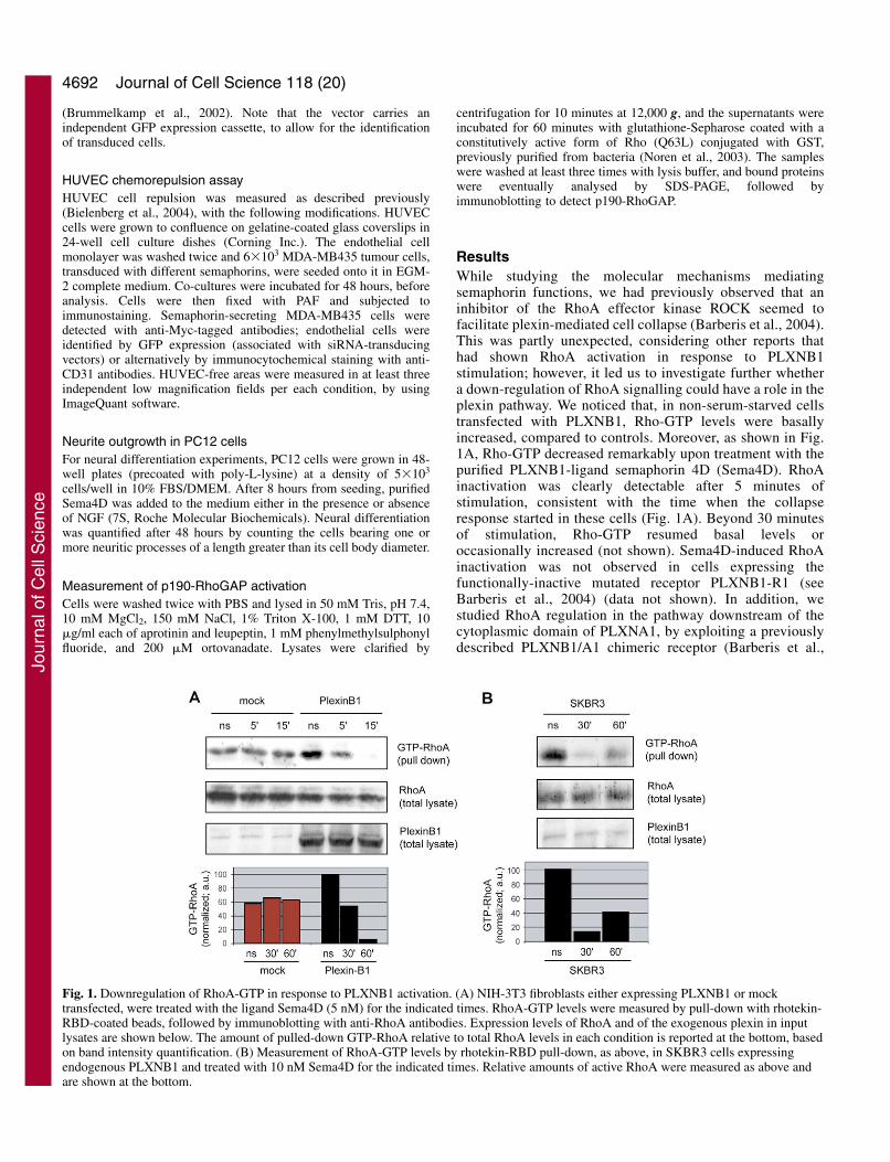
ResultsWhile studying the molecular mechanisms mediatingsemaphorin functions, we had previously observed that aninhibitor of the RhoA effector kinase ROCK seemed tofacilitate plexin-mediated cell collapse (Barberis et al., 2004).This was partly unexpected, considering other reports thathad shown RhoA activation in response to PLXNB1stimulation; however, it led us to investigate further whethera down-regulation of RhoA signalling could have a role in theplexin pathway. We noticed that, in non-serum-starved cellstransfected with PLXNB1, Rho-GTP levels were basallyincreased, compared to controls. Moreover, as shown in Fig.1A, Rho-GTP decreased remarkably upon treatment with thepurified PLXNB1-ligand semaphorin 4D (Sema4D). RhoAinactivation was clearly detectable after 5 minutes ofstimulation, consistent with the time when the collapseresponse started in these cells (Fig. 1A). Beyond 30 minutesof stimulation, Rho-GTP resumed basal levels oroccasionally increased (not shown). Sema4D-induced RhoAinactivation was not observed in cells expressing thefunctionally-inactive mutated receptor PLXNB1-R1 (seeBarberis et al., 2004) (data not shown). In addition, westudied RhoA regulation in the pathway downstream of thecytoplasmic domain of PLXNA1, by exploiting a previouslydescribed PLXNB1/A1 chimeric receptor (Barberis et al.,
Journal of Cell Science 118 (20)
Fig. 1. Downregulation of RhoA-GTP in response to PLXNB1 activation. (A) NIH-3T3 fibroblasts either expressing PLXNB1 or mocktransfected, were treated with the ligand Sema4D (5 nM) for the indicated times. RhoA-GTP levels were measured by pull-down with rhotekin-RBD-coated beads, followed by immunoblotting with anti-RhoA antibodies. Expression levels of RhoA and of the exogenous plexin in inputlysates are shown below. The amount of pulled-down GTP-RhoA relative to total RhoA levels in each condition is reported at the bottom, basedon band intensity quantification. (B) Measurement of RhoA-GTP levels by rhotekin-RBD pull-down, as above, in SKBR3 cells expressingendogenous PLXNB1 and treated with 10 nM Sema4D for the indicated times. Relative amounts of active RhoA were measured as above andare shown at the bottom.
Jour
nal o
f Cel
l Sci
ence

4693p190RhoGAP is required in semaphorin signalling
2004), and observed similar results to those for PLXNB1(supplementary material Fig. S1).
We furthermore demonstrated that purified Sema4D elicitedRhoA inactivation in SKBR3 cells, which express endogenouslevels of PLXNB1 (Fig. 1B). Rho-GTP downregulationoccurred with slower kinetics in these cells, consistent with ourprevious observation that the functional response peaks later inSKBR3 cells compared with fibroblasts overexpressing thereceptor (Barberis et al., 2004).
Altogether, these data indicate that plexin signalling canelicit Rho inactivation. However, is this relevant for thefunctional response mediated by semaphorins? Indirectevidence of this came from the observation that NIH-3T3fibroblasts expressing constitutively active RhoQ63L, ortreated with lysophosphatidic acid (LPA) to increase Rho-GTPlevels, do not undergo cell collapse in response to PLXNB1stimulation (Barberis et al., 2004).
It is known that the downregulation of Rho-GTP levels islargely mediated by p190-RhoGAP [p190A (Vincent andSettleman, 1999)], an activator of the intrinsic GTPase activityof Rho, acting under control of receptor-mediated signals(referred to as p190 hereafter). Therefore, to directly assesswhether p190 has a role in plexin signalling, we studied thepreviously described cell collapsing response (Barberis et al.,
2004) in cells deprived of p190 by expression of targeted smallinterfering RNA (siRNA) sequences. NIH-3T3 cells expressingPLXNB1 and undergoing collapse in response to Sema4Dwere engineered by means of lentiviral vectors to expresssiRNAs designed to selectively inactivate p190 transcripts ortargeted to an unrelated sequence as control. Cells expressingp190-targeted siRNAs were found to have reduced levels ofp190-RhoGAP (Fig. 2A) and were refractory to Sema4D-induced cell collapse (Fig. 2B), whereas cells expressingcontrol siRNAs displayed an efficient functional response,indistinguishable from that of parental cells. Intriguingly, wefound that p190 is also required downstream of the cytoplasmicdomain of PLXNA1, since the functional response mediatedby the chimeric receptor PLXNB1/A1 (Barberis et al., 2004)is impaired in p190-deficient fibroblasts (Fig. 2B, right panel).Therefore, our results indicate that p190-RhoGAP is requiredin the signalling pathway downstream of plexins of both the Aand B subfamilies. Analogous results were obtained by testingsemaphorin-induced collapse of COS cells similarly deprivedof p190 by expression of targeted siRNA (data not shown).
We have previously shown that the inhibition of integrin-based adhesion is a pivotal step in semaphorin signalling. Wetherefore tested whether the regulatory function of plexins onintegrin-mediated adhesion is dependent on p190. As shown in
Fig. 2. p190 Rho-GAP is required to mediate plexin signalling in fibroblasts. (A) Immunoblots of total cell lysates of NIH-3T3 fibroblastsexpressing PLXNB1 and engineered with siRNAs targeted to p190 transcript or to an unrelated sequence. The filter was probed with specificantibodies directed against p190, PLXNB1 and actin. The endogenous levels of p190 are greatly reduced in NIH-3T3 fibroblasts by expressionof targeted siRNA, while plexin expression is unaffected. (B) NIH-3T3 fibroblasts expressing PLXNB1 (or PLXNB1/A1) and engineered withp190-targeted siRNAs (or controls) were grown on glass coverslips and subjected to treatment with 5 mM Sema4D for 15 minutes. Cell werethen analysed by immunofluorescence with EC-6.9 antibodies directed against the extracellular domain of PLXNB1. Scale bar: 20 �m. Theaverage fraction of collapsed cells was determined in each condition (Barberis et al., 2004) and it is shown on the right. Plexin-dependentcollapse response is greatly impaired in the absence of p190. (C) The attachment of the same cells as above to fibronectin-coated wells wasassayed, in the presence or absence of 5 nM Sema4D. After 30 and 120 minutes, adherent cells were fixed and stained with crystal violet. Celladhesion was eventually quantified by eluting the dye and measuring the absorbance at 595 nm. (D) In a similar experiment as described in C,we scored the average fraction of fibroblasts spread on fibronectin, after 1 hour incubation with or without the indicated amounts of Sema4D(see Barberis et al., 2004).
Jour
nal o
f Cel
l Sci
ence

4694
Fig. 2C, while Sema4D significantly inhibited the adhesion tofibronectin of control cells, it was much less effective on cellsexpressing reduced levels of p190. More importantly, controlcells could not extend lamellipodia and did not spread on thesurface in the presence of the semaphorin, while the spreadingof p190-deficient fibroblasts was significantly affected only athigh ligand concentrations (Fig. 2D).
Notably, we could not obtain complete abrogation of p190expression in NIH-3T3 fibroblasts; this may explain why the
functional response, although severely impaired, was not fullyblocked in these cells. We therefore turned to immortalizedfibroblasts derived from p190–/– mice (Brouns et al., 2000) forfurther analysis. Notably, it was shown that these cells behavesimilarly to wild-type counterparts in assays of cellproliferation, cell adhesion to the ECM or cell migration(Brouns et al., 2000); moreover, Rho-GTP levels are notbasally increased in these cells, presumably because ofadaptation (Wennerberg et al., 2003). Fig. 3A shows that these
Journal of Cell Science 118 (20)
Fig. 3. Re-expression of p190 Rho-GAP in gene-deficient cells restores the functional response to plexin activation. (A) Fibroblasts derivedfrom wild-type or from p190–/– mouse embryos (Brouns et al., 2000) were engineered to express PLXNB1 (see B for protein expressionanalysis) and then treated with 10 mM Sema4D for 1 hour to test their collapse response (in analogy to Fig. 2B). Scale bar: 20 �m. (B)PLXNB1-expressing p190–/– knockout fibroblasts were engineered to express exogenous p190 or its inactive mutant p190RA (both conjugatedwith GFP). Protein expression levels were analysed by immunoblotting with specific antibodies. Hsp90 expression was determined as loadingreference. (C) Merged fluorescence images of the cells described in B after treatment with 10 nM Sema4D for 30 minutes to induce cellularcollapse: the red channel reveals PLXNB1, detected with specific antibodies (EC-6.9), while the green channel shows p190-GFP. Co-expressionof PLXNB1 with p190, but not with its inactive mutant, is required and sufficient to rescue the functional response to Sema4D in these cells.The asterisk marks a cell expressing PLXNB1 but not p190, which is insensitive to the ligand. Scale bar: 20 �m. The fraction of GFP-positivecollapsed cells in each condition was counted and it is shown at the bottom. (D) p190-deficient fibroblasts expressing PLXNB1, and furthertransfected with p190-GFP or its inactive mutant p190RA-GFP (the same shown in A), were grown on fibronectin-coated coverslips and treatedwith 5 nM Sema4D for 5 minutes. Cells were then fixed and focal adhesions revealed in the red channel with anti-paxillin antibodies. After thisshort stimulation, very few cells underwent collapse [consistent with that described in 3T3 fibroblasts (see Barberis et al., 2004)], however,focal adhesions were disassembled in most of the cells expressing functionally competent p190-RhoGAP (identified by GFP expression). Incontrast, semaphorin stimulation had no effect in cells lacking p190 or expressing the mutant devoid of RhoGAP activity (p190RA). Scale bar:20 �m. The fraction of GFP-positive cells containing focal adhesion was counted and it is shown at the bottom. (E) Affinity purification ofRhoA-GTP (by rhotekin-RBD pull-down) from equal amounts of protein lysates of p190–/– fibroblasts expressing PLXNB1 and the indicatedexogenous p190 proteins, after treatment with 10 nM Sema4D for 15 minutes. Plexin-mediated Rho inactivation is abrogated in the absence ofp190, but it is rescued by expression of exogenous p190-GFP in the gene-deficient cells. The mutated form of p190 (p190RA), lacking GAPactivity, is unable to rescue the function. The effect was quantified by measuring the relative amounts of active RhoA in each condition (bandintensity of RhoA-GTP versus total RhoA) and it is shown at the bottom.
Jour
nal o
f Cel
l Sci
ence

4695p190RhoGAP is required in semaphorin signalling
cells failed to undergo collapse upon Sema4Dstimulation, in contrast to embryo fibroblasts derivedfrom wild-type animals and similarly engineered toexpress PLXNB1. Moreover, plexin activation in p190-deficient fibroblasts, unlike that seen in wild-type cells(Barberis et al., 2004), did not induce the disassembly ofintegrin-based focal adhesions, nor did it inhibit cell-spreading on fibronectin (supplementary material Fig. S2and data not shown). A brief treatment with 10 �MY27632 (a selective ROCK inhibitor), however, couldinduce major actin remodelling in these cells, asexpected, indicating that the downstream mechanismsregulating cytoskeletal dynamics are preserved (data notshown).
To provide evidence that the lack of p190 is specificallyresponsible for abrogating plexin function in p190–/–
cells, we determined whether re-establishing itsexpression could rescue the functional responses. Wetherefore engineered gene-deficient fibroblasts to expressGFP-fusion proteins of either wild-type p190 or itsinactive form p190RA (Tatsis et al., 1998), carrying theR1283A point mutation in the GAP domain (Fig. 3B). Thecollapse response triggered by plexins was recovered bythe exogenous expression of p190-RhoGAP; by contrast,the mutant form p190RA was ineffective, indicating thatthe Rho-GAP activity of p190 is required in the plexinsignalling pathway (Fig. 3C). By immunostaining withanti-paxillin antibodies (Fig. 3D), we furthermoredemonstrated that the expression of wild-type p190 (butnot p190RA) in gene-deficient fibroblasts is sufficient tomediate the disassembly of integrin-based focal adhesivestructures, which precedes the collapse response inducedby plexin signalling (see Barberis et al., 2004). Thisstrongly suggests that the RhoGAP activity of p190mediates the inhibition of integrin function induced bysemaphorins. Consistent with this conclusion, we alsofound that cellular Rho-GTP levels where unchangedupon plexin activation in p190-deficient cells, while thismechanism was clearly rescued upon re-expression ofwild-type, but not mutated, p190 (Fig. 3E).
We have previously shown that a functional responseto purified Sema4D is also observed in epithelial cellsexpressing endogenous levels of its receptor PLXNB1(Barberis et al., 2004). Therefore, we assayed the functionalrequirement for p190-RhoGAP in SKBR3 mammarycarcinoma cells. To this end, the downregulation of p190protein was achieved by lentiviral-mediated expression ofsmall interfering RNAs (siRNA) selectively targeted to thecorresponding transcript (Fig. 4A). Fig. 4B shows that p190-depleted SKBR3 cells cannot undergo collapse in response toSema4D, unlike parental cells (Barberis et al., 2004). Inaddition, the previously described disassembly of adhesivestructures and inhibition of chemotaxis elicited by Sema4D inthese cells (Barberis et al., 2004) was severely impaired uponsiRNA-mediated knockdown of p190 (Fig. 4C). Theexpression of control siRNA, directed against an unrelatedsequence, did not have any effect.
Semaphorins are known to regulate endothelial cell migrationand angiogenesis. In particular, it has been shown that Sema3Aand Sema3F mediate repulsion of primary endothelial cells(HUVEC) and inhibit angiogenesis (Serini et al., 2003;
Bielenberg et al., 2004), via the function of neuropilins andplexins of subfamily A and their regulatory activity on integrin-mediated adhesion. To test whether these semaphorin-mediatedfunctions also depend on p190, we exploited a modified versionof the chemorepulsion assay in co-culture, described byBielenberg and coworkers (Bielenberg et al., 2004). We seededMDA-MB435 tumour cells, which had been engineered tosecrete different semaphorins (see Fig. 5A), onto confluentmonolayers of HUVEC cells that were either wild-type ordefective for p190-RhoGAP (due to expression of p190-targeted siRNAs, see Fig. 5B). Tumour cells expressingSema3F induced repulsion of wild-type endothelial cells[consistent with that reported by Bielenberg et al. (Bielenberget al., 2004)], as well as Sema3A-expressing cell, which wereextremely effective in our experiments (Fig. 5C, see alsosupplementary material Fig. S3). Sema4D-expressing cellsinduced a weaker repulsion of HUVECs, whereas mock-transfected cells intercalated with endothelial cells withoutinducing any substantial repulsion. Importantly, the down-expression of p190 significantly reduced the retraction of
Fig. 4. p190 is required for multiple functional responses mediated bySema4D in epithelial cells expressing endogenous PLXNB1. (A) Selectivedownregulation of p190 protein in SKBR3 cells by siRNA-mediatedtechnology, demonstrated by immunoblotting with specific antibodies. Theexpression of endogenous PLXNB1 is unchanged. (B) Focal complexdisassembly (revealed by vinculin immunostaining) and cellular collapsemediated by 1 hour treatment with 10 nM Sema4D was abrogated in p190-depleted SKBR3 cells. The functional response was unaffected by siRNAstargeting an unrelated sequence. The micrographs show resultsrepresentative of at least three independent experiments. Scale bar: 40 �m.(C) Chemotactic migration of SKBR3 treated with siRNAs (as above) wasassessed in Transwell® inserts, in the presence of 0.2 nM heregulin-�1(HRG), with or without 5 nM Sema4D. After 6 hours, the cells that hadmigrated across the porous membrane were stained with Crystal Violet.Cell migration was quantified by eluting the dye and measuring absorbanceat 595 nm. Results shown are the average of two independent experiments,performed in duplicate. The expected inhibition of directional cellmigration is lost in p190-deficient cells.
Jour
nal o
f Cel
l Sci
ence

4696
endothelial cells in the presence of any of thesemaphorins. We quantified this effect by measuringthe HUVEC-free surface (Fig. 5D; see Materials andMethods for quantification method). Our resultssuggest that the directional retraction of endothelialcells from areas containing semaphorins depends onthe activity of p190-RhoGAP, potentially mediatinga localized release of stable cell-substrate adhesions.
Beyond its role in cell migration, the RhoGAPactivity of p190 has been implicated in neurite outgrowth inneuroblasts in vitro (Brouns et al., 2001; Troller et al., 2004).Intriguingly, among the functions mediated by semaphorins(including Sema4D) is the ability to promote neurite outgrowth
from PC12 neural cells (Fujioka et al., 2003). We thereforeanalysed whether p190 might be required to mediate thisresponse to Sema4D, by knocking down its expression throughsiRNA-mediated technology. Notably, we did not observe
Journal of Cell Science 118 (20)
Fig. 5. p190 is required for semaphorin-inducedrepulsion of primary endothelial cells. (A) MDA-MB435mammary carcinoma cells were engineered to expressmyc-tagged Sema3F, Sema3A, Sema4D or mocktransfected. The secretion of the semaphorins in theconditioned medium as demonstrated byimmunoblotting with anti-myc antibodies. (B) HUVECcells were engineered to express siRNAs targeted top190 transcript or to an unrelated sequence. Theexpression levels of p190 in cell lysates were thenanalysed by immunoblotting with specific antibodies.(C) siRNA-expressing HUVECs analysed as in B weregrown to confluence on glass coverslips. 6�103 MDA-MB435 tumour cells engineered to express the indicatedsemaphorins (as shown in A) were then seeded onto themonolayer of endothelial cells. After 48 hours of co-culture, the cells were fixed and analysed byimmunofluorescence with anti-myc antibodies to revealsemaphorin-expressing cells. HUVECs were identifiedby GFP expression (associated with siRNA-expressionvectors). Scale bar: 200 �m. In other experiments, theendothelial cell monolayers were revealed byimmunocytochemistry with anti-CD31 antibodies(supplementary material Fig. S3). (D) HUVEC-freeareas were identified by GFP expression or CD31positivity (as described above) and measured byImageQuant software. At least three independent lowmagnification fields were analysed, by two separateinvestigators, for each experimental point. The tableshows average values.
Fig. 6. p190 is required for Sema4D-induced neurite outgrowth. PC12 neuroblasts were grown for 48 hours in the presence of 50 ng/ml NGF,with or without 1 nM Sema4D, and then fixed and photographed. Scale bar: 20 �m. The bar chart shows the mean percentage of cells withneurites (extending for at least one cell diameter) counted in three separate fields. Results are representative of two independent experiments.As discussed in the text, PC12 neuroblasts displayed minimal (if any) functional response to treatment Sema4D only (not shown). However,Sema4D-dependent synergism with NGF-induced neuritogenesis is abrogated in cells depleted of p190 by targeted expression of siRNAs.
Jour
nal o
f Cel
l Sci
ence

4697p190RhoGAP is required in semaphorin signalling
significant phenotypic changes in PC12 cells expressingsiRNAs targeted to p190 transcript, compared with cellsexpressing control siRNAs. Consistent with findingsreported by Fujioka and coworkers, control PC12neuroblasts displayed minimal (if any) functional responseto treatment with Sema4D only (not shown), while purifiedSema4D synergized with nerve growth factor (NGF)induced neurite outgrowth in PC12 cells expressing controlsiRNAs. In contrast, as shown in Fig. 6, semaphorinactivity was completely lost in cells undergoing siRNA-mediated p190 down-expression. Altogether, these resultspoint to p190-RhoGAP activation as a general mechanismrequired for plexin signalling in different cells.
We then asked whether p190 is physically associatedwith semaphorin receptor complexes. In co-immunoprecipitation experiments, we demonstrated thatboth PLXNA1 and PLXNB1 specifically associate withp190-RhoGAP, and this requires the cytoplasmic domainof the receptor (Fig. 7A). The association appears to beconstitutive upon protein overexpression. We thus tested theassociation of PLXNB1 with endogenous p190 and found that,while basally low, it was transiently increased upon stimulationwith the ligand Sema4D (Fig. 7B).
It was reported previously that the functional activation ofp190-RhoGAP is indicated by the increased ability to associatewith its substrate Rho-GTP (Noren et al., 2003). Therefore, weused Sepharose beads coated with Rho-Q63L (a constitutivelyactive form always bound to GTP) to pull down functionallyactivated p190-RhoGAP from lysates of cells treated withSema4D. As shown in Fig. 7C, upon plexin activation, theassociation between p190 and its substrate was significantlyincreased. This is consistent with evidence that p190 mediatesthe down-regulation of Rho-GTP induced by plexins.
Discussion Semaphorins control a wide range of biological functions.Moreover, some of them can induce antagonistic responses in
different cellular populations or experimental settings. Forexample, the same semaphorin can either repel neuronalprocesses (axons or dendrites) or attract them and induce theiroutgrowth (e.g. Kolodkin et al., 1993; Polleux et al., 2000;Swiercz et al., 2002; Schwamborn et al., 2004; Masuda et al.,2004). In a similar manner, the same semaphorin can eitherinhibit directional cell migration and induce apoptosis orpromote cell migration and cell survival (e.g. Giordano et al.,2002; Toyofuku et al., 2004; Barberis et al., 2004). This isconsistent with an ability of semaphorins to trigger a range ofintracellular pathways. For most described functionalresponses, it has been shown that plexins are a requiredcomponent of the receptor complex. However, other signallingreceptors can associate with plexins and have been implicatedin various biological outcomes.
The signalling pathways mediated by the conservedcytoplasmic domain of plexins have not been fully elucidated.In fact, although several putative signal transducers can interactwith plexins, for only a few of them has a ligand-dependent
Fig. 7. p190 is recruited to plexins, and its GAP activity isinduced upon receptor activation. (A) p190-GFP and eitherPLXNB1 or PLXNA1 (VSV-tagged) specifically co-precipitatefrom lysates of co-transfected 293T cells, whereas a truncatedform of PLXNB1 lacking the cytoplasmic domain (PlexinB1-�IC) does not associate with p190. Immunoprecipitations wereperformed using either anti-tag antibodies (GFP for p190 andanti-VSV for plexins, respectively) or non-related serum (nrs).Western blots were probed with the indicated anti-tag orprotein-specific antibodies. Note that PLXNB1 is larger thanother plexins (approx. 300 kDa), while PlexinB1-�IC andPLXNA1 are almost identical in size (approx. 220 kDa).(B) Co-immunopurification of endogenous p190 withPLXNB1 transfected in 293T cells is induced after 5 minutesstimulation with 5 nM Sema4D. The expression levels of p190and PLXNB1 in cell lysates are shown at the bottom.(C) Functionally active p190 was pulled-down (by means ofconstitutive active RhoQ63L-GST coated beads) from lysatesof 3T3 fibroblasts expressing PLXNB1 and treated with 5 nMSema4D for the indicated times. Immunoblotting with specificantibodies was used to detect p190 in pull-downs and in totallysates (included as loading controls). As measured by banddensity quantification, the level of activated p190 transientlyincreases upon plexin activation.
Jour
nal o
f Cel
l Sci
ence

4698
regulation been shown (Pasterkamp and Kolodkin, 2003).While this manuscript was in preparation, it was reported thatthe cytoplasmic domain of PLXNB1 associates with theactivated form of the small GTPase R-Ras, and promotes itsinactivation through an intrinsic R-Ras-GAP activity (Oinumaet al., 2004). This is quite unique, since all other known GAPproteins are recruited from the cytoplasm to protein complexesin the plasma membrane. In this paper, we provide strongevidence that the functional response mediated by plexinsrequires p190-RhoGAP activity. The evidence that Rho-GTPdownregulation by p190 is required to mediate plexin functionis at least three-fold: (1) multiple functional responses to plexinsignalling, elicited in fibroblasts, tumour epithelial and primaryendothelial cells, and related to control of cell adhesion and cellmotility are abrogated in the absence of p190-RhoGAP; (2)neurite outgrowth mediated by semaphorin stimulation of PC12neuroblasts is abrogated upon knockdown of p190-RhoGAP;(3) the functional response to semaphorins is rescued by re-expressing p190 in gene-deficient cells, but not by re-expressinga mutated form devoid of GAP activity. Furthermore, weprovided evidence that functional responses mediated bydifferent semaphorins, as well as by both cytoplasmic domainsof PLXNB1 and PLXNA1, require p190 activity, stronglysuggesting that this signalling pathway is shared by all familymembers. Our evidence that p190 associates with plexins insemaphorin receptor complexes clearly points to a directinvolvement of p190-RhoGAP activity in plexin signalling. Wealso showed that, upon plexin activation, the associationbetween p190 and its substrate Rho-GTP is increased, andaccounts for the observed downregulation of active Rho.Moreover, the kinetics of Rho-GTP downregulation isconsistent with that of the functional response observed infibroblasts engineered with plexins (approx. 10 minutes) and inSKBR3 cells expressing endogenous PLXNB1 (approx. 30minutes). It should be noted that, intriguingly, one of themolecules associated with activated p190 is p120-RasGAP,which can also use R-Ras as a substrate (Kinbara et al., 2003);this could provide an additional mechanism to mediate R-Rasinactivation upon plexin signalling.
It was reported that Rnd1/Rnd3 GTPases interact with p190in vitro (in pull-down assays and in yeast two-hybrid assays)and induce its GAP activity (Wennerberg et al., 2003).Intriguingly, Rnd1 also associates with the cytoplasmic domainof plexins (Rohm et al., 2000), and this seems to be aprerequisite for the recruitment of additional plexin effectormolecules, such as R-Ras or Rho GEFs (Oinuma et al., 2003;Oinuma et al., 2004). Nevertheless, we could not observechanges in the association of p190 to PLXNB1 upon Rnd1overexpression (not shown). Therefore, at the present time, weare not able to determine whether ligand-induced associationof p190 with plexins is direct or depends on additionalintermediary molecules.
In apparent contradiction to our data, others have previouslyreported increased Rho-GTP levels upon activation of plexinsof the B-subfamily. This effect is mediated by Rho ExchangeFactors associated with plexins (PDZ-Rho-GEFs), and requiresthe tyrosine kinase activity of ErbB receptors, induced bysemaphorin treatment (Aurandt et al., 2002; Swiercz et al.,2002; Swiercz et al., 2004). In fact, we found that the cytosoliclevels of active Rho are often increased in cells expressing highlevels of PLXNB1, which may be accounted by kinase-
dependent basal activation of PDZ-RhoGEFs. We have shownhere that soon after receptor stimulation, and consistent withthe time when the collapse response arises, RhoGTP levelsdecrease. This effect is transient, and at later time points activeRho resumes its initial levels (or even increases). Notably, toreveal semaphorin-dependent activation of Rho-GEFs (andRho-GTP upregulation), cells must be subjected to extensiveserum-starvation in order to silence tyrosine kinase activitiesand reduce Rho-GTP basal levels below detection threshold(Swiercz et al., 2002). In these conditions, a further down-regulation of RhoGTP induced by the ligand cannot bedetected. Notably, increased levels of active Rho wereobserved following one hour in presence of Sema4D (Swierczet al., 2002), which is long after we observed Rho-GTPdownregulation (approx. 10 minutes), and long after cellularcollapse is triggered.
Altogether, the above evidence indicates that Rho-GTPdown-regulation and up-regulation elicited by plexins of the Bsubfamily may derive from two independently regulatedpathways. However, how can they both have a role insemaphorin signalling? One bit of direct evidence that Rhoactivation may be required for certain functions is the findingthat the axonal collapse induced by Sema4D in hippocampalneurons was blocked by Rho and ROCK inhibitors (Swiercz etal., 2002). Nonetheless, we have recently shown that ROCKactivity is not required for Sema4D-mediated cellular collapse(Barberis et al., 2004), and this is consistent with resultspreviously reported for growth cone collapse in response toSema3A and Sema3F (Jin et al., 1997; Kuhn et al., 1999;Arimura et al., 2000; Atwal et al., 2003; Turner et al., 2004).In fact, direct Rho activation might only occur downstream ofplexins of the B subfamily, which associate with RhoGEFs,and therefore it is unlikely to play a role in the signallingpathway downstream of other semaphorin receptors.Interestingly, it was recently reported that the downregulationof Rho-GTP levels correlates with PLXNA1-mediatedinhibition of endocardial cell migration (Toyofuku et al., 2004).Here we found that p190-RhoGAP is required to mediateplexin signalling in a range of cells, and that it is required forplexin-induced Rho downregulation. Moreover, we show thata mutant form of p190 that specifically lacks GAP activity andcannot downregulate Rho-GTP is unable to mediate plexinsignalling. The functional requirement for p190 is observed notonly in plexin over-expressing cells, but also in immortalizedepithelial and neuronal cells, as well as in primary endothelialcells, which express endogenous semaphorin receptors. Thus,we propose that a transient inhibition of Rho signalling is acommon mechanism required for plexin-mediated cellcollapse, for the regulation of cell adhesion and cell migration,and in neurite outgrowth induced by semaphorins. However,Rho-GTP upregulation may be selectively elicited by plexinsof the B subfamily as an independent signalling pathway,involved in specific functional responses.
Rho-GTP downregulation mediated by p190 may account forF-actin depolymerization and cytoskeletal rearrangementstypically observed in semaphorin-treated cells and axonalgrowth cones. In addition, we showed here that p190 is requiredfor the early inhibition of integrin function mediated by plexins,which is crucially implicated in the control of cell migration andangiogenesis. How can p190-RhoGAP mediate this effect?Integrin-based complexes are very dynamic structures, as
Journal of Cell Science 118 (20)
Jour
nal o
f Cel
l Sci
ence

4699p190RhoGAP is required in semaphorin signalling
suggested by the fact that both inhibition of adhesion and theinability to break adhesive complexes can hamper cellmigration (Webb et al., 2002). Interestingly, low levels of activeRho at the leading edge guarantee a fast turnover of newlyformed adhesive sites, to allow changes in the direction ofmigration induced by environmental guidance cues. However,a major inhibition of Rho signalling at the cell membrane (suchas that mediated by p190 activation) can hamper the stability ofall adhesive complexes, by weakening their connection with thecontractile actin cytoskeleton (Burgstaller et al., 2004). Whenpresented non-directionally, these signals induce retraction ofall existing pseudopodia, or result in the appearance of randompoorly organized cell protrusions, and eventually lead toimpaired migration (Tatsis et al., 1998; Fincham et al., 1999;Worthylake et al., 2003). However, when the same signals arepresented locally in a polarized manner, as we showed here inthe endothelial cell co-culture assay, they elicit directional cellrepulsion, probably explained by the selective retraction ofcellular processes closer to the factor inhibiting cell-substrateadhesion (such as the semaphorins).
It remains to be determined whether Rho-GAP activity is alsoimportant for semaphorin-mediated axon guidance in vivo. Itshould be noted that the prevailing concept is that Rhoactivation hampers axonal outgrowth, while Rho inhibitionpromotes it. A localized turnover of Rho signalling, however,might be required for the dynamic reshaping of growth conessteering away from repelling signals and towards attractiveones. The demonstration of localized defects in axonpathfinding in p190 knockout mice (Brouns et al., 2000) isconsistent with such a hypothesis and merits furtherinvestigation. Intriguingly, at least three semaphorins (Sema3A,Sema3E and Sema4D) can induce both axonal collapse andneurite outgrowth in different neuronal cells (Sakai et al., 1999;Schwamborn et al., 2004; Masuda et al., 2004). While axonalcollapse and cellular collapse are fast functional responsesthought to share certain effector mechanisms, the signallingpathways involved in semaphorin-mediated neurite outgrowthwere not known. Intriguingly, Rho inactivation by p190 caninduce neurite outgrowth by disassembling the cortical actinnetwork (Brouns et al., 2001), and in this paper we provide thefirst evidence that the neurite outgrowth induced bysemaphorins requires p190-RhoGAP. Based on our results, wethus conclude that p190-Rho-GAP is recruited to plexins uponligand stimulation and is a pivotal mediator of multiplefunctional responses mediated by semaphorins.
We wish to thank M. Shwartz and K. Burridge for generouslyproviding expression constructs for Rhotekin-RBD-GST, and Rho-Q63L-GST and p190-R1283A-GFP, respectively. We thank G. Scita,H. Kikutani, A. Kolodkin, S. Giordano, G. Gilestro and L. Trusolinofor comments and advice, and M. De Petrini, L. Palmas and R. Albanofor help. This work was supported by funds from the ItalianAssociation for Cancer Research-AIRC (to L.T. and P.M.C.) and fromthe EMBO Young Investigator Programme (to L.T.).
ReferencesArimura, N., Inagaki, N., Chihara, K., Menager, C., Nakamura, N.,
Amano, M., Iwamatsu, A., Goshima, Y. and Kaibuchi, K. (2000).Phosphorylation of collapsin response mediator protein-2 by Rho-kinase:Evidence for two separate signaling pathways for growth cone collapse. J.Biol. Chem. 273, 23973-23980.
Atwal, J. K., Singh, K. K., Tessier-Lavigne, M., Miller, F. D. and Kaplan,
D. R. (2003). Semaphorin 3F antagonizes neurotrophin-inducedphosphatidylinositol 3-kinase and mitogen-activated protein kinase kinasesignaling: a mechanism for growth cone collapse. J. Neurosci. 23, 7602-7609.
Aurandt, J., Vikis, H. G., Gutkind, J. S., Ahn, N. and Guan, K. L. (2002).The semaphorin receptor plexin-B1 signals through a direct interaction withthe Rho-specific nucleotide exchange factor, LARG. Proc. Natl. Acad. Sci.USA 99, 12085-12090.
Barberis, D., Artigiani, S., Casazza, A., Corso, S., Giordano, S., Love, C.A., Jones, E. Y., Comoglio, P. M. and Tamagnone, L. (2004). Plexinsignaling hampers integrin-based adhesion, leading to Rho-kinaseindependent cell rounding, and inhibiting lamellipodia extension and cellmotility. FASEB J. 18, 592-594.
Behar, O., Golden, J. A., Mashimo, H., Schoen, F. J. and Fishman, M. C.(1996). Semaphorin III is needed for normal patterning and growth ofnerves, bones and heart. Nature 383, 525-528.
Bernards, A. and Settleman, J. (2004). GAP control: regulating theregulators of small GTPases. Trends Cell Biol. 14, 377-385.
Bielenberg, D. R., Hida, Y., Shimizu, A., Kaipainen, A., Kreuter, M., Kim,C. C. and Klagsbrun, M. (2004). Semaphorin 3F, a chemorepulsant forendothelial cells, induces a poorly vascularized, encapsulated, nonmetastatictumor phenotype. J. Clin. Invest. 114, 1260-1271.
Bito, H., Furuyashiki, T., Ishihara, H., Shibasaki, Y., Ohashi, K., Mizuno,K., Maekawa, M., Ishizaki, T. and Narumiya, S. (2000). A critical rolefor a Rho-associated kinase, p160ROCK, in determining axon outgrowth inmammalian CNS neurons. Neuron 26, 431-441.
Brouns, M. R., Matheson, S. F., Hu, K. Q., Delalle, I., Caviness, V. S.,Silver, J., Bronson, R. T. and Settleman, J. (2000). The adhesion signalingmolecule p190 RhoGAP is required for morphogenetic processes in neuraldevelopment. Development 127, 4891-4903.
Brouns, M. R., Matheson, S. F. and Settleman, J. (2001). p190 RhoGAP isthe principal Src substrate in brain and regulates axon outgrowth, guidanceand fasciculation. Nat. Cell Biol. 3, 361-367.
Brown, C. B., Feiner, L., Lu, M. M., Li, J., Ma, X., Webber, A. L., Jia, L.,Raper, J. A. and Epstein, J. A. (2001). PlexinA2 and semaphorin signalingduring cardiac neural crest development. Development 128, 3071-3080.
Brummelkamp, T. R., Bernards, R. and Agami, R. (2002). A system forstable expression of short interfering RNAs in mammalian cells. Science296, 550-553.
Burbelo, P. D., Finegold, A. A., Kozak, C. A., Yamada, Y. and Takami, H.(1998). Cloning, genomic organization and chromosomal assignment of themouse p190-B gene. Biochim. Biophys. Acta 1443, 203-210.
Burgstaller, G. and Gimona, M. (2004). Actin cytoskeleton remodelling vialocal inhibition of contractility at discrete microdomains. J. Cell Sci. 117,223-231.
Bussolino, F., Di Renzo, M. F., Ziche, M., Bocchietto, E., Olivero, M.,Naldini, L., Gaudino, G., Tamagnone, L., Coffer, A. and Comoglio, P.M. (1992). Hepatocyte growth factor is a potent angiogenic factor thatstimulates endothelial cell motility and growth. J. Cell Biol. 119, 629-641.
Conrotto, P., Corso, S., Gamberini, S., Comoglio, P. M. and Giordano, S.(2004). Interplay between scatter factor receptors and B plexins controlsinvasive growth. Oncogene 23, 5131-5137.
De Palma, M. and Naldini, L. (2002). Transduction of a gene expressioncassette using advanced generation lentiviral vectors. Methods Enzymol.346, 514-529.
Driessens, M. H., Hu, H., Nobes, C. D., Self, A., Jordens, I., Goodman, C.S. and Hall, A. (2001). Plexin-B semaphorin receptors interact directly withactive Rac and regulate the actin cytoskeleton by activating Rho. Curr. Biol.11, 339-344.
Eickholt, B. J., Mackenzie, S. L., Graham, A., Walsh, F. S. and Doherty,P. (1999). Evidence for collapsin-1 functioning in the control of neural crestmigration in both trunk and hindbrain regions. Development 126, 2181-2189.
Etienne-Manneville, S. and Hall, A. (2002). Rho GTPases in cell biology.Nature 420, 629-635.
Feiner, L., Webber, A. L., Brown, C. B., Lu, M. M., Jia, L., Feinstein, P.,Mombaerts, P., Epstein, J. A. and Raper, J. A. (2001). Targeted disruptionof semaphorin 3C leads to persistent truncus arteriosus and aortic archinterruption. Development 128, 3061-3070.
Fincham, V. J., Chudleigh, A. and Frame, M. C. (1999). Regulation of p190Rho-GAP by v-Src is linked to cytoskeletal disruption duringtransformation. J. Cell Sci. 112, 947-956.
Fujioka, S., Masuda, K., Toguchi, M., Ohoka, Y., Sakai, T., Furuyama, T.and Inagaki, S. (2003). Neurotrophic effect of Semaphorin 4D in PC12cells. Biochem. Biophys. Res. Commun. 301, 304-310.
Jour
nal o
f Cel
l Sci
ence
4700
Giordano, S., Corso, S., Conrotto, P., Artigiani, S., Gilestro, G., Barberis,D., Tamagnone, L. and Comoglio, P. M. (2002). The semaphorin 4Dreceptor controls invasive growth by coupling with Met. Nat. Cell Biol. 4,720-724.
Hu, H., Marton, T. F. and Goodman, C. S. (2001). Plexin B mediates axonguidance in Drosophila by simultaneously inhibiting active Rac andenhancing RhoA signaling. Neuron 32, 39-51.
Jin, Z. and Strittmatter, S. M. (1997). Rac1 mediates collapsin-1-inducedgrowth cone collapse. J. Neurosci. 17, 6256-6263.
Kawasaki, T., Kitsukawa, T., Bekku, Y., Matsuda, Y., Sanbo, M., Yagi, T.and Fujisawa, H. (1999). A requirement for neuropilin-1 in embryonicvessel formation. Development 126, 4895-4902.
Kinbara, K., Goldfinger, L. E., Hansen, M., Chou, F. L. and Ginsberg, M.H. (2003). Ras GTPases: integrins’ friends or foes? Nat. Rev. Mol. Cell Biol.4, 767-776.
Kolodkin, A. L., Matthes, D. J. and Goodman, C. S. (1993). The semaphoringenes encode a family of transmembrane and secreted growth cone guidancemolecules. Cell 75, 1389-1399.
Kuhn, T. B., Brown, M. D., Wilcox, C. L., Raper, J. A. and Bamburg, J.R. (1999). Myelin and collapsin-1 induce motor neuron growth conecollapse through different pathways: inhibition of collapse by opposingmutants of rac1. J. Neurosci. 19, 1965-1975.
Luo, Y., Raible, D. and Raper, J. A. (1993). Collapsin: a protein in brain thatinduces the collapse and paralysis of neuronal growth cones. Cell 75, 217-227.
Maestrini, E., Tamagnone, T., Longati, P., Cremona, O., Gulisano, M.,Bione, S., Tamanini, F., Neel, B. G., Toniolo, D. and Comoglio, P. M.(1996). A family of transmembrane proteins with homology to the MET-hepatocyte growth factor receptor. Proc. Natl. Acad. Sci. USA 93, 674-678.
Masuda, K., Furuyama, T., Takahara, M., Fujioka, S., Kurinami, H. andInagaki, S. (2004). Sema4D stimulates axonal outgrowth of embryonicDRG sensory neurones. Genes Cells 9, 821-829.
Moreno-Flores, M. T., Martin-Aparicio, E., Martin-Bermejo, M. J.,Agudo, M., McMahon, S., Avila, J., Diaz-Nido, J. and Wandosell, F.(2003). Semaphorin 3C preserves survival and induces neuritogenesis ofcerebellar granule neurons in culture. J. Neurochem. 87, 879-890.
Noren, N. K., Arthur, W. T. and Burridge, K. (2003). Cadherin engagementinhibits RhoA via p190RhoGAP. J. Biol. Chem. 278, 13615-13618.
Oinuma, I., Katoh, H., Harada, A. and Negishi, M. (2003). Directinteraction of Rnd1 with Plexin-B1 regulates PDZ-RhoGEF-mediated Rhoactivation by Plexin-B1 and induces cell contraction in COS-7 cells. J. Biol.Chem. 278, 25671-25677.
Oinuma, I., Ishikawa, Y., Katoh, H. and Negishi, M. (2004). TheSemaphorin 4D receptor Plexin-B1 is a GTPase activating protein for R-Ras. Science 305, 862-865.
Pasterkamp, R. J. and Kolodkin, A. L. (2003). Semaphorin junction: makingtracks toward neural connectivity. Curr. Opin. Neurobiol. 13, 79-89.
Pasterkamp, R. J., Peschon, J. J., Spriggs, M. K. and Kolodkin, A. L.(2003). Semaphorin 7A promotes axon outgrowth through integrins andMAPKs. Nature 424, 398-405.
Perrot, V., Vazquez-Prado, J. and Gutkind, J. S. (2002). Plexin B regulatesrho through the guanine nucleotide exchange factors leukemia-associatedRho GEF (LARG) and PDZ-RhoGEF. J. Biol. Chem. 277, 43115-43120.
Polleux, F., Morrow, T. and Ghosh, A. (2000). Semaphorin 3A is achemoattractant for cortical apical dendrites. Nature 404, 567-573.
Raftopoulou, M. and Hall, A. (2004). Cell migration: Rho GTPases lead theway. Dev. Biol. 265, 23-32.
Ren, X. D., Kiosses, W. B. and Schwartz, M. A. (1999). Regulation of thesmall GTP-binding protein Rho by cell adhesion and the cytoskeleton.EMBO J. 18, 578-585.
Ren, X. D., Kiosses, W. B., Sieg, D. J., Otey, C. A., Schlaepfer, D. D. andSchwartz, M. A. (2000). Focal adhesion kinase suppresses Rho activity topromote focal adhesion turnover. J. Cell Sci. 113, 3673-3678.
Ridley, A. J., Schwartz, M. A., Burridge, K., Firtel, R. A., Ginsberg, M.H., Borisy, G., Parsons, J. T. and Horwitz, A. R. (2003). Cell migration:integrating signals from front to back. Science 302, 1704-1709.
Rohm, B., Rahim, B., Kleiber, B., Hovatta, I. and Puschel, A. W. (2000).The semaphorin 3A receptor may directly regulate the activity of smallGTPases. FEBS Lett. 486, 68-72.
Rottner, K., Hall, A. and Small, J. V. (1999). Interplay between Rac and Rhoin the control of substrate contact dynamics. Curr. Biol. 9, 640-648.
Sakai, T., Furuyama, T., Ohoka, Y., Miyazaki, N., Fujioka, S., Sugimoto,H., Amasaki, M., Hattori, S., Matsuya, T. and Inagaki, S. (1999). Mousesemaphorin H induces PC12 cell neurite outgrowth activating Ras-mitogen-activated protein kinase signaling pathway via Ca(2+) influx. J. Biol. Chem.274, 29666-29671.
Schwamborn, J. C., Fiore, R., Bagnard, D., Kappler, J., Kaltschmidt, C.and Puschel, A. W. (2004). Semaphorin 3A stimulates neurite extension andregulates gene expression in PC12 cells. J. Biol. Chem. 279, 30923-30926.
Schwartz, M. A. and Shattil, S. J. (2000). Signaling networks linkingintegrins and rho family GTPases. Trends Biochem. Sci. 25, 388-391.
Serini, G., Valdembri, D., Zanivan, S., Morterra, G., Burkhardt, C.,Caccavari, F., Zammataro, L., Primo, L., Tamagnone, L., Logan, M. etal. (2003). Class 3 semaphorins control vascular morphogenesis byinhibiting integrin function. Nature 424, 391-397.
Swiercz, J. M., Kuner, R., Behrens, J. and Offermanns, S. (2002). Plexin-B1 directly interacts with PDZ-RhoGEF/LARG to regulate RhoA andgrowth cone morphology. Neuron 35, 51-63.
Swiercz, J. M., Kuner, R. and Offermanns, S. (2004). Plexin-B1/RhoGEF-mediated RhoA activation involves the receptor tyrosine kinase ErbB-2. J.Cell Biol. 165, 869-880.
Takahashi, T. and Strittmatter, S. M. (2001). Plexina1 autoinhibition by theplexin sema domain. Neuron 29, 429-439.
Takahashi, T., Fournier, A., Nakamura, F., Wang, L. H., Murakami, Y.,Kalb, R. G., Fujisawa, H. and Strittmatter, S. M. (1999). Plexin-neuropilin-1 complexes form functional semaphorin-3A receptors. Cell 99,59-69.
Tamagnone, L. and Comoglio, P. M. (2004). To move or not to move?Semaphorin signalling in cell migration. EMBO Rep. 5, 356-361.
Tamagnone, L., Artigiani, S., Chen, H., He, Z., Ming, G. I., Song, H.,Chedotal, A., Winberg, M. L., Goodman, C. S., Poo, M. et al. (1999).Plexins are a large family of receptors for transmembrane, secreted, andGPI-anchored semaphorins in vertebrates. Cell 99, 71-80.
Tatsis, N., Lannigan, D. A. and Macara, I. G. (1998). The function of thep190 Rho GTPase-activating protein is controlled by its N-terminal GTPbinding domain. J. Biol. Chem. 273, 34631-34638.
Toyofuku, T., Zhang, H., Kumanogoh, A., Takegahara, N., Suto, F., Kamei,J., Aoki, K., Yabuki, M., Hori, M., Fujisawa, H. et al. (2004). Dual rolesof Sema6D in cardiac morphogenesis through region-specific association ofits receptor, Plexin-A1, with off-track and vascular endothelial growth factorreceptor type 2. Genes Dev. 18, 435-447.
Troller, U., Raghunath, A. and Larsson, C. (2004). A possible role forp190RhoGAP in PKCepsilon-induced morphological effects. Cell Signal.16, 245-252.
Turner, L. J., Nicholls, S. and Hall, A. (2004). The activity of the plexin-A1receptor is regulated by Rac. J. Biol. Chem. 279, 33199-33205.
Van Aelst, L. and Symons, M. (2002). Role of Rho family GTPases inepithelial morphogenesis. Genes Dev. 16, 1032-1054.
Vincent, S. and Settleman, J. (1999). Inhibition of RhoGAP activity issufficient for the induction of Rho-mediated actin reorganization. Eur. J.Cell Biol. 78, 539-548.
Webb, D. J., Parsons, J. T. and Horwitz, A. F. (2002). Adhesion assembly,disassembly and turnover in migrating cells – over and over and over again.Nat. Cell Biol. 4, E97-E100.
Wennerberg, K., Forget, M. A., Ellerbroek, S. M., Arthur, W. T., Burridge,K., Settleman, J., Der, C. J. and Hansen, S. H. (2003). Rnd proteinsfunction as RhoA antagonists by activating p190 RhoGAP. Curr. Biol. 13,1106-1115.
Winberg, M. L., Tamagnone, L., Bai, J., Comoglio, P. M., Montell, D. andGoodman, C. S. (2001). The transmembrane protein Off-track associateswith Plexins and functions downstream of Semaphorin signaling duringaxon guidance. Neuron 32, 53-62.
Worthylake, R. A. and Burridge, K. (2003). RhoA and ROCK promotemigration by limiting membrane protrusions. J. Biol. Chem. 278, 13578-13584.
Zanata, S. M., Hovatta, I., Rohm, B. and Puschel, A. W. (2002).Antagonistic effects of Rnd1 and RhoD GTPases regulate receptor activityin Semaphorin 3A-induced cytoskeletal collapse. J. Neurosci. 22, 471-477.
Journal of Cell Science 118 (20)
Jour
nal o
f Cel
l Sci
ence