Embed Size (px)
Citation preview

1
p110inhibition overcomes stromal cell-mediated ibrutinib
resistance in mantle cell lymphoma
Running title: Stromal cell-mediated ibrutinib resistance
Jiyu Guan1,2
, Dan Huang1, Konstantin Yakimchuk
1 and Sam Okret
1*
1Department of Biosciences and Nutrition, Karolinska Institutet, Neo, SE-141 83 Huddinge,
Sweden
2Key Laboratory of Zoonosis Research, Ministry of Education, College of Veterinary
Medicine, Jilin University, 5333 Xi'an Road, 130062, Changchun, China,
E-mail: [email protected]
Running title: Stromal cell-mediated ibrutinib resistance
*Corresponding author:
Prof. Sam Okret
Dept. of Biosciences and Nutrition
Karolinska Institutet
Neo, SE-141 83 Huddinge, Sweden
E-mail: [email protected]
Conflicts of Interest: The authors have no conflict of interest to declare.
on April 22, 2020. © 2018 American Association for Cancer Research. mct.aacrjournals.org Downloaded from
Author manuscripts have been peer reviewed and accepted for publication but have not yet been edited. Author Manuscript Published OnlineFirst on February 26, 2018; DOI: 10.1158/1535-7163.MCT-17-0784
2
Abstract
Acquired resistance to cancer drugs is common, also for modern targeted drugs like the
Bruton’s tyrosine kinase (BTK) inhibitor ibrutinib, a new drug approved for the treatment of
the highly aggressive and relapsing mantle cell lymphoma (MCL). The tumor
microenvironment often impacts negatively on drug response. Here we demonstrate that
stromal cells protect MCL cells from ibrutinib-induced apoptosis and support MCL cell
regrowth after drug removal by impairing ibrutinib-mediated down-regulation of
phosphoinositide-3-kinase (PI3K)/AKT signaling. Importantly, the stromal cell-mediated
ibrutinib resistance was overcome in vitro by inhibiting AKT activity using the PI3K catalytic
p110α subunit specific inhibitor BYL719. This was seen both for MCL cell lines and primary
MCL cells. Furthermore, inhibition of p110 activity by BYL719 potentiated the ability of
ibrutinib to inhibit MCL tumor growth in vivo in a mouse xenograft model. The stromal cell-
mediated ibrutinib resistance was found to be due to a direct interaction with MCL cells and
involves the integrin VLA-4, since disrupting stromal cell-MCL cell interaction using a VLA-
4 blocking antibody abrogated the ibrutinib resistance. This suggests that combined treatment
with ibrutinib and a p110α inhibitor, alternatively by disrupting stromal cell-MCL cell
interaction, may be a promising therapeutic strategy to overcome stromal cell-mediated
ibrutinib resistance in MCL.
on April 22, 2020. © 2018 American Association for Cancer Research. mct.aacrjournals.org Downloaded from
Author manuscripts have been peer reviewed and accepted for publication but have not yet been edited. Author Manuscript Published OnlineFirst on February 26, 2018; DOI: 10.1158/1535-7163.MCT-17-0784

3
Introduction
The tumor microenvironment (TME) with its non-malignant cells and stromal components
has a major influence on tumor cell proliferation, survival, dissemination and resistance to
therapy. This includes Non-Hodgkin lymphomas (NHL) (1,2). Furthermore, recent results
suggest that the interaction between the TME and lymphoma cells is bidirectional, for
example by the lymphoma cells secreting cytokines which in turn attract non-malignant cells
like macrophages, immune and stromal cells that influence the tumor cells (2).
Mantle cell lymphoma (MCL), a B cell NHL, which comprises 5-7% of NHLs, and most
often presents itself in an aggressive manner, is considered incurable. In MCL, a constitutive
activation of the B cell receptor (BCR) signaling pathway has been shown to be essential in
MCL pathogenesis. This involves increased levels of phosphorylated (active) Bruton tyrosine
kinase (BTK) and SYK, two key signaling components of the BCR pathway (3-5). BCR
signaling activates several downstream signaling pathways including nuclear factor-B (NF-
B), PI3K/AKT, RAS and mitogen-activated protein kinase (MAPK), which all contribute to
cell survival, proliferation, adhesion and migration of B cells (6,7). Particularly, high
constitutive NF-B activity is seen in both MCL cell lines and patient samples and is
considered to play a central role in the pathogenesis of MCL (8,9). One of the most recently
developed targeted drugs inhibiting the BCR/NF-B signaling is ibrutinib (binds covalently to
the active site of BTK at cysteine 481), which in clinical studies has been shown to have
unprecedented effects on several B cell malignancies (10). Due to its significant efficacy,
specificity and limited side effects, ibrutinib has been approved for treatment of
relapsed/refractory MCL and chronic lymphocytic leukemia (CLL) (11).
Ibrutinib has been evaluated in clinical studies of relapsed/refractory MCL showing an
overall response rate (partial or complete) of 50-68% (12,13). Still, initial primary resistance
(32-50%) or acquired resistance to ibrutinib is common (13-16). Furthermore, most of the
initially responsive MCL patients eventually acquire ibrutinib resistance and relapse, with a
median duration of response limited to 3.5-17.5 months (12,13). Importantly, only in very few
cases of MCL could development of ibrutinib resistance in relapsing MCL patients be
attributed to an acquired mutation in the BTK (the enzyme targeted by ibrutinib), which was
not present before the start of the ibrutinib treatment, that could explain ibrutinib resistance.
For example, it was reported that only 1 out of 8 patients with non-mutated BTK post
ibrutinib treatment experienced durable response >1 year (13). Taken together, acquired BTK
on April 22, 2020. © 2018 American Association for Cancer Research. mct.aacrjournals.org Downloaded from
Author manuscripts have been peer reviewed and accepted for publication but have not yet been edited. Author Manuscript Published OnlineFirst on February 26, 2018; DOI: 10.1158/1535-7163.MCT-17-0784

4
mutations in MCL during ibrutinib treatment seem to explain only a few cases in which
ibrutinib resistance arises. This is indicative that other mechanisms probably are more
relevant in MCL for the development of ibrutinib resistance. Considering the general impact
of the TME on cancer drug sensitivity, it is not unlikely that the TME may play a role in the
development of ibrutinib resistance. In fact, MCL cells have been shown to become resistant
to conventional anti-cancer drugs in the presence of stromal cells (17-20). This suggests that
TME-MCL interaction most likely contributes to drug resistance in vivo, which in turn
underlies the cause for minimal residual disease (MRD) and relapse. However, the
mechanism for acquired resistance to ibrutinib is largely unclear and may be complex. For
example, PI3K/AKT or ERK1/2 activity, rather than BTK activity, may correlate to clinical
response to ibrutinib in MCL (4,21). A more detailed understanding of the mechanism(s) of
ibrutinib insensitivity will be very useful to find ways of overcoming it, thereby improving
therapeutic outcome.
In this study, we have investigated the mechanism for stromal cell-mediated ibrutinib
resistance and demonstrate how it can be overcome by targeting PI3K signaling by inhibiting
the catalytic subunit p110 or by disrupting stromal cell-MCL cell interaction.
on April 22, 2020. © 2018 American Association for Cancer Research. mct.aacrjournals.org Downloaded from
Author manuscripts have been peer reviewed and accepted for publication but have not yet been edited. Author Manuscript Published OnlineFirst on February 26, 2018; DOI: 10.1158/1535-7163.MCT-17-0784
5
Materials and Methods
Chemicals and antibodies
Ibrutinib (PCI-32765, BTK-inhibitor), Idelalisib (CAL101, p110 inhibitor), TGX221
(p110 inhibitor), CZC24832 (p110 inhibitor), and LY294002 (pan-p110// inhibitor)
were obtained from Selleckchem. The structures, characteristics and references to these
compounds can be found on the company homepage (sellleckchem.com). BYL719 (p110
inhibitor) was from Active Biochem. Integrin alpha4/CD49d (VLA-4) antibody (HP2/1) and
mouse IgG Isotype control were from Thermo Fisher Scientific. Anti-Human CD19-APC
(#302212) and anti-human/mouse phospho-AKT (473)-APC (#17-9715-42) antibodies were
obtained from eBioscience. Human BD Fc BlockTM
antibody (#564220) was from BD
PharmingenTM
.
Cell lines and primary MCL cells
Human MCL cell lines Mino and Rec-1 as well as the murine stromal cell line MS-5 were
from Deutsche Sammlung von Mikroorganismen und Zellkulturen Gmbh (DSMZ), Leibzig,
Germany. All the cells in the repository of and provided by DSMZ have undergone
authentication by DNA (STR) profiling. The human follicular dendritic cell (FDC) line HK-
2m was provided by Dr. Y.S. Choi (22). All the cell lines were tested mycoplasma free by the
MycoAlert™ mycoplasma detection kit from Lonza. The primary MCL cells derived by
blood sampling were from Prof. Anders Österborg, Hematology Center, Karolinska
University Hospital, Solna, Sweden. All the cell lines were grown in in RPMI 1640 (GIBCO)
supplemented with 10% FBS (GIBCO) and 100 IU penicillin/mL and 100ug/mL streptomycin
(GIBCO).
Mice, tumor grafting and treatment
NOD/SCID IL-2Rnull
(NOD.Cg-Prkdcscid Il2rgtm1Wjl/SzJ) mice (23) also denoted NSG,
were bred and kept under pathogen free conditions in the animal facility at Karolinska
University Hospital (Huddinge, Sweden) as previously described (24). Xenografts were
obtained by injecting male NSG mice subcutaneously in the right flank with 1x10⁷ MCL cells,
followed by intraperitoneal treatment once daily with vehicle, ibrutinib (5mg/kg/day),
BYL719 (20mg/kg/day) or ibrutinib plus BYL719 from the day when the volume of the
tumors reached 40-60mm3. The tumor growth was measured daily and tumor volume was
on April 22, 2020. © 2018 American Association for Cancer Research. mct.aacrjournals.org Downloaded from
Author manuscripts have been peer reviewed and accepted for publication but have not yet been edited. Author Manuscript Published OnlineFirst on February 26, 2018; DOI: 10.1158/1535-7163.MCT-17-0784

6
calculated as 0.5 x length (mm) x width2 (mm) using a caliper.
In vitro co-culturing
5×10⁴-1×10
5 MCL cells were added per well in a 24-well plate with stromal cells that 3 days
prior had been seeded with 1×10⁵
murine MS-5 stromal or human follicular dendritic cells
(FDC) per well (pre-treated with 10μg/ml mitomycin C (Sigma) for 2.5h). The co-cultures
were then treated with either vehicle or drug for the number of days indicated.
To address the effect of conditioned cell medium on MCL cells, 1×10⁵ murine MS-5 stromal
cells (pre-treated with 10μg/ml mitomycin C (Sigma) for 2.5h) were grown for 3 days in a 24-
well plate where after 5×10⁴ MCL cells were added to an upper hanging cell culture insert
(24-Well Millicell, Millipore). Vehicle or indicated drugs were then added to the medium for
the number of days as stated in the figure legend, where after the MCL cells were harvested
and analyzed.
Analysis of MCL cell proliferation (CFSE and EdU labelling)
Carboxyfluorescein succinimidyl ester (CFSE, Biolegend) labeled MCL cells were
cultured (with or without stromal cells) and treated as indicated. Cell proliferation was
indicated by a reduced CFSE signal (CFSE dilution). Alternatively, cell proliferation was
analyzed by an EdU incorporation assay as described by the manufacturer of Click-iT®
EdU
Alexa Fluor®
488 Flow Cytometry Assay Kit (Thermo Fisher Scientific).
Analysis of MCL cell apoptosis
Harvested cells from the co-cultures were first stained with anti-human CD19-APC
antibody in order to identify the human MCL cells. Apoptosis was determined by FITC-
Annexin V/ 7-Aminoactinomycin D (7-AAD, eBioscience) staining, and apoptosis in the
stained MCL cells was quantified by flow cytometry.
Analysis of AKT activity
Cells were fixed with Intracellular Fixation Buffer (eBioscience), permeabilized with
absolute methanol, blocked with Human BD Fc BlockTM
antibody and stained with pAKT
(473) antibody followed by analysis by flow cytometry.
Flow cytometry assay
on April 22, 2020. © 2018 American Association for Cancer Research. mct.aacrjournals.org Downloaded from
Author manuscripts have been peer reviewed and accepted for publication but have not yet been edited. Author Manuscript Published OnlineFirst on February 26, 2018; DOI: 10.1158/1535-7163.MCT-17-0784

7
A FACS Calibur (Becton-Dickinson) was used for all of the flow cytometry experiments.
For each sample, approximately 104 cells were analyzed and the data was evaluated using
FCS Express5 software. In all figures where the result from one flow cytometric analysis is
shown, this exemplifies a representative result from 2-3 independent experiments.
RNA extraction and quantitative real-time PCR (qPCR)
RNA isolation, cDNA synthesis and qPCR were performed as described previously (25). The
sequences of gene primers used are shown in Supplementary Table S1.
Statistical analysis
Unpaired, two-tailed students t-test or one-way ANOVA (Tukey’s multiple comparison tests)
were performed for the statistical analysis. P<0.05 was considered significant, * P<0.05, **
P<0.01, *** P<0.001. Mean values ± standard deviation (SD) are presented.
Ethical aspects
Animal care was in accordance with the guidelines of Karolinska Institutet and all animal
experiments were performed according to the approval by the local animal ethical committee
(Stockholm South Animal Ethical Committee, approval S61-14). Ethical permission for the
use of primary MCL material was approved by the Regional Ethical Committee in Stockholm
(approval 2010/1478-32) to Prof. Anders Österborg, who provided the cells.
Results
Stromal cells mediate ibrutinib resistance of MCL cells
Both Mino and Rec-1 MCL cells are sensitive to ibrutinib in vitro as determined by
increased apoptosis and inhibition of proliferation following treatment (Supplementary Figs.
S1A, S1B and ref. (8)). To investigate whether ibrutinib sensitivity of MCL cells is affected
by stromal cells, Rec-1 or Mino cells were co-cultured with or without murine MS-5 stromal
cells where after the impact of stromal cells on MCL cell proliferation and apoptosis was
analyzed in the CD19 positive cell population (Fig. 1A). The co-cultured MCL cells displayed
reduced growth inhibition by ibrutinib (200nM or 500nM) compared to cells grown alone
on April 22, 2020. © 2018 American Association for Cancer Research. mct.aacrjournals.org Downloaded from
Author manuscripts have been peer reviewed and accepted for publication but have not yet been edited. Author Manuscript Published OnlineFirst on February 26, 2018; DOI: 10.1158/1535-7163.MCT-17-0784

8
assayed as reduced CFSE staining (Figs. 1B and C and Supplementary Fig. S1C). Co-
culturing MS-5 stromal cells with Rec-1 or Mino MCL cells also largely reduced sensitivity
of the MCL cells to ibrutinib-induced cell apoptosis (Figs. 1D and E). The same result was
seen when Rec-1 cells were co-cultured with human FDC stromal cells (Supplementary Fig.
S1D). These results demonstrated that stromal cells strongly impaired MCL cell sensitivity to
ibrutinib. Importantly, if Mino cells which were resistant to ibrutinib-induced cell apoptosis in
the presence of stromal cells were transferred into new wells without stromal cells and
retreated with the same concentrations of ibrutinib, they regained sensitivity to ibrutinib-
induced apoptosis (Supplementary Fig. S2). This confirmed that stromal cells, and not a
tumor cell autonomous mechanism, conferred the resistance. Furthermore, the inability of
stromal cell conditioned media to prevent ibrutinib-induced cell apoptosis suggested that a
direct stromal cell-MCL interaction is the main cause for the resistance (Figs. 1F and G). The
requirement for direct stromal cell-MCL cell interaction for generating ibrutinib resistance
was further tested by the addition of an adhesion molecule VLA-4 blocking antibody to the
Mino-MS-5 cell co-cultures, which resulted in a strong reduction in adhesion of Mino cells to
the stromal cells (Supplementary Fig. S3A). Disrupting the interaction using the VLA-4
blocking antibody sensitized the pro-apoptotic effect of ibrutinib on Rec-1 and Mino cells,
respectively, compared to when treated with ibrutinib+control IgG (Figs. 1H and I). Similar
results were seen when Rec-1 cell interaction with FDC was abrogated by the VLA-4
blocking antibody (Supplementary Fig. S3B). The VLA-4 blocking antibody in itself did not
show any pro-apoptotic effect compared to treatment with the control IgG antibody, neither in
the presence nor absence of stromal cells (Figs. 1H and I, Supplementary Figs. S3B and S3C).
This emphasizes that a direct stromal cell-MCL cell interaction is responsible for the ibrutinib
resistance. Furthermore, the results support the involvement of VLA-4 in MCL cell
attachment to stromal cells.
Stromal cells support MCL cell regrowth after ibrutinib removal
Rec-1 and MS-5 cell co-cultures were treated with ibrutinib as depicted in the experimental
outline Fig. 2A. Rec-1 cell proliferation was largely impaired after 10 days of treatment with
500nM ibrutinib, indicated by a reduction of the EdU signal (Fig. 2B). However, a substantial
recovery of proliferating cells was observed 6 days after ibrutinib removal (Fig. 2B). To
exclude a cell autonomous effect, Rec-1 cells in the absence of MS-5 stromal cells were
on April 22, 2020. © 2018 American Association for Cancer Research. mct.aacrjournals.org Downloaded from
Author manuscripts have been peer reviewed and accepted for publication but have not yet been edited. Author Manuscript Published OnlineFirst on February 26, 2018; DOI: 10.1158/1535-7163.MCT-17-0784
9
treated for 2 days with 500nM ibrutinib where after ibrutinib was removed. In contrast to the
situation when the Rec-1 cells were co-cultured with MS-5 cells, this 2 days ibrutinib
treatment in the absence of stromal cells was sufficient to prevent recurrence of Rec-1 cell
proliferation as analyzed 6 days later (Fig. 2C). Importantly, the lack of Rec-1 cell regrowth
was not due to ibrutinib-induced cell death as no major apoptosis was detected after the 2 day
treatment with ibrutinib (Supplementary Fig. S4A). This was in contrast to when the Rec-1
cells were treated with the pan-PI3K inhibitor LY294002, where a significant increase in
apoptosis was detected (Supplementary Fig. S4A). Furthermore, the reappearance of
proliferating cells after ibrutinib removal originated mainly from the adherent cells and not
from the cells growing in suspension (Fig. 2D). The same result was seen for Mino cells
(Supplementary Fig. S4B). Likewise, inhibiting Mino cell interaction with MS-5 stromal cells
using the VLA-4 blocking antibody during ibrutinib treatment prevented regrowth of the
MCL cells after ibrutinib removal, while cells from treatments with the agents individually
did not (Supplementary Fig. S4C).
Stromal cells impair ibrutinib-mediated down-regulation of PI3K/AKT signaling in
MCL cells
Our initial results showed that ibrutinib-mediated repression of NF-B activity remained
intact in MCL cells when co-cultured with MS-5 stromal cells, as indicated by retained down-
regulation of some typical NF-B target genes (Supplementary Fig. S5A). However, when co-
culturing Mino or Rec-1 cells with MS-5 cells, the latter strongly impaired the ibrutinib-
mediated reduction of pAKT (Ser473) levels as compared to when MCL were grown in the
absence of stromal cells (Figs. 3A and B). Similar result was seen when Rec-1 cells were
incubated with human FDC (Supplementary Fig. S5B). These results suggest that stromal
cells impair ibrutinib-mediated down-regulation of AKT activity.
p110 inhibition enhances ibrutinib-mediated down-regulation of pAKT levels and
helps to overcome stromal cell-mediated ibrutinib resistance in vitro.
In order to test if the stromal cell-mediated impaired ability of ibrutinib to reduce AKT
activity involved PI3K/AKT signaling, we screened different PI3K isoform specific inhibitors
for their ability to restore Mino cell sensitivity to ibrutinib-induced apoptosis in MS-5 stromal
on April 22, 2020. © 2018 American Association for Cancer Research. mct.aacrjournals.org Downloaded from
Author manuscripts have been peer reviewed and accepted for publication but have not yet been edited. Author Manuscript Published OnlineFirst on February 26, 2018; DOI: 10.1158/1535-7163.MCT-17-0784
10
cell-Mino cell co-cultures. When treating the co-cultures with increasing concentration of
PI3K catalytic subunit inhibitors BYL719 (p110), TGX221 (p110), CZC24832 (p110) or
CAL101 (p110) in the absence or presence of 500nM ibrutinib, we observed that combined
treatment with BYL719 most effectively restored the ability of ibrutinib to induce Mino cell
apoptosis (Fig. 4A). A weaker effect was observed by CAL101 (Fig. 4A, compare the degree
of apoptosis ± ibrutinib at 5M BYL719 and 20M CAL101 which show the same degree of
apoptosis in the absence of ibrutinib). Treatment with TGX221 or CZC24832 did not
effectively restore ibrutinib sensitivity of the Mino cells. 5M BYL719 also effectively
restored Rec-1 cell sensitivity to ibrutinib in the presence of MS-5 stromal cells
(Supplementary Fig. S6A). Importantly, BYL719 also enhanced the ability of ibrutinib to
induce apoptosis in 3 out of 4 cases when primary MCL cells were co-cultured with MS-5
stromal cells (Fig. 4B). Similarly to the sensitivity of the Mino MCL cells to the various p110
catalytic PI3K subunit inhibitors in the presence of stromal cells, the sensitivity of the Mino
cells in the absence of stromal cells was most profound to BYL719. A weaker sensitivity to
CAL101 was seen, while the Mino MCL cells were largely resistant to CZC24832 and
TGX221 (Supplementary Fig. S6B). Furthermore, combined ibrutinib+BYL719 treatment of
Mino cells in the presence of MS-5 stromal cells resulted in at least an additive reducing
effect on pAKT levels compared to single drug treatment (Fig. 4C). Similar results were seen
for Rec-1 cells co-cultured with FDC (Supplementary Fig. S6C).
To investigate the effect of combined ibrutinib+BYL719 treatment on MCL cell
proliferation and regrowth, cells were treated as outlined in Fig. 2A. Upon treatment with
500nM ibrutinib alone, restart of Mino cell proliferation was observed 6 days after the
ibrutinib was removed. In contrast, restart of proliferation of Mino cells that initially had been
treated with ibrutinib+BYL719 was largely impaired after drug removal (Fig. 4D). The same
result was seen for Rec-1 cells (Supplementary Fig. S6D). The impaired regrowth of the Mino
cells correlated to reduced pAKT levels in the cells (Fig. 4E).
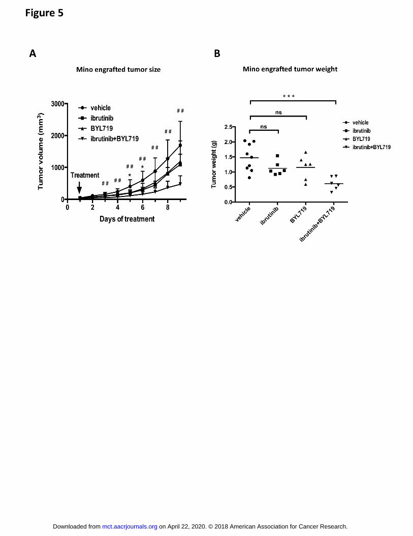
p110 inhibition helps to overcome stromal cell-mediated ibrutinib resistance in vivo.
To test whether BYL719 also can enhance the ibrutinib effect in vivo, Mino cells were
grafted to immunocompromised mice and tumor growth was measured following treatment
with 5mg/kg/day of ibrutinib or 20mg/kg/day of BYL719 alone or in combination. BYL719
on April 22, 2020. © 2018 American Association for Cancer Research. mct.aacrjournals.org Downloaded from
Author manuscripts have been peer reviewed and accepted for publication but have not yet been edited. Author Manuscript Published OnlineFirst on February 26, 2018; DOI: 10.1158/1535-7163.MCT-17-0784

11
treatment alone did not show significant inhibition as compared to vehicle treatment at any
time point while ibrutinib only significantly inhibited tumor growth at early time points (Fig.
5A). However, when ibrutinib and BYL719 were administered together, a significant
inhibition of tumor growth was observed, also at the later treatment period when none of the
drugs alone significantly inhibited tumor growth (Fig. 5A). Tumor weight at the endpoint
confirmed the effects (Fig. 5B). Mouse weight was not affected by the treatments
(Supplementary Fig. S7A). Furthermore, when we analyzed mRNA expression of some
typical NF-B target genes from the above tumor tissues, we found that ibrutinib alone still
effectively repressed gene expression, while repression by BYL719 alone was absent or minor
(Supplementary Fig. S7B). For the NF-B target genes analyzed, no enhanced repression was
seen when the cells were treated with ibrutinib+BYL719 compared to when cells were treated
with ibrutinib alone.
Discussion
Tumor recurrence due to development of resistance to cancer drugs is an obstacle for long-
term treatment effects. This is also the case for the BTK inhibitor ibrutinib, a recently FDA
approved targeted drug for relapsing MCL and CLL. In some recent clinical studies it was
shown that development of ibrutinib resistance in MCL is not uncommon (13,15,16).
Furthermore, in the study by Martin et al. (13) it was shown that development of ibrutinib
resistance was associated with failed improvement of MCL outcome also when treated with
other front-line drugs. This demonstrates the need to find ways to overcome ibrutinib
resistance. Considering that intrinsic mechanisms, e.g. BTK mutations, explaining ibrutinib
resistance are rare in MCLs (13), alternative mechanisms are likely responsible. Tumor
resistance to cancer drugs have to a large extend been attributed to the TME, thereby
contributing to MRD and relapse (26). In this report we describe that stromal cells protect
MCL cells from ibrutinib effects and that ibrutinib sensitivity was established if TME-
mediated “ibrutinib resistant” MCL cells were re-treated with ibrutinib following removal of
stromal cells. This demonstrates a non-autonomous tumor cell mechanism as responsible for
the resistance. This observation also resembles the finding in other cancers, e.g. in
on April 22, 2020. © 2018 American Association for Cancer Research. mct.aacrjournals.org Downloaded from
Author manuscripts have been peer reviewed and accepted for publication but have not yet been edited. Author Manuscript Published OnlineFirst on February 26, 2018; DOI: 10.1158/1535-7163.MCT-17-0784
12
glioblastoma, that recruited stromal cells after radiation facilitate tumor regrowth (27).
Furthermore, although stromal cell-derived soluble factors have been attributed to targeted
drug resistance in cancers (28,29), we show that a direct contact between MCL and stromal
cells is central in conferring the ibrutinib resistance, since growth of MCL in stromal cell
conditioned media was nonsufficient to effectively confer ibrutinib resistance.
Our results which show a stromal cell-mediated impairment of the ability of ibrutinib to
repress pAKT levels in the MCL cells are in line with described association between
PI3K/AKT signaling and resistance to other drugs used in treatment of cancers (30,31). In
contrast, we demonstrate that ibrutinib-mediated repression of NF-B target genes remained
fully functional in MCL cells also when co-cultured with MS-5 stromal cells. This shows that
TME-mediated ibrutinib resistance is not due to abrogated NF-B activity. This is supported
by an observation that ibrutinib still can down-regulate BTK activity in ibrutinib resistant
MCL cells (4). That the PI3K catalytic subunit p110 specific inhibitor BYL719 restored the
ability of ibrutinib to induce apoptosis in both MCL cell lines and in 3 out of 4 primary MCL
leukemic cell samples supports that PI3K/AKT signaling is involved in the stromal cell-
mediated ibrutinib resistance. Furthermore, we also observed that ibrutinib+BYL719 pre-
treated MCL cells (in the presence of stromal cells) showed impaired capacity for regrowth
after drug removal. This correlated to reduced pAKT levels. However, whether this represents
a more permanent inhibition or just a shorter delay in proliferation restart was not examined in
detail. Importantly, we also showed that BYL719 treatment enhanced the ability of ibrutinib
to inhibit Mino cell derived tumor growth in vivo in a xenograft mouse model. Likewise,
PI3K signaling was recently also demonstrated to play a role in activated B cell like Diffuse
Large B-cell Lymphoma (ABC-DLBCL) as inhibition of both p110 and p110 was required
to efficiently repress tumor growth of ABC-DLBCL in a mouse model (32). Furthermore,
dual inhibition of p110/ synergized with ibrutinib in causing remission in the DLBCL
models.
Additional support for using a p110 inhibitor in the treatment of MCL can be derived
from data by Psyrri et al. (33), who showed that in 68% of the cases of primary MCL and in
several MCL cell lines, increased PIK3CA expression due gene amplification is present,
resulting in enhanced AKT activity. Notably, we found that inhibition of p110 was more
effective than inhibition of p110 in sensitizing cells to ibrutinib in the presence of stromal
cells, while inhibition of p110 or p110 did not influence ibrutinib sensitivity. This together
on April 22, 2020. © 2018 American Association for Cancer Research. mct.aacrjournals.org Downloaded from
Author manuscripts have been peer reviewed and accepted for publication but have not yet been edited. Author Manuscript Published OnlineFirst on February 26, 2018; DOI: 10.1158/1535-7163.MCT-17-0784

13
with the observation that MCL tumor cells with increased p110 expression can sustain
constitutive PI3K signaling despite p110 inhibition (34), further supports a rationale for
targeting p110 in MCL. However, although the PI3K/AKT signaling pathway seems to
have a key role in stromal cell-mediated ibrutinib resistance, other signaling pathways may
contribute (35).
Adhesion molecules, including VLA-4, are highly expressed on MCL cells and have a central
role in MCL-stromal cell interaction (36). As demonstrated by us (Supplementary Fig. 3A)
and others (17), blocking VLA-4 disrupts the stromal-MCL cell interaction. Likewise,
Herman et al. (37) described that ibrutinib partially blocked CLL cell adhesion to stroma or
stromal components and may reduce VLA-4-dependent pro-survival signals in the TME.
Importantly, we here demonstrate that disruption of MCL-stromal cell interaction by blocking
VLA-4 led to regained ibrutinib sensitivity, supporting that the stromal cell-mediated ibrutinib
resistance involves a direct stromal cell-MCL cell interaction. Similarly, it has been shown
that VLA-4 blocking can overcome stromal cell-mediated rituximab resistance of NHL cells
in vitro (38). Furthermore, a high VLA-4 expression has been shown to be associated in
leukemia with adverse outcome and shorter survival (39,40). Although not demonstrated in
this report, a direct MCL-stromal cell interaction which involves VLA-4 has been reported to
activate PI3K/AKT signaling (35,41). Combined with our results, this may explain why a
direct stromal-MCL cell interaction results in ibrutinib resistance. This is consistent with
another finding showing that VLA-4 blocking antibody treatment combined with the
cytotoxic drug Ara-C enhanced reduction of pAKT levels (42). Furthermore, it was
demonstrated very recently that in MCL cells that had been selected for ibrutinib resistance
(and were devoid of BTK mutations), ibrutinib was unable to reduce PI3K/AKT signaling in
comparison to sensitive MCL cells (35). In addition, it was also demonstrated that co-
culturing MCL cells with stromal cells resulted in an upregulated expression of integrin 1
(subunit of VLA-4). Moreover, a reduction of AKT in MCL cells lead to reduced integrin 1
expression. Notably, it has been shown that PI3K signaling activates VLA-4 in the lymph
node niche (43). These results show that PI3K/AKT signaling is a central hub for both inside-
out as well as outside-in signaling in MCL-stromal cell interaction (35). It has also been
demonstrated that the transcriptional program of mesenchymal stromal cells in the bone
marrow microenvironment is altered by leukemia cells (44), further supporting a bidirectional
cross-talk between the cancer and the TME cells, with the lymphoma cells reshaping the TME
and the TME supporting lymphoma cell proliferation and survival, contributing to the
on April 22, 2020. © 2018 American Association for Cancer Research. mct.aacrjournals.org Downloaded from
Author manuscripts have been peer reviewed and accepted for publication but have not yet been edited. Author Manuscript Published OnlineFirst on February 26, 2018; DOI: 10.1158/1535-7163.MCT-17-0784
14
ibrutinib resistance. Although we did not observe an effect following p110 inhibition on
stromal cell survival (Supplementary Fig. S8), a possible impact of altered PI3K/AKT
signaling in the stromal cells on stromal cell-lymphoma cell cross-talk cannot be excluded.
Indeed, it has been suggested that inhibition of PI3K/AKT signaling in cells of the TME
could contribute to the effects seen by PI3K inhibitors (45). Taken together, these results
support that PI3K/AKT signaling has a central role in stromal cell-mediated ibrutinib
resistance and support our results that targeted inhibition of PI3K/AKT signaling or MCL-
stromal cell interaction can overcome stromal cell-mediated ibrutinib resistance.
Acknowledgements
We thank Prof. Yong Sung Choi and Prof. Anders Österborg for providing us with the human
FDC line and primary MCL cells, respectively. We thank Dr. Gergely Talaber, Xiaogang
Feng, Dr. Peng Zhang and Dr. Hongya Han for providing experimental advice and Dr.
Mattias Berglund for valuable comments on the manuscript.
This research was supported by funding’s from Cancerfonden (Swedish Cancer Society, CAN
2014/593 and CAN2017/404) to S. Okret and Barncancerfonden (Swedish Childhood Cancer
Foundation, PR2014-00135 and PR2016-0038) to S. Okret. Scholarships from China
Scholarship Council (CSC) supported J. Guan (2011617056) and D. Huang (201600160069),
respectively.
on April 22, 2020. © 2018 American Association for Cancer Research. mct.aacrjournals.org Downloaded from
Author manuscripts have been peer reviewed and accepted for publication but have not yet been edited. Author Manuscript Published OnlineFirst on February 26, 2018; DOI: 10.1158/1535-7163.MCT-17-0784

15
References
1. Olson OC, Joyce JA. Microenvironment-mediated resistance to anticancer therapies. Cell research 2013;23:179-81.
2. Scott DW, Gascoyne RD. The tumour microenvironment in B cell lymphomas. Nat Rev Cancer 2014;14:517-34.
3. Pighi C, Gu TL, Dalai I, Barbi S, Parolini C, Bertolaso A, et al. . Phospho-proteomic analysis of mantle cell lymphoma cells suggests a pro-survival role of B-cell receptor signaling. Cell Oncol (Dordr) 2011;34:141-53.
4. Ma J, Lu P, Guo A, Cheng S, Zong H, Martin P, et al. . Characterization of ibrutinib-sensitive and -resistant mantle lymphoma cells. Br J Haematol 2014;166:849-61.
5. Cinar M, Hamedani F, Mo Z, Cinar B, Amin HM, Alkan S. Bruton tyrosine kinase is commonly overexpressed in mantle cell lymphoma and its attenuation by Ibrutinib induces apoptosis. Leuk Res 2013;37:1271-7.
6. Kurosaki T, Shinohara H, Baba Y. B cell signaling and fate decision. Annu Rev Immunol 2010;28:21-55.
7. Dal Porto JM, Gauld SB, Merrell KT, Mills D, Pugh-Bernard AE, Cambier J. B cell antigen receptor signaling 101. Mol Immunol 2004;41:599-613.
8. Rahal R, Frick M, Romero R, Korn JM, Kridel R, Chan FC, et al. . Pharmacological and genomic profiling identifies NF-kappaB-targeted treatment strategies for mantle cell lymphoma. Nat Med 2014;20:87-92.
9. Saba NS, Liu D, Herman SE, Underbayev C, Tian X, Behrend D, et al. . Pathogenic role of B-cell receptor signaling and canonical NF-kappaB activation in mantle cell lymphoma. Blood 2016;128:82-92.
10. Tucker DL, Rule SA. Ibrutinib for mantle cell lymphoma. Future Oncol 2016;12:477-91. 11. Kapoor A. Ibrutinib: A comprehensive review of a promising drug. Clinical Cancer
Investigation Journal. Volume 5; 2016. p 203-7. 12. Wang ML, Rule S, Martin P, Goy A, Auer R, Kahl BS, et al. . Targeting BTK with ibrutinib in
relapsed or refractory mantle-cell lymphoma. N Engl J Med 2013;369:507-16. 13. Martin P, Maddocks K, Leonard JP, Ruan J, Goy A, Wagner-Johnston N, et al. . Postibrutinib
outcomes in patients with mantle cell lymphoma. Blood 2016;127:1559-63. 14. Stephens DM, Spurgeon SE. Ibrutinib in mantle cell lymphoma patients: glass half full?
Evidence and opinion. Ther Adv Hematol 2015;6:242-52. 15. Zhang SQ, Smith SM, Zhang SY, Lynn Wang Y. Mechanisms of ibrutinib resistance in chronic
lymphocytic leukaemia and non-Hodgkin lymphoma. Br J Haematol 2015;170:445-56. 16. Wang ML, Blum KA, Martin P, Goy A, Auer R, Kahl BS, et al. . Long-term follow-up of MCL
patients treated with single-agent ibrutinib: updated safety and efficacy results. Blood 2015;126:739-45.
17. Kurtova AV, Tamayo AT, Ford RJ, Burger JA. Mantle cell lymphoma cells express high levels of CXCR4, CXCR5, and VLA-4 (CD49d): importance for interactions with the stromal microenvironment and specific targeting. Blood 2009;113:4604-13.
18. Medina DJ, Goodell L, Glod J, Gelinas C, Rabson AB, Strair RK. Mesenchymal stromal cells protect mantle cell lymphoma cells from spontaneous and drug-induced apoptosis through secretion of B-cell activating factor and activation of the canonical and non-canonical nuclear factor kappaB pathways. Haematologica 2012;97:1255-63.
19. Lwin T, Lin J, Choi YS, Zhang X, Moscinski LC, Wright KL, et al. . Follicular dendritic cell-dependent drug resistance of non-Hodgkin lymphoma involves cell adhesion-mediated Bim down-regulation through induction of microRNA-181a. Blood 2010;116:5228-36.
on April 22, 2020. © 2018 American Association for Cancer Research. mct.aacrjournals.org Downloaded from
Author manuscripts have been peer reviewed and accepted for publication but have not yet been edited. Author Manuscript Published OnlineFirst on February 26, 2018; DOI: 10.1158/1535-7163.MCT-17-0784

16
20. Zhang L, Yang J, Qian J, Li H, Romaguera JE, Kwak LW, et al. . Role of the microenvironment in mantle cell lymphoma: IL-6 is an important survival factor for the tumor cells. Blood 2012;120:3783-92.
21. Chiron D, Di Liberto M, Martin P, Huang X, Sharman J, Blecua P, et al. . Cell-cycle reprogramming for PI3K inhibition overrides a relapse-specific C481S BTK mutation revealed by longitudinal functional genomics in mantle cell lymphoma. Cancer Discov 2014;4:1022-35.
22. Park CS, Choi YS. How do follicular dendritic cells interact intimately with B cells in the germinal centre? Immunology 2005;114:2-10.
23. Shultz LD, Lyons BL, Burzenski LM, Gott B, Chen X, Chaleff S, et al. . Human lymphoid and myeloid cell development in NOD/LtSz-scid IL2R gamma null mice engrafted with mobilized human hemopoietic stem cells. J Immunol 2005;174:6477-89.
24. Yakimchuk K, Hasni MS, Guan J, Chao MP, Sander B, Okret S. Inhibition of lymphoma vascularization and dissemination by estrogen receptor beta agonists. Blood 2014;123:2054-61.
25. Talaber G, Yakimchuk K, Guan J, Inzunza J, Okret S. Inhibition of estrogen biosynthesis enhances lymphoma growth in mice. Oncotarget 2016;7:20718-27.
26. McMillin DW, Negri JM, Mitsiades CS. The role of tumour-stromal interactions in modifying drug response: challenges and opportunities. Nature reviews. Drug discovery 2013;12:217-28.
27. Kioi M, Vogel H, Schultz G, Hoffman RM, Harsh GR, Brown JM. Inhibition of vasculogenesis, but not angiogenesis, prevents the recurrence of glioblastoma after irradiation in mice. The Journal of clinical investigation 2010;120:694-705.
28. Straussman R, Morikawa T, Shee K, Barzily-Rokni M, Qian ZR, Du J, et al. . Tumour micro-environment elicits innate resistance to RAF inhibitors through HGF secretion. Nature 2012;487:500-4.
29. Wilson TR, Fridlyand J, Yan Y, Penuel E, Burton L, Chan E, et al. . Widespread potential for growth-factor-driven resistance to anticancer kinase inhibitors. Nature 2012;487:505-9.
30. Berns K, Horlings HM, Hennessy BT, Madiredjo M, Hijmans EM, Beelen K, et al. . A functional genetic approach identifies the PI3K pathway as a major determinant of trastuzumab resistance in breast cancer. Cancer cell 2007;12:395-402.
31. Yano S, Wang W, Li Q, Matsumoto K, Sakurama H, Nakamura T, et al. . Hepatocyte growth factor induces gefitinib resistance of lung adenocarcinoma with epidermal growth factor receptor-activating mutations. Cancer research 2008;68:9479-87.
32. Paul J, Soujon M, Wengner AM, Zitzmann-Kolbe S, Sturz A, Haike K, et al. . Simultaneous Inhibition of PI3Kdelta and PI3Kalpha Induces ABC-DLBCL Regression by Blocking BCR-Dependent and -Independent Activation of NF-kappaB and AKT. Cancer Cell 2017;31:64-78.
33. Psyrri A, Papageorgiou S, Liakata E, Scorilas A, Rontogianni D, Kontos CK, et al. . Phosphatidylinositol 3'-kinase catalytic subunit alpha gene amplification contributes to the pathogenesis of mantle cell lymphoma. Clinical cancer research : an official journal of the American Association for Cancer Research 2009;15:5724-32.
34. Iyengar S, Clear A, Bodor C, Maharaj L, Lee A, Calaminici M, et al. . P110alpha-mediated constitutive PI3K signaling limits the efficacy of p110delta-selective inhibition in mantle cell lymphoma, particularly with multiple relapse. Blood 2013;121:2274-84.
35. Zhao X, Lwin T, Silva A, Shah B, Tao J, Fang B, et al. . Unification of de novo and acquired ibrutinib resistance in mantle cell lymphoma. Nature communications 2017;8:14920.
36. Shain KH, Dalton WS, Tao J. The tumor microenvironment shapes hallmarks of mature B-cell malignancies. Oncogene 2015;34:4673-82.
37. Herman SE, Mustafa RZ, Jones J, Wong DH, Farooqui M, Wiestner A. Treatment with Ibrutinib Inhibits BTK- and VLA-4-Dependent Adhesion of Chronic Lymphocytic Leukemia Cells In Vivo. Clin Cancer Res 2015;21:4642-51.
38. Mraz M, Zent CS, Church AK, Jelinek DF, Wu X, Pospisilova S, et al. . Bone marrow stromal cells protect lymphoma B-cells from rituximab-induced apoptosis and targeting integrin
on April 22, 2020. © 2018 American Association for Cancer Research. mct.aacrjournals.org Downloaded from
Author manuscripts have been peer reviewed and accepted for publication but have not yet been edited. Author Manuscript Published OnlineFirst on February 26, 2018; DOI: 10.1158/1535-7163.MCT-17-0784
17
alpha-4-beta-1 (VLA-4) with natalizumab can overcome this resistance. Br J Haematol 2011;155:53-64.
39. Gattei V, Bulian P, Del Principe MI, Zucchetto A, Maurillo L, Buccisano F, et al. . Relevance of CD49d protein expression as overall survival and progressive disease prognosticator in chronic lymphocytic leukemia. Blood 2008;111:865-73.
40. Shalapour S, Hof J, Kirschner-Schwabe R, Bastian L, Eckert C, Prada J, et al. . High VLA-4 expression is associated with adverse outcome and distinct gene expression changes in childhood B-cell precursor acute lymphoblastic leukemia at first relapse. Haematologica 2011;96:1627-35.
41. Matsunaga T, Takemoto N, Sato T, Takimoto R, Tanaka I, Fujimi A, et al. . Interaction between leukemic-cell VLA-4 and stromal fibronectin is a decisive factor for minimal residual disease of acute myelogenous leukemia. Nat Med 2003;9:1158-65.
42. Kim YR, Eom KS. Simultaneous Inhibition of CXCR4 and VLA-4 Exhibits Combinatorial Effect in Overcoming Stroma-Mediated Chemotherapy Resistance in Mantle Cell Lymphoma Cells. Immune network 2014;14:296-306.
43. Garmy-Susini B, Avraamides CJ, Desgrosellier JS, Schmid MC, Foubert P, Ellies LG, et al. . PI3Kalpha activates integrin alpha4beta1 to establish a metastatic niche in lymph nodes. Proceedings of the National Academy of Sciences of the United States of America 2013;110:9042-7.
44. Kim JA, Shim JS, Lee GY, Yim HW, Kim TM, Kim M, et al. . Microenvironmental remodeling as a parameter and prognostic factor of heterogeneous leukemogenesis in acute myelogenous leukemia. Cancer research 2015;75:2222-31.
45. Okkenhaug K, Graupera M, Vanhaesebroeck B. Targeting PI3K in Cancer: Impact on Tumor Cells, Their Protective Stroma, Angiogenesis, and Immunotherapy. Cancer discovery 2016;6:1090-105.
on April 22, 2020. © 2018 American Association for Cancer Research. mct.aacrjournals.org Downloaded from
Author manuscripts have been peer reviewed and accepted for publication but have not yet been edited. Author Manuscript Published OnlineFirst on February 26, 2018; DOI: 10.1158/1535-7163.MCT-17-0784

18
Figure legends
Figure 1. Bone marrow stromal cells mediate ibrutinib resistance of MCL cells.
(A) shows the outline of analysis of MCL cell proliferation and apoptosis by flow cytometry.
For analysis of apoptosis, collected cells were stained with anti-human CD19-APC antibody
in order to distinguish the human MCL cells from the stromal cells. Cell proliferation was
assayed by staining the MCL cells with CFSE dye prior to incubation with stromal cells or by
labelling cells with EdU 2.5 h before harvesting where after cells were stained with Alexa
Fluor 488-azide according to the manufactures manual. In the cases of assaying cell
proliferation by CFSE, lymphoma cell proliferation is indicated by CFSE dilution. For
analysis of cell apoptosis, the cells were stained with Annexin V-FITC. (B-C) 5×10⁴ CFSE
labeled Rec-1 or Mino MCL cells were added per well in a 24-well plate that 3 days prior had
been seeded or not seeded with murine MS-5 stromal cells (pre-treated with 10μg/ml
mitomycin C for 2.5h). Cells were then treated with either vehicle or ibrutinib (200nM or
500nM) for 10 days where after cells were collected and labeled with anti-human CD19-APC
antibody. Cell proliferation of the MCL cells was determined by flow cytometry analysis
based on the dilution of the CFSE signal. (D-E) Rec-1 or Mino MCL cells grown with or
without MS-5 stromal cells were treated with either vehicle or ibrutinib (200nM or 500nM)
for 10 days where after cells were collected. Apoptosis was determined in CD19 positive
lymphoma cells by flow cytometry followed by Annexin V staining. (F-G) 5×10⁴
Rec-1 or
Mino MCL cells grown with or without conditioned medium from MS-5 stromal cell cultures
were treated with either vehicle or ibrutinib (200nM and 500nM) for 10 days where after cells
were collected and CD19 positive cells were analyzed for apoptosis by Annexin V staining.
Data analysis for Figs. 1B-G was performed by unpaired, two-tailed students t-test, and error
bars show the SD (n=3, * P<0.05, ** P<0.01, *** P<0.001, ns, not significant). (H-I) Co-
cultures of Rec-1 or Mino cells with MS-5 stromal cells were treated for 10 days with IgG
(control, 4µg/ml), ibrutinib (500nM)+IgG, anti-VLA-4 blocking antibody (4µg/ml), or
ibrutinib+anti-VLA-4 blocking antibody where after cell apoptosis of the MCL cells was
determined by analyzing CD19 positive cells by Annexin V staining. Data analysis was
performed by one-way ANOVA Tukey’s test (n=3, *** P<0.001).
on April 22, 2020. © 2018 American Association for Cancer Research. mct.aacrjournals.org Downloaded from
Author manuscripts have been peer reviewed and accepted for publication but have not yet been edited. Author Manuscript Published OnlineFirst on February 26, 2018; DOI: 10.1158/1535-7163.MCT-17-0784

19
Figure 2. Stromal cells support Rec-1 cell regrowth after ibrutinib treatment is terminated.
(A) Experimental outline. MCL cells were seeded onto MS-5 stromal cells and the cells were
then treated with either vehicle or ibrutinib (500nM) for 10 days. On day 10, cells were
incubated with EdU for 2.5h before cell harvesting, where after cells were collected and
labeled with anti-human CD19-APC antibody, followed by Alexa Fluor 488-azide staining. In
a parallel set of wells, ibrutinib was removed and fresh medium without ibrutinib was added
and cells were cultured for an additional 6 days, followed by labeling and staining as
described above. (B) Rec-1 cells co-cultured with stromal cell were treated as described in (A)
above. Cell proliferation of CD19 positive Rec-1 cells were analyzed by EdU/Alexa Fluor
488-azide staining by flow cytometry. (The weakly EdU/Alexa Fluor 488-azide stained peak
(intensity ≈101) represents non-proliferating cells while the EdU/Alexa Fluor 488-azide peak
(intensity 102-10
3) represents proliferating cells (cells in S-phase). The figure shows one out
of three independent experiments with similar results. (C) Rec-1 cells grown in the absence of
MS-5 stromal cells were treated with ibrutinib for 2 days where after ibrutinib was removed
and the cells were cultured for another 6 days. Cell proliferation was analyzed following
EdU/Alexa Fluor 488-azide staining by flow cytometry. The figure shows one out of three
independent experiments with similar results. (D) Rec-1 cells co-cultured with MS-5 stromal
cells as described in A were treated with either vehicle or ibrutinib (200nM or 500nM) for 10
days where after CD19 positive cells in suspension or adherent to stromal cells were analyzed
for EdU/Alexa Fluor 488-azide staining by flow cytometry (left column). In a parallel set of
experiments, adherent and suspension cells were separated on day 10 and the cells were
allowed to continue to grow in the absence of ibrutinib for 6 days and analyzed as above
(right column) at day 16. The bar figures to the right show quantitative data and data analysis
was performed by unpaired, two-tailed students t-test, and error bars show the SD (n=3, ***
P<0.001; ns, not significant).
Figure 3. Stromal cells impair ibrutinib-mediated down-regulation of pAKT levels in MCL
cells.
(A) MCL cells were labeled with CFSE before co-culturing with MS-5 stromal cells in order
to distinguish MCL cells from MS-5 cells. After treatment, cells were collected and stained
with an anti-pAKT-APC antibody. CFSE positive MCL cells were gated and analyzed for
on April 22, 2020. © 2018 American Association for Cancer Research. mct.aacrjournals.org Downloaded from
Author manuscripts have been peer reviewed and accepted for publication but have not yet been edited. Author Manuscript Published OnlineFirst on February 26, 2018; DOI: 10.1158/1535-7163.MCT-17-0784
20
pAKT staining by flow cytometry. (B) 5×10⁴ Mino or Rec-1 cells were first labeled with
CFSE and then added to 24-well plates pre-seeded with or without stromal cells. The cells
were then treated with either vehicle or ibrutinib (500nM) for 5 days. On day 5, all the cells
were collected, washed, fixed and stained for pAKT using an anti-pAKT-APC antibody.
pAKT levels in CFSE labelled cells were determined by flow cytometry (left). The bar figures
to the right show the relative change in pAKT level (median florescence intensity (MFI))
when treated with ibrutinib in the presence or absence of MS-5 stromal cells. Data analysis
was performed by unpaired, two-tailed students t-test, and error bars show the SD (n=3, ***
P<0.001).
Figure 4. p110 inhibition enhances ibrutinib-mediated down-regulation of pAKT levels and
helps to overcome stromal cell-mediated ibrutinib resistance in vitro.
(A) Mino cell apoptosis in the presence of MS-5 stromal cells after treatment with increasing
concentrations of specific p110 catalytic subunit PI3K inhibitors alone or together with
500nM ibrutinib for 10 days. BYL719 (p110 specific inhibitor), CAL101 (p110δ specific
inhibitor); TGX-221 (p110β specific inhibitor) and CZC24832 (p110 specific inhibitor). Cell
labeling and analysis of apoptosis were performed as described in Fig. 1A. For each
treatment, experiments were performed in triplicates. The Annexin V staining signal of gated
CD19 positive cells (Mino cells) was analyzed by flow cytometry. (B) 1x10⁶ cells from 4
different MCL patient samples were co-cultured with MS-5 stromal cells and treated with
vehicle, ibrutinib (500nM) and BYL719 (5M) alone or in combination for 10 days.
Apoptosis was analyzed by Annexin V staining of gated CD19 positive MCL patient cells by
flow cytometry. The percentage of apoptotic cells for each condition is shown in the bar
graphs. (C) The bar graph shows pAKT levels in Mino cells as analyzed by flow cytometry
after 10 days of drug treatment in the presence of MS-5 stromal cells as indicated in the
figure. (D and E) Analysis of cell proliferation (D) and pAKT levels (E), respectively, of the
MCL cells after drug removal (experimental outline as in Fig. 2A). The figures show results
from cells collected day 16, i.e. 6 days after the drug(s) were removed. Data analysis in Figs.
4C-E was performed by one-way ANOVA Tukey’s test, and error bars show the SD (n=3, *
P<0.05, ** P<0.01, *** P<0.001).
on April 22, 2020. © 2018 American Association for Cancer Research. mct.aacrjournals.org Downloaded from
Author manuscripts have been peer reviewed and accepted for publication but have not yet been edited. Author Manuscript Published OnlineFirst on February 26, 2018; DOI: 10.1158/1535-7163.MCT-17-0784

21
Figure 5. Treatment with the p110 inhibitor BYL719 augments the efficiency of ibrutinib to
inhibit MCL tumor growth in vivo.
1x10⁷ Mino cells were engrafted subcutaneously into NSG mice. Treatment started when the
tumors were first palpable with daily injections with vehicle, ibrutinib (5mg/kg), BYL719
(20mg/kg) or a combination thereof. Intraperitoneal injections were given once a day and
tumor volume was measured daily (A) and tumor weight at the day of sacrifice (B) was
analyzed. * vehicle vs. ibrutinib, # vehicle vs. ibrutinib/BYL719. Absent of a symbol or ns
mean that there is no statistical significance. Data analysis was performed by one-way
ANOVA Tukey’s test, and error bars show the SD of 6 to 9 mice per group (##
P<0.01, *
P<0.05, *** P<0.001).
on April 22, 2020. © 2018 American Association for Cancer Research. mct.aacrjournals.org Downloaded from
Author manuscripts have been peer reviewed and accepted for publication but have not yet been edited. Author Manuscript Published OnlineFirst on February 26, 2018; DOI: 10.1158/1535-7163.MCT-17-0784

Published OnlineFirst February 26, 2018.Mol Cancer Ther Jiyu Guan, Dan Huang, Konstantin Yakimchuk, et al. resistance in mantle cell lymphoma
inhibition overcomes stromal cell-mediated ibrutinibαp110
Updated version
10.1158/1535-7163.MCT-17-0784doi:
Access the most recent version of this article at:
Material
Supplementary
http://mct.aacrjournals.org/content/suppl/2018/02/24/1535-7163.MCT-17-0784.DC1
Access the most recent supplemental material at:
Manuscript
Authorbeen edited. Author manuscripts have been peer reviewed and accepted for publication but have not yet
E-mail alerts related to this article or journal.Sign up to receive free email-alerts
Subscriptions
Reprints and
To order reprints of this article or to subscribe to the journal, contact the AACR Publications
Permissions
Rightslink site. Click on "Request Permissions" which will take you to the Copyright Clearance Center's (CCC)
.http://mct.aacrjournals.org/content/early/2018/02/24/1535-7163.MCT-17-0784To request permission to re-use all or part of this article, use this link
on April 22, 2020. © 2018 American Association for Cancer Research. mct.aacrjournals.org Downloaded from
Author manuscripts have been peer reviewed and accepted for publication but have not yet been edited. Author Manuscript Published OnlineFirst on February 26, 2018; DOI: 10.1158/1535-7163.MCT-17-0784