Embed Size (px)
Citation preview

Volume 6 • Issue 3 • 1000233Biochem Pharmacol, an open access journalISSN:2167-0501
Research Article Open Access
Darwish et al., Biochem Pharmacol (Los Angel) 2017, 6.3DOI: 10.4172/2167-0501.1000233
Biochemistry & Pharmacology: Open Access Bioc
hem
istry
& Pharmacology: Open Access
ISSN: 2167-0501
AbstractBackground: Aberrant activation of the Ras/Raf-1/extracellular-regulated kinase (ERK) pathway has been shown
to be involved in the progression of hepatocellular carcinoma (HCC). However, the mechanism of dysregulation of ERK activation is poorly understood. This study aimed to investigate the inhibitory effect of novel 3D-organocopper supramolecular coordination polymer (SCP) on invasive potential of hepatocellular carcinoma (HCC) cells and to explore the underlying mechanism. Chemoprevention could represent an important means to inhibit the process of hepatocarcinogenesis.
Methods: The inhibitory effect of SCP compound on hepatoma cells was evaluated by the examination of HCC chemically induced liver tissues and specific pathway inhibition was examined by immunohistochemistry.
Results: Recently, we identified the newly synthesized SCP as a potent inhibitor of the Ras/Raf-1/ERK pathway. In this study, we found that the expression levels of ERK1 and-2 in chemically induced HCC tissue was frequently decreased in response to SCP treatment, comparing with those untreated with SCP compound. Moreover, ERK expression levels in HCC tissue were frequently correlated with the incidence of tumor invasion and metastasis. Increased expression of beta catenin in liver tissue, which was associated with increased ERK activation. P-ERK1/2 overexpression also reduced the expression of E-cadherin, which play important roles in tumor invasion and metastasis.
Conclusion: We concluded that SCP can effectively inhibit the invasive potential of ERK signaling pathway and ERK could be not only a novel prognostic factor but also a new therapeutic target for human HCC.
The Role of Novel 3d-Copper Cyanide Supramolecular Coordination Polymer in Epithelial Mesenchymal Transition Inhibition in Hepatocellular CarcinomaNoura M Darwish1*, Fahad A Al-Dhabaan2, Ahmed Sultan1, Ahmed Malki1 and Mohamed Soliman Elshikh3
1Department of Biochemistry, Faculty of Science, Alexandria University, Alexandria, Egypt2Biology Department, Science and Humanities College, Sharqa University, Alquwayiyah, KSA3Botany and Microbiology Department, College of Science, King Saud University, Riyadh, Saudi Arabia
*Corresponding author: Darwish NM, Department of Biochemistry, Faculty of Science, Alexandria University, Alexandria, Egypt, Tel: 00201148145525; E-mail: [email protected]
Received: November 09, 2017; Accepted: November 17, 2017; Published November 27, 2017
Citation: Darwish NM, Al-Dhabaan FA, Sultan A, Malki A, Elshikh MS (2017) The Role of Novel 3d-Copper Cyanide Supramolecular Coordination Polymer in Epithelial Mesenchymal Transition Inhibition in Hepatocellular Carcinoma. Biochem Pharmacol (Los Angel) 6: 233. doi: 10.4172/2167-0501.1000233
Copyright: © 2017 Darwish NM, et al. This is an open-access article distributed under the terms of the Creative Commons Attribution License, which permits unrestricted use, distribution, and reproduction in any medium, provided the original author and source are credited.
Keywords: Ras; Raf-1; ERK; SCP; HCC
Abbreviations: ΜM: Micromolar; ANOVA: Analysis of Variance; DMF: Dimethylformamide; DMSO: Dimethyl Sulfoxide; EMT: Epithelial Mesenchymal Transition; ERK: Extracellular Regulated Kinase; H: Hour; PBS: Phosphate Buufer Saline; SCP: Supramolecular Coordination Polymer; SD: Standard Deviation; SEM: Standard Error of Mean; USA: United States of America; DEN: Diethylnitrosamine; CCl4: Carbon Tetrachloride
IntroductionHepatocellular carcinoma HCC is the third leading cause of
cancer-related death worldwide and primary cause of death in patients with liver cirrhosis [1]. Highest HCC prevalence occurs in Africa and Asia, but HCC incidence is rising also in Western countries, due to the increasing rates of alcoholic liver disease and hepatitis C [2,3]. Currently, survival remains poor for most patients with hepatocellular carcinoma, which is due to the aggressiveness of the lesions at the time of diagnosis and the lack of an effective therapy [4]. Similar to other solid tumors, genetic and epigenetic events are implicated in the development of HCC and result in aberrantly activated pathways [5,6]. Hepatocarcinogenesis is characterized by the deregulation of the balance between proliferation and cell death by apoptosis, with decrease in some proapoptotic signals, and up-regulation of antiapoptotic signals [7].
Extracellular-related kinase (ERK1 and ERK2) pathway is an attractive target or therapeutic intervention in cancer due to its integral role in the regulation of cancer cell proliferation, invasiveness, and survival [8-10]. The activity of ERK1/2 has been implicated in the regulation of embryonic morphogenesis, cell proliferation, tumor transformation, metastasis, apoptosis and other tumor-promoting cellular activities [11]. Hyper-activated Ras-ERK1/2 pathway induces
EMT through the loss of epithelial cell-specific features such as the down regulation of E-cadherin expression, therefore promoting cell motility and invasive behaviour in cancer-associated EMT and is required for the induction of motile and invasive behaviour of epithelial and carcinoma cells [12,13]. The down regulation of E-cadherin is balanced by the increased expression of mesenchymal neural cadherin (N-cadherin), which results in a 'cadherin switch' that alters cell adhesion [14]. Through this switch, the transitioning cells lose their association with epithelial cells and acquire an affinity for mesenchymal cells through c N-cadherin interactions; these interactions are weaker than E-cadherin interactions and facilitate cell migration and invasion [15]. N-cadherin connects to the cytoskeleton through β-catenin [16].
All cancers share common hallmarks such as proliferation, invasion and metastasis that play important roles in tumor promotion and progression [17]. Dysregulation of E-cadherin has been reported to contribute to cancer progression [18]. It is reported that decreased expression of E-cadherin is associated with malignant progression in various kinds of cancer including liver cancer [19-21].
Citation: Darwish NM, Al-Dhabaan FA, Sultan A, Malki A, Elshikh MS (2017) The Role of Novel 3d-Copper Cyanide Supramolecular Coordination Polymer in Epithelial Mesenchymal Transition Inhibition in Hepatocellular Carcinoma. Biochem Pharmacol (Los Angel) 6: 233. doi: 10.4172/2167-0501.1000233
Page 2 of 9
Biochem Pharmacol, an open access journalISSN:2167-0501 Volume 6 • Issue 3 • 1000233
induction at a concentration of 300 μM. In this study SCP compound was selected to investigate its role as a new anticancer compound. The structure of the compound is shown in Figure 1.
Animals
Weanling male albino rats 6 to 8 weeks old were obtained from Central Animal House, Faculty of Medicine, Alexandria University, Alexandria, Egypt. All animals were housed four to a plastic cage and had free access to water and diet under controlled environmental conditions of humidity 50 ± 10%, lighting 12 h light/dark cycle and temperature 23 ± 2°C. Animals were handled, ethically treated and sacrificed according to the rules and instructions of Ethical Committee for Animal Care of the Alexandria University in accordance with the Egyptian National Law on animal care and use.
Experimental design
A total of 60 rats were randomized and divided into three groups. All groups except group I control were given oral injections of DEN in saline 200 mg/kg body weight under ether anaesthesia. After 2 weeks of DEN administration, the carcinogenic effect was promoted by the administration of CCl4 0.5 mL/kg body weight, dissolved in sunflower oil, administered thrice a week by intra-gastric tube and phenobarbital PB, phenobarbital-Na; Sigma, St. Louis, MO, USA, tap water was replaced by 0.05% of PB solution after a 12-h fasting, daily as described [23]. After 24 weeks post exposure to DEN and CCl4, rats in groups III received a twice per week oral injection of 90 mg/kg b.wt. of synthesized compound for 4 weeks. All animals were inspected at least twice weekly. Body weight was measured at intervals throughout the study. At the end of experiments, the body weight of each rat was taken before sacrifice and all of the rats were killed by cervical dislocation under ether anaesthesia after an overnight fast. Blood was collected to determine levels of tumor markers and allowed to clot before centrifugation at 1000×g for 10 min at 4°C to separate serum. The liver tissue was washed twice with ice cold 0.1 M phosphate buffer saline PBS, 1:9, pH 7.4, blotted, dried and weighed. The relative liver weight was calculated as the percentage ratio of liver weight to the body weight. A small portion of the tissue was fixed in formalin for histological examination. The remaining tissue was stored at −20°C for not more than 12 h before analysis [24,25].
Toxicity studies
Acute toxicity study was performed for SCP compound according to the arithmetical method of Karber. About 30 white albino female rats weighing approximately 70 g were procured from a random bred colony in the animal house of Faculty of Medicine of Alexandrai University, Alexandrai, Egypt). The rats were caged in plastic cages (5/cage) in a controlled environment (ambient temperature, 25.0 ± 2.0°C and with a 12 h light/darkness cycle) with free access to food and water. They were allowed to acclimatize for 2 weeks during which the weight gained was between 90 g to 100 g. The animals were kept fasting for overnight only on water, after which the compound was administered orally. The animals were observed for first 2 hours and then at 6th and 24th hour for any toxic symptoms. The number of rats that survived were noted after 24 h and then maintained for the further 13 days with daily observation for any further toxicity.
Biochemical determinations
The activities of aspartate transaminase (AST) and alaninetransaminase (ALT) were estimated by the method of Reitman and Frankel [26], while alkaline phosphatase (ALP) and γ-glutamyltransferase (γ GT)
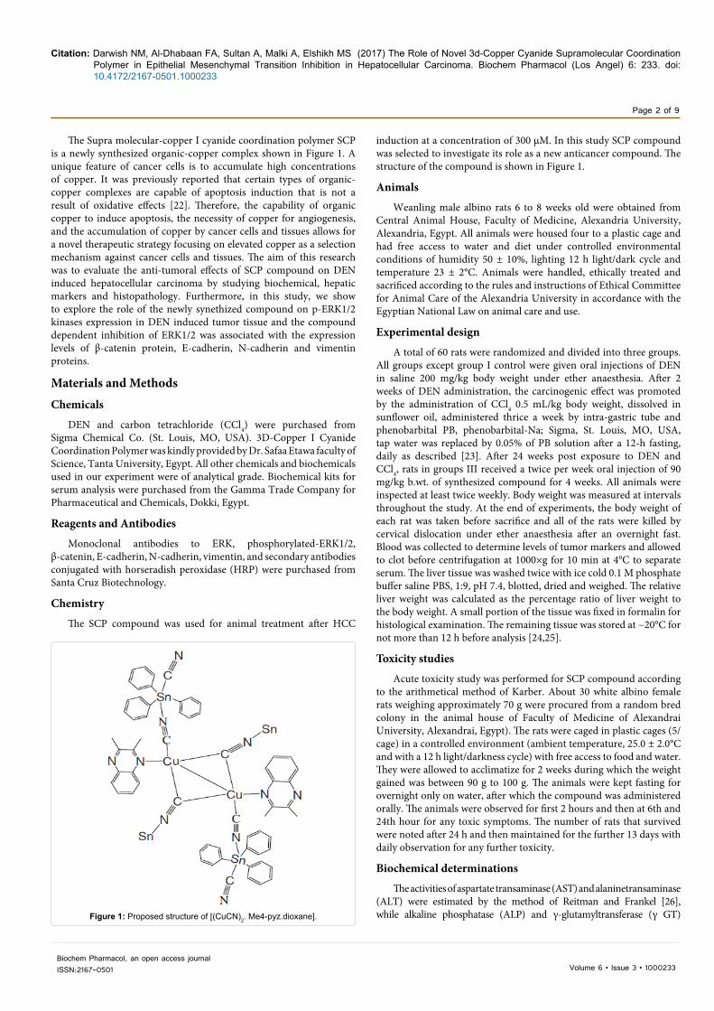
The Supra molecular-copper I cyanide coordination polymer SCP is a newly synthesized organic-copper complex shown in Figure 1. A unique feature of cancer cells is to accumulate high concentrations of copper. It was previously reported that certain types of organic-copper complexes are capable of apoptosis induction that is not a result of oxidative effects [22]. Therefore, the capability of organic copper to induce apoptosis, the necessity of copper for angiogenesis, and the accumulation of copper by cancer cells and tissues allows for a novel therapeutic strategy focusing on elevated copper as a selection mechanism against cancer cells and tissues. The aim of this research was to evaluate the anti-tumoral effects of SCP compound on DEN induced hepatocellular carcinoma by studying biochemical, hepatic markers and histopathology. Furthermore, in this study, we show to explore the role of the newly synethized compound on p-ERK1/2 kinases expression in DEN induced tumor tissue and the compound dependent inhibition of ERK1/2 was associated with the expression levels of β-catenin protein, E-cadherin, N-cadherin and vimentin proteins.
Materials and MethodsChemicals
DEN and carbon tetrachloride (CCl4) were purchased from Sigma Chemical Co. (St. Louis, MO, USA). 3D-Copper I Cyanide Coordination Polymer was kindly provided by Dr. Safaa Etawa faculty of Science, Tanta University, Egypt. All other chemicals and biochemicals used in our experiment were of analytical grade. Biochemical kits for serum analysis were purchased from the Gamma Trade Company for Pharmaceutical and Chemicals, Dokki, Egypt.
Reagents and Antibodies
Monoclonal antibodies to ERK, phosphorylated-ERK1/2, β-catenin, E-cadherin, N-cadherin, vimentin, and secondary antibodies conjugated with horseradish peroxidase (HRP) were purchased from Santa Cruz Biotechnology.
Chemistry
The SCP compound was used for animal treatment after HCC
Figure 1: Proposed structure of [(CuCN)2. Me4-pyz.dioxane].

Citation: Darwish NM, Al-Dhabaan FA, Sultan A, Malki A, Elshikh MS (2017) The Role of Novel 3d-Copper Cyanide Supramolecular Coordination Polymer in Epithelial Mesenchymal Transition Inhibition in Hepatocellular Carcinoma. Biochem Pharmacol (Los Angel) 6: 233. doi: 10.4172/2167-0501.1000233
Page 3 of 9
Biochem Pharmacol, an open access journalISSN:2167-0501 Volume 6 • Issue 3 • 1000233
were estimated by methods of King and Armstrong [27] and Szasz’s [28], respectively. Serum concentrations of albumin, total bilirubin were determined as described by Kaplan, quantitative estimation of tumor marker α-feto protein (AFP) was based on solid phase enzyme linked immunosorbent assay (ELISA) using the UBI MAGIWELL (USA) enzyme immunoassay kit [29-31].
Histopathological assessment
Liver sections were made immediately from the liver of different groups of rats, fixed in 10% formalin, dehydrated in gradual ethanol (50%-100%), cleared in xylene, and embedded in paraffin. Sections 4 µm to 5 µm thick were prepared and the pathological changes were observed microscopically after staining with hematoxylin and eosin (H–E) as described by Carleton [32]. The histopathological slides were examined and photographs were taken with a digital stereomicroscope (Olympus, B061). All slides were reviewed by the same pathologist.
Immunohistochemistry
Immunostaining was carried out using a standard protocol [33]. Formalin-fixed, paraffin-embedded tissues 4 µm were baked for 30 min, deparaffinized in xylene, and rehydrated in a graded series of ethanol solutions. Tissue sections were incubated with primary anti-p-ERK (phosphoThr202/Tyr204, Santa Cruz Biotechnology), anti-E-cadherin, anti β-catenine and anti E-cadherin antibodies (Santa Cruz Biotechnology) at 4°C overnight. Incubation with appropriate secondary antibody was followed by direct diaminobenzidine staining and light counterstaining with hematoxylin.
Western blot for detection of protein expression
Western blot was performed as described previously [34] to detect protein expression levels. The target protein was then probed using (ERK1/2, P-ERK1/2, β-catenin, E-cadherin, N-cadherin and vimentin, 1:1000, Santa Cruz, CA, USA) antibody with β-actin, served as an internal control.
Statistical analysis
Statistical analysis was performed using Student’s t-test to compare the two groups and one-way analysis of variance (ANOVA) was used with Tukey-Kramer multiple comparisons test as a post ANOVA test when the three groups were compared. Significant differences among means were estimated at p<0.05. The results were expressed as mean ± S.E.M. Values were analyzed using the SPSS version 16.
ResultsGross examination
After all experimental procedures, no abnormal appearance, macroscopic tumors or nodules were visible on the surface and sections of liver of control group, while protruding masses from the surface of the liver, characteristic nodules, thickening hardening of liver borders and enlargement of liver was obviously observed in livers of rats in group П. Smooth surface of the livers in the SCP compound treated group. All rats in the DEN group had developed diffusive HCC in cirrhotic livers (Figure 2).
Toxicity study
To assess the preventive effect of SCP compound on development and progression of HCC, SCP compound was examined for toxic effects. The results of this study are shown in Table 1. Over the study
duration of 14 day, there were no deaths recorded in animals given 100 mg/kg body weight oral dose of SCP compound. During the observation period, animals did not produce any variations in the general appearance and motor activity and there was no instant death in rats tested during the period of observation. Oral administration of SCP compound at all given doses (higher than 100 mg/kg) caused death in the tested groups during the first 24 h of observation period. Median lethal dose LD50 was calculated using the following equation: LD50=Least lethal dose-Σ (a×b)/N, where N is the number of animals in each group, a the dose difference and b the mean mortality (mortality in (2nd+1st)/2.
Body weight (initial and final), mean and relative liver weight
In comparison to final body weight of normal control group of rats (301 ± 6.489 g), a significant decrease (217 ± 4.02 g, p<0.01) in body weight of untreated group (group II) following treatment of DEN, CCl4 and PB while SCP treated group (group III) (90 mg/kg body weight) showed significant gain (p<0.05) when compared to untreated group (group II) (Table 2). A significant increase in mean liver weight (12.4 ± 0.72 g, p<0.001) and in relative liver weight (5.75 ± 0.39 g/100 g body weight, p<0.001) in untreated group (group II) was observed when compared to mean liver weight and relative liver weights of negative control group (group I) (7.6 ± 0.58 g, 2.51 ± 0.15 g/100 g body weight) respectively. However, administration of 90 mg/kg SCP compound significantly reduced the liver weight (9.8 ± 0.46 g, p<0.05) and relative liver weight (3.68 ± 0.20 g/100 g body weight P<0.05) compared to (9.8 ± 0.466 g and 5.75 ± 0.39 g/100 g body weight) respectively in untreated group (group II) (Table 2). Therefore SCP administration proved to prevent DEN induced body weight loss and liver weight increase). Table 2 shows the body, liver and relative liver weights of control and experimental groups of rats.
Figure 2: Gross examination of liver organ at the end of experiment. (A) Macroscopic appearance of the liver in an animal of control group; (B) Macroscopic appearance of the liver in an animal injected with DEN, PB and CCl4 showing protruding nodules on the liver surface ( arrows); (C) Macroscopic appearance of livers in the group treated with SCP compound. Note the finely granular aspect of the liver surface in B, compared to the smooth surface of the liver in the SCP compound treated group.
Group Dose (mg/kg)
Number of rats
No. of animals
dead
Dose difference
(a)
Mean mortality
(b)
Probit (a × b)
1 Control 5 0 0 0 02 100 5 0 100 0 03 200 5 1 100 0.5 504 300 5 1 100 1 1005 400 5 2 100 1.5 1506 500 5 3 100 2.5 250
Table 1: LD50 determination by arithmetic method of Karbar (Sum of the product=550, LD50=200-(550/5)=90 mg/kg).
Citation: Darwish NM, Al-Dhabaan FA, Sultan A, Malki A, Elshikh MS (2017) The Role of Novel 3d-Copper Cyanide Supramolecular Coordination Polymer in Epithelial Mesenchymal Transition Inhibition in Hepatocellular Carcinoma. Biochem Pharmacol (Los Angel) 6: 233. doi: 10.4172/2167-0501.1000233
Page 4 of 9
Biochem Pharmacol, an open access journalISSN:2167-0501 Volume 6 • Issue 3 • 1000233
Biochemical estimations
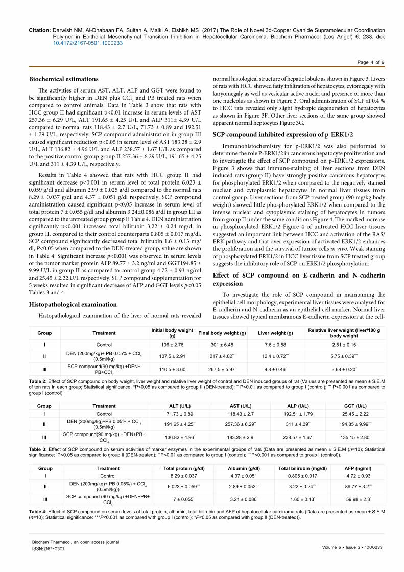
The activities of serum AST, ALT, ALP and GGT were found to be significantly higher in DEN plus CCl4 and PB treated rats when compared to control animals. Data in Table 3 show that rats with HCC group II had significant p<0.01 increase in serum levels of AST 257.36 ± 6.29 U/L, ALT 191.65 ± 4.25 U/L and ALP 311± 4.39 U/L compared to normal rats 118.43 ± 2.7 U/L, 71.73 ± 0.89 and 192.51 ± 1.79 U/L, respectively. SCP compound administration in group III caused significant reduction p<0.05 in serum level of AST 183.28 ± 2.9 U/L, ALT 136.82 ± 4.96 U/L and ALP 238.57 ± 1.67 U/L as compared to the positive control group group II 257.36 ± 6.29 U/L, 191.65 ± 4.25 U/L and 311 ± 4.39 U/L, respectively.
Results in Table 4 showed that rats with HCC group II had significant decrease p<0.001 in serum level of total protein 6.023 ± 0.059 g/dl and albumin 2.99 ± 0.025 g/dl compared to the normal rats 8.29 ± 0.037 g/dl and 4.37 ± 0.051 g/dl respectively. SCP compound administration caused significant p<0.05 increase in serum level of total protein 7 ± 0.055 g/dl and albumin 3.24±0.086 g/dl in group III as compared to the untreated group group II Table 4. DEN administration significantly p<0.001 increased total bilirubin 3.22 ± 0.24 mg/dl in group II, compared to their control counterparts 0.805 ± 0.017 mg/dl. SCP compound significantly decreased total bilirubin 1.6 ± 0.13 mg/dl, P<0.05 when compared to the DEN-treated group, value are shown in Table 4. Significant increase p<0.001 was observed in serum levels of the tumor marker protein AFP 89.77 ± 3.2 ng/ml and GGT194.85 ± 9.99 U/L in group II as compared to control group 4.72 ± 0.93 ng/ml and 25.45 ± 2.22 U/L respectively. SCP compound supplementation for 5 weeks resulted in significant decrease of AFP and GGT levels p<0.05 Tables 3 and 4.
Histopathological examination
Histopathological examination of the liver of normal rats revealed
normal histological structure of hepatic lobule as shown in Figure 3. Livers of rats with HCC showed fatty infiltration of hepatocytes, cytomegaly with karyomegaly as well as vesicular active nuclei and presence of more than one nucleolus as shown in Figure 3. Oral administration of SCP at 0.4 % to HCC rats revealed only slight hydropic degeneration of hepatocytes as shown in Figure 3F. Other liver sections of the same group showed apparent normal heptocytes Figure 3G.
SCP compound inhibited expression of p-ERK1/2
Immunohistochemistry for p-ERK1/2 was also performed to determine the role P-ERK1/2 in cancerous hepatocyte proliferation and to investigate the effect of SCP compound on p-ERK1/2 expressions. Figure 3 shows that immune-staining of liver sections from DEN induced rats (group II) have strongly positive cancerous hepatocytes for phosphorylated ERK1/2 when compared to the negatively stained nuclear and cytoplasmic hepatocytes in normal liver tissues from control group. Liver sections from SCP treated group (90 mg/kg body weight) showed little phosphorylated ERK1/2 when compared to the intense nuclear and cytoplasmic staining of hepatocytes in tumors from group II under the same conditions Figure 4. The marked increase in phosphorylated ERK1/2 Figure 4 of untreated HCC liver tissues suggested an important link between HCC and activation of the RAS/ERK pathway and that over-expression of activated ERK1/2 enhances the proliferation and the survival of tumor cells in vivo. Weak staining of phosphorylated ERK1/2 in HCC liver tissue from SCP treated group suggests the inhibitory role of SCP on ERK1/2 phosphorylation.
Effect of SCP compound on E-cadherin and N-cadherin expression
To investigate the role of SCP compound in maintaining the epithelial cell morphology, experimental liver tissues were analyzed for E-cadherin and N-cadherin as an epithelial cell marker. Normal liver tissues showed typical membranous E-cadherin expression at the cell-
Group Treatment Initial body weight (g) Final body weight (g) Liver weight (g) Relative liver weight (liver/100 g
body weight
I Control 106 ± 2.76 301 ± 6.48 7.6 ± 0.58 2.51 ± 0.15
II DEN (200mg/kg)+ PB 0.05% + CCl4 (0.5ml/kg) 107.5 ± 2.91 217 ± 4.02** 12.4 ± 0.72*** 5.75 ± 0.39***
III SCP compound(90 mg/kg) +DEN+ PB+CCl4
110.5 ± 3.60 267.5 ± 5.97* 9.8 ± 0.46* 3.68 ± 0.20*
Table 2: Effect of SCP compound on body weight, liver weight and relative liver weight of control and DEN induced groups of rat (Values are presented as mean ± S.E.M of ten rats in each group; Statistical significance: *P<0.05 as compared to group II (DEN-treated); ** P<0.01 as compared to group I (control); *** P<0.001 as compared to group I (control).
Group Treatment ALT (U/L) AST (U/L) ALP (U/L) GGT (U/L)I Control 71.73 ± 0.89 118.43 ± 2.7 192.51 ± 1.79 25.45 ± 2.22
II DEN (200mg/kg)+PB 0.05% + CCl4 (0.5ml/kg) 191.65 ± 4.25** 257.36 ± 6.29** 311 ± 4.39** 194.85 ± 9.99***
III SCP compound(90 mg/kg) +DEN+PB+ CCl4
136.82 ± 4.96* 183.28 ± 2.9* 238.57 ± 1.67* 135.15 ± 2.80*
Table 3: Effect of SCP compound on serum activities of marker enzymes in the experimental groups of rats (Data are presented as mean ± S.E.M (n=10); Statistical significance: *P<0.05 as compared to group II (DEN-treated); ** P<0.01 as compared to group I (control); *** P<0.001 as compared to group I (control)).
Group Treatment Total protein (g/dl) Albumin (g/dl) Total bilirubin (mg/dl) AFP (ng/ml)I Control 8.29 ± 0.037 4.37 ± 0.051 0.805 ± 0.017 4.72 ± 0.93
II DEN (200mg/kg)+ PB 0.05%) + CCl4 (0.5ml/kg)) 6.023 ± 0.059*** 2.89 ± 0.052*** 3.22 ± 0.24*** 89.77 ± 3.2***
III SCP compound (90 mg/kg) +DEN+PB+ CCl4
7 ± 0.055* 3.24 ± 0.086* 1.60 ± 0.13* 59.98 ± 2.3*
Table 4: Effect of SCP compound on serum levels of total protein, albumin, total bilirubin and AFP of hepatocellular carcinoma rats (Data are presented as mean ± S.E.M (n=10); Statistical significance: ***P<0.001 as compared with group I (control); *P<0.05 as compared with group II (DEN-treated)).

Citation: Darwish NM, Al-Dhabaan FA, Sultan A, Malki A, Elshikh MS (2017) The Role of Novel 3d-Copper Cyanide Supramolecular Coordination Polymer in Epithelial Mesenchymal Transition Inhibition in Hepatocellular Carcinoma. Biochem Pharmacol (Los Angel) 6: 233. doi: 10.4172/2167-0501.1000233
Page 5 of 9
Biochem Pharmacol, an open access journalISSN:2167-0501 Volume 6 • Issue 3 • 1000233
Figure 3: Photomicrographs of liver specimens stained with H&E. (A1, A2: Liver from control rat (group I) showing normal liver histology with remarkable central vein (A1 × 10 and A2 × 40); B1, B2: Adenoma formation and fatty change of hepatocytes with atypical arrangement of hepatic lobule, lymphocytic infiltration, congestion of hepatic sinusoids and bile duct proliferation (B1 × 10 and B2 × 40); C2: Higher magnification from C1 showing prominent nucleoli and enlarged hepatocytes with mitotic activity (X100); D1: Loss of architecture and neoplastic cells arranged in lobules separated by fibrous septa and focal areas of hydropic degeneration (X10); D2, D3: Higher magnification from D1 showing sever hydropic degeneration of hepatocytes showing irregular enlarged loss of lobular architecture and hyperchromatic nuclei (X40); E, F: Liver sections from SCP treated group (group Ш) rats showing coagulative necrosis of hepatocytes with pyknotic nuclei and congested central veins (X10). G: Degenerative changes and necrosis of hepatic parenchyma necrosis and hepatocytes maintaining near normal liver architecture (X10)).
Figure 4: Immunohistochemical staining pattern of p-ERK1/2 in experimental liver tissues (X40); A: Negative nuclear and cytoplasmic staining in normal liver tissue; B, C: Intense cytoplasmic and nuclear staining of cancerous hepatocytes in DEN induced group (group II); D: Very weak staining in SCP compound treated liver tissue from group III.
Citation: Darwish NM, Al-Dhabaan FA, Sultan A, Malki A, Elshikh MS (2017) The Role of Novel 3d-Copper Cyanide Supramolecular Coordination Polymer in Epithelial Mesenchymal Transition Inhibition in Hepatocellular Carcinoma. Biochem Pharmacol (Los Angel) 6: 233. doi: 10.4172/2167-0501.1000233
Page 6 of 9
Biochem Pharmacol, an open access journalISSN:2167-0501 Volume 6 • Issue 3 • 1000233
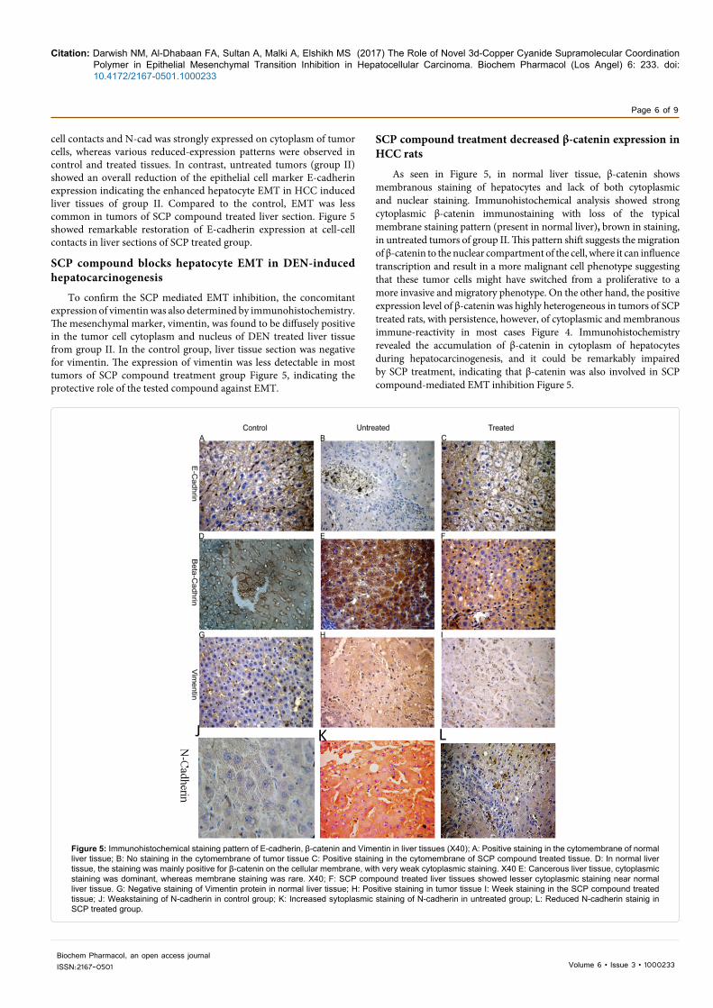
cell contacts and N-cad was strongly expressed on cytoplasm of tumor cells, whereas various reduced-expression patterns were observed in control and treated tissues. In contrast, untreated tumors (group II) showed an overall reduction of the epithelial cell marker E-cadherin expression indicating the enhanced hepatocyte EMT in HCC induced liver tissues of group II. Compared to the control, EMT was less common in tumors of SCP compound treated liver section. Figure 5 showed remarkable restoration of E-cadherin expression at cell-cell contacts in liver sections of SCP treated group.
SCP compound blocks hepatocyte EMT in DEN-induced hepatocarcinogenesis
To confirm the SCP mediated EMT inhibition, the concomitant expression of vimentin was also determined by immunohistochemistry. The mesenchymal marker, vimentin, was found to be diffusely positive in the tumor cell cytoplasm and nucleus of DEN treated liver tissue from group II. In the control group, liver tissue section was negative for vimentin. The expression of vimentin was less detectable in most tumors of SCP compound treatment group Figure 5, indicating the protective role of the tested compound against EMT.
SCP compound treatment decreased β-catenin expression in HCC rats
As seen in Figure 5, in normal liver tissue, β-catenin shows membranous staining of hepatocytes and lack of both cytoplasmic and nuclear staining. Immunohistochemical analysis showed strong cytoplasmic β-catenin immunostaining with loss of the typical membrane staining pattern (present in normal liver), brown in staining, in untreated tumors of group II. This pattern shift suggests the migration of β-catenin to the nuclear compartment of the cell, where it can influence transcription and result in a more malignant cell phenotype suggesting that these tumor cells might have switched from a proliferative to a more invasive and migratory phenotype. On the other hand, the positive expression level of β-catenin was highly heterogeneous in tumors of SCP treated rats, with persistence, however, of cytoplasmic and membranous immune-reactivity in most cases Figure 4. Immunohistochemistry revealed the accumulation of β-catenin in cytoplasm of hepatocytes during hepatocarcinogenesis, and it could be remarkably impaired by SCP treatment, indicating that β-catenin was also involved in SCP compound-mediated EMT inhibition Figure 5.
Figure 5: Immunohistochemical staining pattern of E-cadherin, β-catenin and Vimentin in liver tissues (X40); A: Positive staining in the cytomembrane of normal liver tissue; B: No staining in the cytomembrane of tumor tissue C: Positive staining in the cytomembrane of SCP compound treated tissue. D: In normal liver tissue, the staining was mainly positive for β-catenin on the cellular membrane, with very weak cytoplasmic staining. X40 E: Cancerous liver tissue, cytoplasmic staining was dominant, whereas membrane staining was rare. X40; F: SCP compound treated liver tissues showed lesser cytoplasmic staining near normal liver tissue. G: Negative staining of Vimentin protein in normal liver tissue; H: Positive staining in tumor tissue I: Week staining in the SCP compound treated tissue; J: Weakstaining of N-cadherin in control group; K: Increased sytoplasmic staining of N-cadherin in untreated group; L: Reduced N-cadherin stainig in SCP treated group.
Citation: Darwish NM, Al-Dhabaan FA, Sultan A, Malki A, Elshikh MS (2017) The Role of Novel 3d-Copper Cyanide Supramolecular Coordination Polymer in Epithelial Mesenchymal Transition Inhibition in Hepatocellular Carcinoma. Biochem Pharmacol (Los Angel) 6: 233. doi: 10.4172/2167-0501.1000233
Page 7 of 9
Biochem Pharmacol, an open access journalISSN:2167-0501 Volume 6 • Issue 3 • 1000233
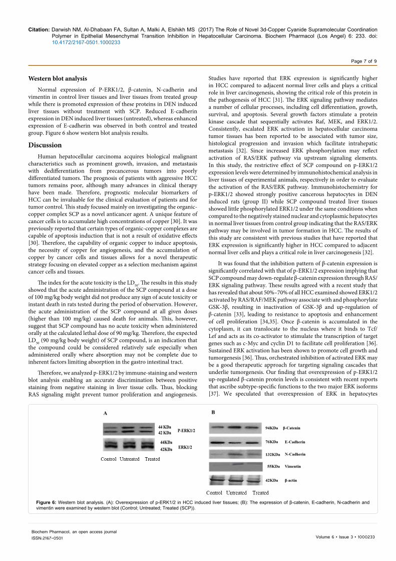
Western blot analysis
Normal expression of P-ERK1/2, β-catenin, N-cadherin and vimentin in control liver tissues and liver tissues from treated group while there is promoted expression of these proteins in DEN induced liver tissues without treatment with SCP. Reduced E-cadherin expression in DEN induced liver tissues (untreated), whereas enhanced expression of E-cadherin was observed in both control and treated group. Figure 6 show western blot analysis results.
DiscussionHuman hepatocellular carcinoma acquires biological malignant
characteristics such as prominent growth, invasion, and metastasis with dedifferentiation from precancerous tumors into poorly differentiated tumors. The prognosis of patients with aggressive HCC tumors remains poor, although many advances in clinical therapy have been made. Therefore, prognostic molecular biomarkers of HCC can be invaluable for the clinical evaluation of patients and for tumor control. This study focused mainly on investigating the organic-copper complex SCP as a novel anticancer agent. A unique feature of cancer cells is to accumulate high concentrations of copper [30]. It was previously reported that certain types of organic-copper complexes are capable of apoptosis induction that is not a result of oxidative effects [30]. Therefore, the capability of organic copper to induce apoptosis, the necessity of copper for angiogenesis, and the accumulation of copper by cancer cells and tissues allows for a novel therapeutic strategy focusing on elevated copper as a selection mechanism against cancer cells and tissues.
The index for the acute toxicity is the LD50. The results in this study showed that the acute administration of the SCP compound at a dose of 100 mg/kg body weight did not produce any sign of acute toxicity or instant death in rats tested during the period of observation. However, the acute administration of the SCP compound at all given doses (higher than 100 mg/kg) caused death for animals. This, however, suggest that SCP compound has no acute toxicity when administered orally at the calculated lethal dose of 90 mg/kg. Therefore, the expected LD50 (90 mg/kg body weight) of SCP compound, is an indication that the compound could be considered relatively safe especially when administered orally where absorption may not be complete due to inherent factors limiting absorption in the gastro intestinal tract.
Therefore, we analyzed p-ERK1/2 by immune-staining and western blot analysis enabling an accurate discrimination between positive staining from negative staining in liver tissue cells. Thus, blocking RAS signaling might prevent tumor proliferation and angiogenesis.
Studies have reported that ERK expression is significantly higher in HCC compared to adjacent normal liver cells and plays a critical role in liver carcinogenesis, showing the critical role of this protein in the pathogenesis of HCC [31]. The ERK signaling pathway mediates a number of cellular processes, including cell differentiation, growth, survival, and apoptosis. Several growth factors stimulate a protein kinase cascade that sequentially activates Raf, MEK, and ERK1/2. Consistently, escalated ERK activation in hepatocellular carcinoma tumor tissues has been reported to be associated with tumor size, histological progression and invasion which facilitate intrahepatic metastasis [32]. Since increased ERK phosphorylation may reflect activation of RAS/ERK pathway via upstream signaling elements. In this study, the restrictive effect of SCP compound on p-ERK1/2 expression levels were determined by immunohistochemical analysis in liver tissues of experimental animals, respectively in order to evaluate the activation of the RAS/ERK pathway. Immunohistochemistry for p-ERK1/2 showed strongly positive cancerous hepatocytes in DEN induced rats (group II) while SCP compound treated liver tissues showed little phosphorylated ERK1/2 under the same conditions when compared to the negatively stained nuclear and cytoplasmic hepatocytes in normal liver tissues from control group indicating that the RAS/ERK pathway may be involved in tumor formation in HCC. The results of this study are consistent with previous studies that have reported that ERK expression is significantly higher in HCC compared to adjacent normal liver cells and plays a critical role in liver carcinogenesis [32].
It was found that the inhibition pattern of β-catenin expression is significantly correlated with that of p-ERK1/2 expression implying that SCP compound may down-regulate β-catenin expression through RAS/ERK signaling pathway. These results agreed with a recent study that has revealed that about 50%–70% of all HCC examined showed ERK1/2 activated by RAS/RAF/MEK pathway associate with and phosphorylate GSK-3β, resulting in inactivation of GSK-3β and up-regulation of β-catenin [33], leading to resistance to apoptosis and enhancement of cell proliferation [34,35]. Once β-catenin is accumulated in the cytoplasm, it can translocate to the nucleus where it binds to Tcf/Lef and acts as its co-activator to stimulate the transcription of target genes such as c-Myc and cyclin D1 to facilitate cell proliferation [36]. Sustained ERK activation has been shown to promote cell growth and tumorgenesis [36]. Thus, orchestrated inhibition of activated ERK may be a good therapeutic approach for targeting signaling cascades that underlie tumorgenesis. Our finding that overexpression of p-ERK1/2 up-regulated β-catenin protein levels is consistent with recent reports that ascribe subtype-specific functions to the two major ERK isoforms [37]. We speculated that overexpression of ERK in hepatocytes
Figure 6: Western blot analysis. (A): Overexpression of p-ERK1/2 in HCC induced liver tissues; (B): The expression of β-catenin, E-cadherin, N-cadherin and vimentin were examined by western blot (Control; Untreated; Treated (SCP)).
Citation: Darwish NM, Al-Dhabaan FA, Sultan A, Malki A, Elshikh MS (2017) The Role of Novel 3d-Copper Cyanide Supramolecular Coordination Polymer in Epithelial Mesenchymal Transition Inhibition in Hepatocellular Carcinoma. Biochem Pharmacol (Los Angel) 6: 233. doi: 10.4172/2167-0501.1000233
Page 8 of 9
Biochem Pharmacol, an open access journalISSN:2167-0501 Volume 6 • Issue 3 • 1000233
causes’ hyperproliferative cell, leading to tumorigenicity. In our study, expression levels of ERK1/2 were simultaneously increased in HCC tumors. These findings suggest that the reduction of both ERK1/2 is a crucial event in the prevention of experimental HCC. Additionally, ERK activation might be necessary for the activation of angiogenesis in HCC β-catenin signaling.
Hallmarks of EMT include the loss of E-cadherin expression, upregulation of N-cadherin and the expression of mesenchymal markers, such as vimentin, in epithelial cells [38]. E-cadherin is the epithelial-specific cell–cell adhesion molecule that has been recognized as an important biomarker of tumor differentiation [39]. Decreased E-cadherin immune-reactivity correlates with a lack of differentiation, tumor aggressiveness and inhibited formation of nascent junctional complexes among HCC cells [40]. In the present study, we have compared E-cadherin and β-catenin immune-staining between tumors in livers of DEN induced rats and that in liver tissue of their control counterparts to determine what specific prognostic information can be obtained. Results of immunohistochemical analysis showed cytoplasmic overexpression of β-catenin and loss of E-cadherin expression in untreated liver sections when compared to normal control ones. Loss of E-cadherin expression at cytomembraneous cellular junctions permits free β-catenin to accumulate and associate with the LEF-1 transcription factor to facilitate cell proliferation [36]. SCP compound has shown to restore cytomembraneous E-cadherin expression and reduce cytoplasmic β-catenin expression suggesting that SCP compound may have a protective effect against EMT.
To evaluate whether the vimentin expression is a potential factor of metastasis in HCC, formalin-fixed and paraffin-embedded liver sections from control, untreated and SCP treated groups were used for immunohistochemical detection of vimentin. vimentin, a cytoplasmic intermediate filament, is characteristic of mesenchymal cells and is not usually expressed in epithelial cells. The atypical expression of vimentin in epithelial cancer cells may be associated with local invasiveness and the potential for metastasis [41]. The aberrant overexpression of vimentin and its relation to tumor metastasis have been reported in HCC [42].
In the present study, vimentin overexpression was found in liver sections of untreated group while reduced vimentin expression was found in SCP compound treated liver tissues when compared to untreated ones. Examples of vimentin-positive and negative tumors are shown in Figure 5. These results indicated that HCC cells surviving in vivo without SCP compound treatment have increased metastatic potential and a possible opposite effect of SCP compound treatment. This finding, combined with up-regulation of β-catenin expression in untreated tumor cells and reduced β-catenin expression in SCP compound treated tissue suggests that tumor cells surviving in vivo acquired intrinsic characteristics that facilitated EMT and that SCP compound may decrease the metastatic potential of liver cancer cells in vivo.
These results are consistent with those of other studies [43,44], so RAS/ERK pathway activation in HCC can induces breakage of the cadherin/catenin complex through the tyrosine phosphorylation pathway mediated by ERK, resulting in depression of cell-cell adhesion and an increasing trend for tumor invasion; and the up-regulation of ERK expression can relax cell-cell or cell-extracellular matrix adhesion and enhance cell migration from HCC [36], accompanied by the lower expression of E-cadherin and abnormal expression of β-catenin, leading to HCC with more invasive power.
These results in accordance with that of western blot suggests that that the RAS/ERK pathway may be involved in tumor invasion and metastasis in HCC, and is consistent with results of previous studies [45].
In summary, the data presented in this study reveal the importance of ERK expression in the positive regulation of HCC cell proliferation, invasion and metastasis. We have concluded that the expression level of β catenin is a potential prognosis marker and the inhibition of ERK phosphorylation could be a novel, therapeutic way of dealing with HCC.
ConclusionSCP compound can effectively inhibit the invasive potential of
ERK pathway in HCC cells by altering EMT, inhibition of beta catenin expression may play a significant role in this process.
Ethical ApprovalThe study protocol was approved by Alexandria University, Faculty
of Science Ethics Committee (REC number: PSU25112012) and by the Supervisors; Ahmed S Sultan, Ahmed Malki.
Availability of Data and MaterialsAll materials and data are available and sharing is available.
Competing InterestsAuthors have declared that no competing interests exist.
FundingAll study material was supplied by Corresponding author. There
was no additional funding for this study.
Consent for PublicationAuthors and corresponding authors have reviewed this paper and
approved it.
ContributorshipAuthors completed the study protocol and were the main organizer
of data collection drafting and revising the manuscript. Noura Darwish wrote the article and guarantees the paper. All authors contributed to the discussion and reviewed the manuscript and helped in designing the study and protocol and engaged in a critical discussion of the draft manuscript. All authors agreed on the final version of the manuscript.
AcknowledgementsGrateful and deepest thanks are extended to Prof. Dr. Safaa Etwa for giving
me the compound under investigation and for always being available when I need help. I thank supervisors for providing all the practical support to the study.
References
1. Sangiovanni AR, Del NE, Fasani P, DeFazio C, Ronchi G, et al. (2004) Increased survival of cirrhotic patients with a hepatocellular carcinoma detected during surveillance. Gastroenterology 126: 1005-1014.
2. Bruix J, Boix L, Sala M, Llovet JM (2004) Focus on hepatocellular carcinoma. Cancer Cell 5: 215-219.
3. Bosch FX, Ribes J, Díaz M, Cléries R (2004) Primary liver cancer: Worldwide incidence and trends. Gastroenterology 127: S5-S16.
4. Llovet JM, Burroughs A, Bruix J (2003) Hepatocellular carcinoma. The Lancet 362: 1907-1917.
5. Villanueva A, Newell P, Chiang D, Friedman S, Llovet J (2007) Genomics and signaling pathways in hepatocellular carcinoma. Seminars in Liver Disease 27: 55-76.

Citation: Darwish NM, Al-Dhabaan FA, Sultan A, Malki A, Elshikh MS (2017) The Role of Novel 3d-Copper Cyanide Supramolecular Coordination Polymer in Epithelial Mesenchymal Transition Inhibition in Hepatocellular Carcinoma. Biochem Pharmacol (Los Angel) 6: 233. doi: 10.4172/2167-0501.1000233
Page 9 of 9
Biochem Pharmacol, an open access journalISSN:2167-0501 Volume 6 • Issue 3 • 1000233
6. Farazi PA, DePinho RA (2006) Hepatocellular carcinoma pathogenesis: From genes to environment. Nature Reviews Cancer 6: 674-687.
7. Fabregat I, Roncero C, Fernández M (2007) Survival and apoptosis: A dysregulated balance in liver cancer. Liver International 27: 155-162.
8. Wilhelm SM, Carter C, Tang L, Wilkie D, McNabola A, et al. (2004) BAY 43-9006 exhibits broad spectrum oral antitumor activity and targets the RAF/MEK/ERK pathway and receptor tyrosine kinases involved in tumor progression and angiogenesis. Cancer Research 64: 7099-7109.
9. Lewis TS, Shapiro PS, Ahn NG (1998) Signal Transduction through MAP Kinase Cascades. Advances in Cancer Research 49: 139.
10. Park HJ, Kim BC, Kim SJ, Choi KS (2002) Role of MAP kinases and their cross-talk in TGF-β1–induced apoptosis in FaO rat hepatoma cell line. Hepatology 35: 1360-1371.
11. Owens DM, Keyse SM (2007) Differential regulation of MAP kinase signalling by dual-specificity protein phosphatases. Oncogene 26: 3203-3213.
12. Oikonomou E, Koc M, Andera L, Sasazuki T, Shirasawa S, et al. (2011) BRAF and RAS oncogenes regulate Rho GTPase pathways to mediate migration and invasion properties in human colon cancer cells: A comparative study. Mol Cancer 110: 118.
13. Doehn U, Hauge C, Frank SR, Jensen CJ, Duda K, et al. ( 2009) RSK is a principal effector of the RAS-ERK pathway for eliciting a coordinate promotile/invasive gene program and phenotype in epithelial cells. Mol Cell 35: 511-522.
14. Gulhati P, Bowen KA, Liu J, Stevens PD, Rychahou PG, et al. (2011) mTORC and mTORC regulate EMT, motility and metastasis of colorectal cancer via RhoA and Rac1 signaling pathways. Cancer Res 71: 3246-3256.
15. Wheelock MJ, Shintani Y, Maeda M, Fukumoto Y, Johnson KR (2008) Cadherin switching. J Cell Sci 121: 727-735.
16. Theveneau E, Mayor R (2012) Cadherins in collective cell migration of mesenchymal cells. Curr Opin Cell Biol 24: 677-684.
17. Hanahan D, Weinberg RA (2000) Differential regulation of MAP kinase signalling by dual-specificity protein phosphatases. Cell 100: 57-70.
18. Paredes J, Figueiredo J, Albergaria A, Oliveira P, Carvalho J, et al. (2012) Epithelial E- and P-cadherins: Role and clinical significance in cancer. Biochim Biophys Acta. 1826: 297-311.
19. Berx G, van Roy F (2009) Involvement of members of the cadherin superfamily in cancer. Cold Spring Harbor Perspectives in Biology 1: a003129-a003129.
20. Lee S, Kim WH, Jung HY, Yang MH, Kang GH (2002) Aberrant CpG island methylation of multiple genes in intrahepatic cholangiocarcinoma. Am J Pathol 161: 1015-1022.
21. Matsumura T, Makino R, Mitamura K (2001) Frequent down-regulation of E-cadherin by genetic and epigenetic changes in the malignant progression of hepatocellular carcinomas. Clin Cancer Res 7: 594-599.
22. Diez M, Arroyo M, Cerdan FJ, Muñoz M, Martin MA, et al. (1989) Serum and tissue trace metal levels in lung cancer. Oncology 46: 230-234.
23. Viktória F, Csaba H, Béla K, János G, Szilvia M, et al. (2005) Bone disorders in experimentally induced liver disease in growing rats. World J Gastroenterol 11: 7169-7173.
24. Bansal AK, Bansal M, Soni G, Bhatnagar D (2005) Protective role of vitamin E pretreatment on N-nitrosodiethylamine induced oxidative stress in rat liver. Chem Biol Interact 156: 101-111.
25. Turner R (1965) Quantal responses. Calculation of ED50. In: Screening methods in pharmacology, Academic Press, New York, pp: 61-63.
26. Reitman S, Frankel S (1957) A colorimetric method for the determination of serum glutamic oxalacetic and glutamic pyruvic transaminases. Am J Clin Pathol 28: 56-63.
27. King EJ, Armstrong AR (1980) Practical Clinical Biochemistry. In: Varley H, Gowenlock AH, Bell M (eds) 5th edn. London: Welliam Heinemann Medical Books Ltd. pp. 897-899.
28. Szasz G (1969) A kinetic photometric method for serum gamma-glutamyltranspeptidase. Clin Chem 15: 124-136.
29. Doumas BT, Watson WA, Biggs HG (1971) Albumin standards and the measurement of serum albumin with bromcresol green. Clin Chim Acta 31: 87-96.
30. Jendrassik L, Grof P (1938) Colorimetric method of determination of bilirubin. Biochem Z 297: 81-82.
31. Sell S, Beckar FF (1978) Alpha feto protein. Natl Cancer Inst. 60: 19-26.
32. Carleton H (1979) Histological techniques. 4th edn. London, Oxford University Press. pp: 9-432.
33. Gao Q, Qiu SJ, Fan J, Zhou J, Wang XY, et al. (2007) Intratumoral balance of regulatory and cytotoxic T cells are associated with prognosis of hepatocellular carcinoma after resection. J Clin Oncol 25: 2586-2593.
34. Yang MC, Chang CP, Lei HY (2010) Induction of liver fibrosis in a murine hepatoma model by thioacetamide is associated with enhanced tumor growth and suppressed antitumor immunity. Lab Invest. 90: 1782-1793.
35. Kolch W (2005) Coordinating ERK/MAPK signaling through scaffolds and inhibitors. Nat Rev Mol Cell Biol 6: 827-837.
36. Ding Q, Xia W, Liu JC, Yang JY, Lee DF, et al. (2005) ERK associates with and primes GSK-3beta for its inactivation resulting in up-regulation of beta-catenin. Mol Mol Cell 19: 159-170.
37. Old WM, Shabb JB, Houel S, Wang H, Couts KL, et al. (2009) Functional proteomics identifies targets of phosphorylation by B-Raf signaling in melanoma. Mol Cell 34: 115-131.
38. Yang J, Mani SA, Donaher JL, Ramaswamy S, Itzykson RA, et al. (2004) Twist, a master regulator of morphogenesis, plays an essential role in tumor metastasis. Cell 117: 927-939.
39. Vlaminck K, Vakaet L, Mareel M, Fiers W, Van RF (1991) Genetic manipulation of E-cadherin expression by epithelial tumor cells reveals an invasion suppressor role. Cell 66: 107-119.
40. Du GS, Wang JM, Lu JX, Ma CQ, Du JT, et al. (2009) Expression of atypical protein kinase c-iota, E-cadherin and β-catenin related to invasion and metastasis in hepatocellular carcinoma. Ann Surg Oncol. 16: 1578-1586.
41. Vuoriluoto K, Haugen H, Kiviluoto S, Mpindi JP, Nevo J, et al. (2011) Vimentin regulates EMT induction by Slug and oncogenic H-Ras and migration by governing Axl expression in breast cancer. Oncogene 30: 1436-1448.
42. Park MY, Kim KR, Park HS, Park BH, Choi HN, et al. (2007) Expression of the serum response factor in hepatocellular carcinoma: Implications for epithelial-mesenchymal transition. Int J Oncol 31: 1309-1315.
43. Cui J, Zhou X, Liu Y, Tang Z (2001) Mutation and overexpression of the beta-catenin gene may play an important role in primary hepatocellular carcinoma among Chinese people. J Cancer Res Clin Oncol 127: 577-581.
44. Reddy KB, Nabha SM, Atanaskova N (2002) Role of MAP kinase in tumor progression and invasion. Cancer Metastasis Rev 22: 395-403.
45. Darwish NM, Sultan A, Malki A, Khamis H, Zaidy M (2016) In vitro antihepatoma activity of novel 3D-Copper Cyanide supramolecular coordination polymers. J AOAC Int 99:1240-1246.


![NAME : MOHAMED DARWISH MOHAMED DARWISH TELEPHONE … · 2017-09-11 · [Mohamed Darwish-CV] HIJJA4R2017 Page 5 of 14 Client Relationship • Develop and maintain good working relationship](https://img.dokumen.tips/doc/110x75/5ed8355c0fa3e705ec0e0970/name-mohamed-darwish-mohamed-darwish-telephone-2017-09-11-mohamed-darwish-cv.jpg)