Embed Size (px)
Citation preview

1
UNIVERSITY "LUCIAN BLAGA" - SIBIU
FACULTY OF MEDICINE "VICTOR PAPILIAN" - SIBIU
"PHLEBOGRAPHY IN EVALUATION OF CHRONIC
VENOUS INSUFFICIENCY "
SUPERVISOR:
PROF. DR. DAN SABĂU
STUDENT:
DR. CIPRIAN RADU ŞOFARIU
PROF. DR. ROMEO ELEFTERESCU

2
MOTTO:
„ TRUE SCIENTIST KNOWS HOW TO FIND IN HIS
EXPERIMENTS ANSWER TO WHAT HE SEARCH, BUT TO
SEEK A RESPONSE FOR WHAT HE DIDN’T ASKED”
CLAUDE BERNARD

3
Definition:
Chronic venous insufficiency (van der Molen) is a clinical syndrome with different
etiopathogeny, prognosis and therapeutic that is subsequent to chronical disturbances of
venous circulation, especially in the lower limbs, resulting in significant changes of interstitial
space, lymphatics and skin.
Chronic venous insufficiency includes two subdivisions clinically very similar, but
different in terms of etiopathogenesis, therapeutics and prognosis:
1. Superficially chronic venous insufficiency, representing the late stage of
superficially veins insufficiency and varicose disease, sometimes surnamed as "varicose
symptomatic complex” the inadequacies of junctions of the large veins, internal and external
saphenous, and / or perforating vein insufficiency.
All of these conditions can be treated successfully, so that failure superficially chronic
venous has a good prognosis.Varicose veins are not the actual cause of failure superficially
chronic venous insufficiency (L. Gherasim), but the more likely to have a common cause with
them.
2. Chronic deep venous insufficiency is the postphlebitic syndrome, the consequence
of occlusions and deep vein functional disorders.
The causes of these disorders can be suppressed in exceptional cases, so that chronic
deep venous insufficiency postphlebitic syndrome not can be cured and requires long term
treatment. It often puts problems for both: patient and doctor.

4
Ultrasound and Phlebographical Diagnosis of Peripheral Chronic Venous Insufficiency
1. PURPOSE
Determination of radio-imaging characteristic aspects of peripheral chronic venous
insufficiency under and over fascial and assess the role of ultrasound and phlebography in its
diagnosis.
2. OBJECTIVES
Analysis of relevant epidemiological factors in causing varicose veins and deep vein
thrombosis (endogenous: anthropological, anatomical and physiological, genetically,
constitutional, sex, age, endocrine, pregnancy, obesity, health status and exogenous: physical
and social).
Morphological analysis of static and dynamic aspects of ultrasound, color Doppler and
spectral in varicose disease, DVT and post thrombotic syndrome, compared with
phlebography.
Establish the role of the radio-imaging methods in the diagnosis of Chronic Venous
Insufficiency - CVI.
3. MATERIALS AND METHODS
Patient selection:
People who presented for ultrasound and phlebography investigation in the Laboratory
of Radiology and Medical Imaging from Pediatric Hospital of Sibiu, Radiology and Medical
Imaging Laboratory of Sibiu’s County Emergency Hospital, Clinic "Body and soul-Prof. Dr.
Ilie Craciun " Clinic “Medica Nursing Plus” with clinical diagnosis of varicose veins, deep
vein thrombosis and post thrombotic syndrome.
Diagnostic methods used:
For analysis were used groups of patients (432 patients only with ultrasound, 54 patients
only with phlebography examination, and 87 patients with ultrasound followed by
phlebography, 10 controls for ultrasound examinations, and 3 for the phlebography).
Ultrasound examinations were performed using several types of ultrasound (mostly
listed first):
- Siemens Acuson X500,
- Siemens Acuson X300,
- Hi Vision Hitachi EUB 5500 (Hitachi Medical Corporation, Kashiwa, Japan).
Phlebography examinations were conducted with two types of devices: Philips
Telediagnost (Philips Eindhoven, Netherlands, 2000), digital fluoroscopy with Easy Vision
imaging station type and Swissray ddr Multi-System (Hochdorf, Switzerland, 2000 ) rontgen
complete digital machine.
Were examined patients with clinical diagnosis of
acute and chronic venous thrombosis
varicose disease
venous valvular insufficiency
vein stenosis
pseudo aneurysms of veins
at the request of patients with subjective complaints of venous disease

5
Method and data collection period: longitudinal prospective study: January 2004-
October 2011.
Standard ultrasound examination consisted of:
Write of patient personal data on:
-age,
-sex,
-area of origin,
-profession,
-personal history of veins pathology
-family history of veins pathology
-clinical signs
The clinical examination performed by the radiologist
Systematic ultrasound exploration of the veins of the lower limb examined
simultaneously with the contra lateral limb veins with parameters and images recording
Analysis and outcome of the result
Transducers were used with the wide dynamic range of:
- 5-10 MHz for Siemens ultrasound machines
- 6.5-13 MHz for Hitachi ultrasound machine.
Examination technique was adapted according to the explored segment, age, and
muscle mass and subcutaneous tissue thickness.
For evaluation of upper limb veins were used frequencies between 5 and 10 MHz
Examinations began with axial sections thus identifying the simultaneous venous and arterial
structures and allow the pressure and compressibility testing, followed by longitudinal sections
needed to study blood flow characteristics and searching obstruction. Upper limb position
varied from anatomical position for forearm and 2/3 distal of the arm, the examination in
abduction of 1/3 proximal of the and underarm and back into anatomical position with the
head rotating to the opposite subclavian vein for subclavian vein exploration.
Protocol to investigate upper limb veins (38), modified as sense of examination:
Hand veins:
a. forearm veins to identify the origins of cephalic vein and basilic vein, median vein
trajectory
b. Envelope elbow: median basilic vein and median cephalic vein, cephalic vein, basilic
vein and brachial veins
c. cephalic vein in the arm and pectoral groove and confluence to subclavian v.
d. basilic vein in the arm and confluence to axillary v.
e. subclavian vein (morphology, dynamic data)
Examination of leg veins has been made with two types of transducers:
- Low frequency convex 3.5-5MHz for IVC (Inferior Vena Cava) and vessels located
in the pelvis
Common iliac veins and internal and external iliac veins and
- Linear transducers with frequencies between 5 and 10 MHz for the rest of the veins
Leg.
This was a close examination of contiguous up to the knee, after which followed some
reference levels (38):
a. common iliac veins
b. external iliac veins
c. common femoral vein
d. sapheno-femoral junction and greater saphenous vein’s trajectory, tributaries,
communicating veins
e. superficial femoral vein (in upper third, medium and lower third of Hunter channel)

6
f. popliteal vein
g. sapheno-popliteal junction and small saphenous vein’s trajectory, tributaries, vv.
communicating
h. posterior tibial veins
i. anterior tibial veins
Static examination was performed involving the venous system venous trunks able
basal view to characterize them: lumen, wall, track, spontaneous flow and its parameters using
Doppler techniques. Modulation-induced respiratory and adjacent arteries were also pursued in
particular linked to the functioning valves.
Examination of dynamic morphological and functional behavior of veins intended to
highlight the application of different diagnostic maneuvers for blood flow augmentation
(muscle compression upstream of the explored site, passive or active rising of the member,
foot finger movement) or to stop it (abdominal or muscle compression downstream of the
explored site, the Valsalva maneuver, cough, tourniquet application). Muller maneuver was
mainly used for valvular reflux view.
Note that all ultrasound examinations were performed by one physician reviewer,
radiologist, in the person of the author of this thesis.
Standard phlebography examination consisted of:
Write of patient personal data similar to that of ultrasound.
Prior preparation of patients consisted of:
- Assessment of serum creatinine,
- Informing them of the procedure,
- Administration of antihistaminic medication (antiH1) the day and morning prior
examination and
- Injection IV a dose of hydrocortisone succinat sodic1-2mg/Kg 5-10 minutes prior to
administration of contrast.
Performing venous puncture raised frequently problems, marked edema, poor state of
superficial venous bed, resulting sometimes delaying of the investigation.
Patient and medical staff will be protecting from radiation with aprons, small aprons
and cervical collars. Patients positioning of examined member in anatomical position. The
examination tables allow quick mobilization regions of interest manually. Tourniquet
application and injection of contrast substance will be followed by acquisition of serial
radiographs, or movie.
For evaluation of lower limb venous system were applied technique of anterograde
combined and complete phlebography (Cid Dos Santos) and variants with changes mainly in
the amount of contrast injected and no use anticoagulant especially in pediatric patients.
Sometimes we practiced tilt table examination of the Philips Telediagnost to
generate a dilation of leg veins and get a slight delay in transit of the contrast.
In some cases underwent reinjection of a new dose of approximately 30 ml contrast for
a better assessment of valvular system in conditions of stasis, expiration and increased intra-
abdominal pressure.
Consideration of the existence of relevant epidemiological factors in causing varicose
veins and deep vein thrombosis (endogenous: anthropological, anatomical and physiological,
genetically, constitutional, sex, age, endocrine, pregnancy, obesity, health status and
exogenous: physical and social).
Although the studies were performed on samples from patients relatively limited
compared with prospective and retrospective analyzes involving thousands of subjects and
investigated cases were selected by doctors, clenched for a few necessary considerations of
epidemiology of the disease on the venous system.
Distribution of studied patients by age and sex:

7
It may be noted that most patients suffering with venous system diseases are grouped
around five decades, six and seven of life with a peak between 51 and 60 ages.
Part of vein diseases are studied in medical pathology (cardiologists and internists):
DVT and superficial thrombophlebitis, post thrombotic syndrome. Varicose disease, venous
dysplasia, trauma and tumors as well as some of the complications of venous disease belong
surgical pathology.
This large chapter of vein diseases requires a multidisciplinary approach. Radiologists,
physicians specialized in functional explorations; laboratory doctors bring their important
contribution to the diagnosis and treatment monitoring.
Opie (59) established an incidence of approximately 5.2% of chronic venous
insufficiency on a group of 500 patients, taking into account only those subjects who backflow
valve of the common femoral vein is associated with clinical symptoms, other patients with
reflux following to be monitored.
Our study demonstrated the following:
The incidence of varicose veins (truncal varicose and spider) by Brand (81),
Carpentier (82), Chiesa (83) and London (84) was approximately 17% for men and 31% in
women.
The incidence of varicose veins in the studied groups was 71% of which 46% women
and 25% male, with a ratio of about 2:1 in favor of females, similar to the Chiesa's multicenter
study.
Patients belonging to urban or rural are difficult to determine because the address
mentioned in identity card that assigns the label of rural area to approx. 23% of patients
require some major adjustments.
Large-scale migration of population from villages to cities during communism and its
depopulation after 1989 looking for a job outside the country, through bankruptcy mono
industrial cities which captured rural labor (commuters) and practicing subsistence agriculture
have left Romanian villages population aging and economic theft.
Low accessibility to the doctor in these localities, reduced importance of aesthetics in
old age and hard physical work underlying predominantly urban distribution of patients
investigated.
Men
0
10
20
30
40
50
1-10 ages
11-20 ages
21-30 ages
31-40 ages
41-50 ages
51-60 ages
61-70 ages
71-80 ages
peste 80 ani
Men
Women

8
Superficially chronic venous insufficiency present in several clinical forms: edema,
and ocher or purple dermatitis, stasis eczema, varicose dermal hypodermitis, white Milian
atrophy, culminating with venous ulcers healed or active, appropriate CEAP clinical stages III-
VI, is 26% of patients studied.
As the Pathogenesis of varicose veins, venous walls meiopragia (meiopragie: it. -
reduced functional capacity and reactivity) has a proven genetic influence of the high
percentage of patients with a family history of hydrostatic varicose veins among relatives of I
degree.
Transmission appears to be polygenic predisposition and not limited to reduced
synthesis of collagen and endo parietal hexoseamine that increase distensibility (85).
FOXC2 gene mutation, 16q24 is found in common distensibility of varicose veins and
lymphedema.
Among patients with a family history of varicose disease 37% of them said they
inherited the disease from both parents.
Note that disease transmission to offspring has a high percentage in patients with
varicose disease group, so 47% of them have children with clinical manifestations in various
stages: from spider veins to varicose veins with edema and trophic changes.
Pregnancy plays an important role in varicose veins or in their emphasis on women
susceptible, 54% of patients considering pregnancy as a trigger point. The higher incidence of
varicose veins in multiparous women compared with nuliparous: ratio 3:1 and tend to balance
the ratio men: women after menopause, indicates the involvement of female hormones in the
genesis of varicose veins.
Parvu and Gherasim (2.3) consider that large amounts of estrogen secretion induce
connective tissue laxity and increased venous walls distensibilitaty.
Oral contraceptives cause endodermic varicose veins. Endocrine status during
pregnancy may lead to opening of fistula arterial-venous fistula responsible for venous
congestion.
Hypervolemia of pregnancy and impairing blood flow in leg veins due to large
abdominal vein compression by gravid uterus and increased blood flow in ovarian veins are
factors that cause lower limb venous distension.
The hypothesis of primary varicose veins of the legs genesis, through arterial venous
shunts is based on the finding of increased oxygen saturation and a higher temperature in
blood from varicose segments than in that of non varicose veins or in mixed venous blood.
Occupational factors play an important role in the genesis of varicose veins. The
professional tier included in our study was a large: the profession involved prolonged
standing: doctors (predominantly surgical specialties), priests, waiters, teachers, hairdressers,
and workers in assembling lines, and those characterized by an excessive sedentary: cashier,
clerk, and IT.
Profession remains the only adjuvant in the context of genetic predisposition
existence. There are patients with varicose disease do not subscribe to the panoply prone
professions.
However, even in the professions likely, most do not develop disease.
Professional predisposing factor is a necessary but not sufficient to trigger disease.
On the other hand short exercises during working hours that make the muscle pump
function or venous channels that favor reduced risk of developing varicose veins even the
professionals likely.
Inactivity and obesity are reflected in the development of varicose veins in people
susceptible because any such heaviness mechanical blood flow in both veins and in the deep
subcutaneous (ascites, pregnancy, abdominal tumors, various compression, thrombosis) lead to
the production of varicose veins by increasing intra abdominal pressure.

9
Endogenous factors: Exogenous factors: 1. anthropological - biped resort 1. Physical (geography, microclimate) 2. Anatomical and physiological 2. Labor. 3. Genetically 4. Constitutional type 5. Sex 6. Age 7. Endocrine factors 8. Pregnancy 9. Obesity 10. Health
Clinical classification (CEAP) of patients with varicose disease:
Class 0 - no visible or palpable signs; Class 1 - spider or reticular veins; Class 2 - varicose veins; Class 3 - edema; Class 4 - skin changes due to venous disease (pigmentation, eczema, venous lipodermo
0
10
20
30
40
50
CEAP 0 28%
CEAP I 1%
CEAP II 41%
CEAP III 15%
CEAP IV 10%
CEAP V 7%
CEAP VI 1%
Clinical classification CEAP
Distribution of male patients with venous disease according to body mass index
sub 18.49 underweight
18.5-24.99 normal weight
25-29.99 over weight
30-34.99 obezity grade I
35-39.99 obezity grade II
peste 40 morbid obezity

10
sclerosis); Class 5 - skin changes as defined above with healed ulceration; Class 6 - skin changes as defined above with active ulceration.
Most patients in the study group were classified into class 2 (varicose veins). A
significant percentage of patients were presented for evaluation of imaging techniques to
conduct appropriate therapeutic approach.
The presence of a high percentage of patients with trophic changes and varicose ulcer
reveals some persistent gaps in the health education of patients, in their adherence to
treatment, and in the absence of uniform, standardized, but also appropriate to each patient
therapeutic strategies.
Backflow of valves
Constantly have revealed sapheno femoral junction with ostium’s valve.
In 14% of cases the same ostium of greater saphenous vein arch and superficial
epigastric vein was noticed, in this situation common ostium and saphenous vein arch were
provided with their own valves.
Valves were identified consistently present in the superficial femoral vein FV
proximal end and femoral vein before penetration in the adductor’s channel.
In 81% of cases was identified a valve above sapheno-femoral junction and one below
the inguinal ligament.
A valve often identified (96% of cases) was located in the popliteal vein, without
precise positioning.
Morphological ultrasound examination of the distal femoral vein in the groin is
difficult for reasons of equipment, not very deep penetrating of linear transducers, decreases
image sharpness especially in obese patients, and no possibility of total compression of the
vein to subjects with well represented muscles.
Generally run out portion of adductors channel, near to the distal ring is relatively easy
to exam, the vein is sometimes better viewed at this level than the artery.

11
This backflow presence at SFJ valves in the long saphenous vein in patients with
varicose disease:
Ostium’s reflux can be caused by functional valves insufficiency in the context of
increasing size of SFJ with incomplete closure of the valve (most cases) or congenital or
acquired defects of valves cusps.
Reflux can be modulated respiratory and / or by arterial pulse.
Depending on the flow may be variable from fine reflux induced by the dynamic
maneuvers to highlight reflux with a massive peak at maneuvers that induce intra abdominal
increased pressure.
0%
50%
100%
SFJ commun ostium
CFV above j. between FV and DFV PV FV
100%
14%
81% 96%
74%
Valves distribution
SFJ backflow
Without reflux 18%
Dynamic induced small reflux12%
Respiratory and arterial modulated reflux 39%
Permanently backflow 29%

12
Complete reversal of flow direction (massive reflux), respiratory modulated with a
defect of the anterior cusps of the SFJ valve seen in color flow.
Illustration of massive reflux in duplex Doppler: two-dimensional view reveals a slow
flow with haematic macro aggregates, "smoke flow", which is reversed at some point become
vortex. Spectral Doppler image illustrates high amplitude positive waves (flow speed of about
40 cm / sec, corresponding to respiratory modulation)
The incompetence of saphenous-femoral junction’s valves is correlated with the
clinical severity of varicose veins, CEAP advanced stages are associated with massive reflux
which was missing in the early stages of disease.
Correlation between level of ostial reflux and severity of varicose disease illustrated
by CEAP classification:
0
20
40
60
80
100
120
CEAP I CEAP II CEAP III CEAP IV CEAP V CEAP VI
Without reflux
Dynamic induced small reflux
Respiratory and arterial modulated reflux
Permanently backflow
13
Anatomic variants of the sapheno-femoral junction:
Sapheno-femoral junction may take different aspects: from the classic version of a
solitary ostium of the long saphenous arch proposed by Blanchemaison (86) to the existence
of a common sapheno-epigastric core (morpho-functional unit often highlight ultrasound).
In terms of sensitivity of ultrasound to detect variations caused by sapheno-femoral
junction and its tributaries is difficult to make a real estimate because anatomical studies
doesn’t agree themselves there is very large differences between different authors. For
example Prof. Jose Ciucci’s (88) appearance on of the Medical University of Buenos Aires, in
a study published in 2010 by a group of 100 subjects, at necropsy, only 9% said that they had
the classic scheme of sapheno-femoral junction. On the other hand Professor Jesus Sanchez,
Spain in 2001 published an intraoperatively study by a group of 136 patients: the classical
scheme of sapheno-femoral junction is identified in 46% of subjects.
FV duplication Hypoplastic popliteal vein replaced by
the muscle branches veins
56%
14% 2.95%
20%
3.06% 0.33% 3.27% 0.33% 0%
10%
20%
30%
40%
50%
60%
Anatomic variants of the sapheno-femoral junction

14
Perforating veins play an important role in hemodynamic status of the lower limbs,
being a system of "locks" with two valve, ensuring a unidirectional flow, facilitated by
muscle pump, from the surface low pressure system to the deep high pressure system.
Although the sensitivity of ultrasound in detecting perforating veins is lower than the
phlebography, B-mode and duplex examination bring additional information about flow sense
and flow speed.
In the study group were identified through ultrasound examinations pathologic
perforating veins changes about for 37.4% of subjects with varicose disease and near for
47.2% of patients which had a history of superficial thrombophlebitis or deep vein
thrombosis, with no significant difference between the sexes.
Comparing the two methods of investigation ultrasound and phlebography can say
that the identification of perforating veins from group of interest Cockett and Boyd was in
similar percentages.
0.00%
10.00%
20.00%
30.00%
Dodd's veins
Boyd's veins
Cockett 4 veins
Cockett 3 veins
Cockett 2 veins Cockett 1
veins
10.34% 8.62%
20.68%
29.30%
18.96%
12.07%
Distribution of ventral medial veins identified by ultrasound
0.00%
10.00%
20.00%
30.00%
Dodd's veins
Boyd's veins
Cockett 4 veins
Cockett 3 veins
Cockett 2 veins
Cockett 1 veins
Hunter's veins
21.27%
8.50% 14.90% 14.90%
19.14%
12.76%
8.50%
Distribution of ventral medial perforating veins identifing by phlebography

15
Although performance of phlebography is superior to ultrasound in terms of discovery
perforating veins according to location, slightly more difficult with ultrasound, It should be
noted that not all perforating veins detected by phlebography are pathologically altered.
At a routine ultrasound perforating veins without pathological changes detection is
difficult. We may conclude that the discovery of aperforating veins with ultrasound should
raise a question to examiner.
Size variations must be correlated with other aspects: number, serpiginos or linear
course, Baun configuration, flow direction: normally superficial to deep, flow sense changes
as a result of dynamic maneuvers application, body mass index, presence of pathological
changes in the superficial or deep venous system, existence of clinical signs attributable to
peripheral venous system disease.
An important aspect in the perforating veins was to assess the competence of the
valvular system indirectly by detecting the sense of flow using color Doppler examination.
0.00%
5.00%
10.00%
15.00% 14.50%
5.80%
10.10% 10.10%
13.40%
8.70%
5.80%
13.40%
7.20% 5.80% 5.80%
Distribution of perforating vein identified by phlebogrphy according to location
7%
24%
69%
Distribution of perforating veins according to caliber
Under 2mm
Between 2 and 3mm
Over 3 mm

16
Doppler spectrum is less sensitive to establish flow sense due to the continuing need to
adapt and rapidly changing Doppler angle, positivity or negativity of the waves at small
changes in sense of perforating veins.
It’s watched the flow sense both spontaneously and after application of dynamic
maneuvers, careful to avoid false positive results generated by perforating veins serpiginos
trajectory or inadequate adjustment of Doppler angle to vessel trajectory.
Complete reversal of flow sense on color flow and spectral Doppler
In terms of establishing a correlation between the topography of perforating vein
valves and their incompetence should be noted that most of them belong to Cockett 3, 2, 1
perforating veins located about 15-20cm over internal malleola 76% of cases. Lower
percentages were recorded in the third upper leg 15% (Boyd and Cockett 4) and 9% in 1/2
lower thigh (Dodd and Hunter).
27%
39%
9%
24.50%
Assesing the competence of the perforating veins by detecting the sense of flow
Normal flow
Spontaneous partial backflow
Flow reverse at dynamic maneuvers
Backflow
17
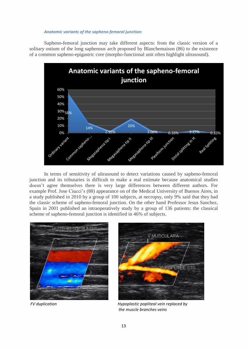
As you can see the severity of varicose veins is closely related to impairment of
perforating vein valves; number of subjects with incompetent perforating veins is greater in
stages 4, 5 and 6 of the clinical classification.
0%
50%
100%
CEAP 0 CEAP 1 CEAP 2 CEAP 3
CEAP 4 CEAP 5
CEAP6
1% 3% 28%
54%
83% 90% 98%
Correlation between CEAP classification and Cockette 3,2,1 perforating veins with
incompetent valves

18
Perforating veins on phlebography stands out much easier than ultrasound, where the
device performance, during which it may grant atention, ecographer’s versatility can influence
their detection.
Perforating veins are easier to quantify like morphology, number and classification
Baun on phlebography, but the snse of flow, and their velocityies, insufficient valves are easy
to quantify at ultrasound exam.
Like Boyd perforating veins connecting long saphenous vein tributaries in the region
under internal condylar with popliteal vein, tibial and peroneal veins, there is a constant
group of veins, most doubles (Baun type II and III) located postero-external linking vein
tributaries to lesser saphenous vein or communicating between two saphenous and peroneal
veins.
Like these three groups is similar others located at the junction of 1/3 top and 1/3
average 1/3 medium and the junction of the third medium and 1/3 lower, somewhat similar
vein Cockett 4, 3 and 2, but located laterally. These perforating veins open in extreme
situations: advanced varicose disease, DVT.

19
In terms of diagnosis phlebography and ultrasound can tell whether it is primitive
varicose veins, secondary varicose veins or symptomatic for a deep venous malformation.
In varicose veins with abnormal location on external front or back of thigh,
phlebography reveals communicating vein place which maintains backflow and require to be
interrupted.
Varicose on greater saphenous „Smoke flow” in swelling of the sinus
Trombophlebitis
In group of patients with varicose disease examined, 10.47% of them had changes of
acute thrombophlebitis (variable echogenic image into vessels associated with Celsius
described clinic signs), while only 7% had variable echogenic images without acute
inflammatory phenomena-old thrombophlebitis.
Patients with recurrent thrombophlebitis associated with varicose veins,
thrombophlebitis here excluding recurrent tumor, or those associated to Burger disease, 4
cases were relatively few 0.92%; recurrence of disease due to the late presentation at
physician and long evolution thanks to lack adherence at drug therapy or surgical treatment
refusal.
7%
31%
3%
69%
Extensia tromboflebitelor
Extinse pe mare parte din traiectul unei safene (peste50%)
Cu interesarea a doua segmente (de obicei in proximitatea genunchiului)
Cu interesarea ambelor sisteme superficial si profund

20
In DVT ultrasound B-mode discover next changes:
Thrombus presence into vessels as a hypoechoic – or anechoic image with thin
echogenic rim
Vessel compression test positive
An important sign is the significant increase in caliber of thrombosed vessel
toward the contralateral vein,
Immobility of venous valves in an intermediate position
Lack of venous dilatation at dynamic maneuvers (compression downstream,
increasing venous pressure)
Slow flow with emphasizing spontaneous the flow echogenicity "smoke flow"
Changes in the soft tissues: edema (from slight increasing of hypoderm
echogenicity to the presence of fluid bays more visible near fascia), cellulitis,
hematoma (hypo-echogenic recently, hyper echogenic-old)
Spectral Dupplex exam reveals in DVT:
Spontaneous or induced absence of Doppler waves in thrombosed vessel.
If is a partially obstructive thrombus, or a partially permeable channel It can be
detect an changed Doppler signal than contralateral vein (reduced amplitude
waves, the absence of their respiratory and / or heart modulation) (Keogan)
(115). There is no flow changes at dynamic maneuvers (compression or leg
raising does not increase flow upstream). These changes occur gradually
depending on the degree of obstruction or on the caliber of free channels.
Color flow Doppler findings in DVT:
Full or partially absence ("color gap") of the lumen filled with color, viewed on
perpendicular sections spontaneously and after dynamic maneuvers. (Prandoni)
(106)
Assess the degree of obstruction on axial sections
Assessment of cranial limit of thrombus on longitudinal sections
Measuring the working lumen illustrating the slow flow by Power / Angio
Doppler and the fastest by Color Flow
Power Doppler examination is a slight tendency to overestimate the size of the
patent channel.

21
DVT highly incomplete sinus opacification Sinus thrombus
Phlebography findings: absence or partial opacification of vascular lumens / removed,
irregular wall contour, hemodynamic response of the superficial system, enlargement of
perforating veins.
CT appearance of sinuous channel of repermeabilisation of right femoral vein in
Hunter channel, sinuous veins accompanying small arms. Magna saphenous varicose vein
with

22
POSTTHROMBOTYC SYNDROME
According to Douz, quoted by Dudea and collaborators, ultrasound posttrombotic
syndrome aims to highlight four vascular syndromes confounding in varying degrees:
venous obstruction syndrome (thrombus, absence of flow)
restrictive syndrome (diameter reduction and vein plasticity by fibrosis)
repermeabilization syndrome (spontaneous and prolonged venous reflux due to
destruction of venous valves)
alternate syndrome, hemodynamic and morphological changes of the
superficial venous system (high flow velocities, flow, non modulated).
View iliac axis. Postthrombotic syndrome iliac shaft with fine repermeabilization
channel comparative aspect between phlebography with classic image (right) and
phlebography with digital subtraction image(left).
On chronic dialysis patients, contrast venography has played an important role in
terms of evaluating the venous system before performing the arteriovenous fistula, maturation
and its functionality and last but not least, complications occurred: thrombosis, pulmonary
embolism secondary to thrombosis, aneurysm, theft syndrome (development of collateral
flow) with the affected limb ischemia, bacterial infection (from local inflammatory signs to
endocarditis).
Lately, with the ultrasound machine evolution, phlebography was placed in obscurity,
with only limited guidance in assessing clenched for a few arteriovenous fistulas.
EVALUATION OF ARTERIOVENOUS FISTULA
Ultrasound provides to surgeon sufficient informations to achieve a successful
intervention, the morphology of vessels concerned: trajectory (location, depth) caliber, arterial
wall (atheromatosis presence at chronic dialysed patients, heavy smokers, dyslipidemia - all
of them make difficult surgical procedure) and venous, and especially data on blood
hemodynamics (speed, sense of flow).
In addition to superior dynamic data than phlebography, ultrasound has clenched for a
few undeniable advantages: examination of both system of vessels: the vein and the artery,
absence of irradiation - repeatability for monitoring the evolution, absence of contrast

23
administration (contrast = side effects and may induce chronic renal failure), relatively easy
accessibility.
An artery with 3mm minimum size and minimum systolic speed 40-50cm/sec, and a
vein caliber minimum 3-3.5mm, with a good heart are prerequisites to achieve a mature
functional arteriovenous fistula.
Phlebography’s advantages consists in detection of all small tributaries of the vein
anastomosed which can lead to the syndrome of theft and to highlight cranial portions of the
cephalic and subclavian veins less accessible to ultrasound examination in obese or well
represented muscle mass patients, and all venous tributaries of the armpit.
Arteriovenous radial-cephalic mature fistula (B-mode and spectral Doppler appearance)
Normal and pathological appearance of arteriovenous fistula

24
CONCLUSIONS
Ultrasound is the imaging method of first choice in exploring the peripheral venous
system due to accessibility, repeatability, noninvasive nature, lack of contraindications and
side effects.
Ultrasound imaging provides semiological informations necessary and sufficient to
carry out differential and positive diagnosis of venous disease, allowing assessment of their
progress with or without treatment.
Ultrasound is a method available for screening and diagnosis of suffering venous leg
giving relevant information about both hemodynamic (speed, sense of flow, functioning
valves, perforating veins) and the morphological appearance.
Exploring limits of ultrasound we could mention the fact that ultrasound is laborious,
subjective, operator dependent, time-consuming and closely linked with the device
performance. Another drawback is given unsatisfactory exploring of deeper or overlapping
gaseous structures (confluence of common iliac vein, distal segment of inferior Cava vein).
Postthrombotic syndrome and varicose disease were the most numerous indications for
phlebography, there are also cases where the confounding clinical signs between two entities
make arbitrary it’s including into one or other class of disease, because many of varicose
disease had postthrombotic syndromes, or potthrombotic syndrome developed secondary
varicose disease on superficially venous system.
Phlebography examination is higher sensitive (ca. 28%) in detecting of characteristic
changes of postthrombotic syndrome (parietal irregularities, filling gaps, absence of
opacification of all vascular lumen) occurred with the smaller vessels of the lower leg (both
intramuscular lakes and venous axes of calf: posterior tibial and peroneal veins).
Phlebography represent a map of the venous system easily examined, viewed, and
flaunted in a treatment room, operating theater, most accessible for physician documentation
than bushy, descriptive, but more complex – ultrasound examination, whose information
about hemodynamic and mechanical of valves allowing for a coherent treatment strategies.
Phlebography limits of exploration due to the fact that it requires a minimally invasive
maneuver usually performed on a member of edema, administration of iodinated contrast
media, with its risks (anaphylaxis, shock, vasovagal reactions, renal dysfunction in patients
predisposed to chronic renal failure).
It was said that is the gold standard phlebography examination of the venous system. It
is difficult to appreciate which imaging methods deserve this title because if digital
subtraction phlebography gives details of the morphological channges and partly related to the
hemodynamic, ultrasonography is sufficient in most cases for before and after therapy
evaluation of the venous system, giving superior hemodynamic data to any radio-imaging
methods.
Indications of conventional phlebography examination as result of this study:
- Congenital malformation
- Anatomical variants
- Patients difficult to explore by ultrasound (profoundly vascular structures at obese
persons, excessive pilosity)
- When ultrasound was inconclusive due to own technique (inadequate adjustment of
speed at EUB 5500, reduced depth sensitivity combined with artifacts difficult to mitigate for
X300)
- For an easier examination of upper limb veins, especially humeral vein thrombosis,
axillary, cephalic and their tributaries

25
- Examination of arteriovenous fistulas for complications in addition for surgical
correction
- Some DVT and varicose disease treatment preceding act, authorizing doctors to
request for clarification of issues related to the expansion point of thrombi before
thrombolysis or thrombectomy, more precise quantification of repermeabilization channels,
location and trajectory of accessory saphenous, location and size of perforating veins,
identifying veins for bypass.
- Edema of unspecified nature of the lower limb phlebography and lymphography,
both are methods that can trance positive diagnosis.
Diagnostic algorithm of patients with disorders of the venous system:
DVT
History should be focused on family history, personal history and on installing
symptoms way. Personal history need to identify risk factors.
clinical
Laboratory methods of investigation:
Combine radio-imaging methods to measure the specific degradation products of
fibrin.
Certain DVT Probably DVT Posibility DVT
Sure clinical sygns Sugestive local sygns Inconclusive local sygns
+/- Pulmonary embolism associated risk factors
↓ ↓ ↓
————————————————————————————
↓
Ultrasound exam
↓ ↓
Pozitive→DVT Negative/inconculsive
↓ ↓
Digital subtraction ascending phlebography
↓
Pozitive→DVT Negative/inconculsive
↓
D-dimer ↑
Antithrombin III <85%
Fibrinopeptides A ↑
Fragment E ↑
Apolipoprotein A↑
↓ ↓
Pozitive→ DVT Negative → DVT absent or small
In the absence of digital subtraction angiography is practiced conventional
phlebography distal segments of the upper or lower limb phlebography and MRI / CT
(114,115) for proximal segments, pelvis, trunk, where the contrast substance is washed
quickly, carefully aiming the source images.

26
References:
1. Partsch H., Editorial, Phebolymphology, nr. 44, 2004, 231-267;
2. Gherasim L., Pârvu V., Bolile venelor periferice, venelor cave şi vaselor
limfatice ale membrelor, în Tratat de medicinǎ internǎ sub redacţia Pǎun R., Editura Medicalǎ,
Bucureşti, 1994, partea IV, 174;
3. Gherasim L., Pârvu V., Bolile venelor, în Medicinǎ internǎ sub redacţia
Gherasim L.,, vol. II, Editura Medicalǎ, Bucureşti, 1996, 1 67-1071;
4. Sinelnikov R. D., Atlas de anatomie umana, vol II, Ed. Mir, Moscova, 1989
374-465;
5. Meyer G. T., Histology on line,University of western Australia, 2008;
6. Saunders P. C., si colab., Vein graft arterializations causes differential
activation of mitogen-activated protein kinases, J. Thorac. Cardiovasc Surg. 2004;127:1276-
1284;
7. Guyton C. Arthur, Fiziologie, ediţia a V-a, Editura Medicală Amaltea
Bucureşti sub licenţă W. B. Saunders, 1996, 1 4-117, 180-187;
8. Intaglietta M., Zweifach W. B. “Microcirculation basis in fluid exchange”
bio.med.phis. 1974; 11:111;
9. Landis E. M., Pappenheimer J. R. Exchange of substances through the capillary
wall. In: Handbook of Physiology Circulation. Washington: Am Physiol Soc 1963;
10. Grama M., Fiziologie, vol. I, Editura Hermann Press, Sibiu, 1994, 30-42;
11. Manville N., Discuss the importance of the Frank-Starling mechanism and
discuss the balancing between the cardiac and vascular function curves, Campus Medicine,
214, 2008;
12. Pop D. Popa I., Socoteanu I., Fiziopatologia circulaţiei venoase, în Fiziologia şi
fiziopatologia hemodinamicii, sub redacţia Teodorescu-Exarcu I., Editura Medicalǎ,
Bucureşti, 1985, 92 ;
13. Brînzeu P., Gavrilescu Şt., Angiografia în practica medicală, Ed. Facla,
1977,185 – 225;
14. Reinharez D. Pigmentation following sclerosis. Phlebologie 1983;36: 337-344;
15. Gerson L., "The Treatment of Varicose Veins, A Critical Study of Choice of
Method", Angiology, The Journal of Vascular Diseases, vol. 13, No. 16,1962 pp. 260-264;
16. Malan E, Puglionisi A Congenital angiodysplasias of the extremities. Note I.
Generalities and classification: venous dysplasias. J Cardiovasc Surg 5:239:87-130;
17. Lev M, Saphir O. Endophlebohypertrophy and phlebosclerosis. I. The popliteal
vein. . AMA Arch Pathol. 1951;51:154–178;
18. Harrison - Principii de Medicina interna ed. 14, Editura Teora Bucuresti 2003;
19. Meissner M. H., Strandness E., Pathophysiology and natural history of acute
deep venous thrombosis, Rutherford's Vascular Surgery. 2005;
20. Thomas D. P., Merton R. E., Hockley D. J., The effect of stasis on the venous
endothelium: an ultrastructural study. Br J Haematol. Sep 1983; 55(1):113-22.
21. Anderson F. A. Jr., Wheeler H. B., Goldberg R. J., et al. The prevalence of risk
factors for venous thromboembolism among hospital patients. Arch Intern Med. Aug
1992;152(8):1660-4
22. Schafer A. I., Hypercoagulable states: molecular genetics to clinical practice.
Lancet. Dec 24-31 1994;344(8939-8940):1739-42;

27
23. Dahlback B. Inherited thrombophilia: resistance to activated protein C as a
pathogenic factor of venous thromboembolism. Blood. Feb 1995; 85(3): 607-14;
24. Weitz J. I., Middeldorp S., Geertz W., et al. Thrombophilia and anticoagulation
drugs. American Society of Hematology Education Book. 2004;
25. Leizorovicz A, Haugh M. C., Chapuis F. R., et al. Low molecular weight
heparin in prevention of perioperative thrombosis. BMJ. Oct 17 1992;305(6859):913-20;
26. Kahn S., et others, Determinants and Time Course of the Postthrombotic
Syndrome after Acute Deep Venous Thrombosis, Annals of Internal Medicine, 18 November
2008 | Volume 149 Issue 10 | 698-707;
27. Barbara Binder; Helmut Karl Lackner; Wolfgang Salmhofer; Susanne
Kroemer; Jasmina Custovic; Rainer Hofmann-Wellenhof, Association Between Superficial
Vein Thrombosis and Deep Vein Thrombosis of the Lower Extremities, Arch Dermatol 2009;
145: 753-757;
28. Forsea D., Popescu R., Popescu C. M., Compendiu de dermatologie şi
venerologie, Editura Tehnicǎ, Bucureşti, 1998, 258-267;
29. Cohen B. A., Lehmann C. U., Derm Atlas online, John Hopkins University,
2009;
30. Craig F., Weiss R., Venous Insufficiency, e-Medicine, 1.10.2004;
31. Lee D. C., Mann K. G., Blood, 1987, 70, suppl. 1, 361;
32. Perdue G. D. Jr., Smith R. B. III, Diseases of peripheral veins and the venae
cavae, in The heart, red. Hurst J. W., V-th edition, McGraw-Hill Bock Company, New York,
1982, 1508.
33. D. Boda, G. Bucur, Letitia Bucur, Adriana Diaconeasa, Tania Cristodulo, H.
Rus, Sanda Marta Popescu, Calcinoza flebopatica - aspecte clinice, diagnostic si tratament,
EMCB, 2003;
34. Bolosiu H. D., Semiologie Medicala, editura MEDEX Cluj Napoca, 1997,
35. Deac M., Boloşiu H. D., Maniţiu I., Semiologia aparatului cardiovascular,
Tiparul Tipografiei Eparhiale Sibiu, 1994, 77-81;
36. Angelescu N., Tratat de patologie chirurgicala, Editura Medicala, Bucuresti,
2001, 2224-2262;
37. Ivan V., Clasificări în patologia venoasă a membrelor inferioare, Revista
română de flebologie, nr. 1, 2 , 46-49;
38. Badea R.I., Dudea S. M., Mircea A. P. si colab., Tratat de ultrasonografie
clinica, vol. I - III, Ed. Medicala, Bucuresti, 2006;
39. Brkljacic B., Bozidar S., Ante G., Leonardo P., Andrija H., Color duplex
Ultrasonography of lower extremities veins types of findings, Radiol. Oncol., 2001, 35(2): 83-
8;
40. Birdwell B. G., Raskob G. E., Whitsett T. L., Durica S. S., Comp P. C., George
J. N., et al. Clinical validity of normal compression ultrasonography in outpatients suspected
of having deep vein thrombosis. Ann Intern Med 1998;128: 1-7;
41. Rose S. C., Zwiebel W.J., Nelson B. D., Symptomatic Lower Extremity Deep
Venous Thrombosis: Accuracy, Limitations and Role of Color Dupplex Flow Imaging in
Diagnosis – Radiology, 1990, 175: 639-645;
42. Lensing A. W .A., Prandoni P, Buller H. R., Casara D., Cogo A., Cate J. W.,
Lower extremity venography with iohexol: results and complication. Radiology
1990;177:503–5;
43. Redman H. C., Deep venous thrombosis: is contrast venography still the
diagnostic gold standard? Radiology 1988;168:277–8;
44. Suresh Vedantham, Deep Venous Thrombosis: The Opportunity at Hand,
American Journal of Roentgenology, AJR 2009; 193:922-927;
28
45. Kinmonth JB. The limphatics: diseases, limphography and surgery. London:
Edward Arnold Ltda; 1972
46. Pillari G., Zito J., Chang J. B., Cohen JR, Greenspan B., Gersten K., et al.
Lower extremity swelling: computerized tomography following negative venography.
Cardiovasc Intervent Radiol 1987; 10:261–3;
47. Stehling M. K., Rosen M. P., Weintraub J., Kim D., Raptopoulos V., Spiral CT
venography of the lower extremity. AJR 1994;163:451–3;
48. Shah A. A., Buckshee N, Yankelevitz D. F., Henschke C. I. Assessment of
deep venous thrombosis using routine pelvic CT. AJR 1999;173:659–63;
49. Spritzer C. E., Arata M. A., Freed K. S., Isolated pelvic deep venous
thrombosis: relative frequency as detected with MR imaging. Radiology 2001;219:521–5;
50. Para J. E., Mora F., Villegas G., Romero-Vecchione E., Vasquez J., Desarrolo
de un pletismografo de impedancia para evaluar la hyperemia reactiva en el antebrazo, Acta
Cientifica Venezoelana, 2003, 54:2-11;
51. Tovey C., Radiology of Deep Vein Thrombosis, bmj.com, 3 Jun 2003
52. Ginsberg J. S,, Kearon C., Douketis J., Turpie A. G., Brill-Edwards P., Stevens
P., et al. The use of D-dimer testing and impedance plethysmographic examination in patients
with clinical indications of deep vein thrombosis. Arch Intern Med 1997;157: 1077-81;
53. Kelly J., Rudd A., Lewis R. R., Hunt B. J., Plasma D-dimers in the diagnosis of
venous thromboembolism. Arch Int Med 2002;162: 747-56;
54. Pittet J. L., de Moerloose P., Reber G., Durand C., Villard C., Piga N., et al.
VIDAS D-dimer: fast quantitative ELISA for measuring D-dimer in plasma. Clin Chem
1996;42: 410-5;
55. Wells P. S., Brill-Edwards P., Stevens P., Panju A., Patel A., Douketis J., et al.
A novel and rapid whole-blood assay for D-dimer in patients with clinically suspected deep
vein thrombosis. Circulation 1995;91: 2184-7;
56. Wells P. S., Anderson D. R., Rodger M., Forgie M., Kearon C., Dreyer J., et al.
Evaluation of D-dimer in the diagnosis of suspected deep-vein thrombosis. N Engl J Med. Sep
25 2003;349(13):1227-35.
57. Hirsh J., M. D.; Hoak J., Management of Deep Vein Thrombosis and
Pulmonary Embolism, Circulation, 1996; 93:2212-2245;
58. Kirsten R. L., Surgical repair of a venous valve, Straub Clin. Proc., 1968, 34,
41-43;
59. Joffe HV, Kucher N, Tapson VF, Goldhaber SZ. Upper-extremity deep vein
thrombosis: a prospective registry of 592 patients. Circulation. Sep 21 2004;110(12):1605-
11Wilson, N. M., Rutt, D. L. and Browse, N. L. (1991), In situ venous valve construction.
British Journal of Surgery, 78: 595–600. doi: 10.1002/bjs.1800780525
60. Rhodes JM, Cho JS, Gloviczki P, Mozes G, Rolle R, Miller VM. Thrombolysis
for experimental deep venous thrombosis maintains valvular competence and vasoreactivity. J
Vasc Surg. Jun 2000;31(6):1193-205.
61. Michota F, Merli G. Anticoagulation in special patient populations: are special
dosing considerations required?. Cleve Clin J Med. Apr 2005;72 Suppl 1:S37-42.
62. McGarray LJ, Stokes ME, Thompson D. Outcomes of Thromboprophylaxis
with enoxaparin vs. Unfractionated Heparin in Medical Inpatients: A Retrospective Database
Analysis. Thromb J; 2006:Sep 27;4(1):17.
63. Stephen Th., Compression bandaging in the treatment of venous leg ulcers,
World Wide Wounds, sep. 1998;
64. Proebstle T. M., Sandhofer M., Kargl A., Gul D., Rother W., Knop J., Thermal
damage of the inner vein wall during endovenous laser treatment: key role of energy
absorption by intravascular blood. Dermatol Surg. Jul 2002;28(7):596-600
29
65. Mordon S. R., Wassmer B., Zemmouri J., Mathematical modeling of
endovenous laser treatment (ELT). Biomed Eng Online. 2006;5:26
66. Anderson R. R., Endovenous Laser: Mechanism of Action. Presented at the
Annual Meeting of the American Academy of Dermatology. San Francisco, California, USA.
March 3-7, 2006.
67. Disselhoff B. C., Rem A. I., Verdaasdonk R. M., Kinderen D. J., Moll F. L.,
Endovenous laser ablation: an experimental study on the mechanism of action. Phlebology.
2008;23(2):69-76
68. Khilnani N. M., Grassi C. J., Kundu S., D'Agostino H. R., Khan A. A.,
McGraw J.K., Multi-society consensus quality improvement guidelines for the treatment of
lower-extremity superficial venous insufficiency with endovenous thermal ablation from the
Society of Interventional Radiology, Cardiovascular Interventional Radiological Society of
Europe, American College of Phlebology and Canadian Interventional Radiology Association.
J Vasc Interv Radiol. Jan 2010;21(1):14-31.
69. Timperman P. E., Sichlau M., Ryu R. K., Greater energy delivery improves
treatment success of endovenous laser treatment of incompetent saphenous veins. J Vasc
Interv Radiol. Oct 2004;15(10):1061-3
70. Desmyttere J, Grard C, Wassmer B, Mordon S. Endovenous 980-nm laser
treatment of saphenous veins in a series of 500 patients. J Vasc Surg. Dec 2007;46(6):1242-7.
71. Proebstle T. M., Gul D., Lehr H. A., Kargl A., Knop J. Infrequent early
recanalization of greater saphenous vein after endovenous laser treatment. J Vasc Surg. Sep
2003;38(3):511-6.
72. Almeida J. I., Raines J. K. Radiofrequency ablation and laser ablation in the
treatment of varicose veins. Ann Vasc Surg. Jul 2006;20(4):547-52
73. Almeida J. I., Kaufman J., Göckeritz O., Chopra P., Evans M. T., Hoheim D.
F., et al. Radiofrequency endovenous ClosureFAST versus laser ablation for the treatment of
great saphenous reflux: a multicenter, single-blinded, randomized study (RECOVERY study).
J Vasc Interv Radiol. Jun 2009;20(6):752-9.
74. Puggioni A., Kalra M., Carmo M., Mozes G., Gloviczki P., Endovenous laser
therapy and radiofrequency ablation of the great saphenous vein: analysis of early efficacy
and complications. J Vasc Surg. Sep 2005;42(3):488-93
75. Mekako A. I., Hatfield J., Bryce J., Lee D., McCollum P. T., Chetter I., A
nonrandomised controlled trial of endovenous laser therapy and surgery in the treatment of
varicose veins. Ann Vasc Surg. Jul 2006;20(4):451-7.
76. Rasmussen L. H., Bjoern L., Lawaetz M., Blemings A., Lawaetz B., Eklof B.
Randomized trial comparing endovenous laser ablation of the great saphenous vein with high
ligation and stripping in patients with varicose veins: short-term results. J Vasc Surg. Aug
2007;46(2):308-15.
77. Zwiebel J. W., Extremity venous examination: Technical Considerations-in:
Zwiebel W.J., Introduction to Vascular Ultrasonography, W.B. Saunders Co, Philadelphia,
2000; 311-327;
78. C.A.D Leguy, E.M.H Bosboom, A.P.G Hoeks, R.N Planken, F.N. van de
Vosse, Assessment of local blood pressure and flow in arteries using ultrasound
79. Opie J. C., Sos P. J., Izdebski Th., Shacket R., Alpern J., Umer A., An
Autogenous Solution for Chronic Venous Insufficiency, Vascular Disease Management, vol.
7, oct. 2011, 204-209
80. Brand F. N., Dannenberg A. L., Abbott R. D., Kannel W. B., The
epidemiology of varicose veins: the Framingham Study, Am J Prev Med. 1988 Mar-
Apr;4(2):96-101;

30
81. Carpentier P. H., Maricq H. R, Biro C., Ponçot-Makinen C. O., Franco A.,
Prevalence, risk factors, and clinical patterns of chronic venous disorders of lower limbs: a
population-based study in France, J Vasc Surg. 2004 Oct;40(4):650-9;
82. Chiesa R., Marone E. M., Limoni C., Volonté M., Schaefer E., Petrini O.,
Chronic venous insufficiency in Italy: the 24-cities cohort study, Eur J Vasc Endovasc Surg.
2005 Oct; 30(4):422-9;
83. Nick J. M. London, Roddy Nash, Varicose veins, BMJ 320 : 1391 doi:
10.1136/bmj.320.7246.1391 (Published 20 May 2000);
84. Andrew T., Spector T. D., Linkage to the FOXC2 region of chromosome 16 for
varicose veins in otherwise healthy, unselected sibling pairs, Journal of Medical Genetics,
2005, vol. 42, no3, pp. 235-239
85. Blanchemaison P. H., Greney P. H., Camponovo J., Atlas de anatomía de las
venas superficiales del miembro inferior. Volumen II: El territorio de la vena safena interna.
Publicaciones Médicas de Laboratorios Servier; Madrid, 1997. Pag: 5-47.
86. Tsai S., Dubovoy A., Wainess R., Severe chronic venous insufficiency:
magnitude of the problem and consequences. Ann Vasc Surg. 2005;19:705-11.
87. Ciucci J. L., Amore M. A., Casal F., Iroulart J. A., Variaciones anatomica del
arco de la vena safena magna, Revista Argentina de Anatomía Online 2010 (Julio – Agosto –
Septiembre), Vol. 1, Nº 3, pp. 81 – 116.
88. Jesús Sánchez. Bases Anatómicas para la Disección Quirúrgica de los Cayados
Safenos. El Confluente Safenofemoral. Anales de Cirugía Cardíaca y Vascular 2000; 6 (2) :
72 – 79.
89. Fischer R., Fulleman N.H., Alder W. A propos d'un dogme phlébologique sur
les localisations des perforantes de Cockett. Phlébologie 1992; 45: 207-12.
90. Gillot C. Anatomie chirurgicale des perforantes de la jambe. Phlébologie 1987;
40: 563-74.
91. Staubesand J., Stemmer R. Études anatomiques sur la constance des
perforantes de Cockett. Phlébologie 1987; 40: 599-604.
92. Baun Jim, Communicating (perforator) Veins, The Prosono Library online,
2006
93. Wesley P. Stuart, Donald J. Adam, Paul L. Allan, C.Vaughan Ruckley,
Andrew W. Bradbury: The relationship between the number, competence, and diameter of
medial calf perforating veins and the clinical status in healthy subjects and patients with
lower-limb venous disease, Journal of Vascular Surgery, Volume 32, Issue 1, July 2000,
Pages 138-143
94. Labropoulos N, Mansour M. A., Kang S. S., et al. New insights into perforator
vein incompetence.Eur J Vasc Endovasc Surg. 1999 Sep; 18(3):228-34.
95. Stuart W. P., Lee A. J., Allan P. L., et al. Most incompetent calf perforating
veins are found in association with superficial venous reflux. J Vasc Surg. 2001 Nov;
34(5):774-8.
96. Leguy C. A., Bosboom E. M., Hoeks A. P., Van de Vosse F. N., Assessment of
blood volume flow in slightly curved arteries from a single velocity profile
97. Lutter K. S., Kerr T. M., Roedersheimer L. R., et al., Superficial
thrombophlebitis diagnosed by duplex scanning. Surgery. Jul 1991;110(1):42-6
98. Johnson G., DePalma R. G., Superficial thrombophlebitis: diagnosis and
management. In: Rutherford's Vascular Surgery. Vol 1. 5th ed. Philadelphia, Pa: WB
Saunders; 1998:1979, section XIX
99. Marc D. Silverstein; John A. Heit; David N. Mohr; Tanya M. Petterson; W.
Michael O'Fallon; L. Joseph Melton III, Trends in the Incidence of Deep Vein Thrombosis
and Pulmonary Embolism, Arch Intern Med. 1998;158:585-593;

31
100. Kaushal (Kevin) Patel; Barry E Brenner, Deep Venous Thrombosis,
Medscape, 16.11.2011
101. Useche J. N., de Castro A. M., Galvis G. E., Mantilla R. A., Ariza A., Use of
US in the evaluation of patients with symptoms of deep venous thrombosis of the lower
extremities. Radiographics. Oct 2008;28(6):1785-97.
102. SEVITT S, GALLAGHER N. Venous thrombosis and pulmonary embolism. A
clinico-pathological study in injured and burned patients. Br J Surg. Mar 1961;48:475-89.
103. Prandoni P., Mannucci P. M., Deep-vein thrombosis of the lower limbs:
diagnosis and management. Baillieres Best Pract Res Clin Haematol. Sep 1999;12(3):533-54.
104. Killewich L. A., Bedford G. R., Strandness D. E., Dupplex Scanning in the
Diagnosis of Deep Venous Thrombosis: Limitations and Methods for Improing Diagnostic
Accuracy-Dinamic Cardiovascular Imaging, 1989; 33-38.
105. Douzat M., Ultrasonographie vasculaire diagnostique: Theorie et Practique –
Ed. Vigot, Paris, 1995
106. Douzat M., Laroche J. P., De Bray J. M., Deklunder G., Winsberg F., Duplex
and Colour- Coded Doppler Examination for the Diagnosisi of Lower Limb Veins and
Arteries – in: Labs K. H., Jager K. A., Fitzgerald D. E., Woodcock J. P., Neuerburgheusler D.,
Diagnostic Vascular Ultrasound, Edward Arnold, London, 1992: 183-209
107. Keogan M. T., Paulson E. K., Paine S. S., Hertzberg B. S., Carroll B.A.,
Bilateral Lower extremity Evaluation of Deep Venous Thrombosis with Color Flow and
Compression Sonography- J. Ultrasound Med., 1994; 13:115-118
108. Prandoni P., Prins M. H., Lensing A. W., Ghirarduzzi A., Ageno W., Imberti
D., et al. Residual thrombosis on ultrasonography to guide the duration of anticoagulation in
patients with deep venous thrombosis: a randomized trial. Ann Intern Med. May 5
2009;150(9):577-85.
109. Baxter G. M., Duffy P., MacKechnie S., Colour Doppler ultrasound of the
post-phlebitic limb: Sounding a cautionary note, Clinical Radiology, Volume 43, Issue 5,
Pages 301-304
110. Sevitt S., The mechanisms of canalisation in deep vein thrombosis. J Pathol.
Jun 1973;110(2):153-65.
111. Monreal M., Martorell A., Callejas J. M., Valls R., Llamazares J. F., Lafoz E.,
et al. Venographic assessment of deep vein thrombosis and risk of developing post-thrombotic
syndrome: a prospective study. J Intern Med. Mar 1993;233(3):233-8.
112. Neglen P., Raju S. A., comparison between descending phlebography and
duplex Doppler investigation in the evaluation of reflux in chronic venous insufficiency: a
challenge to phlebography as the "gold standard". J Vasc Surg. Nov 1992;16(5):687-93.
113. Coche E. E., Hamoir X. L., Hammer F. D., Hainaut P., Goffette P. P., Using
dual-detector helical CT angiography to detect deep venous thrombosis in patients with
suspicion of pulmonary embolism: diagnostic value and additional findings. AJR Am J
Roentgenol. Apr 2001;176(4):1035-9
114. Li W, Salanitri J., Tutton S., Dunkle E. E., Schneider J. R., Caprini J. A., et al.
Lower extremity deep venous thrombosis: evaluation with ferumoxytol-enhanced MR
imaging and dual-contrast mechanism--preliminary experience. Radiology. Mar
2007;242(3):873-81.
115. Schiff R. L., Kahn S. R., Shrier I., Strulovitch C., Hammouda W., Cohen E., et
al. Identifying orthopedic patients at high risk for venous thromboembolism despite
thromboprophylaxis. Chest. Nov 2005;128(5):3364-71.
116. Ginsberg J. S., Turkstra F., Buller H. R., MacKinnon B., Magier D., Hirsh J.
,Postthrombotic syndrome after hip or knee arthroplasty: a cross-sectional study. Arch Intern
Med. Mar 13 2000;160(5):669-72

32
117. Arfvidsson B., Eklof B., Kistner R.,L., Masuda E. M., Sato D. T., Risk factors
for venous thromboembolism following prolonged air travel. Coach class thrombosis.
Hematol Oncol Clin North Am. Apr 2000;14(2):391-400, ix.
118. Monreal M., Lafoz E., Casals A., Inaraja L., Montserrat E., Callejas J. M., et al.
Occult cancer in patients with deep venous thrombosis. A systematic approach. Cancer. Jan
15 1991;67(2):541-5.
119. Rickles F. R., Levine M., Edwards R. L,. Hemostatic alterations in cancer
patients. Cancer Metastasis Rev. Nov 1992;11(3-4):237-48.
120. Levine M. N., Gent M., Hirsh J., Arnold A., Goodyear M. D., Hryniuk W., et
al. The thrombogenic effect of anticancer drug therapy in women with stage II breast cancer.
N Engl J Med. Feb 18 1988;318(7):404-7.
121. Korelitz B. I., Sommers S. C., Responses to drug therapy in ulcerative colitis.
Evaluation by rectal biopsy and histopathological changes. Am J Gastroenterol. Nov
1975;64(5):365-70.
122. Vandenbrouke J. P., Bloemenkamp K. W., Rosendaal F. R., Helmerhorst F.
M., Incidence of venous thromboembolism in users of combined oral contraceptives. Risk is
particularly high with first use of oral contraceptives. BMJ. Jan 1 2000;320(7226):57-8.
123. Gasparis A. P., Tsintzilonis S., Labropoulos N., Extraluminal lipoma with
common femoral vein obstruction: a cause of chronic venous insufficiency. J Vasc Surg. Feb
2009;49(2):486-90.