Embed Size (px)
Citation preview
Utility of the Virtual Liver Parenchymal Perfusion Area
Using a Commercially Available Workstation and
Liver Analysis Software in Conventional Transatrerial
Chemoembolization for Hepatocellular Carcinoma
Mitsuhiro Kinoshita1, Katsuya Takechi1, Yuta Arai2, Ryozo Shirono1,
Yoshihiro Nagao3, Shoichi Izumi3, Shiori Noda2, Shoichiro Takao2,
Seiji Iwamoto2, Masafumi Harada2
Japanese Red Cross Society
Tokushima Red Cross Hospital1 Department of Radiology, Tokushima Red Cross Hospital2 Department of Radiology, Tokushima University Hospital3 Department of Radiological Technology, Tokushima Red Cross Hospital
P-431
※This study has been accepted for publication in CVIR.
(23 July 2018 [Epub ahead of print] )
(Received: 17 April 2018/ Accepted: 17 July 2018)

Conflict of Interest
➢ We have no conflicts of interest to declare.
Japanese Red Cross Society
Tokushima Red Cross Hospital

Background ①
➢ Transarterial chemoembolization (TACE) is an effective therapeutic option for unresectable hepatocellular carcinoma (HCC) [1, 2].
➢ The success of TACE is determined by the identification of the tumor-feeding vessel and complete embolization of the entire tumor, including the safety margin [3, 4].
➢ Development of automated tumor-feeder detection (AFD) software for angiography now facilitates easy detection of tumor-feeding vessels on 3-dimensional (3-D) workstations [4-7]. However, AFD software does not show the optimal catheter position, and it is often difficult to determine the territory covered by the tumor-feeder, including the normal area.
Japanese Red Cross Society
Tokushima Red Cross Hospital

Background ②
➢ Virtual parenchymal perfusion (VPP) software shows the
arterial territory [3, 4] in TACE, but this is prototype
software that is not yet commercially available.
➢ Therefore, we used a commercially available 3-D
workstation and liver analysis software to estimate the
liver parenchymal perfusion area [5].
➢ This is a specific application of this workstation for
operative simulations using the dominant region
extraction function of the portal vein and other veins [6-9].
Japanese Red Cross Society
Tokushima Red Cross Hospital

Purpose
➢ The purpose of this retrospective study
was to evaluate the accuracy of the
virtual liver parenchymal perfusion area
using this workstation and software in
conventional TACE (cTACE) for HCC.
Japanese Red Cross Society
Tokushima Red Cross Hospital

Materials and MethodsPatient selection
➢ Between July 2016 and June 2017, 144 TACE procedures
were performed at Tokushima Red Cross Hospital.
➢ The inclusion criteria for this study were as follows.
1. cTACE performed in the sub-segmental hepatic arteries or
more distally (superselective or ultraselective cTACE) and
not via extrahepatic collateral vessels.
2. Newly developed HCC.
3. Follow-up by non-contrast-enhanced computed
tomography (CT) that showed a clearly embolic area of
each HCC.
Japanese Red Cross Society
Tokushima Red Cross Hospital
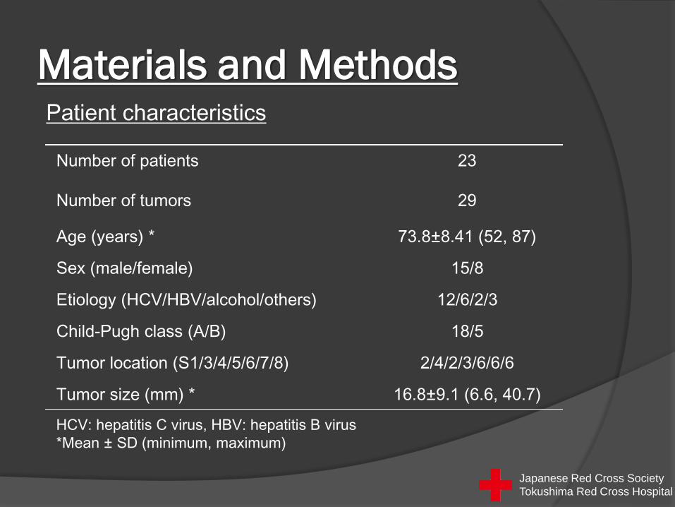
Materials and MethodsPatient characteristics
Number of patients 23
Number of tumors 29
Age (years) * 73.8±8.41 (52, 87)
Sex (male/female) 15/8
Etiology (HCV/HBV/alcohol/others) 12/6/2/3
Child-Pugh class (A/B) 18/5
Tumor location (S1/3/4/5/6/7/8) 2/4/2/3/6/6/6
Tumor size (mm) * 16.8±9.1 (6.6, 40.7)
HCV: hepatitis C virus, HBV: hepatitis B virus
*Mean ± SD (minimum, maximum)
Japanese Red Cross Society
Tokushima Red Cross Hospital

Materials and MethodsEstimation and evaluation of real embolic area
➢ The real embolic area (REA) was defined as that in which
iodized oil accumulated on follow-up CT images that was
performed 1 week after cTACE.
➢ The volume and maximum cross-sectional areas of
iodized oil accumulation in the tumor in the axial, coronal,
and sagittal planes for the REA were measured by two
observer groups using the workstation.
➢ The mean REA between groups A and B (mREA) was
used for reference.
※ Each group comprised an interventional radiologist and a radiological technician, with 10 years (group A; experts: M.K. and Y.N.) or 5 years (group B; semi-experts: K.T. and S.I.).
Japanese Red Cross Society
Tokushima Red Cross Hospital

Materials and Methods
➢ Group A and B used a commercial 3-D workstation
and liver analysis software (Synapse Vincent ver.
5.2; Fujifilm, Tokyo, Japan) to estimate VEA.
➢ The cone beam CT (CBCT) (XperCT; Philips
Healthcare, Best, The Netherlands) images were
randomized, and there was a 4 week interval
between estimation and evaluation of VEA and final
estimation and evaluation of REA to reduce bias.
Estimation and evaluation of virtual embolic area
Japanese Red Cross Society
Tokushima Red Cross Hospital
Materials and Methods
➢ The process of estimation and evaluation of VEA was as follows.
1. Non-selective CBCT during hepatic arteriography (CBCTHA)
data were transferred to the workstation.
Estimation and evaluation of virtual embolic area
Japanese Red Cross Society
Tokushima Red Cross Hospital
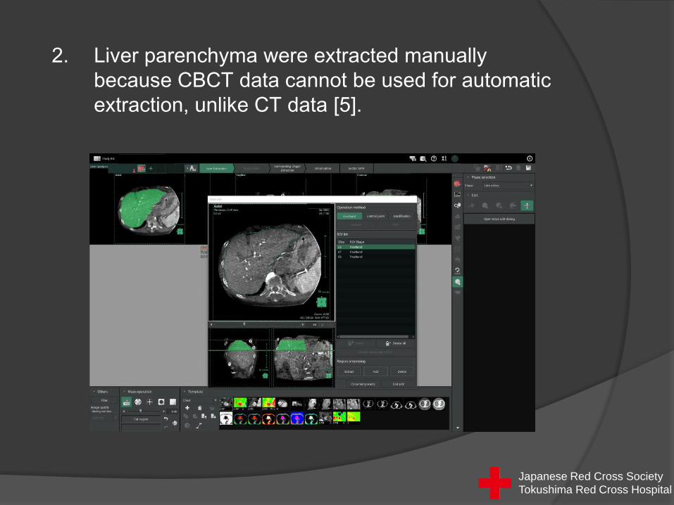
2. Liver parenchyma were extracted manually
because CBCT data cannot be used for automatic
extraction, unlike CT data [5].
Japanese Red Cross Society
Tokushima Red Cross Hospital

3. Major hepatic arteries were extracted manually, in
addition to minor ones extracted. Eventually, volume
rendering of the hepatic arteries was created.
Japanese Red Cross Society
Tokushima Red Cross Hospital

4. A simulated injection point for each tumor feeder was selected
via the 3-D arteriogram extracted from non-selective CBCTHA
with reference to spot radiographs obtained during cTACE.
Japanese Red Cross Society
Tokushima Red Cross Hospital
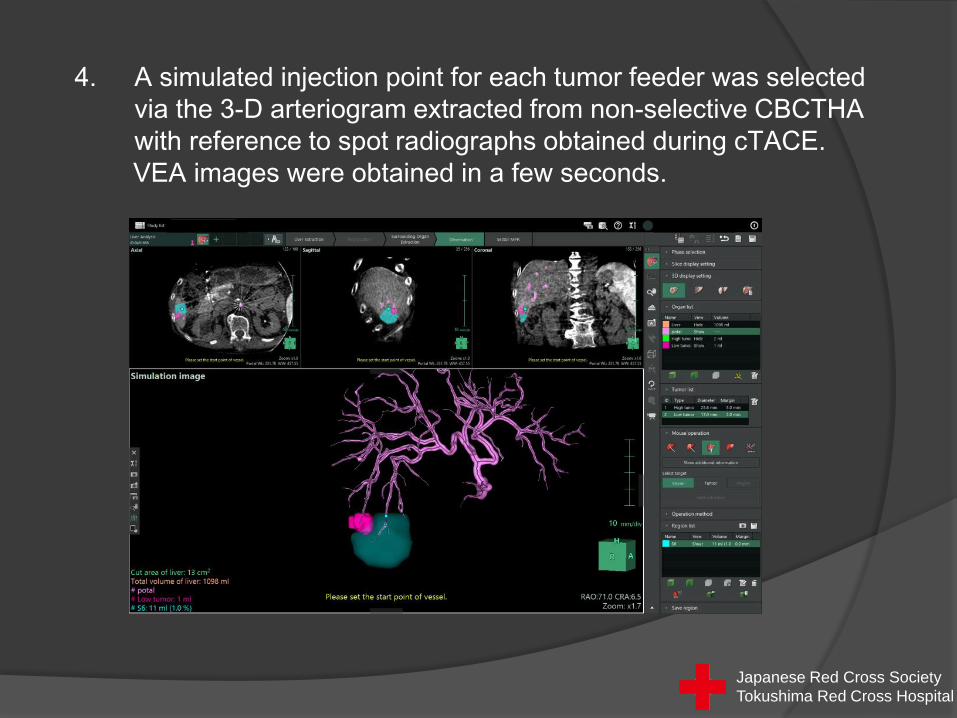
4. A simulated injection point for each tumor feeder was selected
via the 3-D arteriogram extracted from non-selective CBCTHA
with reference to spot radiographs obtained during cTACE.
VEA images were obtained in a few seconds.
Japanese Red Cross Society
Tokushima Red Cross Hospital
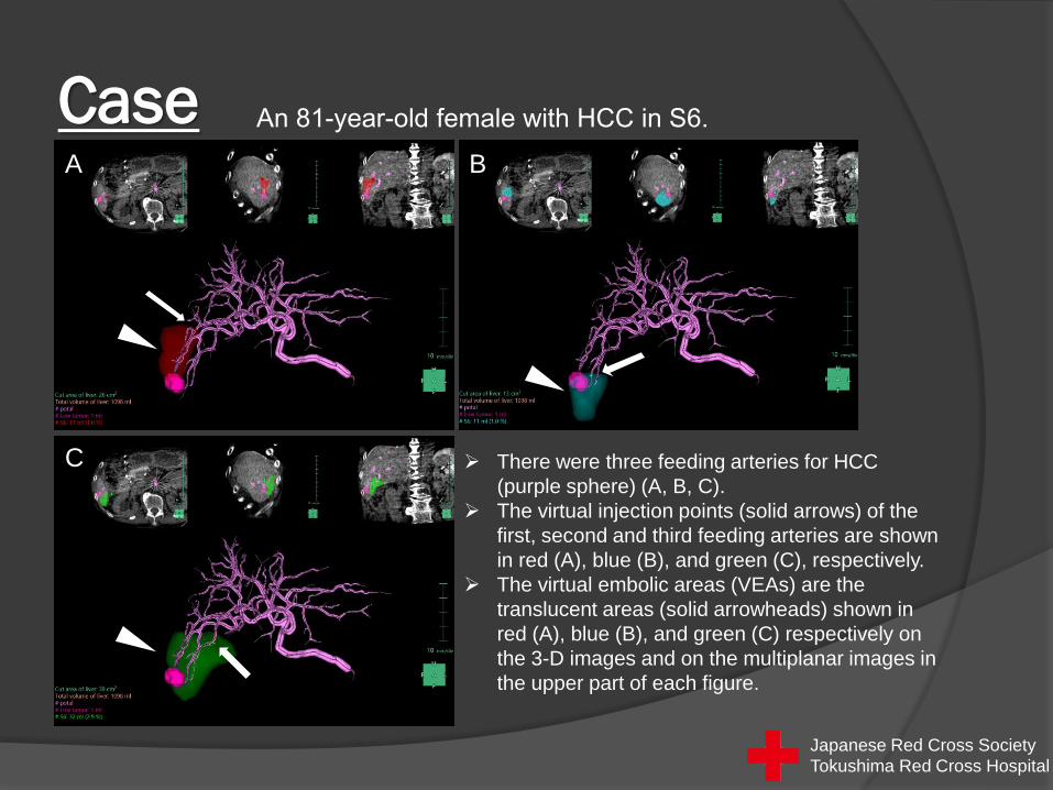
Case An 81-year-old female with HCC in S6.
➢ There were three feeding arteries for HCC
(purple sphere) (A, B, C).
➢ The virtual injection points (solid arrows) of the
first, second and third feeding arteries are shown
in red (A), blue (B), and green (C), respectively.
➢ The virtual embolic areas (VEAs) are the
translucent areas (solid arrowheads) shown in
red (A), blue (B), and green (C) respectively on
the 3-D images and on the multiplanar images in
the upper part of each figure.
A B
C
Japanese Red Cross Society
Tokushima Red Cross Hospital

Case
➢ The total VEA is shown in one figure, with each volume of the VEA in the
lower left of the figure.
➢ The total processing time required from the time the CBCT image was
available to the user to completion of estimation of VEA was about 30 minutes.
An 81-year-old female with HCC in S6.
Japanese Red Cross Society
Tokushima Red Cross Hospital
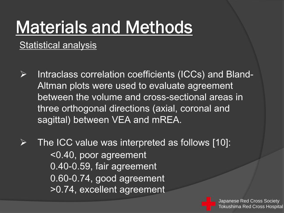
Materials and MethodsStatistical analysis
➢ Intraclass correlation coefficients (ICCs) and Bland-
Altman plots were used to evaluate agreement
between the volume and cross-sectional areas in
three orthogonal directions (axial, coronal and
sagittal) between VEA and mREA.
➢ The ICC value was interpreted as follows [10]:
<0.40, poor agreement
0.40-0.59, fair agreement
0.60-0.74, good agreement
>0.74, excellent agreementJapanese Red Cross Society
Tokushima Red Cross Hospital
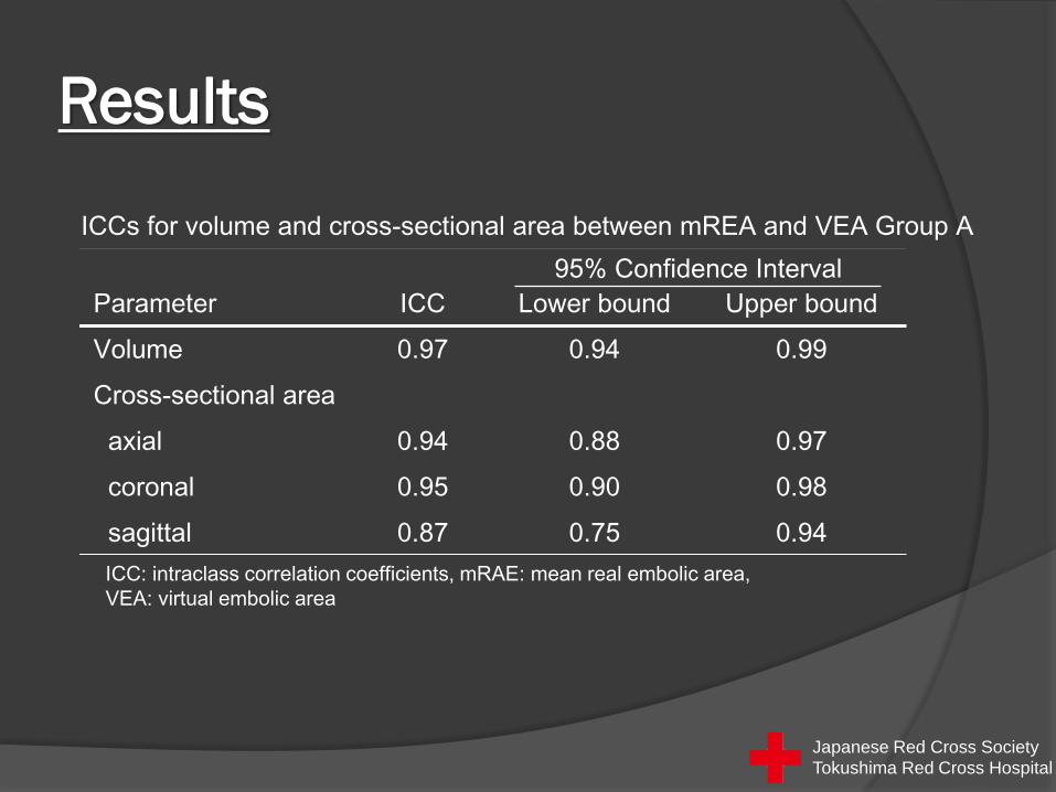
Results
ICCs for volume and cross-sectional area between mREA and VEA Group A
Parameter ICC Lower bound Upper bound
Volume 0.97 0.94 0.99
Cross-sectional area
axial 0.94 0.88 0.97
coronal 0.95 0.90 0.98
sagittal 0.87 0.75 0.94
95% Confidence Interval
ICC: intraclass correlation coefficients, mRAE: mean real embolic area,
VEA: virtual embolic area
Japanese Red Cross Society
Tokushima Red Cross Hospital

Results
ICCs for volume and cross-sectional area between mREA and VEA Group B
Parameter ICC Lower bound Upper bound
Volume 0.88 0.77 0.94
Cross-sectional area
axial 0.88 0.77 0.94
coronal 0.83 0.68 0.92
sagittal 0.74 0.52 0.87
95% Confidence Interval
ICC: intraclass correlation coefficients, mRAE: mean real embolic area,
VEA: virtual embolic area
Japanese Red Cross Society
Tokushima Red Cross Hospital
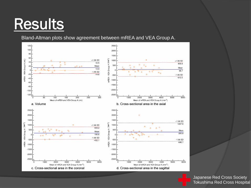
ResultsBland-Altman plots show agreement between mREA and VEA Group A.
Japanese Red Cross Society
Tokushima Red Cross Hospital
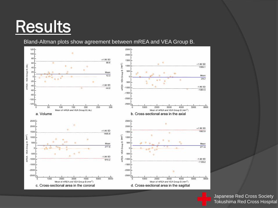
ResultsBland-Altman plots show agreement between mREA and VEA Group B.
Japanese Red Cross Society
Tokushima Red Cross Hospital

Results
➢ The ICCs for volume and cross-sectional area between
mREA and VEA showed excellent agreement, except for
the sagittal plane in group B, which was classified as
good agreement.
➢ Moreover, the lower bounds of the 95% CI indicated
excellent agreement, except for the coronal and sagittal
planes in group B, which were in good agreement.
➢ Bland-Altman plots between VEA and mREA for volume
and cross-sectional area showed no systematic biases.
Japanese Red Cross Society
Tokushima Red Cross Hospital

Discussions ①➢ If the vascular territory of tumor-feeding arteries can be
determined before chemoembolization, the optimal
catheter position including the whole tumor and the safety
margin for chemoembolization can be determined with
reference to VEA.
➢ Moreover, it may contribute to the therapeutic effects,
reduce side effects and the preservation of liver reserve,
because it is possible to intensively inject more anticancer
drug- iodized oil emulsion into the tumor and safety margin
and to reduce its distribution and embolic area to normal
liver parenchyma. In addition, reference to VEA may
contribute to decreases in radiation exposure, contrast
medium and procedure time.
Japanese Red Cross Society
Tokushima Red Cross Hospital
➢ A great improvement in estimating the virtual parenchymal
perfusion area is required for practical clinical use. Shortening of
the time required to prepare a simulation is needed.
Customization of the workstation made it possible to create VEA
in about 30 minutes. Automatic extraction of liver parenchyma
and hepatic arteries from CBCT data would shorten this time to
less than half, in addition to improving accuracy and
standardizing results.
➢ In conclusion, VEA based on CBCT data using a commercially
available 3-D workstation and liver analysis software in cTACE
can be displayed using color coding and clear divisions, and VEA
showed good agreement with REA retrospectively. This method
can be useful for estimating the embolic area in cTACE.
Discussions ②
Japanese Red Cross Society
Tokushima Red Cross Hospital
1) Llovet JM, Bruix J. Systematic review of randomized trials for unresectable hepatocellular cacinoma:
chemoembolization improves survival. Hepatology 2003; 37:429-442.
2) Lo CM, Ngan H, Tso WK, Liu CL, Lam CM, Poon RT, Fan ST, Wong J. Randomized controlled trial of
transcatheter lipiodol chemoembolization for unresectable hepatocellular carcinoma. Hepatology 2002;
35:1164-1171.
3) Miyayama S, Yamashiro M, Nagai K, Yokka A, Yoshida M, Sakuragawa N. Performance of novel virtual
parenchymal perfusion software visualizing embolized areas of transcatheter arterial chemoembolization for
hepatocellular carcinoma. Hepatol Res 2017; 47: 446-454.
4) Derbel H, Kobeiter H, Pizaine G, Ridouani F, Luciani A, Radaelli A, Van der Sterren W, Chiaradia M, Tacher V.
Accuracy of a cone-beam CT virtual parenchymal perfusion algorithm for liver cancer targeting during intra-
arterial therapy. J Vasc Interv Radiol 2017 Nov 27. [Epub ahead of print].
5) Ogawa C, Minami Y, Morita M, Noda T, Arasawa S, Izuta M, Kubo A, Matsunaka T, Tamaki H, Shibatoge M,
Kudo M. Prediction of embolization area after conventional transcatheter arterial chemoembolization for
hepatocellular carcinoma using SYNAPSE VINCENT. Dig Dis 2016; 34: 696-701.
6) Saito S, Yamanaka J, Miura K, Nakao N, Nagao T, Sugimoto T, Hirano T, Kuroda N, Iimuro Y, Fujimoto J. A
novel 3D hepatectomy simulation based on liver circulation: application to liver resection and transplantation.
Hepatology 2005; 41: 1297-1304.
7) Takamoto T, Hashimoto T, Ogata S, Inoue K, Maruyama Y, Miyazaki A, Makuuchi M. Planning of anatomical
liver segmentectomy and subsegmentectomy with 3-demensional simulation software. Am J Surg. 2013; 206:
530-538.
8) Ohshima S. Volume analyzer SYNAPSE VINCENT for liver analysis. J Hepatobiliary Pancreat Sci 2014; 21:
235-238.
9) Oshiro Y, Yano H, Mitani J, Kim S, Kim J, Fukunaga K, Ohkohchi N. Novel 3-dimensional virtual hepatectomy
simulation combined with real-time deformation. World J Gastroenterol 2015; 21: 9982-9992.
10) Oppo K, Leen E, Angerson WJ, Cooke TG, McArdle CS. Doppler perfusion index: an interobserver and
intraobserver reproducibility study. Radiology 1998; 208: 453-457.
References
Japanese Red Cross Society
Tokushima Red Cross Hospital